case report pseudohypertension-like presentation: an...
TRANSCRIPT

Case ReportPseudohypertension-Like Presentation: An Exceptionally RarePresentation in an Athletic Female Patient with Morphea
Ahmed Al-Imam1,2
1Novel Psychoactive Substances Research Unit, University of Hertfordshire Doctoral College, Hertfordshire University, Hatfield, UK2Faculty of Medicine, University of Baghdad, Baghdad, Iraq
Correspondence should be addressed to Ahmed Al-Imam; [email protected]
Received 9 October 2016; Revised 11 December 2016; Accepted 18 December 2016
Academic Editor: Alireza Firooz
Copyright © 2016 Ahmed Al-Imam. This is an open access article distributed under the Creative Commons Attribution License,which permits unrestricted use, distribution, and reproduction in any medium, provided the original work is properly cited.
Introduction. Pseudohypertension is a condition whichmainly occurs due to thickening-calcification of tunica intima of the arterialwall, leading to a faulty measurement of the intra-arterial blood pressure. To the best of our knowledge, this is the first case reportin literature, of a pseudohypertension-like presentation in association with Morphea en plaque. Case Presentation. This is a rarepresentation of a young athletic female and a professional tennis player, with pseudohypertension-like presentation. The patienthad a traumatic injury to the right elbow joint; the injury occurred during a professional tennis match. The injury was managedby immobilization, physiotherapy, and Low-Level Laser Therapy. Soon after that, the patient had a circumscribed sclerotic ivoryplaque affecting the skin of the right cubital fossa.The histopathology analysis, together with the serological-hematological tests andthe clinical picture, along with positive Osler’s signs, leads to the conclusive diagnosis of Morphea en plaque.The peculiar anatomiclocalization of the plaque anterior to the brachial artery leads to faulty blood pressure measurement as recorded by mercurialsphygmomanometer. Conclusion. This unique presentation of Morphea en plaque carries an important message in relation to thebasic medical practice and in relation to the accurate measurement of the vital signs.
1. Introduction
Pseudohypertension, also known as the noncompressibilityartery syndrome, occurs when there is a faulty-recordedelevation of blood pressure in a normal individual or anexaggerated elevation of blood pressure in an already hyper-tensive individual, as measured via sphygmomanometer.This vascular phenomenon occurs mostly in advanced agedue to calcification-thickening of the arterial vascular wall,the tunica intima layer to be specific, which makes theartery difficult to be compressed and/or occluded by thesphygmomanometer. This condition can be clinically testedusing Osler’s maneuver, and accordingly a patient is to bediagnosed with pseudohypertension when Osler’s sign ispositive [1, 2]. In this case presentation, a faulty high bloodpressure, a pseudohypertension-like phenomenon in a youngand athletic female, is reported as a result of a circumscribedplaque of Morphea (Morphea en plaque), developing inthe cutaneous tissue overlying the brachial artery in theregion of the right cubital fossa. This pathology occurred
following a traumatic injury to the right elbow joint, withconsequent physiotherapy and management with Low-LevelLaser Therapy (LLLT).
This phenomenon results in an elevated cuff pressurecompared with intra-arterial measurements and is foundprimarily in advanced age. In a retrospective study (Klemanet al., 2013), conducted on patient attending a hypertensionclinic, it was found that 7% of the patients had pseudohyper-tension [3]. In a parallel cohort study (Collins et al., 2013),the prevalence of systolic pseudohypertension in patientsundergoing Cardiac Catheterization was detected in 10.8%of patients, and the majority were of advanced age [4]. Inrelation to the clinical examination of pseudohypertension,Osler’s maneuver is to be performed for assessing the pal-pability of pulsation of the radial and/or brachial arterydistal to a point of the suspected arterial pathology. Patientswho are Osler’s sign positive have falsely elevated blood-pressure readings, in the range of 10 to 54mmHg betweencuff pressure and intra-arterial pressure [5, 6].
Hindawi Publishing CorporationCase Reports in Dermatological MedicineVolume 2016, Article ID 7027352, 3 pageshttp://dx.doi.org/10.1155/2016/7027352
2 Case Reports in Dermatological Medicine
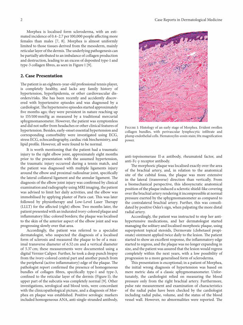
Morphea is localised form scleroderma, with an esti-mated incidence of 0.4–2.7 per 100,000 people affectingmorefemales than males [7, 8]. Morphea is almost uniformlylimited to those tissues derived from the mesoderm, mainlyreticular layer of the dermis.Theunderlying pathogenesis canbe partially attributed to an imbalance of collagen productionand destruction, leading to an excess of deposited type-1 andtype-3 collagen fibres, as seen in Figure 1 [9].
2. Case Presentation
Thepatient is an eighteen-year-old professional tennis player,is completely healthy, and lacks any family history ofhypertension, hyperlipidemia, or other cardiovascular dis-orders/risks. She has been recently and accidently discov-ered with hypertensive episodes and was diagnosed by acardiologist.Thehypertensive episodes started approximatelyfive months ago; they were persistent in nature reaching upto 155/100mmHg as measured by a traditional mercurialsphygmomanometer. However, the patient was symptomlessand did not suffer from headaches or other clinical features ofhypertension. Besides, early-onset essential hypertension andcorresponding comorbidity were investigated using ECG,stress ECG, echocardiography, cardiac risk biochemistry, andlipid profile. However, all were found to be normal.
It is worth mentioning that the patient had a traumaticinjury to the right elbow joint, approximately eight monthsprior to the presentation with the assumed hypertension,the traumatic injury occurred during a tennis match, andthe patient was diagnosed with multiple ligaments injuryaround the elbow and proximal radioulnar joint, specificallythe lateral collateral ligament and the annular ligament. Thediagnosis of the elbow joint injury was confirmed by clinicalexamination and radiography usingMRI imaging, the patientwas advised to limit her daily activities, and the elbow wasimmobilised by applying plaster of Paris cast. This was laterfollowed by physiotherapy and Low-Level Laser Therapy(LLLT) for the affected (right) elbow. Two months later, thepatient presentedwith an indurated ivory-colored plaque andinflammatory lilac-colored borders; the plaque was localisedto the skin of the anterior aspect of the elbow joint and wasprogressing slowly over that area.
Accordingly, the patient was referred to a specialistdermatologist, who suspected the diagnosis of a localisedform of sclerosis and measured the plaque to be of a max-imal transverse diameter of 6.52 cm and a vertical diameterof 5.37 cm; these measurements were documented using adigital Vernier Caliper. Further, he took a deep punch biopsyfrom the ivory-colored central part and another punch fromthe peripheral (active inflammatory) edge of the plaque. Thepathologist report confirmed the presence of homogeneousbundles of collagen fibres, specifically type-1 and type-3,confined to the reticular layer of the dermis (Figure 1); theupper part of the subcutis was completely normal [9]. Otherinvestigations, serological and blood tests, were concordantwith the clinicopathological picture, and a diagnosis of Mor-phea en plaque was established. Positive serologic markersincluded homogeneous ANA, anti-single-stranded antibody,
Figure 1: Histology of an early stage of Morphea. Evident swollencollagen bundles, with perivascular lymphocytic infiltrate andplump endothelial cells. Hematoxylin-eosin stain; 10xmagnificationpower.
anti-topoisomerase II-𝛼 antibody, rheumatoid factor, andanti-Fc-𝛾 receptor antibody.
Themorphoeic plaque was localised exactly over the areaof the brachial artery, and, in relation to the anatomicalsite of the cubital fossa, the plaque was more extensivein the lateral (transverse) direction than vertically. Froma biomechanical perspective, this idiosyncratic anatomicalposition of the plaque induced a sclerotic shield-like coveringover the brachial artery rendering it incompressible at normalpressure exerted by the sphygmomanometer as compared tothe contralateral brachial artery. Further, this was consoli-dated by positive Osler’s sign, when palpating the more distalradial artery.
Accordingly, the patient was instructed to stop her anti-hypertensive medications, and her dermatologist startedmanaging the solitary and localised morphoeic plaque, usingsuperpotent topical steroids, Dermovate (clobetasol propi-onate) ointment applied twice daily to the lesion. The patientstarted to show an excellent response, the inflammatory edgestarted to regress, and the plaque was no longer expanding insize, and the patient was assured that the lesion would regresscompletely within the next years, with a low possibility ofprogression to a more generalised form of scleroderma.
This presentation is exceptional; in a patient of Morphea,the initial wrong diagnosis of hypertension was based onmere metric data of a classic sphygmomanometer. Unfor-tunately, the cardiologist relied on measuring the bloodpressure only from the right brachial artery. Furthermore,pulse rate measurement and examination of characteristicsof the radial pulse have been checked by the cardiologistincluding radial pulse, volume, and the status of the bloodvessel wall. However, no abnormalities were reported. The
Case Reports in Dermatological Medicine 3
effective collaboration and proper attention to the patientdermatologic condition and within the context of interdis-ciplinary thinking of clinical medicine lead to the properdiagnosis and management of this young and athletic femalepatient.
The level of evidence of this manuscript is level 5, accord-ing to the Oxford Centre for Evidence-Based Medicine—Levels of Evidence [10].The literature review was done acrossPubMed, the Cochrane Library, Scopus, and Google Scholar.The search strategy was based on a keyword list applied tothese medical databases; the keywords used were as follows:“Pseudohypertension”; “Arterial Pressure”; “Osler’s sign”;“Localised Scleroderma”; “Morphea”; “Collagen”; and “Low-Level Light Therapy”. A combination of these keywordswas also applied using Boolean Operators [11]. Surprisingly,only a single published manuscript appeared, when thekeywords “Pseudohypertension AND Morphea” were used,an indicator of the extreme paucity of the published scientificliterature [12].
3. Discussion
This is an exceptionally rare case presentation of a young andan athletic female; she has beenwrongly diagnosedwith hyper-tension. Later, a proper diagnosis of Morphea and pseudohy-pertension-like presentation was concluded. Pseudohyper-tension usually occurs in advanced age, unlike this presentedcase.
The morphoeic lesion was anatomically localised inrelation to the right cubital fossa, the skin that is immediatelyin front of the brachial artery. The patient correlated theevolution of her skin lesion with a traumatic injury of herright elbow, for which shewas treatedwith physiotherapy andLow-Level Laser Therapy.
The author insight into the evolution of the morphoeicmight be correlated with the use of LLLT. Low-Level LaserTherapy was used for this patient as a therapeutic modalityto rehabilitate and alleviate pain, to restore the traumatisedright elbow joint back to functionality. In the literature,it is documented that LLLT can significantly reduce painand improves health status in chronic joint disorders, butthe disparity in patient samples, treatment procedures, andtrial design led to unconfirmed and heterogeneous levelof evidence [13]. Similarly, there is a significant paucityof verified evidence in the scientific literature, to concludewhether LLLT might induce or be associated with triggeringMorphea in the skin overlying the treated areas.
The keen interdisciplinary approach of the cardiolo-gist, dermatologist, and the dermatopathologist leads to theproper diagnosis and the subsequent successful managementof this patient.Themistake committed by the cardiologist wasto rely on the measurement of the arterial blood pressure ofright brachial artery (unilaterally) while omitting and failingto compare the pressure bilaterally and with the distal radialpulse, representing a basic and important message to allphysicians, medical practitioners, and the paramedical staff.
Competing Interests
There are no conflicts that the author is aware of.
References
[1] M. H. Beers and R. Berkow, Eds., The Merck Manual ofGeriatrics, Merck Research Laboratories, Whitehouse Station,NJ, USA, 2000.
[2] J. D. Spence, “Pseudohypertension,”Hypertension, vol. 59, no. 5,p. e49, 2012.
[3] M. Kleman, S. Dhanyamraju, andW. Difilippo, “Prevalence andcharacteristics of pseudohypertension in patients with resistanthypertension,” Journal of the American Society of Hypertension,vol. 7, no. 6, pp. 467–470, 2013.
[4] N. Collins, S. Quilty, P. Puller et al., “Pevalence of pseudo-hypertension in a contemporary patient cohort undergoingcardiac catheterisation,” Heart, Lung and Circulation, vol. 22,supplement 1, p. S21, 2013.
[5] J. Belmin, J.-M. Visintin, R. Salvatore, C. Sebban, and R. Mou-lias, “Osler’s maneuver: absence of usefulness for the detectionof pseudohypertension in an elderly population,”The AmericanJournal of Medicine, vol. 98, no. 1, pp. 42–49, 1995.
[6] F. H. Messerli, H. O. Ventura, and C. Amodeo, “Osler’s maneu-ver and pseudohypertension,” The New England Journal ofMedicine, vol. 312, no. 24, pp. 1548–1551, 1985.
[7] L. S. Peterson, A. M. Nelson, and W. D. Su, “Classification ofmorphea (localized scleroderma),”MayoClinic Proceedings, vol.70, no. 11, pp. 1068–1076, 1995.
[8] J. V. Nguyen, V. P. Werth, and N. Fett, Morphea, http://emedicine.medscape.com/article/1065782-overview.
[9] N. Fett and V. P. Werth, “Update on morphea: Part I. Epidemi-ology, clinical presentation, and pathogenesis,” Journal of theAmerican Academy of Dermatology, vol. 64, no. 2, pp. 217–228,2011.
[10] Oxford Centre for Evidence-based Medicine, “Levels ofEvidence,” March 2009, http://www.cebm.net/oxford-centre-evidence-based-medicine-levels-evidence-march-2009/.
[11] E. A. Fox and S. Sharan, “A comparison of two methods for softboolean operator interpretation in information retrieval”.
[12] S.M.Weiner, “InnereMedizin 2: Rheumatologie, Immunologie,Nephrologie, Diabetologie und Endokrinologie,” http://www-brs.ub.ruhr-uni-bochum.de/netahtml/HSS/Diss/FrerixMarc/diss.pdf.
[13] J. M. Bjordal, C. Couppe, R. T. Chow, J. Tuner, and E. A.Ljunggren, “A systematic review of low level laser therapy withlocation-specific doses for pain from chronic joint disorders,”Australian Journal of Physiotherapy, vol. 49, no. 2, pp. 107–116,2003.

Submit your manuscripts athttp://www.hindawi.com
Stem CellsInternational
Hindawi Publishing Corporationhttp://www.hindawi.com Volume 2014
Hindawi Publishing Corporationhttp://www.hindawi.com Volume 2014
MEDIATORSINFLAMMATION
of
Hindawi Publishing Corporationhttp://www.hindawi.com Volume 2014
Behavioural Neurology
EndocrinologyInternational Journal of
Hindawi Publishing Corporationhttp://www.hindawi.com Volume 2014
Hindawi Publishing Corporationhttp://www.hindawi.com Volume 2014
Disease Markers
Hindawi Publishing Corporationhttp://www.hindawi.com Volume 2014
BioMed Research International
OncologyJournal of
Hindawi Publishing Corporationhttp://www.hindawi.com Volume 2014
Hindawi Publishing Corporationhttp://www.hindawi.com Volume 2014
Oxidative Medicine and Cellular Longevity
Hindawi Publishing Corporationhttp://www.hindawi.com Volume 2014
PPAR Research
The Scientific World JournalHindawi Publishing Corporation http://www.hindawi.com Volume 2014
Immunology ResearchHindawi Publishing Corporationhttp://www.hindawi.com Volume 2014
Journal of
ObesityJournal of
Hindawi Publishing Corporationhttp://www.hindawi.com Volume 2014
Hindawi Publishing Corporationhttp://www.hindawi.com Volume 2014
Computational and Mathematical Methods in Medicine
OphthalmologyJournal of
Hindawi Publishing Corporationhttp://www.hindawi.com Volume 2014
Diabetes ResearchJournal of
Hindawi Publishing Corporationhttp://www.hindawi.com Volume 2014
Hindawi Publishing Corporationhttp://www.hindawi.com Volume 2014
Research and TreatmentAIDS
Hindawi Publishing Corporationhttp://www.hindawi.com Volume 2014
Gastroenterology Research and Practice
Hindawi Publishing Corporationhttp://www.hindawi.com Volume 2014
Parkinson’s Disease
Evidence-Based Complementary and Alternative Medicine
Volume 2014Hindawi Publishing Corporationhttp://www.hindawi.com