case report metastatic renal cell carcinoma presenting as a … · 2017-02-05 · metastatic renal...
TRANSCRIPT
Case ReportMetastatic Renal Cell Carcinoma Presenting as a ParanasalSinus Mass: The Importance of Differential Diagnosis
Massimo Ralli,1 Giancarlo Altissimi,2 Rosaria Turchetta,2 and Mario Rigante3
1Department of Oral and Maxillofacial Sciences, Sapienza University of Rome, Rome, Italy2Department of Sense Organs, Audiology Section, Policlinico Umberto I, Sapienza University of Rome, Rome, Italy3Department of Otorhinolaryngology, Catholic University of Sacred Heart, Rome, Italy
Correspondence should be addressed to Massimo Ralli; [email protected]
Received 25 July 2016; Revised 15 November 2016; Accepted 14 December 2016; Published 11 January 2017
Academic Editor: Marco Berlucchi
Copyright © 2017 Massimo Ralli et al. This is an open access article distributed under the Creative Commons Attribution License,which permits unrestricted use, distribution, and reproduction in any medium, provided the original work is properly cited.
Metastases in the paranasal sinuses are rare; renal cell carcinoma is the most common cancer that metastasizes to this region. Wepresent the case of a patient with a 4-month history of a rapidly growing mass of the nasal pyramid following a nasal trauma,associated with spontaneous epistaxis and multiple episodes of hematuria. Cranial CT scan and MRI showed an ethmoid massextending to the choanal region, the right orbit, and the right frontal sinus with an initial intracranial extension. Patient underwentsurgery with a trans-sinusal frontal approach using a bicoronal incision combinedwith an anteriormidfacial degloving; histologicalexam was compatible with a metastasis of clear cell renal cell carcinoma. Following histological findings, a total body CT scanshowed a solitary 6 cmmass in the upper posterior pole of the left kidney identified as the primary tumor. Although rare, metastaticrenal cell carcinoma should always be suspected in patients with nasal or paranasal masses, especially if associated with symptomssuggestive of a systemic involvement such as hematuria. A correct early-stage diagnosis ofmetastatic RCC can considerably improvesurvival rate in these patients; preoperative differential diagnosis with contrast-enhanced imaging is fundamental for the correcttreatment and follow-up strategy.
1. Introduction
Renal cell carcinoma (RCC) is the most common kidneycancer, with approximately 35,000 new cases in the USeach year [1]; RCC mainly affects male patients between40 and 60 years old [2]. Common presentation symptomsinclude hematuria (40%), flank pain (40%), and a palpableabdominal mass (25%) [3]. Approximately 30% of patientswith renal cell carcinoma present with metastatic disease[4]; target organs are lung (75%), soft tissues (36%), bone(20%), liver (18%), cutaneous sites (8%), and central nervoussystem (8%) [5, 6]. Metastases in the paranasal sinuses arerare [7]; however, RCC is the most common cancer thatmetastasizes to this region. Prognosis of metastatic RCC ispoor [8]; the survival rate ranges between 15 and 30% at5 years [9] in case of a single metastasis and between 0and 7% in patients with multiple metastases [10]. MetastaticRCC is often resistant to chemotherapy and radiotherapy [11];numerous agents targeting VEGF and non-VEGFR pathways
have been proposed during the last decade for the treatmentof advanced RCC [12–18].
We present the case of a patient with a single, rapidlygrowing mass in the upper portion of the nasal pyramid,with late, postnasal surgery histological diagnosis of renal cellcarcinoma that allowed primary tumor identification.
2. Case Presentation
A 72-year-old man was referred to our institution with a 4-month history of a voluminous mass in the upper portionof the nasal pyramid following a nasal trauma. He had beentreated a few weeks earlier at a different ENT service fora massive spontaneous epistaxis. The patient also reporteda long history of hematuria, previously attributed to renaltuberculosis occurring over 40 years before. At admission,a cranial CT scan showed a large soft tissue ethmoid massextending to the right and left choanal region, the right orbit,the right frontal sinus, and an initial intracranial extension
Hindawi Publishing CorporationCase Reports in OtolaryngologyVolume 2017, Article ID 9242374, 5 pageshttps://doi.org/10.1155/2017/9242374
2 Case Reports in Otolaryngology
(a) (b)
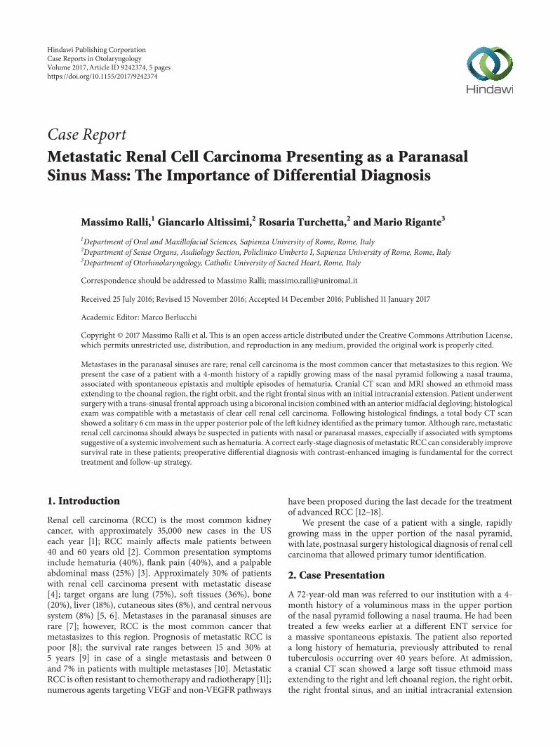
Figure 1: MRI in the axial (a) and sagittal (b) planes showing a soft tissue ethmoid mass extending to the right and left choanal region, theright orbit, the right frontal sinus, and an initial intracranial extension with partial erosion of the crista galli.
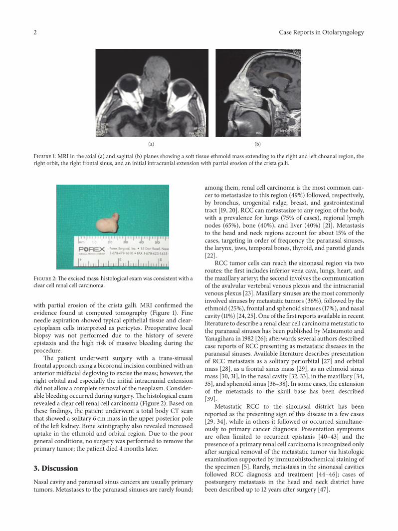
Figure 2:The excised mass; histological exam was consistent with aclear cell renal cell carcinoma.
with partial erosion of the crista galli. MRI confirmed theevidence found at computed tomography (Figure 1). Fineneedle aspiration showed typical epithelial tissue and clear-cytoplasm cells interpreted as pericytes. Preoperative localbiopsy was not performed due to the history of severeepistaxis and the high risk of massive bleeding during theprocedure.
The patient underwent surgery with a trans-sinusalfrontal approach using a bicoronal incision combinedwith ananterior midfacial degloving to excise the mass; however, theright orbital and especially the initial intracranial extensiondid not allow a complete removal of the neoplasm. Consider-able bleeding occurred during surgery.The histological examrevealed a clear cell renal cell carcinoma (Figure 2). Based onthese findings, the patient underwent a total body CT scanthat showed a solitary 6 cm mass in the upper posterior poleof the left kidney. Bone scintigraphy also revealed increaseduptake in the ethmoid and orbital region. Due to the poorgeneral conditions, no surgery was performed to remove theprimary tumor; the patient died 4 months later.
3. Discussion
Nasal cavity and paranasal sinus cancers are usually primarytumors. Metastases to the paranasal sinuses are rarely found;
among them, renal cell carcinoma is the most common can-cer to metastasize to this region (49%) followed, respectively,by bronchus, urogenital ridge, breast, and gastrointestinaltract [19, 20]. RCC can metastasize to any region of the body,with a prevalence for lungs (75% of cases), regional lymphnodes (65%), bone (40%), and liver (40%) [21]. Metastasisto the head and neck regions account for about 15% of thecases, targeting in order of frequency the paranasal sinuses,the larynx, jaws, temporal bones, thyroid, and parotid glands[22].
RCC tumor cells can reach the sinonasal region via tworoutes: the first includes inferior vena cava, lungs, heart, andthe maxillary artery; the second involves the communicationof the avalvular vertebral venous plexus and the intracranialvenous plexus [23].Maxillary sinuses are themost commonlyinvolved sinuses by metastatic tumors (36%), followed by theethmoid (25%), frontal and sphenoid sinuses (17%), and nasalcavity (11%) [24, 25].One of the first reports available in recentliterature to describe a renal clear cell carcinomametastatic tothe paranasal sinuses has been published by Matsumoto andYanagihara in 1982 [26]; afterwards several authors describedcase reports of RCC presenting as metastatic diseases in theparanasal sinuses. Available literature describes presentationof RCC metastasis as a solitary periorbital [27] and orbitalmass [28], as a frontal sinus mass [29], as an ethmoid sinusmass [30, 31], in the nasal cavity [32, 33], in the maxillary [34,35], and sphenoid sinus [36–38]. In some cases, the extensionof the metastasis to the skull base has been described[39].
Metastatic RCC to the sinonasal district has beenreported as the presenting sign of this disease in a few cases[29, 34], while in others it followed or occurred simultane-ously to primary cancer diagnosis. Presentation symptomsare often limited to recurrent epistaxis [40–43] and thepresence of a primary renal cell carcinoma is recognized onlyafter surgical removal of the metastatic tumor via histologicexamination supported by immunohistochemical staining ofthe specimen [5]. Rarely, metastasis in the sinonasal cavitiesfollowed RCC diagnosis and treatment [44–46]; cases ofpostsurgery metastasis in the head and neck district havebeen described up to 12 years after surgery [47].
Case Reports in Otolaryngology 3
The key point in RCC presenting with a sinonasalmetastasis is differential diagnosis with primary tumorssuch as adenocarcinomas, angiofibromas, hemangiopericy-tomas, melanomas, hemangiomas, metastatic tumors fromthe breast and lungs, and, more rarely, systemic diseasessuch as Wegener’s and midline granulomas [48]. In fact, insuch cases diagnostic delays, misdiagnosis, undertreatment,and mismanagement could occur due to (1) the attributionof the mass to a primary sinonasal cancer given the rarenature of sinonasal metastasis or (2) to the overlook ofpresenting symptoms such as recurrent epistaxis, swelling,pain, and nasal obstruction. Hematuria can be consideredas an indicator of RCC; it has been reported that about 10%of patients with RCC with distant metastasis exhibit massivehematuria. However, intermittent hematuria may be presentin 90% of cases [3]. For this reason, patients presenting withnasosinusal tumors also reporting hematuria should alwaysundergo systemic evaluation. Radiological examination withCT scan and, secondly, MRI and angiography are necessaryin assessing the extent of the metastatic lesion. However,it should be considered that RCC metastases have similarradiological appearances to primary malignant lesions ofsinonasal cavities; some indicators of renal origin at CTscan are enhancement, destruction, and lack of tumoralcalcification [6].
In this case, CT scan allowed the identification of aneoformed paranasal sinus mass; however, only histologicalexam identified the mass as a metastasis of RCC and ledto the execution of total body CT scan to identify primarytumor. Although difficult, differential preoperative diagnosisis fundamental for the correct treatment and follow-upstrategy; contrast-enhanced imaging plays a central role sincea preoperative biopsy of the nasal mass may be difficultin these patients due to massive recurring bleeding and, insome cases, may result in only necrotic tissue inconclusiveon histopathology [42].The ENT specialist, therefore, shouldalways suspect metastatic disease from primary sites externalto the head and neck region in patients with hypervascularmass in the nasal cavity or paranasal sinuses and a historyof massive nasal bleeding and should complete preoperativeworkupwith total bodyCT scan. Furthermore, it is importantto remark that metastatic tumors originating from primarykidney masses are highly vascularized and surgeons shouldexpect significant haemorrhage during surgical removal. Oneof the main advantages of a preoperative diagnosis of RCCwhen approaching a patient with sinonasal mass is the prepa-ration for management of severe perioperative bleeding, thusimplementing strategies to optimise the patient’s tolerance tobleeding and to reduce the amount of bleedingmorbidity andmortality.
Prognosis of metastatic RCC is poor; however, a correctearly-stage diagnosis of metastatic disease can considerablyimprove survival rate: literature reports that excision ofsolitary metastatic lesion of renal cell carcinoma followingnephrectomy results in a 41% survival at 2 years and 13%survival at 5 years [48]. The sole excision of the metastaticlesion, instead, significantly lowers survival rate [49]; patientswith multiple metastases have a 5-year survival rate between0 and 7% [10].
Althoughmetastatic RCC is often resistant to chemother-apy and radiotherapy, numerous agents targeting VEGF andnon-VEGFR pathways should be taken into account for thetreatment of advanced RCC. Multitargeted VEGF tyrosinekinase inhibitors (TKIs) include sorafenib [12], sunitinib [13],pazopanib [14], axitinib [15], and bevacizumab [16]; mTORinhibitors include temsirolimus [17] and everolimus [18].Unfortunately, especially in cases of advanced neoplasms,benefits are still time-limited and treatment decisions shouldbe based not only on guidelines but also on clinical con-siderations, such as patient comorbidities, treatment toxicity,prognostic factors, and molecular aspects of disease. In thiscase, the poor general conditions of the patient preventedadditional treatment except for palliative pain management.
In conclusion, metastatic renal cell carcinoma shouldalways be suspected in patients with nasal or paranasalmasses, especially if associated with symptoms suggestive of asystemic involvement such as hematuria; early-stage diagno-sis of metastatic disease can considerably limit perioperativecomplications and improve survival rate.
Competing Interests
The authors declare that they have no competing interests.
References
[1] A. Jemal, R. C. Tiwari, T.Murray et al., “Cancer Statistics, 2004,”CA:ACancer Journal for Clinicians, vol. 54, no. 1, pp. 8–29, 2004.
[2] R. Y. Lim, D. F. Bastug, and B. L. Caldwell, “Metastatic renalcell carcinoma of the nasal septum,”The West Virginia MedicalJournal, vol. 85, no. 4, pp. 143–145, 1989.
[3] D. G. Skinner, C. D. Vermillion, R. C. Pfister, and W. F.Leadbetter, “Renal cell carcinoma,” American Family Physician,vol. 4, no. 4, pp. 89–94, 1971.
[4] R. C. Flanigan, S. C. Campbell, J. I. Clark, and M. M. Picken,“Metastatic renal cell carcinoma,” Current Treatment Options inOncology, vol. 4, no. 5, pp. 385–390, 2003.
[5] J. Singh, V. Baheti, S. S. Yadav, and R. Mathur, “Occult renal cellcarcinoma manifesting as nasal mass and epistaxis,” Reviews inUrology, vol. 16, no. 3, pp. 145–148, 2014.
[6] P. M. Som, K. I. Norton, J. M. Shugar et al., “Metastatichypernephroma to the head and neck,” American Journal ofNeuroradiology, vol. 8, no. 6, pp. 1103–1106, 1987.
[7] M. Ziari, S. Shen, R. J. Amato, and B. S. Teh, “Metastatic renalcell carcinoma to the nose and ethmoid sinus,” Urology, vol. 67,no. 1, pp. 199.e21–199.e23, 2006.
[8] M.H.Ather, N.Masood, andT. Siddiqui, “Currentmanagementof advanced and metastatic renal cell carcinoma,” UrologyJournal, vol. 7, no. 1, pp. 1–9, 2010.
[9] B. Torres Muros, R. Bonilla Parrilla, J. R. Solano Romero, J. G.Rodrıguez Baro, and J. Verge Gonzalez, “Metastasis in maxilarsinus as only manifestation of disseminate renal adenocarci-noma,”Anales Otorrinolaringologicos Ibero-Americanos, vol. 34,no. 3, pp. 231–236, 2007.
[10] E. T. Cheng, D. Greene, and R. J. Koch, “Metastatic renalcell carcinoma to the nose,” Otolaryngology—Head and NeckSurgery, vol. 122, no. 3, p. 464, 2000.
4 Case Reports in Otolaryngology
[11] R. J. Motzer, P. Russo, D. M. Nanus, and W. J. Berg, “Renal cellcarcinoma,” Current Problems in Cancer, vol. 21, no. 4, pp. 185–232, 1997.
[12] B. Escudier, N. Lassau, E. Angevin et al., “Phase I trial ofsorafenib in combination with IFN 𝛼-2a in patients with unre-sectable and/or metastatic renal cell carcinoma or malignantmelanoma,” Clinical Cancer Research, vol. 13, no. 6, pp. 1801–1809, 2007.
[13] R. J. Motzer, M. D. Michaelson, J. Rosenberg et al., “Sunitinibefficacy against advanced renal cell carcinoma,” Journal ofUrology, vol. 178, no. 5, pp. 1883–1887, 2007.
[14] C. N. Sternberg, I. D. Davis, J. Mardiak et al., “Pazopanib inlocally advanced or metastatic renal cell carcinoma: results ofa randomized phase III trial,” Journal of Clinical Oncology, vol.28, no. 6, pp. 1061–1068, 2010.
[15] B. I. Rini, B. Melichar, T. Ueda et al., “Axitinib with or withoutdose titration for first-line metastatic renal-cell carcinoma: arandomised double-blind phase 2 trial,” The Lancet Oncology,vol. 14, no. 12, pp. 1233–1242, 2013.
[16] B. Escudier, A. Pluzanska, P. Koralewski et al., “Bevacizumabplus interferon alfa-2a for treatment of metastatic renal cellcarcinoma: a randomised, double-blind phase III trial,” TheLancet, vol. 370, no. 9605, pp. 2103–2111, 2007.
[17] G. Hudes, M. Carducci, P. Tomczak et al., “Temsirolimus,interferon alfa, or both for advanced renal-cell carcinoma,”NewEngland Journal of Medicine, vol. 356, no. 22, pp. 2271–2281,2007.
[18] R. J.Motzer, B. Escudier, S.Oudard et al., “Efficacy of everolimusin advanced renal cell carcinoma: a double-blind, randomised,placebo-controlled phase III trial,”TheLancet, vol. 372, no. 9637,pp. 449–456, 2008.
[19] P. Sountoulides, L. Metaxa, and L. Cindolo, “Atypical presenta-tions and rare metastatic sites of renal cell carcinoma: a reviewof case reports,” Journal of Medical Case Reports, vol. 5, articleno. 429, 2011.
[20] E. Evgeniou, K. R. Menon, G. L. Jones, H. Whittet, and W.Williams, “Renal cell carcinoma metastasis to the paranasalsinuses and orbit,” BMJ Case Reports, vol. 2012, 2012.
[21] E. E. Lang, N. Patil, R.M.Walsh,M. Leader, andM.A.Walsh, “Acase of renal cell carcinomametastatic to the nose and tongyue,”Ear, Nose andThroat Journal, vol. 82, no. 5, pp. 382–383, 2003.
[22] F. O. Dincbas, B. Atalar, D. C. Oksuz, F. V. Aker, and S. Koca,“Unusual metastasis of renal cell carcinoma to the nasal cavity,”Journal of B.U.ON., vol. 9, no. 2, pp. 201–204, 2004.
[23] M. D. Gottlieb and J. T. Roland Jr., “Paradoxical spread of renalcell carcinoma to the head and neck,” Laryngoscope, vol. 108, no.9, pp. 1301–1305, 1998.
[24] M. Kovacic, A. Krvavica, and M. Rudic, “Renal cell carcinomametastasis to the sinonasal cavity: case report,” Acta ClinicaCroatica, vol. 54, no. 2, pp. 223–226, 2015.
[25] J. M. Bernstein, W.W.Montgomery, and K. Balogh, “Metastatictumors to the maxilla, nose, and paranasal sinuses,” Laryngo-scope, vol. 76, no. 4, pp. 621–650, 1966.
[26] Y. Matsumoto and N. Yanagihara, “Renal clear cell carcinomametastatic to the nose and paranasal sinuses,” Laryngoscope, vol.92, no. 10, part 1, pp. 1190–1193, 1982.
[27] J. J. Homer and N. S. Jones, “Renal cell carcinoma presentingas a solitary paranasal sinus metastasis,” Journal of Laryngologyand Otology, vol. 109, no. 10, pp. 986–989, 1995.
[28] J. W. Jung, S. C. Yoon, D. H. Han, and M. Chi, “Metastatic renalcell carcinoma to the orbit and the ethmoid sinus,” Journal ofCraniofacial Surgery, vol. 23, no. 2, pp. e136–e138, 2012.
[29] T. Ikeuchi, N. Asai, T. Hori et al., “Renal cell carcinoma detectedby metastasis to the frontal sinus: a case report,” Acta UrologicaJaponica, vol. 44, no. 2, pp. 89–92, 1998.
[30] G. K. Maheshwari, H. A. Baboo, M. H. Patel, and G. Usha,“Metastatic renal cell carcinoma involving ethmoid sinus atpresentation,” Journal of Postgraduate Medicine, vol. 49, no. 1,pp. 96–97, 2003.
[31] N. Terada, K. Hiruma, M. Suzuki, T. Numata, and A. Konno,“Metastasis of renal cell cancer to the ethmoid sinus,”Acta Oto-Laryngologica, Supplement, no. 537, pp. 82–86, 1998.
[32] S. Vreugde, R. Duttmann, A. Halama, and P. Deron, “Metastasisof a renal cell carcinoma to the nose and paranasal sinuses,”Acta Oto-Rhino-Laryngologica Belgica, vol. 53, no. 2, pp. 129–131, 1999.
[33] R. Nason and R. L. Carrau, “Metastatic renal cell carcinomato the nasal cavity,” American Journal of Otolaryngology—Headand Neck Medicine and Surgery, vol. 25, no. 1, pp. 54–57, 2004.
[34] B. Torres Muros, J. R. Solano Romero, J. G. Rodrıguez Baro,and R. Bonilla Parrilla, “Maxillary sinus metastasis of renal cellcarcinoma,” Actas Urologicas Espanolas, vol. 30, no. 9, pp. 954–957, 2006.
[35] Y. He, J. Chen, W. Xu et al., “Case report metastatic renal cellcarcinoma to the left maxillary sinus,” Genetics and MolecularResearch, vol. 13, no. 3, pp. 7465–7469, 2014.
[36] S. Koscielny, “The paranasal sinuses as metastatic site of renalcell carcinoma,” Laryngorhinootologie, vol. 78, no. 8, pp. 441–444, 1999.
[37] R. Simo, A. J. Sykes, S. P. Hargreaves et al., “Metastatic renal cellcarcinoma to the nose and paranasal sinuses,” Head and Neck,vol. 22, no. 7, pp. 722–727, 2000.
[38] J. G. Pereira Arias, V. Ullate Jaime, F. Valcarcel Martın etal., “Epistaxis as initial manifestation of disseminated renaladenocarcinoma,” Actas Urologicas Espanolas, vol. 26, no. 5, pp.361–365, 2002.
[39] P. K. Parida, “Renal cell carcinoma metastatic to the sinonasalregion: three case reports with a review of the literature,” Ear,Nose andThroat Journal, vol. 91, no. 11, pp. E11–E16, 2012.
[40] M. Szymanski, A. Szymanska, K. Morshed, and H. Siwiec,“Renal cell carcinoma metastases to nose and paranasal sinusespresenting as recurrent epistaxis,”Wiadomosci Lekarskie, vol. 57,no. 1-2, pp. 94–96, 2004.
[41] H. Lee, H. J. Kang, and S. H. Lee, “Metastatic renal cellcarcinoma presenting as epistaxis,” European Archives of Oto-Rhino-Laryngology, vol. 262, no. 1, pp. 69–71, 2005.
[42] D. R. Nayak, K. Pujary, S. Ramnani, C. Shetty, and P. Parul,“Metastatic renal cell carcinoma presenting with epistaxis,”Indian Journal of Otolaryngology and Head and Neck Surgery,vol. 58, no. 4, pp. 406–408, 2006.
[43] R. Kumar, K. Sikka, R. Kumar, and P. Chatterjee, “Nephrogenicepistaxis,” SingaporeMedical Journal, vol. 55, no. 7, pp. e112–e113,2014.
[44] V. Montoro Martınez, M. Lopez Vilas, M. Gurri Freixa, E.De Dios Oran, J. R. Montserrat Gili, and J. M. Fabra Llopis,“Nasal sinus metastasis of renal carcinoma. A case report,” ActaOtorrinolaringologica Espanola, vol. 50, no. 8, pp. 653–656, 1999.
[45] H. Sawazaki, T. Segawa, K. Yoshida et al., “Bilateral maxillarysinus metastasis of renal cell carcinoma: a case report,” ActaUrologica Japonica, vol. 53, no. 4, pp. 231–234, 2007.
[46] S.-L. Hong, D.-W. Jung, H.-J. Roh, and K.-S. Cho, “Metastaticrenal cell carcinoma of the posterior nasal septum as the firstpresentation 10 years after nephrectomy,” Journal of Oral andMaxillofacial Surgery, vol. 71, no. 10, pp. 1813.e1–1813.e7, 2013.

Case Reports in Otolaryngology 5
[47] G. Fyrmpas, A. Adeniyi, and S. Baer, “Occult renal cell carci-noma manifesting with epistaxis in a woman: a case report,”Journal of Medical Case Reports, vol. 5, article 79, 2011.
[48] M. K. Dineen, R. D. Pastore, L. J. Emrich, and R. P. Huben,“Results of surgical treatment of renal cell carcinoma withsolitary metastasis,” Journal of Urology, vol. 140, no. 2, pp. 277–279, 1988.
[49] D. G. Skinner, R. B. Colvin, C. D. Vermillion, R. C. Pfister,and W. F. Leadbetter, “Diagnosis and management of renalcell carcinoma. A clinical and pathologic study of 309 cases,”Cancer, vol. 28, no. 5, pp. 1165–1177, 1971.

Submit your manuscripts athttps://www.hindawi.com
Stem CellsInternational
Hindawi Publishing Corporationhttp://www.hindawi.com Volume 2014
Hindawi Publishing Corporationhttp://www.hindawi.com Volume 2014
MEDIATORSINFLAMMATION
of
Hindawi Publishing Corporationhttp://www.hindawi.com Volume 2014
Behavioural Neurology
EndocrinologyInternational Journal of
Hindawi Publishing Corporationhttp://www.hindawi.com Volume 2014
Hindawi Publishing Corporationhttp://www.hindawi.com Volume 2014
Disease Markers
Hindawi Publishing Corporationhttp://www.hindawi.com Volume 2014
BioMed Research International
OncologyJournal of
Hindawi Publishing Corporationhttp://www.hindawi.com Volume 2014
Hindawi Publishing Corporationhttp://www.hindawi.com Volume 2014
Oxidative Medicine and Cellular Longevity
Hindawi Publishing Corporationhttp://www.hindawi.com Volume 2014
PPAR Research
The Scientific World JournalHindawi Publishing Corporation http://www.hindawi.com Volume 2014
Immunology ResearchHindawi Publishing Corporationhttp://www.hindawi.com Volume 2014
Journal of
ObesityJournal of
Hindawi Publishing Corporationhttp://www.hindawi.com Volume 2014
Hindawi Publishing Corporationhttp://www.hindawi.com Volume 2014
Computational and Mathematical Methods in Medicine
OphthalmologyJournal of
Hindawi Publishing Corporationhttp://www.hindawi.com Volume 2014
Diabetes ResearchJournal of
Hindawi Publishing Corporationhttp://www.hindawi.com Volume 2014
Hindawi Publishing Corporationhttp://www.hindawi.com Volume 2014
Research and TreatmentAIDS
Hindawi Publishing Corporationhttp://www.hindawi.com Volume 2014
Gastroenterology Research and Practice
Hindawi Publishing Corporationhttp://www.hindawi.com Volume 2014
Parkinson’s Disease
Evidence-Based Complementary and Alternative Medicine
Volume 2014Hindawi Publishing Corporationhttp://www.hindawi.com