case report metastatic basal cell carcinoma accompanying...
TRANSCRIPT
Case ReportMetastatic Basal Cell Carcinoma AccompanyingGorlin Syndrome
Yeliz Bilir,1 Erkan Gokce,2 Banu Ozturk,3 Faik Alev Deresoy,4
Ruken Yuksekkaya,2 and Emel Yaman5
1 Department of Internal Diseases, Faculty of Medicine, Gaziosmanpasa University, 60100 Tokat, Turkey2Department of Radiodiagnostics, Faculty of Medicine, Gaziosmanpasa University, 60100 Tokat, Turkey3 Division of Medical Oncology, Faculty of Medicine, Gaziosmanpasa University, 60100 Tokat, Turkey4Department of Pathology, Faculty of Medicine, Gaziosmanpasa University, 60100 Tokat, Turkey5 Department of Medical Oncology, Faculty of Medicine, Mersin University, 33343 Mersin, Turkey
Correspondence should be addressed to Banu Ozturk; [email protected]
Received 11 April 2014; Revised 15 September 2014; Accepted 29 September 2014; Published 19 November 2014
Academic Editor: Josep M. Ribera
Copyright © 2014 Yeliz Bilir et al.This is an open access article distributed under theCreative CommonsAttribution License, whichpermits unrestricted use, distribution, and reproduction in any medium, provided the original work is properly cited.
Gorlin-Goltz syndrome or basal cell nevus syndrome is an autosomal dominant syndrome characterized by skeletal anomalies,numerous cysts observed in the jaw, and multiple basal cell carcinoma of the skin, which may be accompanied by falx cerebricalcification. Basal cell carcinoma is the most commonly skin tumor with slow clinical course and low metastatic potential. Itsconcomitance with Gorlin syndrome, resulting from a mutation in a tumor suppressor gene, may substantially change morbidityand mortality. A 66-year-old male patient with a history of recurrent basal cell carcinoma was presented with exophthalmus in theleft eye and the lesions localized in the left lateral orbita and left zygomatic area. His physical examination revealed hearing loss,gapped teeth, highly arched palate, and frontal prominence. Left orbital mass, cystic masses at frontal and ethmoidal sinuses, andmultiple pulmonary nodules were detected at CT scans. Basal cell carcinoma was diagnosed from biopsy of ethmoid sinus. Basedon the clinical and typical radiological characteristics (falx cerebri calcification, bifid costa, and odontogenic cysts), the patient wasdiagnosed with metastatic skin basal cell carcinoma accompanied by Gorlin syndrome. Our case is a basal cell carcinoma withaggressive course accompanying a rarely seen syndrome.
1. Introduction
Gorlin syndrome, one of the hereditary syndromes concomi-tant with multiple basal cell carcinoma (BCC), is associatedwith many anomalies observed in several systems [1]. Thisautosomal dominant syndrome is characterized by multipleBCC seen in the early stage of the life, palmar and plan-tar pits, mandibular prognathism, mandibular keratocysts,hypertelorism, falx cerebri calcification, spina bifida, bifidcosta, vertebral abnormalities, ovarian fibroma, cardiac fibro-mas, milia, epidermal cysts, mesenteric cysts, gastric polyps,medulloblastoma, meningioma, and fatal rhabdomyoma [1].Odontogenic keratocysts are characteristic in this syndrome.PTCH (Drosophila patched gen human analogue) is localizedon chromosome 9q22.3-q31 and encodes sonic hedgehog
(SHH) signaling pathway. Autosomal dominant germlinemutations on this gene account for the pathogenesis of Gorlinsyndrome. The inactivation of PTCH gene causes cancerformation, mainly by leading to overexpression in the SHHpathway and basal cell carcinoma [2].
Basal cell carcinoma (BCC) is the most commonly seenskin cancer, which accounts for 80% of the nonmelanomaskin cancers [3]. BCC is most commonly seen between theages of 40 and 80 years old, with the incidence increasingover time. Lifetime risk for BCC was reported to be 1/18in men and 1/20 in women. Environmental and hereditaryfactors have a substantial contribution to the development ofBCC. In its etiology, most likely causal factor is ultraviolet-B(UVB) beams. Clear skin color and prolonged sun exposureare important risk factors. Tumor may show a course with
Hindawi Publishing CorporationCase Reports in Oncological MedicineVolume 2014, Article ID 362932, 7 pageshttp://dx.doi.org/10.1155/2014/362932

2 Case Reports in Oncological Medicine
Figure 1: The facial profile of the patient.
local recurrences, but its potential for metastasis is low [4,5]. The risk for metastasis occurrence is 0.0028–0.55% [6].Majority of the metastases originate from facial BCCs. Mostcommonly seen metastatic sites include lymph node (60%),lungs (42%), bone (20%), and skin (10%) [7]. Incidence ofpulmonary metastasis is approximately 0.1% [8].
Gorlin syndrome is a syndromewith a high risk formalig-nity development at earlier ages, which may be diagnosedduring the childhood based on physical findings. Our caseis a basal cell carcinoma with diffuse lung metastases, whichhas a course with recurrent multiple basal cell carcinomaand which is accompanied by Gorlin syndrome diagnosed atadvanced age.
2. Case Report
The medical history of the 66-year-old male patient, whohas been presented to the hospital with exophthalmus andskin lesions, revealed that he had firstly undergone localexcision due to basal cell skin carcinoma located on his face12 years ago, that new lesions had formed 2 cm below the leftoculonasal junction and in the left nasal root 6 years followingthe first diagnosis, and that the mass has been diagnosedto be a recurrence of BCC that indented medial wall of theleft orbita and that expanded to close bones. The patienthad undergone rhinectomy and reconstructive surgery. Hehad received radiotherapy. Six years after the completion ofthe therapy, he was presented with a lesion near the left eye(Figure 1).
FacialMRI of the patient revealed a dense and large cysticmass lesion that led to an expansion of left frontal sinus byfilling it and that destructed the anterior wall (Figure 2(a)).There were ovoid, dense cystic lesions with uniform contoursin the central part and round, millimetric cystic lesions in theright of the mandibular corpus. A cystic lesion with uniformcontours was observed in the contiguity of the retromolardental roots in the left maxillar bone (Figures 2(a) and 2(b)).
In the left, there were 2 contiguous cystic lesions withdifferent sizes and contents and with regular contours inthe ethmoid air cells, showing indentation to left medialorbita, with a expansile-destructive characteristic (Figures2(c) and 2(d)). A round, dense cystic lesion with expansilregular contour was observed in the right ramus mandibula
(Figure 2(e)). In the hard palate, clefting and morphologyconsistent with highly arched palate were observed (Figures2(a)–2(d)). There was a soft tissue mass with heterogeneousand irregular contour, which contained cystic-necrotic areasinside and which, starting from left auricula, extended toexternal ear tract and to parotid lodge. Another lesionwith similar appearance was localized in the orbital roof(Figure 2). In the contrasted series, we observed a soft tissuemass with heterogeneous contrast and irregular contourwith cystic-necrotic areas inside, which, starting from leftauricula, extended to external ear tract and to parotid lodge(Figures 3(a), 3(b), and 3(c)). Similarly, on the left orbitalroof, there was a soft tissue mass that led to destruction inthe inferior part of the frontal bone (Figure 3(d)). Contrastedneck CT revealed soft tissue lesions and cystic masses thatformed expansion-destruction in the cranial bone structures(Figure 4). Odontogenic keratocysts that were describedsuggested the presence of Gorlin syndrome in the patient.
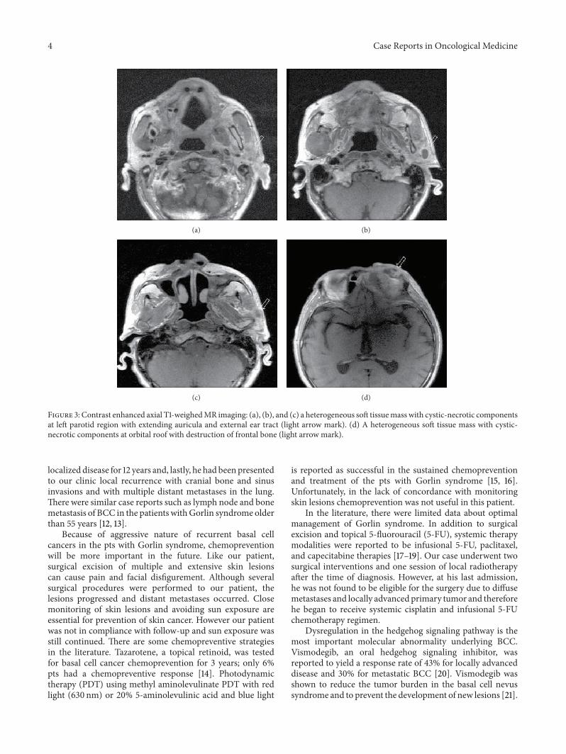
Other findings of Gorlin syndrome were investigated.The patient had hearing loss, gapped teeth, highly archedpalate, and frontal prominence. Brain CT of the patientshowed diffuse lamellar-slightly modular calcifications inthe falx cerebri and focal calcifications in the tentorium(Figure 5). Lung X-ray showed bifid costa anomaly in theright 1st costa. In both lungs, there were the opacities forthe lesions suggesting nodular metastasis with irregular thickcavitary appearance and irregular contours. In the left apex,opacity was increased and rough calcifications were presenton the pleural surfaces (Figure 6(a)). Due to clinical andradiological findings that accompanied BCC lesions, Gorlin-Goltz syndrome was considered.
Thoracic CT performed upon the observation of nodulessuggesting the metastasis on the lung X-ray revealed verynumerous cavitary masses with irregular contours and thickwalls and solid metastatic mass lesions of various sizeswith irregular contours that showed parenchymal spiculatedextensions in both lungs (Figures 6(b)–6(f)).
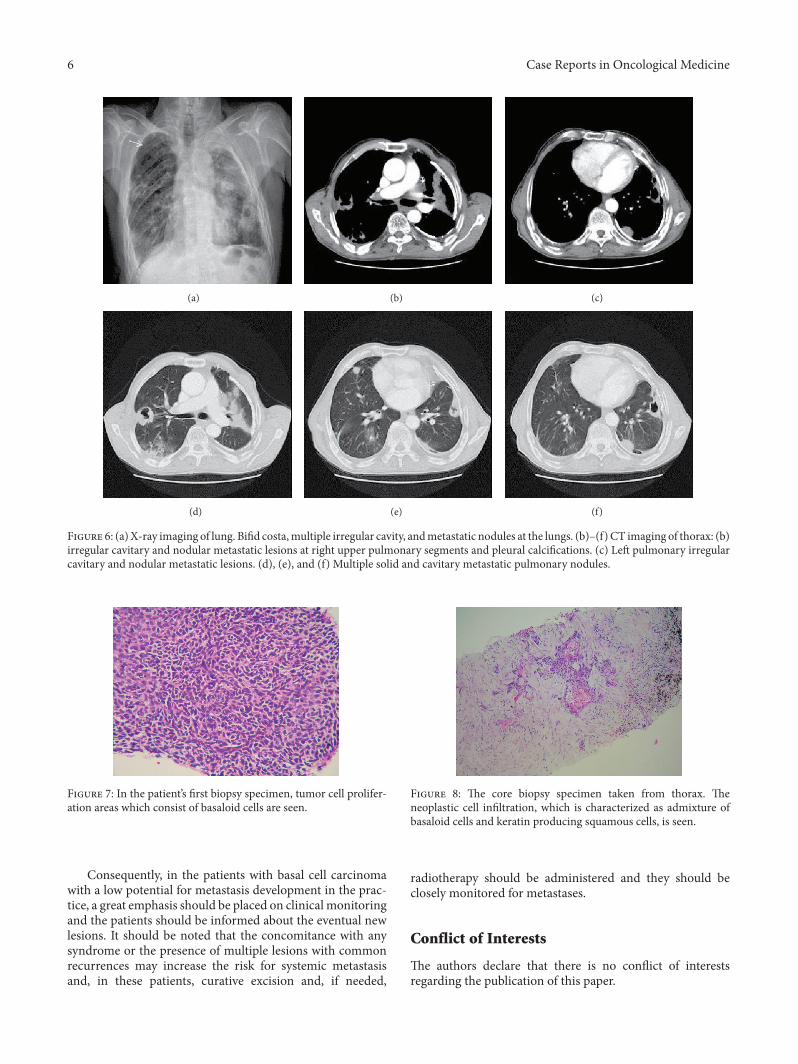
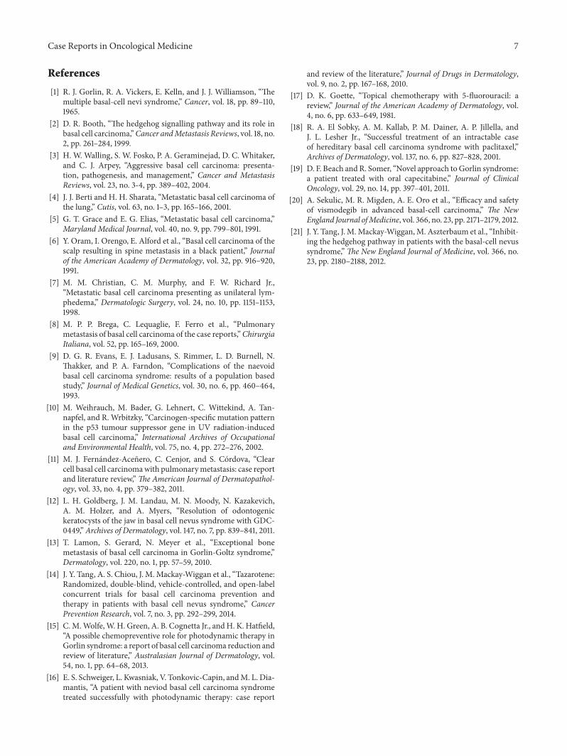
The first biopsy sample obtained from the mass locatedin the ethmoid sinus of the patient was examined (Figure 7).The first biopsy specimen showed a basaloid tumor cellproliferation, which was characterized as basaloid cells withrelatively small, round/oval hyperchromatic nuclei and nar-row cytoplasms. In some areas peripheral palisading nucleiand retraction artifacts were present, which support basalcell carcinoma diagnosis. Squamoid differentiation or keratinproduction findings were not seen in any area. Besides thenext biopsy specimen which was taken from thorax, thebasaloid cell proliferation is admixed with keratin producingsquamous cells and has an infiltrative pattern, which wassimilar to basosquamous carcinoma (Figure 8).
In the light of existing clinical and laboratory findings, thepatient was diagnosed with metastatic BCC accompanied byGorlin syndrome.
3. Discussion
In 1996, Gorlin andGoltz reported in twowomen a syndromecharacterized with multiple basal cell carcinoma, the cysts in

Case Reports in Oncological Medicine 3
(a) (b) (c)
(d) (e) (f)
Figure 2: Coronal T2-weighed MR imaging: (a) a lobular hypodense mass caused destruction and expansion at frontal sinus like a mucoselor keratocyst (arrowmark). Cystic mass at left orbital wall andmandibular corpus (arrowmarked). (b) Dens odontogenic keratocysts at rightmandibular corpus (arrow mark). (c), (d) Multiple cystic lesions with different size and dens at left ethmoidal sinuses (marked arrow). (e) Around, dense cystic lesionwith expansil regular contourwas observed in the rightmandibular ramus. (f) A soft tissuemasswith heterogeneousand irregular contour, which contained cystic-necrotic areas starting from left auricula, extended to external ear tract (arrow mark).
the chin and bifid costa, which they have called after theirname [1]. Gorlin-Goltz syndrome is also called as basal cellnevus syndrome, nevoid basal cell carcinoma syndrome, andWard syndrome [9]. Despite its autosomal dominant trait, thefamilial history of the syndrome is negative in 1/3 cases [9].
BCC accounts for 80% of all skin cancers [3]. This tumororiginates from pluripotential cells located in the basal layerof the epidermis and in the outer sheath of the hair follicle.Although it is seen in any area with the skin, 85% of thecases are seen in head-neck area. Cumulative exposure toultraviolet (UV) is known as the main causal factor in thedevelopment of basal cell carcinoma. UV was found tocause the development of basal cell carcinoma by leading tomutations in p53 and PTCH tumor suppressor genes [10].Furthermore, etiological factors of the BCC developmentinclude having a light colour of hair, eyes, and skin, sunburnduring the childhood and adolescence, ionized radiation,human papilloma virus infections, exposure to inorganicarsenic, infrared radiation, scar tissue, tattoo, sebaceous
nevus, trauma, chronic ulcers, and immunosuppression.Basal cell carcinomas of the skin are commonly seen andslowly growing tumours with rarely seen metastases.
Our patient is a case of basal cell carcinoma who hadthe diagnosis 12 years ago and who underwent 2 resectionsfor local recurrences during that 12-year period. He had UVexposure due to his occupation. In the systemic scans per-formed at the time of the last recurrence, the lesions primarilysuggested the presence of metastasis in bilateral lungs. Inthe literature, there are limited numbers of case reports withbasal cell carcinoma that did systemic metastases and thataccompanied to Gorlin syndrome. Fernandez-Acenero et al.reported a rare casewith clear cell BCCwithGorlin syndromeand lung metastasis and it was emphasized that the recur-rence andmetastatic potential are higher in such patients [11].
In the patients with Gorlin syndrome, there was a ten-dency of developmentmultiple skin cancer withmore aggres-sive clinical course characterised with local and systemicrecurrences. Our patient has been followed up with recurrent
4 Case Reports in Oncological Medicine
(a) (b)
(c) (d)
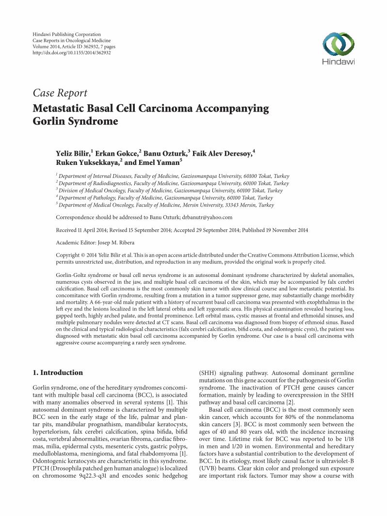
Figure 3: Contrast enhanced axial T1-weighedMR imaging: (a), (b), and (c) a heterogeneous soft tissuemass with cystic-necrotic componentsat left parotid region with extending auricula and external ear tract (light arrow mark). (d) A heterogeneous soft tissue mass with cystic-necrotic components at orbital roof with destruction of frontal bone (light arrow mark).
localized disease for 12 years and, lastly, he had beenpresentedto our clinic local recurrence with cranial bone and sinusinvasions and with multiple distant metastases in the lung.There were similar case reports such as lymph node and bonemetastasis of BCC in the patients withGorlin syndrome olderthan 55 years [12, 13].
Because of aggressive nature of recurrent basal cellcancers in the pts with Gorlin syndrome, chemopreventionwill be more important in the future. Like our patient,surgical excision of multiple and extensive skin lesionscan cause pain and facial disfigurement. Although severalsurgical procedures were performed to our patient, thelesions progressed and distant metastases occurred. Closemonitoring of skin lesions and avoiding sun exposure areessential for prevention of skin cancer. However our patientwas not in compliance with follow-up and sun exposure wasstill continued. There are some chemopreventive strategiesin the literature. Tazarotene, a topical retinoid, was testedfor basal cell cancer chemoprevention for 3 years; only 6%pts had a chemopreventive response [14]. Photodynamictherapy (PDT) using methyl aminolevulinate PDT with redlight (630 nm) or 20% 5-aminolevulinic acid and blue light
is reported as successful in the sustained chemopreventionand treatment of the pts with Gorlin syndrome [15, 16].Unfortunately, in the lack of concordance with monitoringskin lesions chemoprevention was not useful in this patient.
In the literature, there were limited data about optimalmanagement of Gorlin syndrome. In addition to surgicalexcision and topical 5-fluorouracil (5-FU), systemic therapymodalities were reported to be infusional 5-FU, paclitaxel,and capecitabine therapies [17–19]. Our case underwent twosurgical interventions and one session of local radiotherapyafter the time of diagnosis. However, at his last admission,he was not found to be eligible for the surgery due to diffusemetastases and locally advancedprimary tumor and thereforehe began to receive systemic cisplatin and infusional 5-FUchemotherapy regimen.
Dysregulation in the hedgehog signaling pathway is themost important molecular abnormality underlying BCC.Vismodegib, an oral hedgehog signaling inhibitor, wasreported to yield a response rate of 43% for locally advanceddisease and 30% for metastatic BCC [20]. Vismodegib wasshown to reduce the tumor burden in the basal cell nevussyndrome and to prevent the development of new lesions [21].

Case Reports in Oncological Medicine 5
(a) (b)
(c) (d)
Figure 4: Contrast enhanced CT images of neck: (a) cystic mass lesions with regular margins at mandibular corpus (arrow marked). (b) Around cystic lesion with lytic expansil regular contour at right ramus mandible and small cystic lesion at maxillar bone (marked arrow), aheterogeneous contrasted mass at left parotid region (light arrow marked). (c) Two lytic expansil cystic lesions at left ethmoidal cells. (d) Aheterogeneous contrasted mass with irregular margins at subcutaneus tissue of left orbital wall with destruction of frontal bone.
(a) (b)
Figure 5: CT imaging of brain: (a) calcifications at tentorium and mass at frontal sinus with bone destruction and expansion. (b) Diffusecalcification at falx cerebri.
6 Case Reports in Oncological Medicine
(a) (b) (c)
(d) (e) (f)
Figure 6: (a) X-ray imaging of lung. Bifid costa,multiple irregular cavity, andmetastatic nodules at the lungs. (b)–(f) CT imaging of thorax: (b)irregular cavitary and nodular metastatic lesions at right upper pulmonary segments and pleural calcifications. (c) Left pulmonary irregularcavitary and nodular metastatic lesions. (d), (e), and (f) Multiple solid and cavitary metastatic pulmonary nodules.
Figure 7: In the patient’s first biopsy specimen, tumor cell prolifer-ation areas which consist of basaloid cells are seen.
Consequently, in the patients with basal cell carcinomawith a low potential for metastasis development in the prac-tice, a great emphasis should be placed on clinical monitoringand the patients should be informed about the eventual newlesions. It should be noted that the concomitance with anysyndrome or the presence of multiple lesions with commonrecurrences may increase the risk for systemic metastasisand, in these patients, curative excision and, if needed,
Figure 8: The core biopsy specimen taken from thorax. Theneoplastic cell infiltration, which is characterized as admixture ofbasaloid cells and keratin producing squamous cells, is seen.
radiotherapy should be administered and they should beclosely monitored for metastases.
Conflict of Interests
The authors declare that there is no conflict of interestsregarding the publication of this paper.
Case Reports in Oncological Medicine 7
References
[1] R. J. Gorlin, R. A. Vickers, E. Kelln, and J. J. Williamson, “Themultiple basal-cell nevi syndrome,” Cancer, vol. 18, pp. 89–110,1965.
[2] D. R. Booth, “The hedgehog signalling pathway and its role inbasal cell carcinoma,”Cancer andMetastasis Reviews, vol. 18, no.2, pp. 261–284, 1999.
[3] H. W. Walling, S. W. Fosko, P. A. Geraminejad, D. C. Whitaker,and C. J. Arpey, “Aggressive basal cell carcinoma: presenta-tion, pathogenesis, and management,” Cancer and MetastasisReviews, vol. 23, no. 3-4, pp. 389–402, 2004.
[4] J. J. Berti and H. H. Sharata, “Metastatic basal cell carcinoma ofthe lung,” Cutis, vol. 63, no. 1–3, pp. 165–166, 2001.
[5] G. T. Grace and E. G. Elias, “Metastatic basal cell carcinoma,”Maryland Medical Journal, vol. 40, no. 9, pp. 799–801, 1991.
[6] Y. Oram, I. Orengo, E. Alford et al., “Basal cell carcinoma of thescalp resulting in spine metastasis in a black patient,” Journalof the American Academy of Dermatology, vol. 32, pp. 916–920,1991.
[7] M. M. Christian, C. M. Murphy, and F. W. Richard Jr.,“Metastatic basal cell carcinoma presenting as unilateral lym-phedema,” Dermatologic Surgery, vol. 24, no. 10, pp. 1151–1153,1998.
[8] M. P. P. Brega, C. Lequaglie, F. Ferro et al., “Pulmonarymetastasis of basal cell carcinoma of the case reports,”ChirurgiaItaliana, vol. 52, pp. 165–169, 2000.
[9] D. G. R. Evans, E. J. Ladusans, S. Rimmer, L. D. Burnell, N.Thakker, and P. A. Farndon, “Complications of the naevoidbasal cell carcinoma syndrome: results of a population basedstudy,” Journal of Medical Genetics, vol. 30, no. 6, pp. 460–464,1993.
[10] M. Weihrauch, M. Bader, G. Lehnert, C. Wittekind, A. Tan-napfel, and R. Wrbitzky, “Carcinogen-specific mutation patternin the p53 tumour suppressor gene in UV radiation-inducedbasal cell carcinoma,” International Archives of Occupationaland Environmental Health, vol. 75, no. 4, pp. 272–276, 2002.
[11] M. J. Fernandez-Acenero, C. Cenjor, and S. Cordova, “Clearcell basal cell carcinomawith pulmonarymetastasis: case reportand literature review,”The American Journal of Dermatopathol-ogy, vol. 33, no. 4, pp. 379–382, 2011.
[12] L. H. Goldberg, J. M. Landau, M. N. Moody, N. Kazakevich,A. M. Holzer, and A. Myers, “Resolution of odontogenickeratocysts of the jaw in basal cell nevus syndrome with GDC-0449,” Archives of Dermatology, vol. 147, no. 7, pp. 839–841, 2011.
[13] T. Lamon, S. Gerard, N. Meyer et al., “Exceptional bonemetastasis of basal cell carcinoma in Gorlin-Goltz syndrome,”Dermatology, vol. 220, no. 1, pp. 57–59, 2010.
[14] J. Y. Tang, A. S. Chiou, J. M.Mackay-Wiggan et al., “Tazarotene:Randomized, double-blind, vehicle-controlled, and open-labelconcurrent trials for basal cell carcinoma prevention andtherapy in patients with basal cell nevus syndrome,” CancerPrevention Research, vol. 7, no. 3, pp. 292–299, 2014.
[15] C.M.Wolfe,W. H. Green, A. B. Cognetta Jr., andH. K. Hatfield,“A possible chemopreventive role for photodynamic therapy inGorlin syndrome: a report of basal cell carcinoma reduction andreview of literature,” Australasian Journal of Dermatology, vol.54, no. 1, pp. 64–68, 2013.
[16] E. S. Schweiger, L. Kwasniak, V. Tonkovic-Capin, andM. L. Dia-mantis, “A patient with neviod basal cell carcinoma syndrometreated successfully with photodynamic therapy: case report
and review of the literature,” Journal of Drugs in Dermatology,vol. 9, no. 2, pp. 167–168, 2010.
[17] D. K. Goette, “Topical chemotherapy with 5-fluorouracil: areview,” Journal of the American Academy of Dermatology, vol.4, no. 6, pp. 633–649, 1981.
[18] R. A. El Sobky, A. M. Kallab, P. M. Dainer, A. P. Jillella, andJ. L. Lesher Jr., “Successful treatment of an intractable caseof hereditary basal cell carcinoma syndrome with paclitaxel,”Archives of Dermatology, vol. 137, no. 6, pp. 827–828, 2001.
[19] D. F. Beach and R. Somer, “Novel approach to Gorlin syndrome:a patient treated with oral capecitabine,” Journal of ClinicalOncology, vol. 29, no. 14, pp. 397–401, 2011.
[20] A. Sekulic, M. R. Migden, A. E. Oro et al., “Efficacy and safetyof vismodegib in advanced basal-cell carcinoma,” The NewEngland Journal ofMedicine, vol. 366, no. 23, pp. 2171–2179, 2012.
[21] J. Y. Tang, J. M.Mackay-Wiggan,M. Aszterbaum et al., “Inhibit-ing the hedgehog pathway in patients with the basal-cell nevussyndrome,” The New England Journal of Medicine, vol. 366, no.23, pp. 2180–2188, 2012.

Submit your manuscripts athttp://www.hindawi.com
Stem CellsInternational
Hindawi Publishing Corporationhttp://www.hindawi.com Volume 2014
Hindawi Publishing Corporationhttp://www.hindawi.com Volume 2014
MEDIATORSINFLAMMATION
of
Hindawi Publishing Corporationhttp://www.hindawi.com Volume 2014
Behavioural Neurology
EndocrinologyInternational Journal of
Hindawi Publishing Corporationhttp://www.hindawi.com Volume 2014
Hindawi Publishing Corporationhttp://www.hindawi.com Volume 2014
Disease Markers
Hindawi Publishing Corporationhttp://www.hindawi.com Volume 2014
BioMed Research International
OncologyJournal of
Hindawi Publishing Corporationhttp://www.hindawi.com Volume 2014
Hindawi Publishing Corporationhttp://www.hindawi.com Volume 2014
Oxidative Medicine and Cellular Longevity
Hindawi Publishing Corporationhttp://www.hindawi.com Volume 2014
PPAR Research
The Scientific World JournalHindawi Publishing Corporation http://www.hindawi.com Volume 2014
Immunology ResearchHindawi Publishing Corporationhttp://www.hindawi.com Volume 2014
Journal of
ObesityJournal of
Hindawi Publishing Corporationhttp://www.hindawi.com Volume 2014
Hindawi Publishing Corporationhttp://www.hindawi.com Volume 2014
Computational and Mathematical Methods in Medicine
OphthalmologyJournal of
Hindawi Publishing Corporationhttp://www.hindawi.com Volume 2014
Diabetes ResearchJournal of
Hindawi Publishing Corporationhttp://www.hindawi.com Volume 2014
Hindawi Publishing Corporationhttp://www.hindawi.com Volume 2014
Research and TreatmentAIDS
Hindawi Publishing Corporationhttp://www.hindawi.com Volume 2014
Gastroenterology Research and Practice
Hindawi Publishing Corporationhttp://www.hindawi.com Volume 2014
Parkinson’s Disease
Evidence-Based Complementary and Alternative Medicine
Volume 2014Hindawi Publishing Corporationhttp://www.hindawi.com