case report autoresuscitation: a case and discussion of...
TRANSCRIPT
Case ReportAutoresuscitation: A Case and Discussion ofthe Lazarus Phenomenon
Kjartan Eskjaer Hannig,1 Rasmus Wulff Hauritz,1 and Erik Lerkevang Grove2
1Department of Anaesthesiology, Kolding Hospital, Skovvangen 2-8, 6000 Kolding, Denmark2Department of Cardiology, Aarhus University Hospital, Palle Juul-Jensens Boulevard 99, 8200 Aarhus, Denmark
Correspondence should be addressed to Erik Lerkevang Grove; [email protected]
Received 12 April 2015; Accepted 13 May 2015
Academic Editor: William J. Brady
Copyright © 2015 Kjartan Eskjaer Hannig et al. This is an open access article distributed under the Creative Commons AttributionLicense, which permits unrestricted use, distribution, and reproduction in any medium, provided the original work is properlycited.
Lazarus phenomenon or autoresuscitation is a very rare condition defined as delayed unassisted return of spontaneous circulationafter cessation of cardiopulmonary resuscitation. Based on a casewith a 67-year-oldmalewho came back to life after discontinuationof cardiopulmonary resuscitation, we discuss the background and possible countermeasures related to the Lazarus phenomenon.
1. Introduction
Lazarus phenomenon or autoresuscitation (AR) is a very rarecondition defined as delayed unassisted return of sponta-neous circulation (ROSC) after cessation of cardiopulmonaryresuscitation (CPR) [1, 2]. After being first reported by Linkoet al. in 1982 [3], it was later termed the “Lazarus phenome-non” by Bray Jr. in 1993 [4] after the biblical figure Lazarus,whom Jesus supposedly resurrected four days after his deathand burial (Gospel of John Chapter 11: 1–44).
The occurrence of this phenomenon may be widelyunderreported as illustrated by the fact that almost 50% ofFrench emergency physicians claim to have encountered ARin clinical practice [5] and by the statement by Dhanani et al.that more than one-third of Canadian intensivists have seenat least one case of AR [6]. The true incidence remainsunknown.
2. Case Report
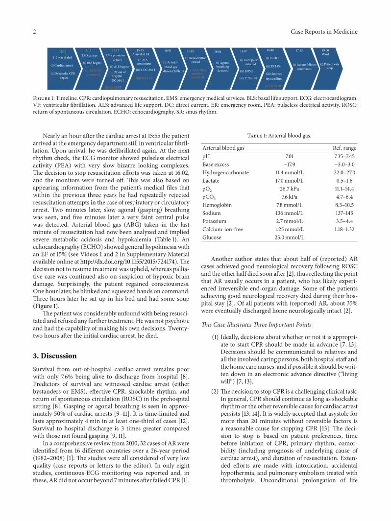
A 67-year-old Caucasian male collapsed with cardiac arrestoutside his home (Figure 1). A nurse, who was caring for thepatient on a daily basis, coincidently passed by and immedi-ately initiated CPR. The emergency medical services (EMS)were called at 15:10 hours and an ambulance arrived at 15:13and the emergency physician at 15:15. On arrival, the initial
rhythmwas ventricular fibrillation. Resuscitation was perfor-med according to the advanced life support (ALS) algorithm[7] including chest compression, ventilation, intubation inthe field, a total of 20 defibrillations, and standard drug admi-nistration, including epinephrine and amiodarone.
The patient had massive comorbidity on the basis of uni-versal atherosclerosis due to 31 pack years of smoking, severehypertension, hypercholesterolemia, and heredity. In 2004,bilateral in situ bypass surgery optimised blood flow to hislower extremities. He was also diagnosed with left ventricularhypertrophy and systolic and diastolic dysfunction with anejection fraction (EF) of 40%. In 2006, the patient underwentcoronary artery bypass graft surgery, complicated by post-operative ventilator therapy for one month, middle cerebralartery infarction, sternal infection, and organic delirium,which elapsed into a psychotic episode and severe depression.In the last three years, the patient had been depending onhaemodialysis three times a week. Furthermore, the medicalhistory included severe chronic obstructive pulmonary dis-ease with a forced expiratory vital capacity of 45%, abdominalaortic aneurism of 5.1 cm, paroxysmal atrial fibrillation,and chronic musculoskeletal pain due to cervical and lum-bal spinal stenosis. His medication comprised carvedilol,ramipril, simvastatin, warfarin, inhaled salmeterol/flutica-sonpropionat, inhaled tiotropium, pantoprazole, paraceta-mol, vitamins and, related to his hemodialysis, alfacalcidol,erythropoietin, phosphate-binding drug, and furosemide.
Hindawi Publishing CorporationCase Reports in MedicineVolume 2015, Article ID 724174, 5 pageshttp://dx.doi.org/10.1155/2015/724174
2 Case Reports in Medicine
15:10 15:13EMS arrives
15:15EMS physician
(i) ALS begins (ii) 20 out of
hospital DC 360 J
(iii) ECG VF
15:55Arrival at ER
(ii) 1 DC 360 J
(iii) ECG VF
16:01 16:02 16:04
(i) Agonal
16:07
(i) Faint pulsedetected
(ii) ROSC
112 was dialed
(i) Cardiac arrest
begins
(i) BLS begins
detected
arrives (i) ALS continuous (i) Arterial
drawn blood gas
(i) Resuscitation ceased
(ii) ECG PEA,monitor
turned off
breathing detected
16:40
(i) ECHO
(ii) EF 15%
(iii) Stunnedmyocardium
(iv) ECG SR
17:15
(i) Patient followscommands
19:00Ward
(i) Patient eatssoup
(ii) Bystander CPR(ii) ECG VF
(iii) P 70–100
(Table 1)
Figure 1: Timeline. CPR: cardiopulmonary resuscitation. EMS: emergencymedical services. BLS: basal life support. ECG: electrocardiogram.VF: ventricular fibrillation. ALS: advanced life support. DC: direct current. ER: emergency room. PEA: pulseless electrical activity. ROSC:return of spontaneous circulation. ECHO: echocardiography. SR: sinus rhythm.
Nearly an hour after the cardiac arrest at 15:55 the patientarrived at the emergency department still in ventricular fibril-lation. Upon arrival, he was defibrillated again. At the nextrhythm check, the ECG monitor showed pulseless electricalactivity (PEA) with very slow bizarre looking complexes.The decision to stop resuscitation efforts was taken at 16.02,and the monitors were turned off. This was also based onappearing information from the patient’s medical files thatwithin the previous three years he had repeatedly rejectedresuscitation attempts in the case of respiratory or circulatoryarrest. Two minutes later, slow agonal (gasping) breathingwas seen, and five minutes later a very faint central pulsewas detected. Arterial blood gas (ABG) taken in the lastminute of resuscitation had now been analyzed and impliedsevere metabolic acidosis and hypokalemia (Table 1). Anechocardiography (ECHO) showed general hypokinesia withan EF of 15% (see Videos 1 and 2 in Supplementary Materialavailable online at http://dx.doi.org/10.1155/2015/724174).Thedecision not to resume treatment was upheld, whereas pallia-tive care was continued also on suspicion of hypoxic braindamage. Surprisingly, the patient regained consciousness.One hour later, he blinked and squeezed hands on command.Three hours later he sat up in his bed and had some soup(Figure 1).
The patient was considerably unfoundwith being resusci-tated and refused any further treatment. Hewas not psychoticand had the capability of making his own decisions. Twenty-two hours after the initial cardiac arrest, he died.
3. Discussion
Survival from out-of-hospital cardiac arrest remains poorwith only 7.6% being alive to discharge from hospital [8].Predictors of survival are witnessed cardiac arrest (eitherbystanders or EMS), effective CPR, shockable rhythm, andreturn of spontaneous circulation (ROSC) in the prehospitalsetting [8]. Gasping or agonal breathing is seen in approx-imately 50% of cardiac arrests [9–11]. It is time-limited andlasts approximately 4min in at least one-third of cases [12].Survival to hospital discharge is 3 times greater comparedwith those not found gasping [9, 11].
In a comprehensive review from 2010, 32 cases of ARwereidentified from 16 different countries over a 26-year period(1982–2008) [1]. The studies were all considered of very lowquality (case reports or letters to the editor). In only eightstudies, continuous ECG monitoring was reported and, inthese, ARdid not occur beyond 7minutes after failedCPR [1].
Table 1: Arterial blood gas.
Arterial blood gas Ref. rangepH 7.01 7.35–7.45Base excess −17.9 −3.0–3.0Hydrogencarbonate 11.4mmol/L 22.0–27.0Lactate 17.0mmol/L 0.5–1.6pO2 26.7 kPa 11.1–14.4pCO2 7.6 kPa 4.7–6.4Hemoglobin 7.8mmol/L 8.3–10.5Sodium 136mmol/L 137–145Potassium 2.7mmol/L 3.5–4.4Calcium-ion-free 1.25mmol/L 1.18–1.32Glucose 25.0mmol/L
Another author states that about half of (reported) ARcases achieved good neurological recovery following ROSCand the other half died soon after [2], thus reflecting the pointthat AR usually occurs in a patient, who has likely experi-enced irreversible end-organ damage. Some of the patientsachieving good neurological recovery died during their hos-pital stay [2]. Of all patients with (reported) AR, about 35%were eventually discharged home neurologically intact [2].
This Case Illustrates Three Important Points
(1) Ideally, decisions about whether or not it is appropri-ate to start CPR should be made in advance [7, 13].Decisions should be communicated to relatives andall the involved caring persons, both hospital staff andthe home care nurses, and if possible it should bewrit-ten down in an electronic advance directive (“livingwill”) [7, 13].
(2) The decision to stop CPR is a challenging clinical task.In general, CPR should continue as long as shockablerhythm or the other reversible cause for cardiac arrestpersists [13, 14]. It is widely accepted that asystole formore than 20 minutes without reversible factors isa reasonable cause for stopping CPR [13]. The deci-sion to stop is based on patient preferences, timebefore initiation of CPR, primary rhythm, comor-bidity (including prognosis of underlying cause ofcardiac arrest), and duration of resuscitation. Exten-ded efforts are made with intoxication, accidentalhypothermia, and pulmonary embolism treated withthrombolysis. Unconditional prolongation of life
Case Reports in Medicine 3
(i) It is a very rare condition.(ii) It is presumably widely underreported.(iii) The precise pathophysiological mechanisms are unknown, but they may include
hyperinflation,myocardial stunning,hyperkalaemia,delayed action of drugs,countershock asystole,unobserved minimal vital signs.
(iv) There are countermeasures as follows:ECG monitoring 10min after prolonged cardiopulmonary resuscitation,avoiding hyperventilation,arterial blood gas as soon as possible,echocardiography and capnography.
Box 1: Facts about the Lazarus Phenomenon.
should not be the goal in itself, rather to achieve asufficient quality of life [13].
(3) An example of the Lazarus phenomenon with itsbackground and possible countermeasures is dis-cussed below (summarized in Box 1).
The pathophysiological mechanisms for AR are poorlyunderstood [1, 2, 15]. Hyperinflation (in obstructive lung dis-ease), myocardial stunning (in acute myocardial infarction),hyperkalaemia (especially in renal failure), delayed action ofdrugs, countershock asystole, and unobserved minimal vitalsigns amongst others have been considered to be the mostcommon mechanisms [1, 2, 15].
Unrecognized dynamic lung hyperinflation can theoreti-cally occur in all patients but is especially seen in patientswithobstructive lung disease (chronic obstructive pulmonarydisease and asthma). It is probably due to rapid manualventilation with inadequate time for exhalation, leading toelevated end-expiratory pressure (auto-PEEP) [2]. Auto-PEEP is caused by air trapping, that is, air entering the lungsand being unable to escape.This gradual increase of intratho-racic pressure leads to decreased venous return to the heart(preload) and subsequently low cardiac output and cardiacarrest, even in the presence of a perfusable cardiac rhythm[1, 2, 7]. The physiology of severe auto-PEEP is similar topericardial tamponade [2]. Decreased venous returnmay alsodelay drug delivery to the central circulation, impeding med-ication action during resuscitation.
Countermeasures. Avoid hyperinflation by bag-mask ventila-tion using an inspiratory time of about 1 second and give onlyenough volume to produce a visible normal chest rise [7].If the patient is intubated and connected to a ventilator, thetidal volume is set at 6mL/kg ideal bodyweight at 10 breaths/minute [7]. If hyperinflation is suspected, intermittent dis-connection of the tracheal tube for 10 seconds [15]may relieveair trapping permitting return of venous flow and spon-taneous circulation [7]. Dynamic hyperinflation increasestransthoracic impedance and with shockable rhythms highershock energies may be considered [7]. Always consider
tension pneumothorax or bilateral pneumothorax in asthma-related cardiac arrest. In skilled hands, lung ultrasound is afaster and more sensitive diagnostic test than chest X-ray [7].
Myocardial stunning is due to ischaemia most oftenbecause of infarction [2]. After brief periods of myocardialischaemia, prolonged myocardial dysfunction can occur, fol-lowed by gradual recovery and improvement in cardiac out-put typically within 2-3 days [7, 15].
Countermeasure. Percutaneous coronary intervention (PCI)should be conducted as soon as possible, when appropriate.
Hyperkalemia prevents adequate efflux of potassium,and hence the resting membrane potential of the myocytesdecreases, leaving themyocardium depolarized and themyo-cytes refractory to further stimulation (unexcitable).The car-diac conduction is slowed and the heart stops in diastolerefractory to resuscitation. Voelckel hypothesized this as amechanism of delayed ROSC [16]. The myocardium is alsoextremely sensitive to hypokalemia, which alters cardiactissue excitability and conduction, andmay inducemalignantventricular arrhythmias, resulting in cardiac arrest.
Countermeasure. ABG taken early (or even in the prehospitalsetting) can confirm suspicion.
Delayed delivery and action of administered medications(like epinephrine) can occur, when drugs are administeredthrough a peripheral vein, as central delivery is slow due toimpaired venous return. Presumably, some older cases of ARcan be attributed to this mechanism, especially due to escala-ting doses of epinephrine used according to historic ALSguidelines [2].
Countermeasure. Use intraosseous line or central line ifpresent.
Transient asystole or PEA following defibrillation of pro-longed VF is a well-known phenomenon and occurs in about60% of patients [2, 17]. Surprisingly, it has a worse prognosisthan primary asystole or PEA. This may be related to directmyocardial injury due to electrical current and because thefibrillating myocardium requires more oxygen and fasterdepletes high-energy phosphate stores [17].
4 Case Reports in Medicine
Countermeasure. Use biphasic instead of monophasic defib-rillators, using only energy levels specified by the manufac-turer.
Unobserved minimal vital signs likely represent anotherexplanation, when the circulation is present again. Pseudo-PEA is the clinical situation in which contractile activity isoccurring, yet it is of minimal magnitude. Often, pulse pal-pation can be challenging or nearly impossible. In such situa-tions, ECHO easily shows if cardiac contractions are present.
Countermeasure. Use ECHO when available. Consider usingcapnography during CPR, which serves three purposes.Firstly, it confirms correct placement of the endotrachealtube, when consistent (blunted) waveforms are presentbeyond the first 6-7 respirations, whereas a “flat capnogram”is indicative of accidental oesophageal intubation [7, 18, 19].Secondly, it reflects pulmonary blood flow and thus cardiacoutput, providing the opportunity of monitoring the efficacyof chest compression [7, 18, 19]. In healthy individuals,the normal range for end-tidal CO
2is 4.65–6.0 kPa (35–
45mmHg). During CPR, an optimal target for end-tidalCO2has not been established [7], but values as high as
possible above 1.35–2.65 kPa (10–20mmHg) are desirable[18]. According to Kodali et al, the initial end-tidal CO
2level
under CPR may divide patients into those likely to achieveROSC (values > 1.35 kPa = 10mmHg) and those not likely toachieve ROSC (values < 1.35 kPa = 10mmHg) [7, 18].Thirdly,a significant abrupt and sustained increase in end-tidal CO
2
during CPR from initial baseline (e.g., increase exceeding1.35 kPa = 10mmHg) may be seen as the first indicator ofROSC. This may precede a palpable pulse [2, 7, 15, 18, 19].
In our case, hyperventilation may be a possible explana-tion, since the patient’s pCO
2was about 10 kPa (75mmHg)
normally and measured under CPR 7.6 kPa (57mmHg),which may be considered as relative hypocapnia. Hypokale-mia was present and must have been worse at the time ofthe cardiac arrest, which was followed by nearly one hour ofCPR causing anaerobe metabolism. A possible explanationmay be that epinephrine stimulates the sodium potassiumpump (Na-K ATPase), causing influx of potassium into thecells. Delayed action of drugs may have been important aswell, since the patient arrived with a small bore peripheralvenous line, which continued to be used during in-hospitalresuscitation. Probably, AR in our case was explained byseveral concurring mechanisms.
Since death is not an event but a process [2], according tothe ALS guidelines, the patient must be observed for a mini-mum of 5 minutes before confirming death [7]. Since mostcases of AR occur within tenminutes, it should be consideredto extend this period to 10 minutes with ECG monitoringbefore certifying death or informing the family [2, 3, 15]. Theabsence of mechanical cardiac function can be confirmed byabsence of central pulse on palpation and absence of heartsounds on auscultation [20]. Supplementary, one or more ofthe following clinical findings can be used: asystole on ECG,absence of pulsatile flow on intra-arterial line, and absenceof contractile activity on ECHO [20]. After 5−10 minutes ofcontinued cardiorespiratory arrest, the absence of pupillaryresponse to light, the absence of corneal reflexes, and absence
of any motor response to supraorbital pressure should beconfirmed [7, 20, 21].
Conflict of Interests
The authors declare that they have no conflict of interests.
References
[1] K. Hornby, L. Hornby, and S. D. Shemie, “A systematic reviewof autoresuscitation after cardiac arrest,” Critical Care Medicine,vol. 38, no. 5, pp. 1246–1253, 2010.
[2] V. Adhiyaman, S. Adhiyaman, and R. Sundaram, “The Lazarusphenomenon,” Journal of the Royal Society of Medicine, vol. 100,no. 12, pp. 552–557, 2007.
[3] K. Linko, P. Honkavaara, and M. Salmenpera, “Recovery afterdiscontinued cardiopulmonary resuscitation,” The Lancet, vol.1, no. 8263, pp. 106–107, 1982.
[4] J. G. Bray Jr., “The Lazarus phenomenon revisited,” Anesthesiol-ogy, vol. 78, no. 5, p. 991, 1993.
[5] D. Gerard, J. Vaux, T. Boche, C. Chollet-Xemard, and J.Marty, “Lazarus phenomenon: knowledge, attitude and prac-tice,” Resuscitation, vol. 84, no. 12, p. E153, 2013.
[6] S. Dhanani, R. Ward, L. Hornby, N. J. Barrowman, K. Hornby,and S. D. Shemie, “Survey of determination of death after car-diac arrest by intensive care physicians,” Critical Care Medicine,vol. 40, no. 5, pp. 1449–1455, 2012.
[7] C. D. Deakin, J. P. Nolan, J. Soar et al., “European resuscitationcouncil guidelines for resuscitation 2010. Section 4. Adultadvanced life support,” Resuscitation, vol. 81, no. 10, pp. 1305–1352, 2010.
[8] C. Sasson, M. A. M. Rogers, J. Dahl, and A. L. Kellermann,“Predictors of survival from out-of-hospital cardiac arrest a sys-tematic review and meta-analysis,” Circulation: CardiovascularQuality and Outcomes, vol. 3, no. 1, pp. 63–81, 2010.
[9] J. J. Clark, M. P. Larsen, L. L. Culley, J. R. Graves, and M. S.Eisenberg, “Incidence of agonal respirations in sudden cardiacarrest,” Annals of Emergency Medicine, vol. 21, no. 12, pp. 1464–1467, 1992.
[10] A. Bang, J. Herlitz, and S. Martinell, “Interaction betweenemergency medical dispatcher and caller in suspected out-of-hospital cardiac arrest calls with focus on agonal breathing.A review of 100 tape recordings of true cardiac arrest cases,”Resuscitation, vol. 56, no. 1, pp. 25–34, 2003.
[11] B. J. Bobrow, M. Zuercher, G. A. Ewy et al., “Gasping duringcardiac arrest in humans is frequent and associated withimproved survival,” Circulation, vol. 118, no. 24, pp. 2550–2554,2008.
[12] M. S. Eisenberg, “Incidence and significance of gasping oragonal respirations in cardiac arrest patients,” Current Opinionin Critical Care, vol. 12, no. 3, pp. 204–206, 2006.
[13] F. K. Lippert, V. Raffay,M.Georgiou, P.A. Steen, andL. Bossaert,“European Resuscitation Council Guidelines for Resuscitation2010. Section 10. The ethics of resuscitation and end-of-lifedecisions,” Resuscitation, vol. 81, no. 10, pp. 1445–1451, 2010.
[14] K. Adelborg, B. Løfgren, and E. L. Grove, “Cardiopulmonaryresuscitation should continue as long as shockable cardiacrhythms persist,” Ugeskrift for Læger, vol. 174, no. 34, pp. 1905–1906, 2012.

Case Reports in Medicine 5
[15] V. Adhiyaman and R. Sundaram, “The Lazarus phenomenon,”The Journal of the Royal College of Physicians of Edinburgh, vol.32, pp. 9–13, 2002.
[16] W. Voelckel and G. Kroesen, “Unexpected return of cardiacaction after termination of cardiopulmonary resuscitation,”Resuscitation, vol. 32, no. 1, pp. 27–29, 1996.
[17] J. T. Niemann, S. J. Stratton, B. Cruz, and R. J. Lewis, “Outcomeof out-of-hospital postcountershock asystole and pulseless elec-trical activity versus primary asystole and pulseless electricalactivity,” Critical Care Medicine, vol. 29, no. 12, pp. 2366–2370,2001.
[18] B. S. Kodali and R. D. Urman, “Capnography during cardiopul-monary resuscitation: current evidence and future directions,”Journal of Emergencies, Trauma, and Shock, vol. 7, no. 4, pp. 332–340, 2014.
[19] K. R. Ward and D. M. Yealy, “End-tidal carbon dioxide mon-itoring in emergency medicine, part 2: clinical applications,”Academic Emergency Medicine, vol. 5, no. 6, pp. 637–646, 1998.
[20] Academy of Medical Royal Colleges, A Code of Practice forthe Diagnosis and Confirmation of Death, 2008, http://www.aomrc.org.uk.
[21] S. D. Shemie, L. Hornby, A. Baker et al., “International guidelinedevelopment for the determination of death,” Intensive CareMedicine, vol. 40, no. 6, pp. 788–797, 2014.

Submit your manuscripts athttp://www.hindawi.com
Stem CellsInternational
Hindawi Publishing Corporationhttp://www.hindawi.com Volume 2014
Hindawi Publishing Corporationhttp://www.hindawi.com Volume 2014
MEDIATORSINFLAMMATION
of
Hindawi Publishing Corporationhttp://www.hindawi.com Volume 2014
Behavioural Neurology
EndocrinologyInternational Journal of
Hindawi Publishing Corporationhttp://www.hindawi.com Volume 2014
Hindawi Publishing Corporationhttp://www.hindawi.com Volume 2014
Disease Markers
Hindawi Publishing Corporationhttp://www.hindawi.com Volume 2014
BioMed Research International
OncologyJournal of
Hindawi Publishing Corporationhttp://www.hindawi.com Volume 2014
Hindawi Publishing Corporationhttp://www.hindawi.com Volume 2014
Oxidative Medicine and Cellular Longevity
Hindawi Publishing Corporationhttp://www.hindawi.com Volume 2014
PPAR Research
The Scientific World JournalHindawi Publishing Corporation http://www.hindawi.com Volume 2014
Immunology ResearchHindawi Publishing Corporationhttp://www.hindawi.com Volume 2014
Journal of
ObesityJournal of
Hindawi Publishing Corporationhttp://www.hindawi.com Volume 2014
Hindawi Publishing Corporationhttp://www.hindawi.com Volume 2014
Computational and Mathematical Methods in Medicine
OphthalmologyJournal of
Hindawi Publishing Corporationhttp://www.hindawi.com Volume 2014
Diabetes ResearchJournal of
Hindawi Publishing Corporationhttp://www.hindawi.com Volume 2014
Hindawi Publishing Corporationhttp://www.hindawi.com Volume 2014
Research and TreatmentAIDS
Hindawi Publishing Corporationhttp://www.hindawi.com Volume 2014
Gastroenterology Research and Practice
Hindawi Publishing Corporationhttp://www.hindawi.com Volume 2014
Parkinson’s Disease
Evidence-Based Complementary and Alternative Medicine
Volume 2014Hindawi Publishing Corporationhttp://www.hindawi.com