case report a puzzle of vestibular physiology in a meniere...
TRANSCRIPT

Case ReportA Puzzle of Vestibular Physiology in a Meniere’s DiseaseAcute Attack
Marta Martinez-Lopez, Raquel Manrique-Huarte, and Nicolas Perez-Fernandez
Department of Otorhinolaryngology, Clinica Universidad de Navarra, University of Navarra,Avenida Pıo XII 36, 31008 Pamplona, Spain
Correspondence should be addressed to Nicolas Perez-Fernandez; [email protected]
Received 10 February 2015; Accepted 24 May 2015
Academic Editor: Guangwei Zhou
Copyright © 2015 Marta Martinez-Lopez et al. This is an open access article distributed under the Creative Commons AttributionLicense, which permits unrestricted use, distribution, and reproduction in any medium, provided the original work is properlycited.
The aimof this paper is to present for the first time the functional evaluation of each of the vestibular receptors in the six semicircularcanals in a patient diagnosed with Meniere’s disease during an acute attack. A 54-year-old lady was diagnosed with left Meniere’sdisease who during her regular clinic review suffers an acute attack of vertigo, with fullness and an increase of tinnitus in her leftear. Spontaneous nystagmus and the results in the video head-impulse test (vHIT) are shown before, during, and after the attack.Nystagmus was initially left beating and a few minutes later an upbeat component was added. No skew deviation was observed. Adecrease in the gain of the vestibuloocular reflex (VOR) and the presence of overt saccades were observed when the stimuli were inthe plane of the left superior semicircular canal. At the end of the crisis nystagmus decreased and vestibuloocular reflex returnedto almost normal. A review of the different possibilities to explain these findings points to a hypothetical utricular damage.
1. Introduction
Meniere’s disease (MD) crises are usually managed bypatients at home or at a primary care facility.The characteris-tic features of the disease occur andusually hearing loss is bestdefined and sometimes audiometry is performed to confirmthe deterioration in hearing loss.
Case reports of vestibular signs are infrequent becauseof the limited availability of equipment at emergency rooms.All of them provide interesting insight into the patho-physiology of the disorder by describing findings in termsof spontaneous and post-head-shake or vibration nystag-mus, caloric test [1], and more recently head-impulse test(HIT) at bedside or assisted with video (vHIT) for theassessment of semicircular canal function [2] and vestibularevoked myogenic potentials (VEMP) for otolithic function[3].
We present a patient in whom an acute spell of vertigohas been seen while being at the hospital and in whichthe spontaneous nystagmus and the assessment of all 6semicircular canals were possible.
2. Case Report
A 54-year-old woman was diagnosed with MD in her leftear. Vertigo spells begun 30 years ago, but after three yearsof activity the disease entered in a quiescent phase. In March2013 vertigo recurred and since then has been unresponsiveto medical treatment. When seen for the first time in July2014, the number of typical vertigo spells in the 6 monthsprevious was 12; also 4 additional ones were of the Tumarkintype, and her functional level score [4] was 4. PTA in theright ear was 5 dBHL and in her left ear 28 dBHL. CTscan and MRI were normal. After complete evaluation shewas treated with intratympanic gentamicin ITG: August 5thand 11th and September 24th. It is interesting to mentionthat the external auditory canal is markedly anfractuousand the approach to the tympanic membrane is difficult;as such myringotomy was done in the anterior part of theinferior hemotympanum and a curved needle was used todeliver the gentamicin close to the round window niche.The treatment provided 1 month without any symptom andgood equilibrium without increased hearing loss. Since then,
Hindawi Publishing CorporationCase Reports in OtolaryngologyVolume 2015, Article ID 460757, 5 pageshttp://dx.doi.org/10.1155/2015/460757
2 Case Reports in Otolaryngology
Before vertigo crisisN1P1(500) [8.2]
0.00 5.00 10.00 15.00 20.00Time (ms)
AsymmetryAnterior: 22%Lateral: 19%
Posterior: 13%
Time (ms)
N1
P1(500) [7.1]
0.00 5.00 10.00 15.00 20.00
0.00 10.00 20.00 30.00 40.00 40.00Time (ms)
cVEMP-le
re: right ear
−140 0 560
300200
0−100
Hea
d an
d ey
e vel
ocity
Left anterior
Mean gain: 0.76Left anterior (ms)
HeadSaccadeVOR
Mean gains
1.00.5 RA
RLLL
LA
RPLP
Hea
d an
d ey
e velo
city 300
200
0−100
−140 0 560
HeadSaccadeVOR
le: left ear
Left lateral
Hea
d an
d ey
e velo
city 300
200
0−100
−140 0 560
Left lateral (ms)Mean gain: 0.83 Head
SaccadeVOR
Left posterior
Left posterior (ms)Mean gain: 0.87
Hea
d an
d ey
e velo
city 300
200
0−100
−140 0 560
HeadSaccadeVOR
Right anterior
−140 0 560
300200
0−100
Hea
d an
d ey
e vel
ocity
Right anterior (ms)Mean gain: 0.98
HeadSaccadeVOR
Right lateral (ms)
Right lateral
Mean gain: 1.03
Right posterior
−140 0 560
300200
0−100
Hea
d an
d ey
e velo
city
Right posterior (ms)Mean gain: 0.76
HeadSaccadeVOR
N1
P1
0.00 10.00 20.00 30.00cVEMP-re
Time (ms)
oVEMP-le oVEMP-re
4𝜇V
40𝜇V40𝜇V
4𝜇V
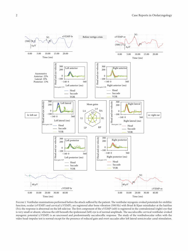
Figure 1: Vestibular examinations performed before the attack suffered by the patient.The vestibularmyogenic evoked potentials for otolithicfunction, ocular (oVEMP) and cervical (cVEMP), are registered after bone vibration (500Hz) with Bruel & Kjaer minishaker at the hairline(Fz); the response is abnormal on the left side/ear. The first component of the oVEMP (n10) is registered in the contralesional (right) eye thatis very small or absent, whereas the n10 beneath the ipsilesional (left) eye is of normal amplitude.The sacculocollic cervical vestibular-evokedmyogenic potential (cVEMP) is an uncrossed and predominantly sacculocollic response. The study of the vestibuloocular reflex with thevideo head-impulse test is normal except for the presence of reduced gain and overt saccades after left lateral semicircular canal stimulation.

Case Reports in Otolaryngology 3
0.59
1.17
0.760.87
0.69
00.20.40.60.811.21.4
1 2 3 4 5 6 7 8 9 51 52 53 65 66 67 70 149
150
Gain of the VOR left ear
∗∗∗
HSCPSCSSC
Figure 2: Gain of the vestibuloocular reflex in the left ear threesemicircular canals at different follow-ups. Day 1 was the day ofthe first treatment with gentamicin intratympanically (that and theothers are represented by ∗). In light grey, the dates the patient feltsteady and free of vertigo spells.
vertigo spells have recurred scaling up in intensity and againone Tumarkin spell has occurred.
The date seen she was in good condition; last vertigo spellwas 2 days before. Tympanicmembrane had healed normally.There was no spontaneous nystagmus with or without visualfixation; neither gaze evoked nor post head-shake. The vHITwas normal in terms of gain of the vestibuloocular reflex(VOR), but overt saccades were clear for yaw-axis leftwardhead impulses as shown in Figure 1, suggesting a small reduc-tion of VOR gain (changes in VOR at the different follows-upregarding the treatments are shown in Figure 2). Vestibularevokedmyogenic potentials (VEMP)were performedwith Fz500Hz vibration delivered with a Bruel & Kjaer minishaker.Results are shown to be normal for the right ear (both oVEMPand cVEMP) and abnormal for the left (both oVEMP andcVEMP) as shown in Figure 1. Hearing loss wasmild to severein her left ear (PTA was 48 dBHL).
After clinical and laboratory evaluation and when thepatient was entering the office, she began to develop a vertigospell and concurrently an intense tinnitus and pressuresensation in her left ear. At that time a left beating nystagmuswith a mean slow phase velocity (mSPV) of 9.7∘/s, andsuppressed by visual fixation was seen under video-Frenzelglasses; 3 minutes latter a mild (mSPV = 4.3∘/s) upbeatcomponent was added (Figure 2). The nystagmus increasedin leftward gaze and there was not skew deviation. In thevHIT there was an abnormal left superior VOR and this (left-anterior right-posterior) LARP plane of testing was repeated3 times, in a period of 10 minutes, and was similar in allof them. The vertigo spell did not stop for 20 minutes andat that time was treated with sulpiride (100mg I.M.). Threehours later the vertigo spell ended and the patient was able toreturn home; nystagmus was significantly reduced and vHITreturned to almost normal (precrisis) level (Figure 3).
3. Discussion
The situation that we present here is indicative of a com-plete, temporary dysfunction in the superior semicircularcanal in the affected side and a unidirectional nystagmus
beating ipsilaterally and upwards; in other words bothhorizontal and vertical components were present. Thesepreviously unreported findings while being in an acutevertigo spell in a patient with Meniere’s disease deserve anexplanation:
(1) While being in the vertigo crisis an acute deficit in theleft superior canal was registered and confirmed not to be anartifact: the low gain was followed in each impulse by clearrefixation saccades which had not been present during theearlier test in the quiescent phase.
(2) As the patient was seen in the very first minuteof the crisis we can say that, in terms of nystagmus, thiswas unidirectional throughout the episode with a verticalcomponent soon added and kept constant. In the case ofunidirectional nystagmus most are paretic and very rarelyirritative [3]. However the nystagmus in MD can changedirections and the order of appearance heterogeneous. Ingeneral, an initial and brief period (<2min) of irritative nys-tagmus is followed by a more prolonged period (20–30min)during which nystagmus changes to paretic, being followedby a more prolonged period (days) of irritative nystagmusalso called “recovery nystagmus.” Different sequences canoccur regarding the period of time the patient is seen. Theorigin for nystagmus has been claimed to be due to theirritative and paralytic action of potassium in the perilymph,respectively, for the first and second periods and to be due toa fast adapting response for the third one [5, 6]. Mechanicaleffects have been hypothesized to explain these findings too[7].
(3) Interestingly the spontaneous nystagmus has littleeffect on the assessment of both horizontal semicircularcanals, either at the beginning or at the end of the crisis,probably because the SPV was so small relative to SPV of theVOR response.
(4) Regarding the treatment performed we can speculatethat although some of the gentamicin could make its wayto the inner ear the amount must be very small accordingto changes in the VOR in the different periods of timethe patient was seen: the function from the horizontal andposterior semicircular canals in the left ear almost did notchange, while that of the superior asmentioned above showedsome fluctuations. This is also supported in the examinationperformed before the crisis: there were signs neither of theexpected acute status nor of the compensated status after anappropriate damage to the left ear: spontaneous right beatingnystagmus, biphasic post-head-shake nystagmus (first rightand after left beating nystagmus).
The strong unidirectional nystagmus first raised thequestion of a deficit from the right side; however no differenceto previous test was seen and during the crisis the patientmentioned neither symptoms pointing to a right side audi-tory deficit nor to pressure or tinnitus. Contrary to this anaggravation of pressure and tinnitus was attributed to herleft ear. The finding of a reduction in right side posteriorcanal function (RP) is thought to occur due to the loss ofconcurrent inhibitory function from the left superior (LA)canal for head impulses in their plane. Alternatively anincrease in the activity from the left horizontal canal couldaccount for the right beating nystagmus; however as same

4 Case Reports in Otolaryngology
300200
0
0 560−100
−140
Left anterior (ms)Hea
d an
d ey
e velo
city
Mean gain: 0.19
Left anterior 300200
0
0 560−100
−140
Right anterior (ms)Hea
d an
d ey
e velo
city
Mean gain: 0.83
Right anterior
300200
0
0 560−100
−140
Left lateral (ms)Hea
d an
d ey
e velo
city
Mean gain: 0.84
Left lateral 300200
0
0 560−100
−140
Right lateral (ms)Hea
d an
d ey
e velo
city
Mean gain: 0.98
Right lateral
300200
0
0 560−100
−140
Left posterior (ms)Hea
d an
d ey
e velo
city
Mean gain: 0.78
Left posterior300200
0
0 560−100
−140
Right posterior (ms)Hea
d an
d ey
e velo
city
Mean gain: 0.66
Right posterior
Mean gains1.0
0.5 RA
RLLL
LA
RPLP
End of vertigo crisisDuring vertigo crisis
Ar
Ab
Ar
Ab
RightLeftUpDown
Position
20
10
0
−10
−20
20
10
0
−10
−20
−30
Left anterior (ms)Mean gain: 0.62
Left anterior
HeadSaccade
VOR
HeadSaccade
VOR
HeadSaccade
VOR
HeadSaccade
VOR
HeadSaccade
VOR
HeadSaccade
VOR
300200
0
0 560−100
−140
300200
0
0 560−100
−140
300200
0
0 560−100
−140
300200
0
0 560−100
−140
300200
0
0 560−100
−140
Right anterior (ms)Hea
d an
d ey
e velo
city300
200
0
0 560−100
−140
Hea
d an
d ey
e velo
city
Mean gain: 0.87
Right anterior
HeadSaccade
VOR
HeadSaccade
VOR
HeadSaccade
VOR
HeadSaccade
VOR
HeadSaccade
VOR
HeadSaccade
VOR
Left lateral (ms)Hea
d an
d ey
e velo
city
Mean gain: 0.84
Left lateral
Right lateral (ms)Hea
d an
d ey
e velo
city
Mean gain: 0.97
Right lateral
Left posterior (ms)Hea
d an
d ey
e velo
city
Mean gain: 0.74
Left posterior
Right posterior (ms)Hea
d an
d ey
e velo
city
Mean gain: 0.63
Right posterior
Mean gains1.0
0.5 RA
RLLL
LA
RPLP
AsymmetryAnterior: 29%Lateral: 13%
Posterior: 15%
AsymmetryAnterior: 77%Lateral: 14%
Posterior: 15%
5 s
Figure 3: Spontaneous nystagmus and results of the video head-impulse test at the initiation of the crisis and at the end. Nystagmus is leftbeating with an upward component and there is a clear decrease in the gain of the vestibuloocular reflex in the plane of the left superiorsemicircular canal and right posterior canal (LARP) and the consequent presence of saccades. After the acute attack the same evaluationshows an increase of the gain and a significant decrease of the nystagmus.
as with the right side, no changes were seen form before toduring the crisis for the function in this canal.
The need to focus on the superior canal is mainly dueto the finding during the crisis but also is supported by aprevious report that mentions marked fluctuations in activityfor the canal function between the quiescent and acuteattack states and in particular after gentamicin treatment [8].In our patient in the different follow-ups in which vHITwas done, a fluctuation in the activity of the left superiorcanal function was registered. In the case of a deficient leftsuperior semicircular canal the expected nystagmus shouldbe a mixed vertical-torsional that from the examiner’s pointof view should be upward and counterclockwise with a small
rightward component on VNG. However in our case onlythe vertical component fits with this finding; the torsionalcomponent is very slow and the horizontal one is the contrary.
A very similar pattern of nystagmus has been describedin patients after surgery for superior semicircular canaldehiscence [9]. In all patients there is an abnormal HITfor impulses in the plane of the treated semicircular canalas expected for a surgery that prevents endolymph flowthrough that canal and abnormal pressure effects relatedto dilation of the canal and in 40% of the patients thiscombines alsowith a deficit in the posterior canal too; in thesepatients nystagmus, in the immediate postoperative period,was beating to the operated side. In the infrequent situation

Case Reports in Otolaryngology 5
of a complete deficit for all the three semicircular canalsnystagmus was typically paretic, beating to the nonoperatedside [10]. For the situation in which an irritative nystagmuswas found an exchange of potassium between endolymphand perilymph was proposed to occur through small leaksin the membranous canal. This finding was also reported byothers who also describe an acute event in a patient withMeniere-like symptoms but with superior semicircular canaldehiscence [11]. The pattern of the spontaneous nystagmuswas similar as irritative both for the horizontal and torsionalcomponents. Concurrent with previous findings the authorspresume an endolymphatic hydrops secondary to the actionof prolapsing dura on membranous labyrinth through largecanal dehiscences.
Another source of horizontal nystagmus has been shownto the utricle. Due to convergence of neural input from theotoliths onto horizontal canal neurons in the vestibular nucleian ipsilesional nystagmus can occur in case of a deficit ofthe utricle [12]. In our case the previously done oVEMPboth at the time of diagnosis (not shown) and at the dayof last follow-up (Figure 1) is concurrent with a deficit fromthe ipsilateral utricle. However in the case of acute vertigoattacks in patients with Meniere’s disease usually there is anenhancement of dynamic utricular function in the affectedear, contrary to what occurs in the case of Lermoyez crisis.We can speculate that this could have occurred in our patientand in particular because the modulation of nystagmusdirectionwas very small when gazewas taken to right/left andup/down. Unfortunately her clinical situation was not goodenough to proceed in more tests while being in the crisis.
As such, a combination of increased pressure in the leftutricle (with concurrent amelioration of the n10 potentialof the oVEMP, not found) generating a left beating nys-tagmus, transmitted to the superior semicircular canal witha concurrent utriculopetal displacement of the cupula (inthe inhibitory direction) generating an upbeating nystagmus,could sum to generate the findings in our patient.
Conflict of Interests
All the authors declare not to have any conflict of interests.
Acknowledgment
The authors wish to thank Professor Dr. I. Curthoys for hisinvaluable comments on the paper original form.
References
[1] H. Fushiki, M. Ishida, S. Sumi, A. Naruse, and Y. Watanabe,“Correlation between canal paresis and spontaneous nystagmusduring early stage of acute peripheral vestibular disorders,”ActaOto-Laryngologica, vol. 130, no. 12, pp. 1352–1357, 2010.
[2] L. Manzari, H. G. MacDougall, A. M. Burgess, and I. S.Curthoys, “New, fast, clinical vestibular tests identify whethera vertigo attack is due to early Meniere’s disease or vestibularneuritis,” Laryngoscope, vol. 123, no. 2, pp. 507–511, 2013.
[3] P. Duwel, L. E. Walther, J. Ilgner, and M. Westhofen, “Time-dependent vestibular function loss of semicircular cannels and
otolith organs in Meniere’s disease,” Laryngo-Rhino-Otologie,vol. 84, no. 8, pp. 589–593, 2005.
[4] “Committee on Hearing and Equilibrium guidelines for thediagnosis and evaluation of therapy in Meniere’s disease,”Otolaryngology—Head and Neck Surgery, vol. 113, no. 3, pp. 181–185, 1995.
[5] T. K.Watanabe, “Nystagmus during an acute attack ofMeniere’sdisese,” ENGReport, pp. 1–3, 1996.
[6] J. A. McClure, J. C. Copp, and P. Lycett, “Recovery nystagmus inMeniere’s disease,” Laryngoscope, vol. 91, no. 10, pp. 1727–1737,1981.
[7] J. Tonndorf, “Vestibular signs and symptoms in Meniere’sdisorder: mechanical considerations,” Acta Oto-Laryngologica,vol. 95, no. 5-6, pp. 421–430, 1983.
[8] L. E. Walther, R. Huelse, K. Blattner, M. B. Bloching, andA. Blodow, “Dynamic change of VOR and otolith functionin intratympanic gentamicin treatment for Meniere’s disease:case report and review of the literature,” Case Reports inOtolaryngology, vol. 2013, Article ID 168391, 5 pages, 2013.
[9] K. L. Janky, M. G. Zuniga, J. P. Carey, and M. Schubert,“Balance dysfunction and recovery after surgery for superiorcanal dehiscence syndrome,” Archives of Otolaryngology—Headand Neck Surgery, vol. 138, no. 8, pp. 723–730, 2012.
[10] J. P. Carey, A.A.Migliaccio, and L. B.Minor, “Semicircular canalfunction before and after surgery for superior canal dehiscence,”Otology and Neurotology, vol. 28, no. 3, pp. 356–364, 2007.
[11] C. Brandolini and G. C. Modugno, “Do signs of natural plug-ging of superior semicircular canal dehiscence exist?”AmericanJournal of Otolaryngology: Head andNeckMedicine and Surgery,vol. 33, no. 2, pp. 268–271, 2012.
[12] L. Manzari, A. M. Burgess, and I. S. Curthoys, “Does unilateralutricular dysfunction cause horizontal spontaneous nystag-mus?” European Archives of Oto-Rhino-Laryngology, vol. 269,no. 11, pp. 2441–2445, 2012.

Submit your manuscripts athttp://www.hindawi.com
Stem CellsInternational
Hindawi Publishing Corporationhttp://www.hindawi.com Volume 2014
Hindawi Publishing Corporationhttp://www.hindawi.com Volume 2014
MEDIATORSINFLAMMATION
of
Hindawi Publishing Corporationhttp://www.hindawi.com Volume 2014
Behavioural Neurology
EndocrinologyInternational Journal of
Hindawi Publishing Corporationhttp://www.hindawi.com Volume 2014
Hindawi Publishing Corporationhttp://www.hindawi.com Volume 2014
Disease Markers
Hindawi Publishing Corporationhttp://www.hindawi.com Volume 2014
BioMed Research International
OncologyJournal of
Hindawi Publishing Corporationhttp://www.hindawi.com Volume 2014
Hindawi Publishing Corporationhttp://www.hindawi.com Volume 2014
Oxidative Medicine and Cellular Longevity
Hindawi Publishing Corporationhttp://www.hindawi.com Volume 2014
PPAR Research
The Scientific World JournalHindawi Publishing Corporation http://www.hindawi.com Volume 2014
Immunology ResearchHindawi Publishing Corporationhttp://www.hindawi.com Volume 2014
Journal of
ObesityJournal of
Hindawi Publishing Corporationhttp://www.hindawi.com Volume 2014
Hindawi Publishing Corporationhttp://www.hindawi.com Volume 2014
Computational and Mathematical Methods in Medicine
OphthalmologyJournal of
Hindawi Publishing Corporationhttp://www.hindawi.com Volume 2014
Diabetes ResearchJournal of
Hindawi Publishing Corporationhttp://www.hindawi.com Volume 2014
Hindawi Publishing Corporationhttp://www.hindawi.com Volume 2014
Research and TreatmentAIDS
Hindawi Publishing Corporationhttp://www.hindawi.com Volume 2014
Gastroenterology Research and Practice
Hindawi Publishing Corporationhttp://www.hindawi.com Volume 2014
Parkinson’s Disease
Evidence-Based Complementary and Alternative Medicine
Volume 2014Hindawi Publishing Corporationhttp://www.hindawi.com