case reort - cardioldepartamentos.cardiol.br/dic/publicacoes/revistadic...63 case reort right atrial...
TRANSCRIPT
63
Case Report
Right Atrial Myxoma: Rare Occurrence of an Uncommon DiseaseEduardo Menti, Vinicius Leite Gonzalez, Ana Paula Susin Osorio, Luciane Durigon CoccoInstituto de Cardiologia do Rio Grande do Sul, Porto Alegre, RS – Brazil
IntroductionCardiac tumors are rare and it is difficult to know their
real incidence, since most cases are incidentally diagnosed in autopsy studies. It is known that cardiac involvement by secondary tumors prevail over those originating in the organ itself1. The origin of metastatic involvement is more frequently from lung cancer, lymphoma, breast cancer and esophagus, commonly by direct invasion or lymphatic dissemination2,3. The most common location of metastasis in the heart is the pericardium (69%), followed by epicardium (34%), myocardium (32%) and endocardium (5%). Pericardial involvement by metastases typically determines few symptoms and there may be extensive involvement even without symptoms. Symptoms are more related to the location of the tumor than with its size4.
Cardiac tumors cause symptoms through four main mechanisms: embolization, blocking, arrhythmias and constitutional symptoms. Embolism is a complication that affects up to 25% of cases and is more frequently associated with small tumors, especially when located in the left atrium and in the aortic valve5. Larger tumors tend to cause complications through flow obstruction, determining symptoms of heart failure and syncope. The most common symptom in these cases is dyspnea followed by chest pain. Constitutional symptoms, such as fever, weight loss and fatigue, are assigned to the substances produced by the tumor, such as interleukin 6, and are especially related to cardiac myxoma2.
Cardiac myxoma is the most common primary tumor of the heart, corresponding to approximately 50% of benign tumors. They occur at a frequency of about 1 case per 2 million people, most often in adults aged 30 to 50, predominantly in women2. Most myxomas are is located in the left atrium, in 75% to 85% of the cases, followed by the right atrium in 15% to 20% and ventricles in 5% to 10%. Echocardiographic evaluation can classify myxomas into two groups: rounded-shaped and solid with fixed surface (52% of cases) and polypoid-shaped, with irregular, moving surface (48% of cases), the latter being more prone to embolic phenomena6.
Echocardiography is the initial diagnostic method for evaluating patients with suspected cardiac tumors, since it is a simple noninvasive inexpensive test that is widely available. Through this test, it is possible to evaluate the location, morphology and mobility of tumors and assess their hemodynamic consequences5.
Case ReportFemale 37-year-old patient with exertional dyspnea
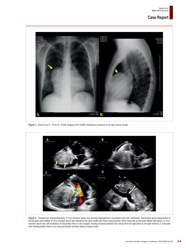
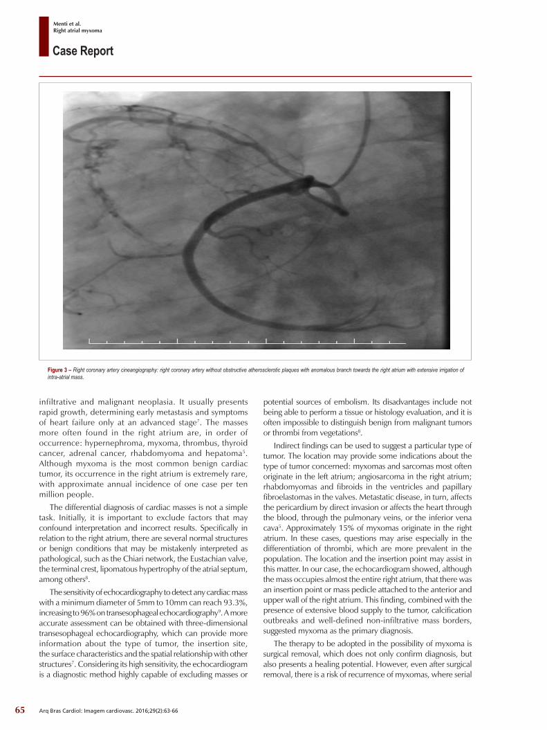
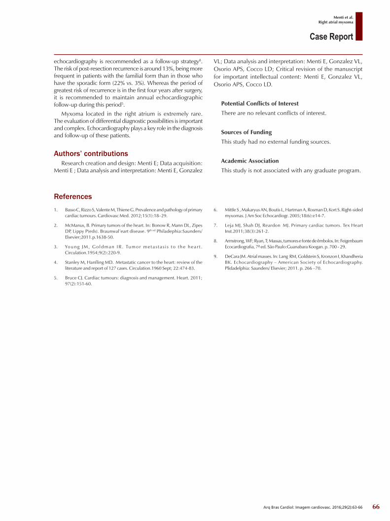
with indolent progression over the years and recent onset of lower limb edema and right hypochondrium pain. The patient had no history of cardiovascular comorbidities. On physical examination, pale emaciated mucous membranes, distended abdomen, painful hepatomegaly, and lower limb edema with formation of bilateral perimalleolar pitting edema. Electrocardiogram suggested right atrial enlargement. Chest X-ray revealed increased middle mediastinum with bulging located on its right and anterior border (Figure 1). Echocardiography revealed right atrial enlargement, containing large mass with maximum diameters of 92 mm x 95 mm with apparent pedicle attached to the anterior right atrial wall, protruding into the right ventricle, with vacuolization inside and flow on Doppler, indicating increased vascularization of the tumor. There was right ventricular diastolic flow obstruction determining maximum transvalvar tricuspid diastolic gradient of 17 mmHg and average gradient of 11 mmHg, associated with signs of systemic congestion, with right atrial pressure estimated at 20 mmHg. There was no impairment of global and segmental systolic function of the ventricles, which had normal diameters (Figure 2). The patient underwent cardiac catheterization to complement preoperative evaluation, which revealed no obstructive coronary atherosclerotic plaques, but confirmed extensive vascularization of the mass through coronary branch from the right coronary artery (Figure 3).
The patient underwent cardiac surgery with resection of a tumor which occupied about 90% of the right atrial cavity. The tumor was pedunculated and was inserted into the right atrial anterior wall as described by preoperative echocardiography. Tricuspid valve surgery and resection of the excess atrial wall borders was necessary. Anatomical and pathological analysis of the tumor confirmed diagnosis of right atrial myxoma. The patient is under outpatient monitoring with general improvement, with no adverse events, and no tumor recurrence.
DiscussionThe occurrence of primary cardiac tumors is thirty times
less frequent than metastatic implants. Primary tumor located in the right atrium is more likely to be some massive,
KeywordsHeart Neoplasms/surgery; Myxoma; Heart Failure;
Echocardiography.
Mailing Address: Eduardo Menti •Rua Regente, 245, Sala 504. Postal Code 90470-170, Bairro Bela Vista, Porto Alegre, RS - BrazilE-mail: [email protected] submitted December 1, 2016; revised December 16, 2016; accepted February 29, 2016.
DOI: 10.5935/2318-8219.20160016
64
Case Report
Menti et al.Right atrial myxoma
Arq Bras Cardiol: Imagem cardiovasc. 2016;29(2):63-66
Figure 2 - Transthoracic echocardiography: A: Four-chamber apical view showing heterogeneous vacuolated mass with calcification, determining apical displacement of the tricuspid valve leaflets; B: Four-chamber apical view directed to the right cavities with mass measurements, which nearly fills up the entire dilated right atrium; C: Four-chamber apical view, with evaluation of intracavitary flow on color Doppler, showing increased diastolic flow velocity from the right atrium to the right ventricle; D: Subcostal view showing dilated inferior vena cava and hepatic branches without masses inside.
Figure 1 - Chest X-ray: A – Front; B – Profile. Bulging of the middle mediastinum observed on its right anterior border.
A B
65
Case Report
Menti et al.Right atrial myxoma
Arq Bras Cardiol: Imagem cardiovasc. 2016;29(2):63-66
Figure 3 – Right coronary artery cineangiography: right coronary artery without obstructive atherosclerotic plaques with anomalous branch towards the right atrium with extensive irrigation of intra-atrial mass.
infiltrative and malignant neoplasia. It usually presents rapid growth, determining early metastasis and symptoms of heart failure only at an advanced stage7. The masses more often found in the right atrium are, in order of occurrence: hypernephroma, myxoma, thrombus, thyroid cancer, adrenal cancer, rhabdomyoma and hepatoma5. Although myxoma is the most common benign cardiac tumor, its occurrence in the right atrium is extremely rare, with approximate annual incidence of one case per ten million people.
The differential diagnosis of cardiac masses is not a simple task. Initially, it is important to exclude factors that may confound interpretation and incorrect results. Specifically in relation to the right atrium, there are several normal structures or benign conditions that may be mistakenly interpreted as pathological, such as the Chiari network, the Eustachian valve, the terminal crest, lipomatous hypertrophy of the atrial septum, among others8.
The sensitivity of echocardiography to detect any cardiac mass with a minimum diameter of 5mm to 10mm can reach 93.3%, increasing to 96% on transesophageal echocardiography9. A more accurate assessment can be obtained with three-dimensional transesophageal echocardiography, which can provide more information about the type of tumor, the insertion site, the surface characteristics and the spatial relationship with other structures7. Considering its high sensitivity, the echocardiogram is a diagnostic method highly capable of excluding masses or
potential sources of embolism. Its disadvantages include not being able to perform a tissue or histology evaluation, and it is often impossible to distinguish benign from malignant tumors or thrombi from vegetations8.
Indirect findings can be used to suggest a particular type of tumor. The location may provide some indications about the type of tumor concerned: myxomas and sarcomas most often originate in the left atrium; angiosarcoma in the right atrium; rhabdomyomas and fibroids in the ventricles and papillary fibroelastomas in the valves. Metastatic disease, in turn, affects the pericardium by direct invasion or affects the heart through the blood, through the pulmonary veins, or the inferior vena cava5. Approximately 15% of myxomas originate in the right atrium. In these cases, questions may arise especially in the differentiation of thrombi, which are more prevalent in the population. The location and the insertion point may assist in this matter. In our case, the echocardiogram showed, although the mass occupies almost the entire right atrium, that there was an insertion point or mass pedicle attached to the anterior and upper wall of the right atrium. This finding, combined with the presence of extensive blood supply to the tumor, calcification outbreaks and well-defined non-infiltrative mass borders, suggested myxoma as the primary diagnosis.
The therapy to be adopted in the possibility of myxoma is surgical removal, which does not only confirm diagnosis, but also presents a healing potential. However, even after surgical removal, there is a risk of recurrence of myxomas, where serial
66
Case Report
Menti et al.Right atrial myxoma
Arq Bras Cardiol: Imagem cardiovasc. 2016;29(2):63-66
echocardiography is recommended as a follow-up strategy8. The risk of post-resection recurrence is around 13%, being more frequent in patients with the familial form than in those who have the sporadic form (22% vs. 3%). Whereas the period of greatest risk of recurrence is in the first four years after surgery, it is recommended to maintain annual echocardiographic follow-up during this period5.
Myxoma located in the right atrium is extremely rare. The evaluation of differential diagnostic possibilities is important and complex. Echocardiography plays a key role in the diagnosis and follow-up of these patients.
Authors’ contributionsResearch creation and design: Menti E; Data acquisition:
Menti E ; Data analysis and interpretation: Menti E, Gonzalez
VL; Data analysis and interpretation: Menti E, Gonzalez VL, Osorio APS, Cocco LD; Critical revision of the manuscript for important intellectual content: Menti E, Gonzalez VL, Osorio APS, Cocco LD.
Potential Conflicts of Interest
There are no relevant conflicts of interest.
Sources of Funding
This study had no external funding sources.
Academic Association
This study is not associated with any graduate program.
1. Basso C, Rizzo S, Valente M, Thiene G. Prevalence and pathology of primary cardiac tumours. Cardiovasc Med. 2012;15(1):18–29.
2. McManus, B. Primary tumors of the heart. In: Bonow R, Mann DL, Zipes DP, Lippy P(eds). Braunwal’eart disease. 9th ed Philadephia:Saunders/Elsevier;2011.p.1638-50.
3. Yo u n g J M , G o l d m a n I R . Tu m o r m e t a s t a s i s t o t h e h e a r t . Circulation.1954;9(2):220-9.
4. Stanley M, Hanfling MD. Metastatic cancer to the heart: review of the literature and report of 127 cases. Circulation.1960 Sept; 22:474-83.
5. Bruce CJ. Cardiac tumours: diagnosis and management. Heart. 2011; 97(2):151-60.
6. Mittle S , Makaryus AN, Boutis L, Hartman A, Rosman D, Kort S. Right-sided myxomas. J Am Soc Echocardiogr. 2005;18(6):e14-7.
7. Leja MJ, Shah DJ, Reardon MJ. Primary cardiac tumors. Tex Heart Inst.2011;38(3):261-2.
8. Armstrong, WF; Ryan, T; Massas, tumores e fonte de êmbolos. In: Feigenbaum Ecocardiografia, 7ª ed. São Paulo:Guanabara Koogan. p. 700 - 29.
9. DeCara JM. Atrial masses. In: Lang RM, Goldstein S, Kronzon I, Khandheria BK. Echocardiography – American Society of Echocardiography. Plidadelphia: Saunders/ Elsevier; 2011. p. 266 –70.
References