cardiovascular system composed of : 1.heart: pumps blood...
TRANSCRIPT
Cardiovascular System
5th LECTURE
Please check our Editing File before studying this lecture
Objectives:• Identify the components of the cardiovascular
system.
• Describe the heart in regard to (position,
chambers, and valves).
• Describe the blood vessels (arteries, veins, and
capillaries).
• Describe the portal system.
• Describe the functional and anatomical end
arteries.
• Describe arteriovenous anastomosis.
• Describe the components blood and its
function.
• Describe sinusoids.
Lecture’s Guide:• Text in pink was only found in the girls’ slides.
• Text in blue was only found in the boys’ slides.
• Text in red is considered important.
• The Dr.’s comments in class are written in green.
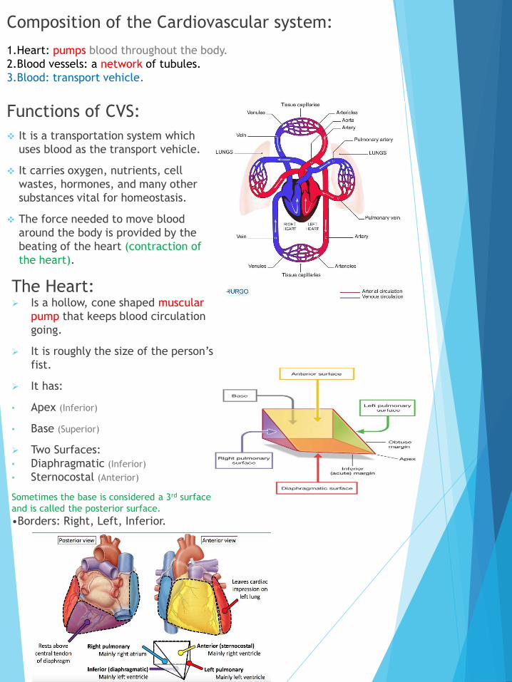
Composition of the Cardiovascular system:
1.Heart: pumps blood throughout the body.
2.Blood vessels: a network of tubules.
3.Blood: transport vehicle.
Functions of CVS:
❖ It is a transportation system which
uses blood as the transport vehicle.
❖ It carries oxygen, nutrients, cell
wastes, hormones, and many other
substances vital for homeostasis.
❖ The force needed to move blood
around the body is provided by the
beating of the heart (contraction of
the heart).
The Heart: ➢ Is a hollow, cone shaped muscular
pump that keeps blood circulation
going.
➢ It is roughly the size of the person’s
fist.
➢ It has:
• Apex (Inferior)
• Base (Superior)
➢ Two Surfaces:
• Diaphragmatic (Inferior)
• Sternocostal (Anterior)
Sometimes the base is considered a 3rd surface
and is called the posterior surface.
•Borders: Right, Left, Inferior.
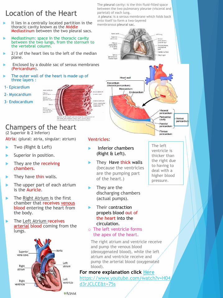
Location of the Heart It lies in a centrally located partition in the
thoracic cavity known as the Middle Mediastinum between the two pleural sacs.
Mediastinum: space in the thoracic cavity between the two lungs, from the sternum to the vertebral column.
2/3 of the heart lies to the left of the median plane.
Enclosed by a double sac of serous membranes (Pericardium).
The outer wall of the heart is made up of three layers :
1- Epicardium
2- Myocardium
3- Endocardium
Champers of the heart (2 Superior & 2 Inferior)
Atria: (plural: atria, singular: atrium)
Two (Right & Left)
Superior in position.
They are the receiving chambers.
They have thin walls.
The upper part of each atrium is the Auricle.
The Right Atrium is the first chamber that receives venousblood entering the heart from the body.
The Left Atrium receives arterial blood coming from the lungs.
Ventricles:
Inferior chambers
(Right & Left).
They Have thick walls
(because the ventricles
are the pumping part
of the heart.)
They are the
discharging chambers
(actual pumps).
Their contraction
propels blood out of
the heart into the
circulation.
o The left ventricle forms
the apex of the heart.
For more explanation click Here
https://www.youtube.com/watch?v=H04
d3rJCLCE&t=75s
The pleural cavity: is the thin fluid-filled space
between the two pulmonary pleurae (visceral and
parietal) of each lung.
A pleura: is a serous membrane which folds back
onto itself to form a two-layered
membranous pleural sac.
The left
ventricle is
thicker than
the right due
to having to
deal with a
higher blood
pressure.
The right atrium and ventricle receive
and pump the venous blood
(deoxygenated blood), while the left
atrium and ventricle receive and
pump the arterial blood (oxygenated
blood).
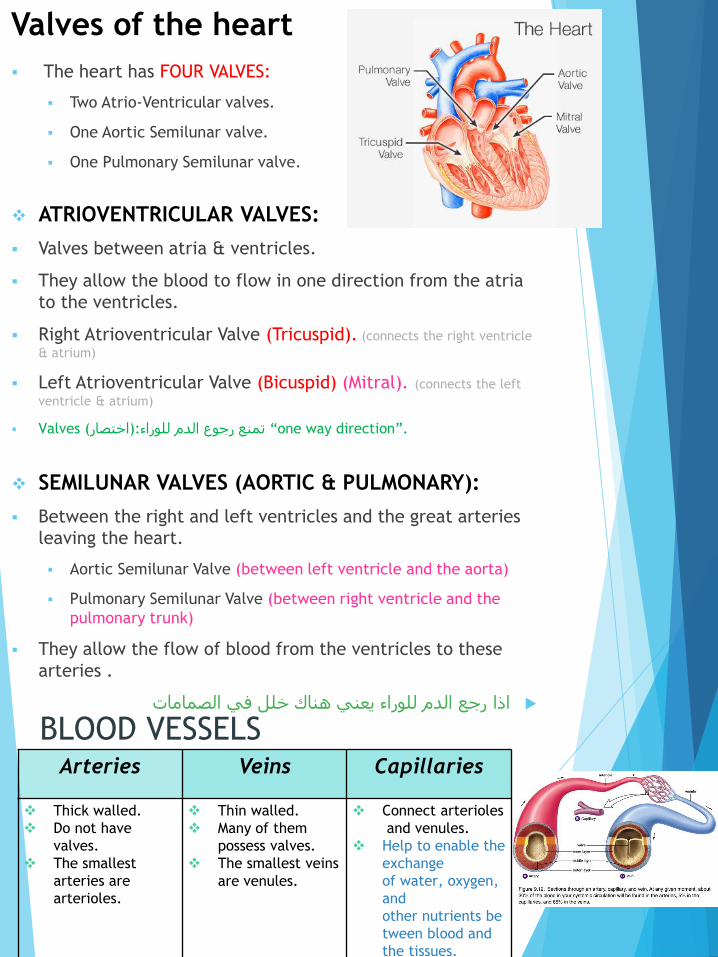
Valves of the heart
▪ The heart has FOUR VALVES:
▪ Two Atrio-Ventricular valves.
▪ One Aortic Semilunar valve.
▪ One Pulmonary Semilunar valve.
❖ ATRIOVENTRICULAR VALVES:
▪ Valves between atria & ventricles.
▪ They allow the blood to flow in one direction from the atria
to the ventricles.
▪ Right Atrioventricular Valve (Tricuspid). (connects the right ventricle
& atrium)
▪ Left Atrioventricular Valve (Bicuspid) (Mitral). (connects the left
ventricle & atrium)
▪ Valves (اختصار):تمنع رجوع الدم للوراء “one way direction”.
❖ SEMILUNAR VALVES (AORTIC & PULMONARY):
▪ Between the right and left ventricles and the great arteries
leaving the heart.
▪ Aortic Semilunar Valve (between left ventricle and the aorta)
▪ Pulmonary Semilunar Valve (between right ventricle and the
pulmonary trunk)
▪ They allow the flow of blood from the ventricles to these
arteries .
رجع الدم للوراء يعني هناك خلل في الصماماتاذا
BLOOD VESSELSArteries Veins Capillaries
❖ Thick walled.
❖ Do not have
valves.
❖ The smallest
arteries are
arterioles.
❖ Thin walled.
❖ Many of them
possess valves.
❖ The smallest veins
are venules.
❖ Connect arterioles
and venules.
❖ Help to enable the
exchange
of water, oxygen,
and
other nutrients be
tween blood and
the tissues.
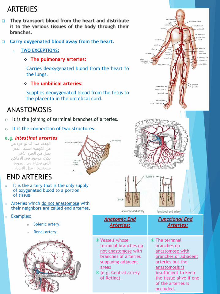
They transport blood from the heart and distribute
it to the various tissues of the body through their
branches.
Carry oxygenated blood away from the heart.
o TWO EXCEPTIONS:
❖ The pulmonary arteries:
Carries deoxygenated blood from the heart to
the lungs.
❖ The umbilical arteries:
Supplies deoxygenated blood from the fetus to
the placenta in the umbilical cord.
ARTERIES
ANASTOMOSIS
o It is the joining of terminal branches of arteries.
o It is the connection of two structures.
e.g. Intestinal arteries
o It is the artery that is the only supply of oxygenated blood to a portion of tissue.
o Arteries which do not anastomose with their neighbors are called end arteries.
o Examples:
o Splenic artery.
o Renal artery.
END ARTERIES
الهدف منه ان لو جزء من من االوعية انسد ،الدم
.يصل من الجزء اآلخريكون موجود في األماكن
اللي تحتاج دمن بصورة .مستمرة ، مثل األمعاء
Anatomic End
Arteries:
Functional End
Arteries:
Vessels whose
terminal branches do
not anastomose with
branches of arteries
supplying adjacent
areas
(e.g. Central artery
of Retina).
The terminal
branches do
anastomose with
branches of adjacent
arteries but the
anastomosis is
insufficient to keep
the tissue alive if one
of the arteries is
occluded.
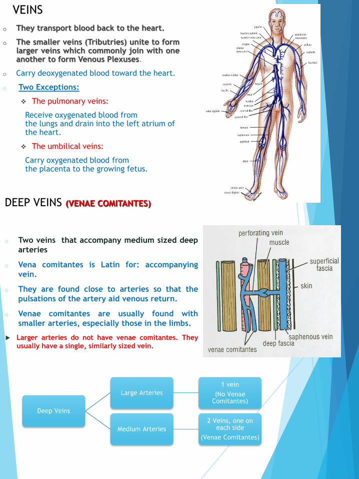
o They transport blood back to the heart.
o The smaller veins (Tributries) unite to formlarger veins which commonly join with oneanother to form Venous Plexuses.
o Carry deoxygenated blood toward the heart.
o Two Exceptions:
❖ The pulmonary veins:
Receive oxygenated blood from the lungs and drain into the left atrium of the heart.
❖ The umbilical veins:
Carry oxygenated blood from the placenta to the growing fetus.
VEINS
DEEP VEINS (VENAE COMITANTES)
o Two veins that accompany medium sized deep
arteries
o Vena comitantes is Latin for: accompanying
vein.
o They are found close to arteries so that the
pulsations of the artery aid venous return.
o Venae comitantes are usually found with
smaller arteries, especially those in the limbs.
Larger arteries do not have venae comitantes. They
usually have a single, similarly sized vein.
Deep Veins
Large Arteries
1 vein
(No Venae Comitantes)
Medium Arteries
2 Veins, one on each side
(Venae Comitantes)

CAPILLARIES
o Microscopic vessels in the form of a network.
o They connect arterioles to venules. (Site of the
union between the arteries and veins)
o They allow the exchange of water, oxygen,
and many other nutrients between blood and
the tissue.
ARTERIOVENOUS ANASTOMOSIS
o Direct connections between arteries and
veins without the intervention of
capillaries.
o Found in tips of the fingers and toes.
SINUSOIDS
• Thin walled blood vessels like capillaries.
• They are wider with irregular cross diameter.
• They are found in:
1. Liver
2. Spleen
3. Bone marrow
4. Pituitary gland
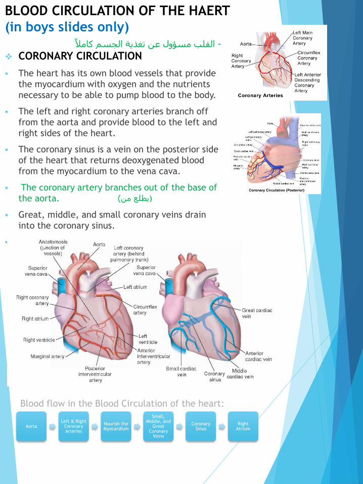
BLOOD CIRCULATION OF THE HAERT
(in boys slides only)القلب مسؤول عن تغذية الجسم كامالً -
❖ CORONARY CIRCULATION
▪ The heart has its own blood vessels that provide
the myocardium with oxygen and the nutrients
necessary to be able to pump blood to the body.
▪ The left and right coronary arteries branch off
from the aorta and provide blood to the left and
right sides of the heart.
▪ The coronary sinus is a vein on the posterior side
of the heart that returns deoxygenated blood
from the myocardium to the vena cava.
▪ The coronary artery branches out of the base of
the aorta.
▪ Great, middle, and small coronary veins drain
into the coronary sinus.
▪ Coronary sinus drains into right atrium.
Aorta Left & Right
Coronary Arteries
Nourish the Myocardium
Small, Middle, and
Great Coronary
Veins
Coronary Sinus
Right Atrium
Blood flow in the Blood Circulation of the heart:
(يطلع من )
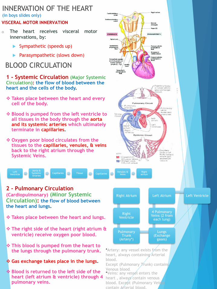
VISCERAL MOTOR INNERVATION
o The heart receives visceral motor
innervations, by:
Sympathetic (speeds up)
Parasympathetic (slows down)
INNERVATION OF THE HEART(in boys slides only)
BLOOD CIRCULATION
1 - Systemic Circulation (Major Systemic Circulation): the flow of blood between the heart and the cells of the body.
❖ Takes place between the heart and every cell of the body.
❖ Blood is pumped from the left ventricle to all tissues in the body through the aorta and its systemic arteries which ultimately terminate in capillaries.
❖ Oxygen poor blood circulates from the tissues to the capillaries, venules, & veins back to the right atrium through the Systemic Veins.
2 - Pulmonary Circulation (Cardiopulmonary) (Minor Systemic Circulation): the flow of blood between the heart and lungs.
❖ Takes place between the heart and lungs.
❖ The right side of the heart (right atrium &
ventricle) receive oxygen poor blood.
❖ This blood is pumped from the heart to the lungs through the pulmonary trunk.
❖ Gas exchange takes place in the lungs.
❖ Blood is returned to the left side of the heart (left atrium & ventricle) through 4 pulmonary veins.
Left Ventricle
Aorta & Systemic Arteries
Capillaries Tissue CapillariesVenules &
VeinsRight
atrium
Right Atrium
Right Ventricle
Pulmonary Trunk
(Artery*)
Lungs (Exchange
gases)
4 Pulmonary Veins (2 from
each lung)
Left Atrium Left Ventricle
*Artery: any vessel exists from the
heart, always containing Arterial
blood.
Except (Pulmonary Trunk) contains
Venous blood.
*Veins: any vessel enters the
heart , always contain venous
blood. Except (Pulmonary Veins)
contain Arterial blood.

3 - Hepatic Portal Circulation: blood flowsfrom the GIT to the liver through the portalvein then to systemic circulation via IVC(inferior vena cava).
BLOOD CIRCULATION (cont.)
Portal circulation System
It is a system of vessels interposed between two capillary beds it occurs when a capillary bed pools into another capillary bed through veins, without first going through the heart.
It takes place in the liver and some endocrine glands (Pituitary gland).
Veins leaving the gastrointestinal tract do not go directly to the heart (because it contains CO2 and digested food).
They pass the Portal Vein.
This vein enters the liver and breaks up into veins of diminishing size which ultimately join capillary like vessels (Sinusoids): first capillary bed.
Venous blood enters the 2nd capillary bed then to smaller veins that leave the liver through hepatic veins.
*A network of capillaries is known
as a capillary bed.

Blood in boys slide only
o Blood is the actual carrier oxygen and nutrients intoarteries.
o Blood is made mostly of plasma, which is a yellowish liquidthat is 90% water.
o Plasma also contains salts, glucose, and other substances.
o Most importantly, plasma contains proteins that carryimportant nutrients to the body’s cells and strengthen thebody’s immune system.
o Blood has 3 main types of blood cells that circulate with theplasma.
Types Of Blood Cells in boys slide only
For more details: watch this video
https://www.youtube.com/watch?v=nPfTQLpUQ3M
PLATELETSHelp blood clot, clotting stops the blood from flowing
out of the body when a vein or artery is broken.
Platelets are also called thrombocytes.
RED BLOOD CELLSCarry oxygen, a healthy adult has about 35 trillion of
them. Red blood cells are also called erythrocytes.
WHITE BLOOD CELLSThese cells, which come in many shapes and sizes, are
vital to the immune system against infections. When
the body is fighting off infection, they increase. White
blood cells are also called leukocytes.
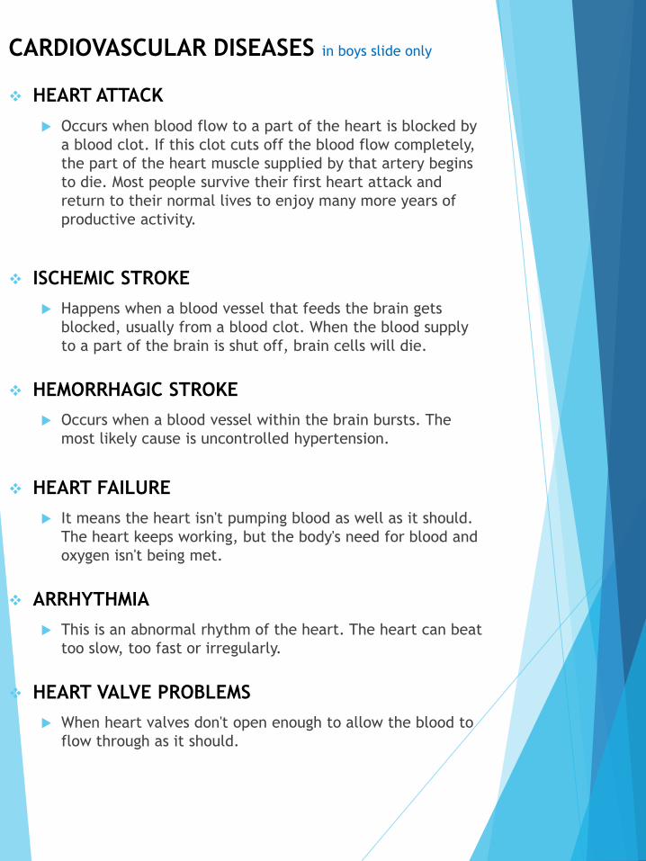
CARDIOVASCULAR DISEASES in boys slide only
❖ HEART ATTACK
Occurs when blood flow to a part of the heart is blocked by
a blood clot. If this clot cuts off the blood flow completely,
the part of the heart muscle supplied by that artery begins
to die. Most people survive their first heart attack and
return to their normal lives to enjoy many more years of
productive activity.
❖ ISCHEMIC STROKE
Happens when a blood vessel that feeds the brain gets
blocked, usually from a blood clot. When the blood supply
to a part of the brain is shut off, brain cells will die.
❖ HEMORRHAGIC STROKE
Occurs when a blood vessel within the brain bursts. The
most likely cause is uncontrolled hypertension.
❖ HEART FAILURE
It means the heart isn't pumping blood as well as it should.
The heart keeps working, but the body's need for blood and
oxygen isn't being met.
❖ ARRHYTHMIA
This is an abnormal rhythm of the heart. The heart can beat
too slow, too fast or irregularly.
❖ HEART VALVE PROBLEMS
When heart valves don't open enough to allow the blood to
flow through as it should.

Summary• The cardiovascular system is a transporting system.
• It is composed of the heart and blood vessels.
• The heart is cone shaped, covered by pericardium and
composed of four chambers.
• The blood vessels are the arteries, veins and capillaries.
• Arteries transport the blood from the heart.
• The terminal branches of the arteries can anastomose
with each other freely or be anatomic or functional end
arteries.
• Veins transport blood back to the heart.
• Sinusoids are a special type of capillaries.
• The portal system is composed of two sets of capillaries.
• It is found in the liver & pituitary gland.
• Capillaries connect the arteries to the veins.
• The portal system is composed of two sets of capillaries.
• The veins from the GIT go to the liver first through the
portal vein.
• Blood is the actual carrier of oxygen and nutrients into
arteries.
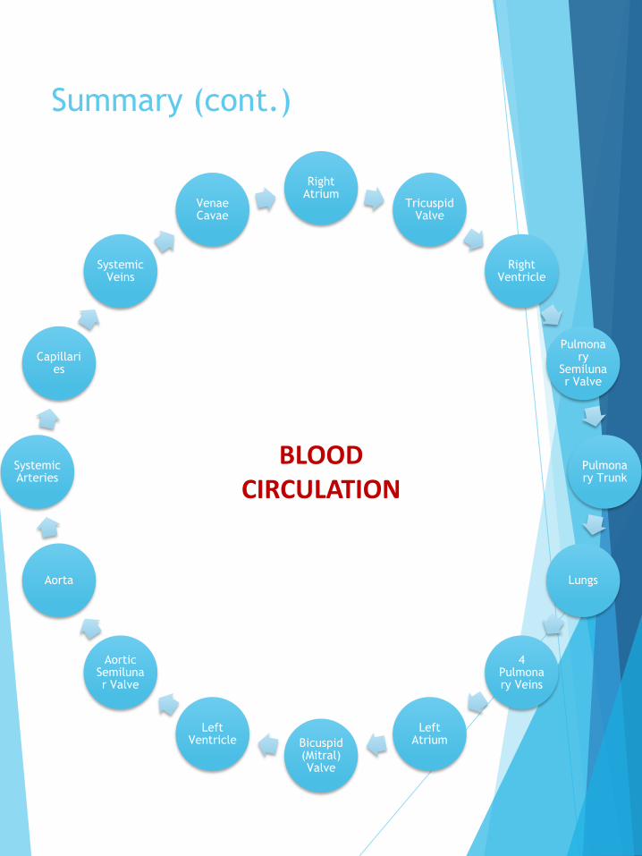
Summary (cont.)
Right Atrium
Tricuspid Valve
Right Ventricle
Pulmonary
Semilunar Valve
Pulmonary Trunk
Lungs
4 Pulmonary Veins
Left AtriumBicuspid
(Mitral) Valve
Left Ventricle
Aortic Semilunar Valve
Aorta
Systemic Arteries
Capillaries
Systemic Veins
Venae Cavae
BLOOD CIRCULATION
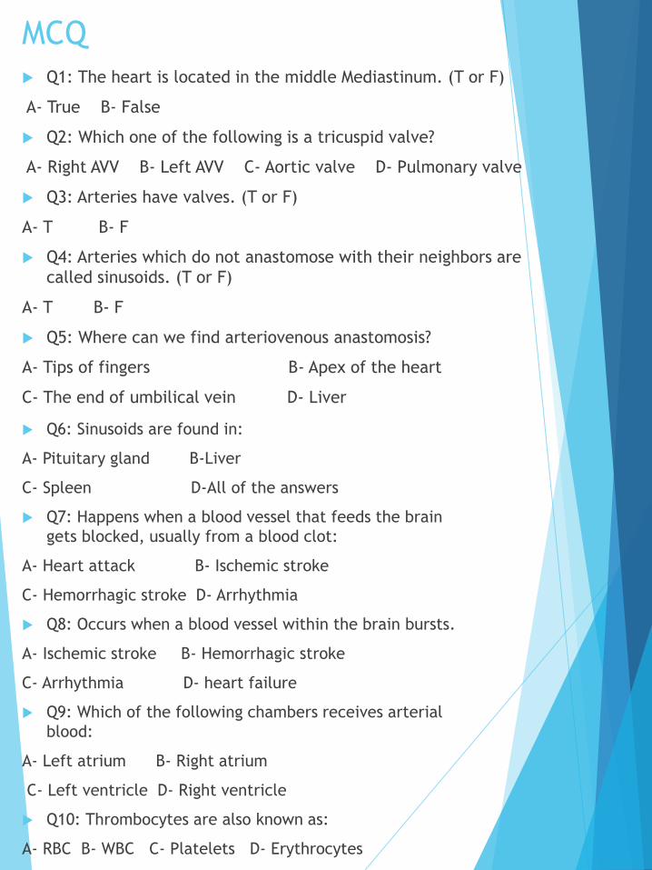
MCQ
Q1: The heart is located in the middle Mediastinum. (T or F)
A- True B- False
Q2: Which one of the following is a tricuspid valve?
A- Right AVV B- Left AVV C- Aortic valve D- Pulmonary valve
Q3: Arteries have valves. (T or F)
A- T B- F
Q4: Arteries which do not anastomose with their neighbors are
called sinusoids. (T or F)
A- T B- F
Q5: Where can we find arteriovenous anastomosis?
A- Tips of fingers B- Apex of the heart
C- The end of umbilical vein D- Liver
Q6: Sinusoids are found in:
A- Pituitary gland B-Liver
C- Spleen D-All of the answers
Q7: Happens when a blood vessel that feeds the brain
gets blocked, usually from a blood clot:
A- Heart attack B- Ischemic stroke
C- Hemorrhagic stroke D- Arrhythmia
Q8: Occurs when a blood vessel within the brain bursts.
A- Ischemic stroke B- Hemorrhagic stroke
C- Arrhythmia D- heart failure
Q9: Which of the following chambers receives arterial
blood:
A- Left atrium B- Right atrium
C- Left ventricle D- Right ventricle
Q10: Thrombocytes are also known as:
A- RBC B- WBC C- Platelets D- Erythrocytes
MCQ Answers 1-A 2-A 3-B 4-B 5-A 6-D 7-B 8-B 9-A 10-C
Faisal Fahad ALsaif ( Team Leader)
Abdulrahman Sulaiman Aldawood
Fahad Aldhowaihy
Abdullah Almeaither
Abdulelah Abdulhadi Aldossari
Saleh Abdullah Almoaiqel
Abdulaziz Mohammed Alabdulkareem
Abdulmajeed Khaled Alwardi
Abdulaziz Ibrahim Aldrgam
Akram Alfandi
Saud Abdulaziz Alghufaily
Mohammed Alquwayfili
Ali Alammari
Sultan Alfuhaid
Zeyad Alkhenizan
Fahad Alshughaithry
Saad Aloqile
Abduljabbar Alyamani
Mohammed Alomar
Abdullah Alqarni
Yazeed Aldossari
Fahad Alfaiz
Team members Lamia Abdullah AlKuwaiz (Team Leader)
Abeer Alabduljabbar
Afnan Abdulaziz Almustafa
Albandari Alshaye
Alfahdah Abdullah Alsaleem
Layan Hassan Alwatban
Majd Khalid Albarrak
Norah Alharbi
Rinad Musaed Alghoraiby
Rawan Mohammad Alharbi
Wafa Alotaibi
Wejdan Fahad Albadrani
Good Luck
https://imgur.com/a/DSjCMMind Map