physiology of the proprioceptors in …ksumsc.com/download_center/2nd/1) neuropsychiatry block/male...
TRANSCRIPT

DR SYED SHAHID HABIBMBBS DSDM PGDCR FCPS
Professor & Consultant Clinical Neurophysiology
Dept. of PhysiologyCollege of Medicine & KKUH
PHYSIOLOGY OF THE PROPRIOCEPTORS IN BALANCE & ITS
PATHWAYS

Identify the major sensory pathways
Describe the components, processes and functions of the sensory pathways
Appreciate the dorsal column system in conscious proprioception
Describe the pathway of spinocerebellar tract in unconscious proprioception from muscles, tendons and joints
Differentiate between sensory and motor ataxia
OBJECTIVES
At the end of this lecture you should be able to:

PROPRIOCEPTION
Latin proprius, meaning "one's own", "individual" and perception, is the sense of the relative position of neighbouring parts of the body and strength of effort being employed in movement.
exteroception, by which one perceives the outside world
interoception, by which one perceives pain, hunger, etc., and the movement of internal organs. MECHANORECEPTORS: which detect
mechanical compression or stretching of the receptor or of tissues adjacent to the receptor
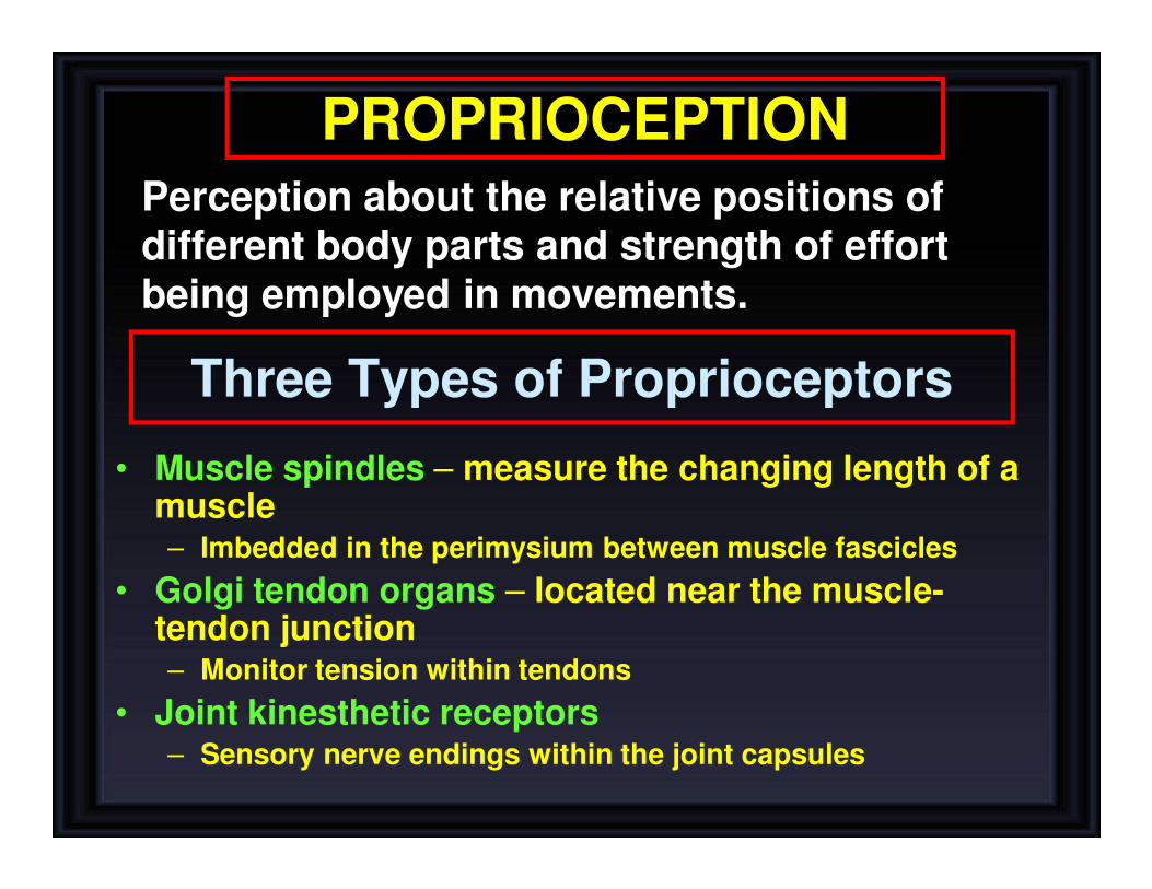
PROPRIOCEPTION
Perception about the relative positions of different body parts and strength of effort being employed in movements.
Three Types of Proprioceptors
• Muscle spindles – measure the changing length of a muscle– Imbedded in the perimysium between muscle fascicles
• Golgi tendon organs – located near the muscle-tendon junction– Monitor tension within tendons
• Joint kinesthetic receptors– Sensory nerve endings within the joint capsules

• Proprioceptors include the muscle spindles, Golgi tendon organs, and joint receptors.
• These provide a sense of body position and allow fine control of skeletal movements
PROPRIOCEPTORS
(1) Static position sense: which means conscious
perception of the orientation of the different parts of the
body with respect to one another, and
(2) Rate of movement sense: also called kinesthesia or
dynamic proprioception

SENSORY RECEPTORS TYPES
• (1) MECHANORECEPTORS: which detect mechanical compression or stretching of the receptor or of tissues adjacent to the receptor eg proprioceptors
• (2) THERMORECEPTORS, which detect changes in temperature, some receptors detecting cold and others warmth.
• (3) NOCICEPTORS (pain receptors), which detect damage occurring in the tissues, whether physical damage or chemical damage eg free nerve endings
• (4) ELECTROMAGNETIC RECEPTORS, which detect light on the retina of the eye eg rods and cones
• (5) CHEMORECEPTORS, which detect taste in the mouth, smell in the nose, oxygen level in the arterial blood, osmolality of the body fluids, carbon dioxide concentration, and perhaps other factors that make up the chemistry of the body. Eg chemo R in carotid bodies

• Proprioceptors
• Brain Stem (Cortico,Rubro,Vestibulo,Reticulo,Olivo,Tectospinal)
• vestibular system (apparatus, nuclei etc)
• Ascending Tracts
• Visual system
• Cerebellum (flocculonodular lobe→dynamicequilibrium, Uvula → Static equilibrium)
• Cerebral cortex (primary cortical center for equilibrium located in the parietal lobe deep in the sylvian fissure)
STRUCTURES CONCERNED WITH PROPRIOCEPTION
SPATIAL ORIENTATION (four inputs)

Adaptation of Receptors
When a continuous sensory stimulus is applied, the receptor responds at a high impulse rate at first and then at a progressively slower rate until finally the rate of action potentials decreases to very few or often to none at all.

1. Rapid adapting or phasic receptors egmeissner’s corpuscles(touch), paciniancorpuscles(vibration)
2. Slowly adapting or tonic receptors egruffini’s (pressure ,skin stretch ) krause’send bulbs,and Merkel’s disks.
3. Non adapting receptors eg Free nerve endings for pain sensation
Adaptation of Receptors

Mechanisms by which Receptors Adapt.
• First, the pacinian corpuscle is a viscoelasticstructure so that after stimulation within few
hundredths of a second, the fluid within the corpuscle redistributes, so that the receptor potential is no longer elicited.
• The second mechanism of adaptation of the pacinian corpuscle, but a much slower one, results from accommodation, which occurs in thenerve fiber itself. This probably results from
progressive “inactivation” of the sodium channels in the nerve fiber membrane

Receptor Potential of the Pacinian Corpuscle
•The receptor potential produced by compression induces a local circuit of current flow that spreads along nerve fiber.
•The frequency of repetitive action potentials transmitted from sensory receptors increases approximately in proportion to the increase in receptor potential
For joint position and vibration sensation(Also Ruffini’sEndings)

1- Conscious proprioception reach the level of cerebral cortex sensory area via dorsal column tract.
2- Subconscious proprioception reach the level of cerebellum via spinocerebellartracts
Where is the location of these tracts?
Types of proprioception

Structure of Proprioceptors

Encapsulated Nerve EndingsMeissner’s corpuscles, Pacinian corpuscles’
Ruffini’s corpuscles, Proprioceptors

Unencapsulated Nerve Endings


NERVE FIBERS CLASSIFICATION • Type A
– Alpha
– Beta
– Gamma
– Delta
• Type C

Reciprocal Excitation of antagonist
Stretch Reflex
Muscle Stretch
Muscle Spindle
Afferent 1a
Spinal Cord (Center)
Efferent αMotor
Extrafusal Fibers
Ms Contration
Over Stretch
Golgi Tendon Organ
Afferent 1b
Spinal Cord (Center)
Efferent αMotor
Extrafusal Fibers
Ms Relaxation(lengthening reaction)
Inhibitory Interneuron
Reciprocal Inhibition of antagonist
Stimulus
Receptor
Afferent
Center
Efferent
Effector
Effect
Inverse Stretch Reflex


SPINAL CORD TRACTS
Ascending Tracts (Sensory)
Descending Tracts (Motor)
Sensory information from receptors throughout most of the body is relayed to the brain by means of ascending tracts of fibers that conduct impulses up the spinal cord.
When the brain directs motor activities, these directions are in the form of nerve impulses that travel down the spinal cord in descending tracts of fibers.

Gray Matter: Organization
• Dorsal half – sensory roots and ganglia
• Ventral half – motor roots
• Dorsal and ventral roots fuse laterally to form spinal nerves
• Four zones are evident within the gray matter – somatic sensory (SS),
visceral sensory (VS), visceral motor (VM), and somatic motor (SM)

White Matter in the Spinal Cord
• Fibers run in three directions – ascending, descending, and transversely
• Divided into three funiculi (columns) – posterior, lateral, and anterior
• Each funiculus contains several fiber tracks
– Fiber tract names reveal their origin and destination
– Fiber tracts are composed of axons with similar functions

SENSORY TRACTS
• DORSAL COLUMN SYSTEM
• ANTEROLATERAL SYSTEM
Each system carries different types ofsensations which are known as MODALITIESlike pain, temperature, fine touch, crudetouch, vibration, proprioception etc

1. Dorsal column pathway- carries signal of fine touch, pressure, vibration , stereognsisand proprioception,
2. Spinothalamic pathway- carries signals of pain, temperature, deep pressure, and course touch.
3,4- Posterior and anterior spinocerebellarpathways- carry subsconciousproprioception. Dorsal gray horn- to lateral column- to medulla oblongata- to pons – tocerebellum.

DORSAL COLUMNMEDIAL LEMNISCAL SYSTEM
1. Touch sensations requiring a high degree of localization of the stimulus
2. Touch sensations requiring transmission of fine gradations of intensity
3. Phasic sensations like vibratory sensations 4. Sensations that signal movement against skin 5. Joints Position sensations (Proprioception)6. Pressure sensations requiring fine degrees of
judgment of intensity 7. Strereognosis

ANTEROLATERAL SYSTEM
1. Pain 2. Thermal sensations, (warmth & cold)3. Crude touch and pressure sensations
capable only of crude localizing ability on the surface of the body
4. Tickle and itch sensations 5. Sexual sensations
Ventral & lateral spinothalamic tracts

ANTEROLATERAL
SYSTEM
DORSAL
COLUMN
SYSTEM

PROPRIOCEPTION FROM HEAD
In this pathway through the brain stem, each medial lemniscus is joined by additional fibers from the sensory nuclei of the trigeminal nerve; these fiberssubserve the same sensory functions for the head that the dorsal column fibers subservefor the body.

Dorsal column lesion
dorsal column pathway
Leftspinal cord injury
Loss of sense of:•touch•proprioception•vibrationin left leg
spinothalamic pathway
Leftspinal cord injury
Loss of sense of:•Touch•Pain•Warmth/coldin right leg
Spinothalamic lesion

Dorsal column damage
• Sensory ataxia
• Patient staggers; cannot perceive position or movement of legs
• Visual clues help movement
Positive Romberg testThe test depends on the integrity of proprioception from the joints of the legs.

FUNCTIONAL DIVISION OF CEREBELLUM

Vestibulocerebellum (Flocculonodular Lobe)— In Association with the Brain Stem and Spinal Cord Control Equilibrium and Postural Movementsduring performance of rapid motions specially with changes in direction
controlling balance between agonist and antagonist muscle contractions of
the spine, hips, and shoulders during rapid changes in body positions as
required by the vestibular apparatus
Spinocerebellum (Vermis+Intermediate Zone)—Feedback Control of Distal Limb Movements by Way of the Intermediate Cerebellar Cortex and the Interposed Nucleusfeedback information from the peripheral parts of the body, especially from the distal proprioceptors of the limbs, telling the cerebellum what actual movements result.
Cerebrocerebellum— Function of the Large Lateral Zone of the Cerebellar Hemisphere to Plan, Sequence, and Time Complex Movements
Role of Cerebellum in Proprioception

Spinocerebellar pathway
• Carries subconscious proprioception signals
• Receptors in muscles & joints
• 1st neuron: enters spinal cord through dorsal root
• 2nd neuron: ascends to cerebellum
• No 3rd neuron to cortex

Spinocerebellar tract damage
• Cerebellar ataxia
– Clumsy movements
– Incoordination of the limbs (intention tremor)
– Wide-based, reeling gait (ataxia)
– Alcoholic intoxication produces similar effects!

Ataxia and Gait Disturbances
• Pathophysiology
– Result from any condition that affects the central and peripheral nervous systems
– Ataxia: Types
• Motor ataxia
• Sensory ataxia

• Motor Ataxia
– Caused by cerebellar disorders
• Intact sensory receptors and afferent pathways
• Integration of proprioception is faulty
• Midline cerebellar lesions cause truncalataxia
• Lateral cerebellar lesions cause limb ataxia
• Thalamic infarcts may cause contralateralataxia with sensory loss
Ataxia and Gait Disturbances

Dermatomes
Dermatome is area on skin supplied by a single spinal nerve


Brown Sequard syndrome
Ipsilateral Loss:• Fine touch, Vibration, Proprioception (Dorsal Column)• Leg Ataxia (Dorsal Spinocerebellar)• Spastic Paresis below lesion (Lat Corticospinal)• Flaccid Paralysis (Vent horn destruction)• Dermatomal Anesthesia (Dorsal Horn destruction)
Contralateral Loss:• Loss of pain and temp (lat Spinothalamic)• Loss of crude touch and Pressure (Vent Spinothalamic)• Minor Contralat Muscle Weakness (Vent Corticospinal)• Leg Ataxia (Vent Spinocerebellar)
HEMISECTION OF SPINAL CORD

• Peripheral sensory lesions (e.g. polyneuropathy) cause
ataxia because there is loss of the sense of joint position -
proprioception. Broad-based, high-stepping, stamping gait
develops.
• This ataxia is made worse by removal of additional sensory
input (e.g. vision) and is worse in the dark. First described in
sensory ataxia of tabes dorsalis, this is the basis
of…………..Romberg's test. Ask the patient to close the eyes
while standing: observe whether the patient becomes
unstable (and prevent falling).
SENSORY ATAXIA