cardiovascular manifestations of inflammatory bowel...
TRANSCRIPT

Review ArticleCardiovascular Manifestations of Inflammatory Bowel Disease:Pathogenesis, Diagnosis, and Preventive Strategies
Diana-Maria Bunu ,1 Cristian-Eugen Timofte ,2 Manuela Ciocoiu ,3
Mariana Floria ,4,5 Claudia-Cristina Tarniceriu,6 Oana-Bogdana Barboi,7,8
and Daniela-Maria Tanase 4,5
1Department of Cardiology, Institute of Cardiovascular Diseases, Timisoara 300310, Romania2Department of Radiology, County Emergency Hospital Timisoara, Timisoara 300723, Romania3Department of Pathophysiology, Faculty of Medicine, “Grigore T. Popa” University of Medicine and Pharmacy,Iasi 700111, Romania4Department of Internal Medicine, “Grigore T. Popa” University of Medicine and Pharmacy, Iasi 700111, Romania53rd Internal Medicine Clinic, “Sf. Spiridon” County Clinical Emergency Hospital Iasi, Iasi, Romania6Department of Morpho-Functional Sciences I, Discipline of Anatomy, Faculty of Medicine, “Grigore T. Popa” University of Medicineand Pharmacy, Iasi 700111, Romania7Institute of Gastroenterology and Hepatology-“Sf. Spiridon” County Clinical Emergency Hospital Iasi, Iasi, Romania8“Grigore T. Popa” University of Medicine and Pharmacy, Iasi 700111, Romania
Correspondence should be addressed to Cristian-Eugen Timofte; [email protected] Mariana Floria; [email protected]
Received 2 September 2018; Revised 18 November 2018; Accepted 6 December 2018; Published 13 January 2019
Academic Editor: Konstantinos Triantafyllou
Copyright © 2019 Diana-Maria Bunu et al. This is an open access article distributed under the Creative Commons AttributionLicense, which permits unrestricted use, distribution, and reproduction in anymedium, provided the original work is properly cited.
Inflammatory bowel disease (IBD) refers to a group of chronic inflammatory diseases that targets mainly the gastrointestinal tract.The clinical presentation of IBD includes both gastrointestinal manifestations and extraintestinal manifestations (EIM). Thereported cardiovascular manifestations in IBD patients include pericarditis, myocarditis, venous and arterial thromboembolism,arrhythmias, atrioventricular block, heart failure, endocarditis, valvulopathies, and Takayasu arteritis. The aim of this article is toreview the available literature about the possible pathogenic mechanisms and determine preventive measures capable of reducingthe incidence and severity of the cardiovascular manifestations. In IBD patients, the incidence of cardiovascular manifestations islow, but higher than that in the general population. Therefore, clinicians should pay attention to any new modification thatmight indicate cardiovascular involvement in IBD patients, and they should consider chronic inflammatory diseases in patientswith cardiac conditions without an evident cause. Considering the role of inflammation in the development of cardiovascularmanifestations, the management should include prevention of flares and maintenance of remission for as long as possible.Preventive measures should also include active screening and strict control of the cardiovascular risk factors in all IBD patients.
1. Introduction
IBD defines a group of chronic inflammatory diseases thattargets predominantly the gastrointestinal tract. IBD canmanifest itself in two major forms: Crohn’s Disease (CD)and Ulcerative Colitis (UC) [1, 2].
The etiopathogenesis of IBD is not yet fully elucidated,but it is known to involve the interaction between four major
components: an aberrant immune system, genetic factors,environmental factors, and intestinal microbiota (therefore,the presence of only one component does not cause the onsetof IBD) [3]. The inflammatory response is mediated byimmune cells (T-helper 1 and T-helper 17 in CD andT-helper 2 in UC), cytokines (Tumor Necrosis Factor-α(TNF-α), transforming growth factor-β, and interleukins-(IL-) 12, IL-17, and IL23), chemokines, reactive oxygen
HindawiGastroenterology Research and PracticeVolume 2019, Article ID 3012509, 14 pageshttps://doi.org/10.1155/2019/3012509

species, neuropeptides, and nonimmune (myeloid, epithe-lial, mesenchymal, lymphoid, neurogenic, and endothelial)cells [4, 5].
The primary immune response to one or more stimuliinduces tissue destruction and proliferation of endothelialand mesenchymal cells, resulting in a secondary immuneresponse that amplifies the already present inflammationand stimulates fibrosis, tissue remodeling, angiogenesis,and lymphangiogenesis [3]. The nonresolving inflamma-tion determines the installation of a vicious cycle ofself-sustaining chronic inflammation and maintenance ofangiogenesis, fibrosis, and tissue destruction processes [6].
The main manifestations in IBD are intestinal (abdomi-nal pain, mucoid or bloody stool, rectal bleeding, and tenes-mus) and systemic (fever, fatigue, loss of appetite, and
weight loss) [7]. IBD can also exhibit a wide range of extrain-testinal manifestations (IBD-associated disorders that affectorgans that are distant to the digestive tract): hepatobiliary,genitourinary, musculoskeletal, respiratory, ophthalmic,cutaneous, and cardiovascular [8, 9].
Cardiovascular manifestations in IBD can be defined byIBD-associated disorders that affect the cardiovascular sys-tem [9]. Cardiovascular manifestations in patients with IBDmostly occur as immune-related consequences and includethe following: pericarditis, myocarditis, venous and arterialthromboembolism, left ventricle impairment, arrhythmiasand conduction disorders, infective endocarditis, valvulopa-thy, and Takayasu arteritis [10]. The pathogenic mechanismsbehind the cardiovascular manifestations are briefly pre-sented in each section and in Table 1.
Table 1: Cardiovascular manifestations in IBD.
Cardiovascular manifestations Possible pathogenic mechanisms References
Pericarditis and myocarditis(i) Immune-mediated myocarditis in IBD as a result of exposure to autoantigens
[40–43](ii) Cardiotoxicity as an adverse effect of the treatment with 5-ASA and its derivatives
Venous thromboembolism
(i) Hypercoagulability induced by the systemic inflammation
[22, 53–61]
(ii) Platelet abnormalities
(iii) Endothelial dysfunction induced by mechanical and systemic factors
(iv) Venous stasis
(v) Acquired risk factors (prolonged hospitalization, surgical interventions,central venous catheters, prolonged immobilization and bed rest, glucocorticoids,smoking, oral contraceptives, vitamin deficiencies, dehydration, hormone replacementtherapy, and hyperhomocysteinemia)
(vi) Genetic risk factors (dysfibrinogenemias, prothrombin gene mutation, factor VLeiden thrombophilia, and deficiency of proteins C, S, and antithrombin)
Arterial thromboembolism
(i) Structural and functional vascular alterations induced by chronic systemicinflammation
[57, 68–78]
(ii) Accelerated development of atherosclerosis and highly unstable atheroscleroticplaques
(iii) Endothelial dysfunction induced by microbial lipopolysaccharides
(iv) Altered gut microbiota
(v) Adipokines
(vi) Calprotectin
(vii) NOD2/CARD15 gene polymorphism
(viii) Dyslipidemia
Heart failure
(i) Myocardial fibrosis secondary to altered collagen metabolism, impaired nitricoxide-mediated vasodilation, and deficiencies of vitamins and essential trace elements
[83–87](ii) Heart muscle atrophy due to prolonged use of corticosteroids,total parenteral nutrition, and chronic inflammatory status
(iii) Myocarditis, endocarditis, and valvulopathy
Arrhythmias and conductiondisorders
(i) Interstitial fibrosis and structural and functional cardiac remodeling[91–95, 99–101](ii) Impaired autonomic nervous system: increased sympathetic and decreased
parasympathetic activity
Endocarditis(i) Bacteremia due to increased transmucosal permeability
[16, 103–106](ii) Predisposing risk factors: immunosuppression, preexistentvalvular heart disease, and central venous catheters
Valvulopathies(i) Myxomatous degeneration
[110, 111](ii) Ascending aorta changes due to chronic systemic inflammation
Takayasu arteritis (i) Genetic risk factors: HLA-A∗24:02, HLA-B∗52:01, and HLA-DRB-1∗15:02 [114, 115]
2 Gastroenterology Research and Practice
2. Epidemiology of Cardiovascular EIM
Frequencies of EIM in IBD range from 6% to 47%, and mul-tiple EIM may concomitantly occur. Moreover, EIM mayoccur prior to the diagnosis of IBD in up to 25% of cases [11].
The cardiovascular disease incidence among IBD patientsis modestly higher than that in the general population [12].This fact is supported by a recent Danish cohort study thatalso observed that the prevalence of traditional cardiovascu-lar risk factors is surprisingly low among IBD patients [13].Cardiovascular involvement rarely occurs in IBD patients,but its incidence should not be ignored, considering theserious impact of the consequences if untreated.
In 2016, Card et al. published their results on the inci-dence of EIM in IBD patients. Their study showed a slightlyincreased diagnosis rate for ischemic heart disease, atrialfibrillation, and hypertension in IBD patients when com-pared to the general population (odds ratio = 1 85 vs. 0.77,1.51 vs. 0.56, and 1.69 vs. 0.51, respectively). There were sim-ilar diagnosis rates between CD and UC patients [14].
Pericarditis represents the most frequent cardiovascularEIM in IBD patients (70% of the total number of cardiovas-cular complications) [15, 16]. Its prevalence is 0.19% amongthe CD patients and 0.23% among the UC patients [17]. Areview of 68 patients with IBD showed that pericarditisoccurs more frequently in male patients with UC [16].
A 16-year Danish nationwide cohort study of 15572 IBDpatients found that myocarditis had a total risk of 4.6 per100000 years of risk in IBD patients. The incidence rate ratiofor myocarditis was 8.3 for CD and 2.6 for UC when com-pared to the general population [18].
Patients with IBD may present with both venous andarterial thromboembolic complications. They present a1.7-5.5-fold greater risk for venous thromboembolism thanthe general population [19]. The Swiss Inflammatory BowelDisease Cohort Study showed a 3.9 prevalence of VTEamong the IBD patients, with a higher incidence among theUC patients when compared to the CD patients (4.7 vs. 3.4,respectively) [20]. Moreover, the mortality associated withthromboembolism is 2 times higher in patients with IBDwhen compared to the general population [21].
There is an increased risk for arterial thromboembolicevents in IBD patients (but to a lesser degree than venousthromboembolism), with similar rates in UC and CDpatients [22]. Patients with IBD have a moderately higherrisk (18%) of arterial thromboembolic events [23]. Thearterial thromboembolic events occur most frequently atthe cardiac (acute myocardial infarction), cerebral (stroke),and intestinal (mesenteric ischemia) levels [12]. Their inci-dence in IBD patients when compared to the general popula-tion is 1.2 times higher for acute myocardial infarction, 3.5times higher for mesenteric ischemia and 1.2 times higherfor stroke [22]. In 2013, a Danish cohort study has shownthat patients with IBD are at an increased risk of arterialthromboembolism especially during flares and episodes ofpersistent disease activity [24]. Feng et al. conducted ameta-analysis study on the risk of ischemic heart disease inpatients with IBD, and they have observed an increased riskin young female patients with CD [25]. An increased risk of
stroke associated with IBD (the risk is greater for UCpatients), independent of gender, was found in themeta-analysis conducted by Katsanos et al. [26]. There is alsoan increased risk of peripheral artery disease (the stenosis inarteries other than those supplying the heart and brain)among IBD patients [27, 28]. Koutroubakis presented in hisliterature review that most cases with the involvement ofthe peripheral arteries presented with thrombosis of theupper and lower limbs and they were associated with signif-icantly increased morbidity and mortality [29].
There are not many studies concerning the prevalence ofheart failure among the IBD patients. However, a recentpopulation-based cohort study showed a twofold higher riskof heart failure in IBD patients (adjusted hazard ratio, 2.03;95% CI, 1.36–3.03) and 2.5-fold increased risk in systemiccorticosteroid users (adjusted hazard ratio, 2.51; 95% CI,1.93–4.57) when compared to the control group. Also, itappears that only patients with UC have a significantly higherrelative risk of heart failure [30]. Another Danish populationcohort study found an increased risk of first heart failure hos-pitalization in patients with IBD, with a 2.5 times higher riskduring the periods of active disease (persistent activity orflares) [31].
The incidence of atrial fibrillation is 11.3% in patientswith IBD versus 0.9% in the general population [32]. A Dan-ish cohort study found a 2 times higher risk of atrial fibrilla-tion in IBD during flares and episodes of persistent activity[33]. It is important to acknowledge the increased risk ofatrial fibrillation in IBD patients since this arrhythmia isassociated with an increased risk of thromboembolism, heartfailure, and mortality, it affects the quality of life and exercisecapacity, and it increases the hospitalization risk [34].
Concerning the endocarditis prevalence in IBD patients,there are some case reports cited in the literature and a retro-spective and prospective case control study from 1992 thatshows an increased prevalence of bacterial endocarditis inpatients with IBD [35, 36].
There have been several case reports cited in the literaturethat presented patients with IBD who developed mitral oraortic regurgitation, but its incidence is very rare [37].
The coexistence of IBD and Takayasu arteritis is alsorare, with just a small number of reported cases in theliterature [38].
3. Myocarditis and Pericarditis
Pericarditis represents the most common cardiovascularcomplication in IBD [13]. Myocarditis can be defined as aninflammation of the myocytes and the interstitial tissue.Occasionally, the pericardiummay also be involved, in whichcase it is called myopericarditis [39].
Patients with IBD have a higher risk for developingmyopericarditis than the general population. This can beexplained by two mechanisms: autoimmune mediationgenerated by exposure to autoantigens and drug toxicity fol-lowing an administration of 5-aminosalicylic acid (5-ASA) orits derivatives [40].
Exposure to autoantigens in an acute episode may causedirect cytotoxicity on myocytes, causing the release of
3Gastroenterology Research and Practice

inflammatory mediators and activation of the immune sys-tem. This sequence of events can lead to acute myocarditis[41]. If myocarditis is not detected in the acute or sub-acute stage and the inflammation is not counteracted,myocardial destruction will continue and patients willdevelop chronic myocarditis [42]. Remodeling processesthat are characteristic of chronic inflammation may causedilation of the cardiac cavities (resulting in systolic dys-function, anomalies of parietal kinetics, and decreasedejection fraction), valvular regurgitation (by rupture ofpapillary muscles), or arrhythmias (due to the fibrosis ofthe conduction system, occurrence of reentry phenomena,and excessive adrenergic stimulation) [43].
The clinical picture for myopericarditis is nonspecific.Patients may present with symptoms similar to those ofthe acute coronary syndrome, heart failure (new onset ordecompensated heart failure), arrhythmias, cardiogenicshock, or sudden death [43]. The occurrence of such clin-ical picture within the first 28 days since the initiation ofthe treatment with 5-ASA or its derivatives raises thesuspicion of drug toxicity [44].
The 12-lead electrocardiogram may be normal, or it mayreveal an ST segment elevation or depression, a negative Twave or rhythm, or conduction disorders [45]. Blood testsmay indicate elevated levels of biomarkers of cardiac injury(troponin, creatine kinase, creatine kinase-MB, alanineaminotransferase, and aspartate aminotransferase), as wellas B-type natriuretic peptide and N-terminal probrainnatriuretic peptide in patients with associated heart failure.Leukocytosis and increased levels of acute-phase reactants(erythrocyte sedimentation rate, C-reactive protein, andfibrinogen) may also be present [46].
Transthoracic echocardiography should be performed inall patients with clinical presentation suggestive for myoper-icarditis. The presence of left ventricular dysfunction, anom-alies of parietal kinetics, low ejection fraction, oraccumulation of pericardial fluid should raise the suspicionof myopericarditis [47]. Angiocoronarography should beperformed in all patients with the clinical picture suggestivefor acute coronary syndrome, since the absence of hemody-namically significant angiographic lesions of the coronaryarteries excludes the diagnosis of myocardial infarction [39].
Cardiovascular magnetic resonance (CMR) offers nonin-vasive characterization of the myocardial tissue, and it canprovide the necessary information for the diagnosis of myo-carditis. CMR diagnostic criteria for myocarditis reveal myo-cardial (regional or global) oedema, myocardial hyperaemia,and focal fibrosis or necrosis with noncoronary artery distri-bution [45]. If patients are hemodynamically stable and CMRis available, then it is recommended to perform CMR beforeendomyocardial biopsy. Endomyocardial biopsy should beperformed in life-threatening conditions, when CMR is notindicated [40].
Endomyocardial biopsy remains the gold standard indiagnosing and establishing the etiology of myocarditis[48]. IBD-associated myocarditis can frequently presentunder two histopathological forms: acute/chronic lympho-cytic myocarditis and giant cell myocarditis (the latter formhas a poor prognosis) [43].
Supportive therapy depends on the patients’ hemody-namic stability. Patients who are hemodynamically unstableshould be redirected to intensive care units that can provideadvanced cardiopulmonary support such as mechanical ven-tilation and extracorporeal membrane oxygenation [46].Hemodynamically stable patients should be monitored in ahospital setting and treated according to the current guide-lines for heart failure with beta-blockers and/or inhibitorsof the renin-angiotensin-aldosterone system [49].
Nonsteroidal anti-inflammatory drugs can be used inpatients with pericardial involvement [46]. Consideringthe high possibility of gastrointestinal toxicity, nonsteroidalanti-inflammatory drugs which selectively inhibit thecyclooxygenase-2 should be recommended [50]. Colchicine,another drug usually prescribed for pericarditis, may causediarrhea as a side effect and, therefore, it can potentially com-plicate the evolution of IBD [15].
Immunosuppressive therapy may be associated with thesupportive therapy, but only after exclusion of infectious eti-ology. The most widely used immunosuppressive agents areimmunoglobulin, corticosteroids, azathioprine, and cyclo-sporine, and the duration of administration ranges from 3to 6 months [51].
Management of myocarditis also includes restriction ofphysical activity during the acute phase and for the following6 months. Recovery signs include improvement of ejectionfraction and parietal kinetics [45].
4. Venous Thromboembolism
Deep vein thrombosis and pulmonary thromboembolismrepresent the most frequent venous thromboembolic compli-cations. But venous thromboembolic events can also occur inthe cerebral, portal, mesenteric, or retinal sites [52].
Venous thromboembolism can be triggered by genetic oracquired factors. Long-term hospitalization, surgical inter-ventions, central venous catheters, prolonged bed rest andimmobilization, corticosteroid therapy, smoking, use of oralcontraceptives, vitamin deficiency, dehydration, hormonereplacement therapy, and hyperhomocysteinemia are amongthe most frequent acquired risk factors for venous thrombo-embolism [53]. Genetic risk factors such as dysfibrinogen-emia, mutations of the prothrombin gene, factor V Leiden,antithrombin, and protein C or S deficiency should beconsidered in patients with IBD who experience recurrentthromboembolic venous events [54].
Virchow’s triad, known to be associated with venousthromboembolism, describes three conditions that predis-pose to thrombosis: hypercoagulability, endothelial dysfunc-tion, and venous stasis [55].
Hypercoagulability is mediated by the inflammatory pro-cess that initiates clotting and interferes with the fibrinolyticsystem, decreasing the anticoagulant activity [56]. Theinflammation in IBD levels can be illustrated by increasedlevels of C-reactive protein and cytokines (the most fre-quently observed cytokines are TNF-α, vascular endothelialgrowth factor, and IL-6) [57]. The reduction in anticoagulantactivity not only increases thrombosis risk but also helps
4 Gastroenterology Research and Practice

maintain the inflammatory status by stimulation of thrombinto produce TNF-α, IL-6, and IL-10 [58].
In addition, the following hemostatic disorders have beenobserved during flares: elevated levels of coagulation factors(V, VIII, von Willebrand, and fibrinogen) and products ofthrombin and fibrin formation, increased markers of vascu-lar endothelial activation, and acquired deficiencies ordysfunction of natural anticoagulants (protein C, protein S,and antithrombin) [22]. Platelet abnormalities (reactivethrombocytosis, reduced mean platelet volume, and aug-mented granular content) also contribute to the hypercoagu-lability. The enhanced activation state of platelets is mediatedby the CD40-CD40 ligand pathway [59].
Endothelial dysfunction in IBD patients (procoagulantsurface of the vascular bed) is a consequence of mechanicaldamage (e.g., intravenous catheters) or activation of thevascular endothelial cells by inflammatory mediators.Inflammation determines the occurrence of the thrombophi-lic effect of the vascular endothelium, and it accentuates theadhesion between the endothelial surface and leukocytes orplatelets [22, 60].
The last condition described by Virchow’s triad is rep-resented by a disturbance of the blood flow, as it is seen inpatients with prolonged immobilization (a common situa-tion encountered during flares in IBD), dehydration, orcentral vein catheters [22, 61].
The clinical picture of venous thromboembolismdepends on the site of the thrombus, and it can range fromasymptomatic to severely symptomatic, but the suspicion ofvenous thromboembolic event is raised when the patienthas an unexplained episode of dyspnea, hypoxia or unilateralleg pain, and swelling [62].
The diagnosis of venous thromboembolic events is basedon appropriate imaging investigations such as compressionultrasound or venography for deep vein thrombosis, ventila-tion/perfusion lung scanning or spiral computed tomogra-phy for pulmonary emboli, and computed tomography forother affected sites [52, 63].
The primary prevention of venous thromboembolism inacutely ill-hospitalized patients includes the prophylacticadministration of one of the following anticoagulants:low-molecular-weight heparin, low-dose-unfractionatedheparin, or fondaparinux [64]. Mechanical thromboprophy-laxis (graduated compression stockings and pneumatic com-pression devices) represents a valid alternative for patientswho present contraindications for anticoagulation [65].
The primary prevention of venous thromboembolism inhospitalized but stable patients includes prophylactic antic-oagulation and management of additional risk factors. Thus,it is intended to maintain the remission, to use intravenouscatheters with caution, to correct vitamin deficiencies anddehydration, and to early mobilize the patients [22].
Treatment of acute venous thromboembolism inpatients with IBD is similar to that in patients withoutIBD. The use of anticoagulants (unfractionated heparinand low-molecular-weight heparin) is recommended formild and moderate venous thrombosis, whereas local orsystemic thrombolysis is recommended for massive veinthrombosis [66].
The secondary prevention of venous thromboembolism(anticoagulation) must be individualized according to eachpatient’s hemorrhagic and thromboembolic risks [65].Long-term anticoagulation using low-molecular-weightheparin, vitamin K antagonists, or novel direct oral antico-agulants is indicated in case of initial unprovoked venousthromboembolic event (in the absence of disease activityor temporary/transient risk factors) [64]. Short-term antic-oagulation (3-6 months) is indicated in case of provokedthromboembolic event, and prophylaxis of disease exacer-bations can also be added. The placement of inferior venacava filters is recommended to patients with high throm-boembolic risk [66].
5. Arterial Thromboembolism
Out of traditional cardiovascular risk factors (male gender,age > 55 years, smoking, obesity, dyslipidemia, arterialhypertension, diabetes mellitus, and chronic kidney disease),advanced age, obesity, and smoking have a reduced preva-lence among the IBD patients [67]. But IBD patients alsopresent nontraditional cardiovascular risks: hyperhomocys-teinemia, leukocytosis, anemia, corticosteroid therapy,thrombocytosis, high levels of C-reactive protein, andincreased erythrocyte sedimentation rate [68].
It is a well-known fact that chronic inflammation andendothelial dysfunction play a role in atherogenesis, one ofthe most important factors involved in arterial thromboem-bolism. C-reactive protein, TNF-α, vascular endothelialgrowth factor, and IL-6 represent molecules involved in bothatherogenesis and IBD, and their increased serum levelsamong the IBD patients confirm the fact that atherogenesisis accelerated among this class of patients [57, 69].
There are multiple mechanisms that contribute to themaintenance of the chronic inflammation. IBD patientsare also characterized by a disrupted intestinal barrierwhich allows microbial products (lipopolysaccharides andother endotoxins) to enter the bloodstream. Lipopolysac-charides are known to induce the expression of proinflam-matory cytokines, to affect the oxidation of low-densitycholesterol, and to activate the macrophages, all of whichcontributing to endothelial dysfunction, foam cell forma-tion, and atherosclerosis [57].
Obesity (when present) also contributes to the inflamma-tory status. The adipose tissue is responsible for producingadipokines: leptin, resistin, and adiponectin (proinflamma-tory cytokines). Mesenteric fat also produces proinflamma-tory cytokines, such as TNF-α and IL-6 [70].
The gut microbiota in IBD is characterized by anabnormal microbial composition (loss of microbial diver-sity) that can induce immunoregulatory pathways andcan mitigate the chronic inflammation [71, 72]. The gutmicrobiota is also implicated in the atherosclerosis processand increased platelet activation via decreased levels of tri-methylamine N-oxide and the induction of expression ofToll-like receptors 2 and 4. Microorganisms can also influ-ence the blood pressure in IBD [73, 74].
Calprotectin is another acute-phase reactant that hasbeen linked to the disease activity in IBD and higher risk
5Gastroenterology Research and Practice

for cardiovascular events. Calprotectin binds to Toll-likereceptor 4, a receptor that amplifies inflammation andatherosclerosis. Also, calprotectin binds to the receptors foradvanced glycation end products (RAGE) which mediatecardiomyocyte dysfunction [75].
The chronic inflammatory process causes structural andfunctional arterial changes. Smooth muscle cell hyperplasiacan be demonstrated by determining carotid intima-mediathickness (a subclinical marker of atherosclerosis). Vascularfibrosis and degradation of elastic fibers that occur in thewalls of large blood vessels determine vascular stiffness(another subclinical marker of atherosclerosis) [60]. Vascularstiffness is not associated with the cardiovascular risk factors,but with the duration of the episodes of disease activity [76].
Moreover, preexisting coronary atheromatous plaquesare more unstable in patients with CD than in the generalpopulation. This can be explained by the common geneticalcharacterization of both CD and atherogenesis by a polymor-phism of the NOD2/CARD15 gene [77]. Thrombus forma-tion and acute coronary syndrome are favored by the factthat, after the atheromatous plaque has cracked, the lipidcore is being exposed to the bloodstream characterized byhypercoagulability and platelet anomalies [58].
IBD patients are also characterized by altered lipidprofiles (low levels of total and high-density cholesteroland high levels of low-density cholesterol), known risk fac-tors for atherosclerosis. The exact mechanism is unknown,but it is thought to be either chronic inflammation ormalabsorption [78].
Thromboembolic arterial complications also includeventricular thrombosis that occurs, most frequently, in theleft ventricle, during flares. Most patients with ventricularthrombosis are asymptomatic, and diagnosis is made pre-dominantly incidental, using imagistic techniques: transtho-racic echocardiography (simple or contrast-enhanced),transoesophageal echocardiography, and CMR [79].
The clinical picture depends on the thrombus location,and it can vary from asymptomatic to intensely symptomatic(e.g., chest pain or heart failure symptoms in myocardialinfarction and pale, cold, and painful extremities in acutelimb ischemia). The probability for arterial thromboembo-lism is high when the patients complain of chest pain ormotor deficits [80–82].
Primary preventive measures of arterial thromboembo-lism include maintaining the remission, strict control ofcardiovascular risk factors, avoiding consumption of oralcontraceptives or hormonal replacement therapy, andadministration of vitamin B6, B12, and folic acid supple-ments in case of hyperhomocysteinemia [19]. Acute manage-ment and secondary prevention of arterial thromboembolismare not different from those in non-IBD patients [80–82].
6. Heart Failure
Acute heart failure can be caused by acute myocardial infarc-tion, myocarditis, pericarditis complicated by tamponade, orendocarditis. Chronic heart failure is caused by valvulopa-thies, untreated/undiagnosed myocarditis, heart muscle
atrophy due to prolonged use of corticosteroids or total par-enteral nutrition, and chronic inflammatory status [83–86].
A chronic inflammatory status associated with IBDaffects collagen metabolism, causing an inadequate collagendeposit in both affected and distant target organs (demon-strated by elevated serum levels of procollagen III peptides)[84]. This, together with secondary microvascular endothe-lial dysfunction, alteration of nitric oxide-mediated vasodila-tion, and deficiencies of vitamins and essential elements,contributes to myocardial fibrosis [85, 86].
Myocardial fibrosis causes left ventricle (LV) impair-ment: both systolic and diastolic [10]. Transthoracic echocar-diography represents the method of choice to diagnose heartfailure and evaluate both systolic and diastolic functions ofLV. LV ejection fraction reflects the systolic function of theLV, but there are new deformation imaging techniques(strain and strain rate) that can detect subtle abnormalitiesin the systolic function of the LV even from the preclinicalstage [49]. Thus, one can see a low LV global longitudinalstrain that can moreover be correlated with the indexes forIBD activity [87–89]. In addition, LV longitudinal strain ratesare also reduced, suggesting delayed LV peak contractility[88, 89]. Another sign of LV systolic dysfunction is the detec-tion of abnormalities of wall kinetics (myocarditis and arte-rial involvement should be taken into consideration) [49].
Diastolic dysfunction can be evidenced in the earlystages by measuring the ratio between early mitral inflowvelocity and early mitral annular diastolic velocity (oneof the most used echocardiographic parameters, a ratiothat reflects the LV filling pressures), which will beincreased (>14) in patients with IBD with subclinical LVdiastolic impairment [90].
7. Arrhythmias and Conduction Disorders
Patients with IBD present a predisposition for atrial and ven-tricular arrhythmias and conduction disturbances [15].
The chronic inflammatory condition found in IBD is thekey element in the pathogenesis of arrhythmias. The chronicinflammatory process mediated by proinflammatory cyto-kines (C-reactive protein, IL-6, and TNF-α) causes, throughischemia and oxidative stress, myocardial destruction that,in time, causes interstitial fibrosis and impairs the intracellu-lar calcium current resulting in structural and electricalremodeling, known determinants of arrhythmias [67]. Dur-ing the active periods of the disease, the enhanced inflamma-tory status will probably trigger the arrhythmia [34].
Chronic inflammation also leads to the occurrence ofautonomic dysregulation (increased sympathetic tone anddecreased parasympathetic tone), resulting in reducedheart rate variability and prolonged QT interval, factorsthat contribute to the development of arrhythmias [92–94].Heart rate variability in patients with IBD correlates withperiods of activity, duration of illness, and inflammatorymarkers [95].
Patients with IBD experienced increased values for cor-rected QT interval and corrected QT dispersion. Theseparameters reflect the ventricular depolarization/repolariza-tion time and conduction heterogeneity at this level. This
6 Gastroenterology Research and Practice

indicates the increased risk of ventricular arrhythmias inpatients with IBD [92, 94]. Obesity, iron deficiency anemiaand electrolyte disturbances (hypokalemia, hypocalcaemia,and hypomagnesaemia), and selenium deficiency amongIBD patients are additional risk factors for ventriculararrhythmias [16, 94].
P-wave dispersion measured on the electrocardiogramreflects the conduction of the sinus electrical stimulus at theatrial level. Increased values are found in IBD patients, andit indicates the heterogeneity of intra-atrial and interatrialconduction as well as discontinuous propagation of electricalimpulses, which predisposes to atrial fibrillation [96].Intra-atrial and interatrial conduction can also be assessedby the Doppler echocardiography, and IBD patients presentan increase in atrial electromechanical delay and a reductionin left atrial mechanical function, changes that correlate withthe disease duration (patients with active disease have signif-icantly higher values than patients in remission, but bothpatients with active disease and in remission have highervalues than the general population) [34, 97]. Chronic inflam-mation also affects the success rate of cardioversion andmaintenance of the sinus rhythm in patients with IBD andatrial fibrillation [98].
Atrioventricular conduction disturbances (completeatrioventricular block, second-degree or first-degree atrio-ventricular block) have been reported in patients withIBD and may occur due to the administration of inflixi-mab, ischemia in the conduction system secondary toinflammation, vasculitis, or microvascular endothelial dys-function [99–101]. Pacemaker implantation is the goldstandard treatment in the complete atrioventricular blockand other symptomatic conduction disorders that affect thegeneral status of the IBD patient (syncope, altered generalstatus, fatigue, and worsening of the heart failure) [102].
Careful ECG monitoring (including 24h Holter ECG),regular determination of serum electrolyte levels, and main-tenance of remission for as long as possible are additionalmeasures that need to be taken into consideration in themanagement of IBD.
8. Endocarditis
Cases of infectious endocarditis are reported in the literaturein patients with IBD. Predisposing risk factors includeimmunosuppressive medication, the presence of centralvenous catheters, and significant preexisting valvulopathies[16]. In addition, IBD patients have an increased risk ofsecondary bacteremia due to increased transmucosal perme-ability and secondary immunosuppression due to thecorticosteroids or other immunosuppressants [103, 104].Microbial agents cited in the literature as being involvedin the etiopathogenesis of infectious endocarditis areEnterococcus faecalis, Enterococcus faecium, Peptostreptococ-cus, Streptococcus bovis, Candida albicans, and Bacteroidesfragilis [36, 105].
There has been reported a case of nonbacterial throm-botic endocarditis (Libman-Sacks endocarditis) in a patientwith IBD. The noninfective endocarditis is associated with a
higher thromboembolic risk, especially in IBD patients whoare characterized by hypercoagulability [106].
Symptoms and signs that suggest the possibility of endo-carditis are fever, heart murmurs, or embolic phenomena.Treatment of infectious endocarditis in patients with IBDdoes not differ from that in patients without IBD [107].
The Advisory Group of the British Cardiac SocietyClinical Practice Committee and Royal College of PhysiciansClinical Effectiveness and Evaluation Unit include the IBDpatients in the high-risk group. Antibiotic prophylaxisshould be mandatory in the case of invasive procedures orcentral venous catheters, especially if the patient has a preex-istent valvulopathy. Furthermore, the use of immunosup-pressive therapy and corticosteroids should be alsominimized [108, 109].
9. Valvulopathies
The most common IBD-related valvulopathies are aorticregurgitation and mitral regurgitation [37].
In IBD, inflammation causes mitral and aortic valvulopa-thies (where blood pressures are high), and excess TNF-αcauses the thickening and shortening of the leaflets, resultingin regurgitation [110]. In addition, there is an overwhelmingfibroblastic healing phenomenon (seen echocardiographi-cally as a subaortic bump) and a thickening of the aorticintima, both conditions leading to an ascending aortic aneu-rysm. Thus, localized lesions of the aortic root also contributeto the occurrence of aortic regurgitation [111]. Another pos-sible mechanism is the myxomatous degeneration (collagendeposition on the valve) resulting in a benign valve prolapseor even mild regurgitation [10].
Other changes that may be secondary to the chronicinflammatory process include aortic aneurysm or ectasia,coronary ostial stenosis, and atrioventricular conduction dis-orders [112].
Early detection of these valvular changes could lead toprevention of flares in IBD or worsening of the valvulopa-thies [113].
10. Takayasu Arteritis
Takayasu arteritis is an autoimmune condition that targetslarge vessel components. The inflammation of the vesselsdetermines fibrosis, stenosis, and thrombosis [114].
Takayasu arteritis and IBD (especially UC) have sev-eral common types of HLA: class I (HLA-A∗24:02 andHLA-B∗52:01) and class II (HLA-DRB-1∗15:02), whichcould explain their coexistence in some patients withIBD [115].
In patients with IBD and Takayasu arteritis, IBD symp-toms are the first to appear. At the same time, in thesepatients, Takayasu arteritis manifestations appear more rap-idly when compared to patients with only Takayasu arteritis,without IBD [116].
The clinical picture includes fatigue, fever, and focalsymptoms (depending on the affected vessel): cervical, max-illary, brachial, humeroscapular, or chest pain, whether ornot accompanied by unilateral or bilateral paraesthesia
7Gastroenterology Research and Practice

[117]. In patients with IBD and Takayasu arteritis, theclinical picture is formed more frequently by constitutionalsymptoms, headache, vertigo, and gastrointestinal symp-toms [118].
Early diagnosis of Takayasu arteritis by means of a thor-ough clinical examination (vascular murmurs, pulse reduc-tion, and hypertension) and noninvasive imaging methods(transthoracic echocardiography, computed tomography,magnetic resonance imaging, and fluoro-D-glucose positronemission tomography) and early therapy contribute to theprevention of Takayasu arteritis complications: aortic regur-gitation, congestive heart failure, renal hypertension, orstroke [38, 116].
Takayasu arteritis treatment includes first-line cortico-steroids. If corticosteroids prove to be ineffective or with-drawal is not possible, immunosuppressants will beadded: methotrexate, cyclophosphamide, azathioprine, ortacrolimus [117]. Biological agents such as infliximab,rituximab, and tocilizumab can also be used in refractorycases, with infliximab being elective in patients with IBD[114]. Cases of arterial stenosis or occlusion may requiresurgery or endovascular surgery [117].
11. Medicines in IBD Treatment andCardiovascular Complications
Pharmacological treatment in IBD includes five mainclasses: aminosalicylates (5-aminosalicylic acid-based com-pounds), corticosteroids, immunosuppressants (azathioprine,mercaptopurines, and methotrexate), calcineurin inhibitor(cyclosporine), and monoclonal antibodies (infliximab, adali-mumab, and certolizumab) [119]. The most frequent cardio-vascular side effects are presented in Table 2.
5-ASA and its derivatives represent one of the mostwidely used drug classes in the treatment of IBD (espe-cially for UC). But myopericarditis is among the sideeffects of these drugs. Four possible mechanisms canexplain the occurrence of these side effects: direct cytotox-icity, cell-mediated hypersensitivity reaction, autoimmuneresponse, or immunoglobulin E-mediated allergic reaction.The first therapeutic measure in myopericarditis secondaryto 5-ASA or its derivative is to discontinue the administra-tion [44]. If the symptoms persist, then corticosteroids willbe administrated and other causes for myopericarditis willbe sought [120]. Rechallenge with an alternative agent maybe attempted only after the patient has been hemodynam-ically stabilized (e.g., mesalazine may be attempted in
patients with myopericarditis caused by sulfasalazine) andonly under close medical supervision due to the possibilityof recurrence of symptomatology [44]. Sinus bradycardiahas been associated with mesalamine administration [121].
The most important side effects of corticosteroids affect-ing the cardiovascular system are hypertension, dyslipidemia,and thromboembolism, all of which contribute to the acceler-ation of atherosclerosis and increase the risk of acutecoronary syndrome [122]. Other side effects are oedema,worsening heart failure, and electrolyte imbalances (hyperna-tremia and hypokalemia) [123]. Preventive measures tolower the side effect incidence include a diet based on low saltand sugar intake, potassium supplementation, and initiationof antihypertensive treatment in case of hypertension withcareful electrolyte monitoring [124]. The contribution of cor-ticosteroid therapy to the reduction of cardiovascular risk iscontroversial. Although corticotherapy reduces the inflam-mation, it is associated with a 2.5 higher risk of cardiovascu-lar events, especially because its long-term side effects(diabetes, hypertension, and obesity) represent well-knowncardiovascular risk factors [103, 125, 126].
It is worth mentioning that in the literature, there havebeen reported cases of atrial fibrillation and prolonged QTinterval that occurred during azathioprine use (but other riskfactors were also present) [92, 127–129]. Other rare cardio-vascular side effects of azathioprine are angina, hypotension,venous thrombosis, and cardiogenic shock [130]. Methotrex-ate administration is associated with hypotension, acute cor-onary syndrome, pericarditis, myocarditis, thromboembolicevents, and methotrexate side effects [131].
Cyclosporine is associated with increased risk of hyper-tension, arrhythmias, acute coronary syndrome, and heartfailure [132].
Biological molecules are associated with increasedarrhythmogenic risks: supraventricular tachycardia, atrialfibrillation, ventricular tachycardia, multiple ectopic ventric-ular beats, bradyarrhythmias, and conduction disturbances[132]. Besides the arrhythmogenic risks, cardiovascularadverse effects include hypotension, hypertension, acute cor-onary syndrome, dyslipidemia, and worsening heart failure.An important contraindication to infliximab is heart failureclass NYHA III and IV [133, 134].
On the other hand, IBD treatment can positively influ-ence the cardiovascular system. For instance, long-termtreatment with immunosuppressants may reduce the aorticwall stiffness and improve the endothelial dysfunction, butit does not influence the peripheral arteries [135]. TNF-αantagonists increase the cardiovascular risk in patients withIBD, while, surprisingly, thiopurine administration and sur-gical interventions could, potentially, decrease the risk[136]. 5-ASA treatment reduces the inflammation status,thereby decreasing the cardiovascular risk [137].
12. Discussion
During the past two decades, there has been growing evi-dence that IBD patients can have cardiovascular manifesta-tions besides other EIM. Even though the increased risk of
Table 2: Cardiovascular side effects of IBD treatment [44, 92,120–134].
(i) Hypertension
(ii) Dyslipidemia
(iii) Accelerated atherosclerosis and acute coronary syndromes
(iv) Thromboembolism
(v) Worsening heart failure
(vi) Arrhythmias
(vii) Pericarditis and myocarditis
8 Gastroenterology Research and Practice
venous thromboembolism in IBD patients has been proven,the prevalence of the rest of cardiovascular EIM is low.
There are studies that deny the moderate risk of arterialthromboembolic events. For instance, Sridhar et al. per-formed a cross-sectional study to observe the association ofdifferent cardiovascular conditions with IBD. They found asignificantly increased risk for mesenteric ischemia (adjustedodds ratio 3.4; 95% CI, 2.9-4.0), venous thromboembolism(adjusted odds ratio, 1.38; 95% CI, 1.25-1.53), and arrhyth-mias (among young females, adjusted odds ratio, 2.05; 95%
CI, 1.72-2.44), but not with any other cardiovascular condi-tions (such as stroke or acute coronary syndrome) [93]. Butcardiovascular mortality is not increased in IBD patientswhen compared to the general population, with no differ-ences between UC and CD [138].
It is difficult to differentiate between true cardiovascularEIM and cardiovascular complications. Considering the rar-ity of cardiovascular EIM and the large number of side effectsassociated with the treatment in IBD, when patients presentwith cardiovascular symptoms and signs, clinicians shouldtry to exclude the possibility of side effects associated withcertain drugs used for treating IBD before establishing thedisease activity as a causal factor.
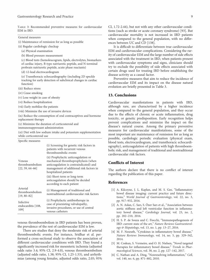
Preventive measures that aim to reduce the incidence ofcardiovascular EIM and its impact on the disease naturalevolution are briefly presented in Table 3.
13. Conclusions
Cardiovascular manifestations in patients with IBD,although rare, are characterized by a higher incidencewhen compared to the general population, and they occurdue to the effects of chronic or acute inflammation, drugtoxicity, or genetic predisposition. Early recognition helpsprevent complications and minimize the impact on thedisease’s natural course. Among the primary preventivemeasures for cardiovascular manifestations, some of themost important are maintenance of remission for as long aspossible, cardiologic periodic evaluation (physical exam,blood tests, electrocardiogram, and transthoracic echocardi-ography), anticoagulation of patients with high thromboem-bolic risk, and management of traditional and nontraditionalcardiovascular risk factors.
Conflicts of Interest
The authors declare that there is no conflict of interestregarding the publication of this paper.
References
[1] A. Kilcoyne, J. L. Kaplan, and M. S. Gee, “Inflammatorybowel disease imaging: current practice and future direc-tions,” World Journal of Gastroenterology, vol. 22, no. 3,pp. 917–932, 2016.
[2] A. N. Aslan, C. Sarı, S. Özer Sarı et al., “Association betweenaortic stiffness and left ventricular function in inflamma-tory bowel disease,” Cardiology Journal, vol. 23, no. 2,pp. 202–210, 2016.
[3] H. S. P. de Souza and C. Fiocchi, “Immunopathogenesis ofIBD: current state of the art,” Nature Reviews Gastroenterol-ogy & Hepatology, vol. 13, no. 1, pp. 13–27, 2016.
[4] M. F. Neurath, “Cytokines in inflammatory bowel disease,”Nature Reviews Immunology, vol. 14, no. 5, pp. 329–342,2014.
[5] M. Coskun, S. Vermeire, and O. H. Nielsen, “Novel targetedtherapies for inflammatory bowel disease,” Trends in Phar-macological Sciences, vol. 38, no. 2, pp. 127–142, 2017.
[6] C. Nathan and A. Ding, “Nonresolving inflammation,” Cell,vol. 140, no. 6, pp. 871–882, 2010.
Table 3: Recommended preventive measures for cardiovascularEIM in IBD.
General measures
(i) Maintenance of remission for as long as possible
(ii) Regular cardiologic checkup
(a) Physical examination
(b) Blood pressure measurement
(c) Blood tests (hemoleucogram, lipids, electrolytes, biomarkersof cardiac injury, B-type natriuretic peptide, and N-terminalprobrain natriuretic peptide, acute phase reactants)
(d) 12-lead electrocardiogram
(e) Transthoracic echocardiography (including 2D speckletracking for early detection of subclinical changes in cardiacfunction)
(iii) Reduce stress
(iv) Cease smoking
(v) Lose weight in case of obesity
(vi) Reduce hospitalization
(vii) Early mobilize the patients
(viii) Minimize the use of invasive devices
(ix) Reduce the consumption of oral contraceptives and hormonereplacement therapy
(x) Minimize the duration of corticosteroid andimmunosuppressant administration
(xi) Diet with low sodium intake and potassium supplementationwhile corticosteroid use
Specific measures
Venousthromboembolism[22, 59, 64–66]
(i) Screening for genetic risk factors inpatients with recurrent venousthromboembolic events
(ii) Prophylactic anticoagulation ormechanical thromboprophylaxis (whenanticoagulation is contraindicated) andmanagement of additional risk factors inhospitalized patients
(iii) Short-term or long-termanticoagulation should be decidedaccording to each patient
Arterialthromboembolism[19, 80–82]
(i) Management of traditional andnontraditional cardiovascular risk factors
Infectiveendocarditis [108,109]
(i) Prophylactic antibiotherapy incase of preexisting valvulopathy,invasive procedures, or use of centralvenous catheters
9Gastroenterology Research and Practice

[7] H. J. Su, Y. T. Chiu, C. T. Chiu et al., “Inflammatory boweldisease and its treatment in 2018: Global and Taiwanesestatus updates,” Journal of the Formosan Medical Association,2018.
[8] J. D. Olpin, B. P. Sjoberg, S. E. Stilwill, L. E. Jensen,M. Rezvani, and A. M. Shaaban, “Beyond the bowel: extrain-testinal manifestations of inflammatory bowel disease,”Radiographics, vol. 37, no. 4, pp. 1135–1160, 2017.
[9] K. Karmiris, A. Avgerinos, A. Tavernaraki et al., “Prevalenceand characteristics of extra-intestinal manifestations in alarge cohort of Greek patients with inflammatory boweldisease,” Journal of Crohn's and Colitis, vol. 10, no. 4,pp. 429–436, 2016.
[10] E. Vizzardi, E. Sciatti, I. Bonadei et al., “Subclinical cardiacinvolvement in Crohn’s disease and ulcerative colitis: anechocardiographic case-control study,” Panminerva Medica,vol. 58, no. 2, pp. 115–120, 2016.
[11] S. R. Vavricka, A. Schoepfer, M. Scharl, P. L. Lakatos,A. Navarini, and G. Rogler, “Extraintestinal manifestationsof inflammatory bowel disease,” Inflammatory Bowel Dis-eases, vol. 21, no. 8, pp. 1982–1992, 2015.
[12] M. Harbord, V. Annese, S. R. Vavricka et al., “The firstEuropean evidence-based consensus on extra-intestinal man-ifestations in inflammatory bowel disease,” Journal of Crohn'sand Colitis, vol. 10, no. 3, pp. 239–254, 2016.
[13] J. Aarestrup, T. Jess, C. J. Kobylecki, B. G. Nordestgaard, andK. H. Allin, “Cardiovascular risk profile among patients withinflammatory bowel disease: a population-based study ofmore than 100000 individuals,” Journal of Crohn's and Coli-tis, 2018.
[14] T. R. Card, S. M. Langan, and T. P. C. Chu, “Extra-gastroin-testinal manifestations of inflammatory bowel disease may beless common than previously reported,” Digestive Diseasesand Sciences, vol. 61, no. 9, pp. 2619–2626, 2016.
[15] N. E. Mitchell, N. Harrison, Z. Junga, and M. Singla, “Heartunder attack: cardiac manifestations of inflammatory boweldisease,” Inflammatory Bowel Diseases, vol. 24, no. 11,pp. 2322–2326, 2018.
[16] K. H. Katsanos and E. V. Tsianos, “The heart in inflammatorybowel disease,” Annals of Gastroenterology, vol. 15, no. 2,pp. 124–133, 2002.
[17] E. H. Park, B. J. Kim, J. K. Huh et al., “Recurrentmesalazine-induced myopericarditis in a patient with ulcera-tive colitis,” Journal of Cardiovascular Ultrasound, vol. 20,no. 3, pp. 154–156, 2012.
[18] H. T. Sørensen and K. M. Fonager, “Myocarditis and inflam-matory bowel disease. A 16-year Danish nationwide cohortstudy,” Danish Medical Bulletin, vol. 44, no. 4, pp. 442–444,1997.
[19] V. P. Tan, A. Chung, B. P. Yan, and P. R. Gibson, “Venousand arterial disease in inflammatory bowel disease,” Journalof Gastroenterology and Hepatology, vol. 28, no. 7,pp. 1095–1113, 2013.
[20] A. Alatri, A. Schoepfer, N. Fournier et al., “Prevalence and riskfactors for venous thromboembolic complications in the Swissinflammatory bowel disease cohort,” Scandinavian Journal ofGastroenterology, vol. 51, no. 10, pp. 1200–1205, 2016.
[21] L. Bollen, N. Vande Casteele, M. Peeters et al., “The occur-rence of thrombosis in inflammatory bowel disease isreflected in the clot lysis profile,” Inflammatory BowelDiseases, vol. 21, no. 11, pp. 2540–2548, 2015.
[22] P. Zezos, G. Kouklakis, and F. Saibil, “Inflammatory boweldisease and thromboembolism,” World Journal of Gastroen-terology, vol. 20, no. 38, pp. 13863–13878, 2014.
[23] S. Singh, H. Singh, E. V. Loftus Jr., and D. S. Pardi, “Risk ofcerebrovascular accidents and ischemic heart disease inpatients with inflammatory bowel disease: a systematicreview and meta-analysis,” Clinical Gastroenterology andHepatology, vol. 12, no. 3, pp. 382–393.e1, 2014.
[24] S. L. Kristensen, O. Ahlehoff, J. Lindhardsen et al., “Diseaseactivity in inflammatory bowel disease is associated withincreased risk of myocardial infarction, stroke and cardiovas-cular death – a Danish nationwide cohort study,” PLoS One,vol. 8, no. 2, p. e56944, 2013.
[25] W. Feng, G. Chen, D. Cai, S. Zhao, J. Cheng, and H. Shen,“Inflammatory bowel disease and risk of ischemic heartdisease: an updated meta‐analysis of cohort studies,” Journalof the American Heart Association, vol. 6, no. 8, articlee005892, 2017.
[26] A. H. Katsanos, M. Kosmidou, S. Giannopoulos et al., “Cere-bral arterial infarction in inflammatory bowel diseases,”European Journal of Internal Medicine, vol. 25, no. 1,pp. 37–44, 2014.
[27] T.-Y. Lin, Y.-G. Chen, C.-L. Lin, W.-S. Huang, andC.-H. Kao, “Inflammatory bowel disease increases the riskof peripheral arterial disease: a nationwide cohort study,”Medicine, vol. 94, no. 52, p. e2381, 2015.
[28] F. Agüero, G. González-Zobl, J. M. Baena-Díez et al., “Preva-lence of lower extremity peripheral arterial disease in individ-uals with chronic immune mediated inflammatorydisorders,” Atherosclerosis, vol. 242, no. 1, pp. 1–7, 2015.
[29] I. E. Koutroubakis, “Therapy insight: vascular complicationsin patients with inflammatory bowel disease,”Nature ClinicalPractice Gastroenterology & Hepatology, vol. 2, no. 6,pp. 266–272, 2005.
[30] S. Aniwan, D. S. Pardi, W. J. Tremaine, and E. V. Loftus Jr,“Increased risk of acute myocardial infarction and heart fail-ure in patients with inflammatory bowel diseases,” ClinicalGastroenterology and Hepatology, vol. 16, no. 10, pp. 1607–1615.e1, 2018.
[31] S. L. Kristensen, O. Ahlehoff, J. Lindhardsen et al., “Inflam-matory bowel disease is associated with an increased risk ofhospitalization for heart failure: a Danish nationwide cohortstudy,” Circulation: Heart Failure, vol. 7, no. 5, pp. 717–722, 2014.
[32] D. J. Pattanshetty, K. Anna, R. D. Gajulapalli, and R. S. R.Sappati-Biyyani, “Inflammatory bowel “cardiac” disease:point prevalence of atrial fibrillation in inflammatory boweldisease population,” Saudi Journal of Gastroenterology,vol. 21, no. 5, pp. 325–329, 2015.
[33] S. L. Kristensen, J. Lindhardsen, O. Ahlehoff et al., “Increasedrisk of atrial fibrillation and stroke during active stages ofinflammatory bowel disease: a nationwide study,” Europace,vol. 16, no. 4, pp. 477–484, 2014.
[34] T. H. Efe, T. Cimen, A. G. Ertem et al., “Atrial electromechan-ical properties in inflammatory bowel disease,” Echocardiog-raphy, vol. 33, no. 9, pp. 1309–1316, 2016.
[35] C. Berkelhammer, J. Andrejic, and A.Mohammed, “Endocar-ditis in Crohn’s disease,” Inflammatory Bowel Diseases,vol. 15, no. 9, pp. 1293-1294, 2009.
[36] G. Kreuzpaintner, D. Horstkotte, A. Heyll, B. Lösse, andG. Strohmeyer, “Increased risk of bacterial endocarditis in
10 Gastroenterology Research and Practice

inflammatory bowel disease,” The American Journal ofMedicine, vol. 92, no. 4, pp. 391–395, 1992.
[37] G. Bragagni, R. Brogna, P. Franceschetti, and G. Zoli, “Car-diac involvement in Crohn’s disease: echocardiographicstudy,” Journal of Gastroenterology and Hepatology, vol. 22,no. 1, pp. 18–22, 2007.
[38] R. Kusunoki, S. Ishihara, M. Sato et al., “Rare case ofTakayasu’s arteritis associated with Crohn’s disease,” Inter-nal Medicine, vol. 50, no. 15, pp. 1581–1585, 2011.
[39] S. Le Mener, S. Huynh-Moynot, J. A. Bronstein,U. Vinsonneau, and Z. Richert, “Les myocardites aiguëscompliquant les maladies inflammatoires chroniques del’intestin : à propos de deux observations,” La Revue deMédecine Interne, vol. 33, no. 10, pp. 583–585, 2012.
[40] A. L. P. Caforio, S. Pankuweit, E. Arbustini et al., “Currentstate of knowledge on aetiology, diagnosis, management,and therapy of myocarditis: a position statement of theEuropean Society of Cardiology Working Group on Myo-cardial and Pericardial Diseases,” European Heart Journal,vol. 34, no. 33, pp. 2636–2648, 2013.
[41] J. Asadi, S. S. Bhandari, and N. Ahmed, “Mesalazine-inducedmyopericarditis in a patient with ulcerative colitis,” EchoResearch and Practice, vol. 5, no. 1, pp. K1–K5, 2017.
[42] F. Dominguez, U. Kühl, B. Pieske, P. Garcia-Pavia, andC. Tschöpe, “Update on myocarditis and inflammatory car-diomyopathy: reemergence of endomyocardial biopsy,”Revista Española de Cardiología, vol. 69, no. 2, pp. 178–187,2016.
[43] W. Bracamonte-Baran and D. Čiháková, “Cardiac autoim-munity: myocarditis,” Advances in Experimental Medicineand Biology, vol. 1003, pp. 187–221, 2017.
[44] G. Brown, “5-Aminosalicylic acid-associated myocarditis andpericarditis: a narrative review,” The Canadian Journal ofHospital Pharmacy, vol. 69, no. 6, pp. 466–472, 2016.
[45] M. Moonen and P. Lancellotti, “La myocardite,” RevueMédicale de Liège, vol. 73, no. 5-6, pp. 269–276, 2018.
[46] I. Kindermann, C. Barth, F. Mahfoud et al., “Update on myo-carditis,” Journal of the American College of Cardiology,vol. 59, no. 9, pp. 779–792, 2012.
[47] V. C. Varnavas, N. Reinsch, M. Perrey et al., “Recurrent lym-phocytic myocarditis in a young male with ulcerative colitis,”European Journal of Medical Research, vol. 19, no. 1, p. 11,2014.
[48] L. T. Cooper, K. L. Baughman, A. M. Feldman et al., “The roleof endomyocardial biopsy in the management of cardiovas-cular disease: a scientific statement from the American HeartAssociation, the American College of Cardiology, and theEuropean Society of Cardiology. Endorsed by the Heart Fail-ure Society of America and the Heart Failure Association ofthe European Society of Cardiology,” Journal of the AmericanCollege of Cardiology, vol. 50, no. 19, pp. 1914–1931, 2007.
[49] P. Ponikowski, A. A. Voors, S. D. Anker et al., “2016 ESCguidelines for the diagnosis and treatment of acute andchronic heart failure,” European Journal of Heart Failure,vol. 18, no. 8, pp. 891–975, 2016.
[50] A. Klein and R. Eliakim, “Non steroidal anti-inflammatorydrugs and inflammatory bowel disease,” Pharmaceuticals(Basel), vol. 3, no. 4, pp. 1084–1092, 2010.
[51] H. P. Schultheiss, U. Kühl, and L. T. Cooper, “The manage-ment of myocarditis,” European Heart Journal, vol. 32,no. 21, pp. 2616–2625, 2011.
[52] G. Van Assche, A. Dignass, B. Bokemeyer et al., “SecondEuropean evidence-based consensus on the diagnosis andmanagement of ulcerative colitis part 3: special situations,”Journal of Crohn's and Colitis, vol. 7, no. 1, pp. 1–33, 2013.
[53] M. Giannotta, G. Tapete, G. Emmi, E. Silvestri, and M. Milla,“Thrombosis in inflammatory bowel diseases: what’s thelink?,” Thrombosis Journal, vol. 13, no. 1, p. 14, 2015.
[54] D. Kohoutova, P. Moravkova, P. Kruzliak, and J. Bures,“Thromboembolic complications in inflammatory boweldisease,” Journal of Thrombosis and Thrombolysis, vol. 39,no. 4, pp. 489–498, 2015.
[55] V. F. Tapson, “Acute pulmonary embolism,” The NewEngland Journal of Medicine, vol. 358, no. 10, pp. 1037–1052, 2008.
[56] D. Owczarek, D. Cibor, M. K. Głowacki, T. Rodacki, andT. Mach, “Inflammatory bowel disease: epidemiology,pathology and risk factors for hypercoagulability,” WorldJournal of Gastroenterology, vol. 20, no. 1, pp. 53–63, 2014.
[57] R. Schicho, G. Marsche, and M. Storr, “Cardiovascular com-plications in inflammatory bowel disease,” Current Drug Tar-gets, vol. 16, no. 3, pp. 181–188, 2015.
[58] F. Scaldaferri, S. Lancellotti, M. Pizzoferrato, and R. DeCristofaro, “Haemostatic system in inflammatory boweldiseases: new players in gut inflammation,” World Journalof Gastroenterology, vol. 17, no. 5, pp. 594–608, 2011.
[59] S. Danese, C. de la Motte, and C. Fiocchi, “Platelets in inflam-matory bowel disease: clinical, pathogenic, and therapeuticimplications,” The American Journal of Gastroenterology,vol. 99, no. 5, pp. 938–945, 2004.
[60] O. A. Hatoum and D. G. Binion, “The vasculature andinflammatory bowel disease: contribution to pathogenesisand clinical pathology,” Inflammatory Bowel Diseases,vol. 11, no. 3, pp. 304–313, 2005.
[61] N. Chande, “Prevention of venous thromboembolism inhospitalized patients with inflammatory bowel disease,”Inflammatory Bowel Diseases, vol. 19, no. 3, pp. 669–671,2013.
[62] S. Alhassan, A. Pelinescu, V. Gandhi, M. Naddour, A. C.Singh, and E. Bihler, “Clinical presentation and risk factorsof venous thromboembolic disease,” Critical Care NursingQuarterly, vol. 40, no. 3, pp. 201–209, 2017.
[63] P. Papay, W. Miehsler, H. Tilg et al., “Clinical presentation ofvenous thromboembolism in inflammatory bowel disease,”Journal of Crohn's and Colitis, vol. 7, no. 9, pp. 723–729, 2013.
[64] G. C. Nguyen, C. N. Bernstein, A. Bitton et al., “Consensusstatements on the risk, prevention, and treatment of venousthromboembolism in inflammatory bowel disease: Canadianassociation of gastroenterology,” Gastroenterology, vol. 146,no. 3, pp. 835–848.e6, 2014.
[65] W. H. Geerts, D. Bergqvist, G. F. Pineo et al., “Prevention ofvenous thromboembolism: American College of ChestPhysicians evidence-based clinical practice guidelines (8thEdition),” Chest Journal, vol. 133, no. 6, Supplement,pp. 381S–453S, 2008.
[66] N. L. Zitomersky, M. Verhave, and C. C. Trenor III, “Throm-bosis and inflammatory bowel disease: a call for improvedawareness and prevention,” Inflammatory Bowel Diseases,vol. 17, no. 1, pp. 458–470, 2011.
[67] C. Rungoe, N. Nyboe Andersen, and T. Jess, “Inflammatorybowel disease and risk of coronary heart disease,” Trends inCardiovascular Medicine, vol. 25, no. 8, pp. 699–704, 2015.
11Gastroenterology Research and Practice

[68] A. J. Yarur, A. R. Deshpande, D. M. Pechman, L. Tamariz,M. T. Abreu, and D. A. Sussman, “Inflammatory bowel dis-ease is associated with an increased incidence of cardiovascu-lar events,” The American Journal of Gastroenterology,vol. 106, no. 4, pp. 741–747, 2011.
[69] A. B. Reiss, N. M. Siegart, and J. De Leon, “Interleukin-6 inatherosclerosis: atherogenic or atheroprotective?,” ClinicalLipidology, vol. 12, no. 1, pp. 14–23, 2017.
[70] J. W. Harper and T. L. Zisman, “Interaction of obesity andinflammatory bowel disease,” World Journal of Gastroenter-ology, vol. 22, no. 35, pp. 7868–7881, 2016.
[71] I. D'Odorico, S. di Bella, J. Monticelli, D. R. Giacobbe,E. Boldock, and R. Luzzati, “Role of fecal microbiotatransplantation in inflammatory bowel disease,” Journal ofDigestive Diseases, vol. 19, no. 6, pp. 322–334, 2018.
[72] C. Manichanh, N. Borruel, F. Casellas, and F. Guarner, “Thegut microbiota in IBD,” Nature Reviews Gastroenterology &Hepatology, vol. 9, no. 10, pp. 599–608, 2012.
[73] A. Wilson, W. A. Teft, B. L. Morse et al., “Erratum to: tri-methylamine-N-oxide: a novel biomarker for the identifica-tion of inflammatory bowel disease,” Digestive Diseases andSciences, vol. 61, no. 1, p. 325, 2016.
[74] W. H. W. Tang, D. Y. Li, and S. L. Hazen, “Dietary metabo-lism, the gut microbiome, and heart failure,” Nature ReviewsCardiology, 2018.
[75] P. Kruzliak, J. Novák, M. Novák, and G. J. Fodor, “Role of cal-protectin in cardiometabolic diseases,” Cytokine & GrowthFactor Reviews, vol. 25, no. 1, pp. 67–75, 2014.
[76] L. Zanoli, S. Rastelli, A. Granata et al., “Arterial stiffness ininflammatory bowel disease: a systematic review andmeta-analysis,” Journal of Hypertension, vol. 34, no. 5,pp. 822–829, 2016.
[77] S. Galluzzo, G. Patti, G. Dicuonzo et al., “Association betweenNOD2/CARD15 polymorphisms and coronary artery dis-ease: a case-control study,” Human Immunology, vol. 72,no. 8, pp. 636–640, 2011.
[78] R. S. R. Sappati Biyyani, B. S. Putka, and K. D. Mullen, “Dys-lipidemia and lipoprotein profiles in patients with inflamma-tory bowel disease,” Journal of Clinical Lipidology, vol. 4,no. 6, pp. 478–482, 2010.
[79] S. Pak, J. Linares, Y. Yatsynovich et al., “Spontaneous ventric-ular thrombosis in patients with inflammatory bowel disease,”Cardiology in the Young, vol. 28, no. 3, pp. 351–353, 2018.
[80] W. J. Powers, A. A. Rabinstein, T. Ackerson et al., “2018guidelines for the early management of patients with acuteischemic stroke: a guideline for healthcare professionals fromthe American Heart Association/American Stroke Associa-tion,” Stroke, vol. 49, no. 3, pp. e46–e110, 2018.
[81] B. Ibanez, S. James, S. Agewall et al., “2017 ESC guidelines forthe management of acute myocardial infarction in patientspresenting with ST-segment elevation: the task force for themanagement of acute myocardial infarction in patients pre-senting with ST-segment elevation of the European Societyof Cardiology (ESC),” European Heart Journal, vol. 39,no. 2, pp. 119–177, 2018.
[82] M. Bala, J. Kashuk, E. E. Moore et al., “Acute mesentericischemia: guidelines of the World Society of EmergencySurgery,” World Journal of Emergency Surgery, vol. 12,no. 1, p. 38, 2017.
[83] K. H. Katsanos, D. K. Christodoulou, K. Pappas, C. Pappas,and E. V. Tsianos, “Electrocardiograph abnormalities in
patients with active inflammatory bowel disease,” Annals ofGastroenterology, vol. 20, no. 4, pp. 275–281, 2007.
[84] M. De Simone, U. Cioffi, E. Contessini-Avesani et al., “Ele-vated serum procollagen type III peptide in splanchnic andperipheral circulation of patients with inflammatory boweldisease submitted to surgery,” BMC Gastroenterology, vol. 4,no. 1, 2004.
[85] O. A. Hatoum, D. G. Binion, M. F. Otterson, and D. D.Gutterman, “Acquired microvascular dysfunction in inflam-matory bowel disease: loss of nitric oxide-mediated vasodila-tion,” Gastroenterology, vol. 125, no. 1, pp. 58–69, 2003.
[86] D. Waśko-Czopnik and L. Paradowski, “The influence ofdeficiencies of essential trace elements and vitamins on thecourse of Crohn’s disease,” Advances in Clinical and Experi-mental Medicine, vol. 21, no. 1, pp. 5–11, 2012.
[87] A. Cincin, M. Sunbul, T. Kivrak et al., “Evaluation of cardiacfunction by two-dimensional speckle tracking echocardiogra-phy in ulcerative colitis patients,” Digestive Diseases andSciences, vol. 59, no. 12, pp. 3004–3011, 2014.
[88] T. Kivrak, M. Sunbul, A. Cincin et al., “Two-dimensionalspeckle tracking echocardiography is useful in early detectionof left ventricular impairment in patients with Crohn’sdisease,” European Review for Medical and PharmacologicalSciences, vol. 20, no. 15, pp. 3249–3254, 2016.
[89] K. O. Hensel, F. E. Abellan Schneyder, L. Wilke, A. Heusch,S. Wirth, and A. C. Jenke, “Speckle tracking stress echocardi-ography uncovers early subclinical cardiac involvement inpediatric patients with inflammatory bowel diseases,” Scien-tific Reports, vol. 7, no. 1, p. 2966, 2017.
[90] S. F. Nagueh, O. A. Smiseth, C. P. Appleton et al., “Recom-mendations for the evaluation of left ventricular diastolicfunction by echocardiography: an update from the AmericanSociety of Echocardiography and the European Associationof Cardiovascular Imaging,” Journal of the American Societyof Echocardiography, vol. 29, no. 4, pp. 277–314, 2016.
[91] O. Gedikli, A. Dogan, I. Altuntas et al., “Inflammatorymarkers according to types of atrial fibrillation,” InternationalJournal of Cardiology, vol. 120, no. 2, pp. 193–197, 2007.
[92] H. A. Bornaun, N. Yılmaz, G. Kutluk et al., “ProlongedP-wave and QT dispersion in children with inflammatorybowel disease in remission,” BioMed Research International,vol. 2017, Article ID 6960810, 7 pages, 2017.
[93] A. R. M. Sridhar, S. Parasa, U. Navaneethan, M. D. Cro-well, and K. Olden, “Comprehensive study of cardiovascu-lar morbidity in hospitalized inflammatory bowel diseasepatients,” Journal of Crohn's and Colitis, vol. 5, no. 4,pp. 287–294, 2011.
[94] D. J. Pattanshetty, R. D. Gajulapalli, K. Anna, and R. S. S.Biyyani, “Prevalence of QT interval prolongation in inflam-matory bowel disease,” The Turkish Journal of Gastroenterol-ogy, vol. 27, no. 2, pp. 136–142, 2016.
[95] T. Engel, S. Ben-Horin, and M. Beer-Gabel, “Autonomic dys-function correlates with clinical and inflammatory activity inpatients with Crohn’s disease,” Inflammatory Bowel Diseases,vol. 21, no. 10, pp. 2320–2326, 2015.
[96] Y. Dogan, A. Soylu, G. A. Eren et al., “Evaluation of QT and Pwave dispersion and mean platelet volume among inflamma-tory bowel disease patients,” International Journal of MedicalSciences, vol. 8, no. 7, pp. 540–546, 2011.
[97] G. Nar, B. Ergul, G. Aksan, and S. Inci, “Assessment of atrialelectromechanical delay and left atrial mechanical functions
12 Gastroenterology Research and Practice

in patients with ulcerative colitis,” Echocardiography, vol. 33,no. 7, pp. 970–976, 2016.
[98] T. T. Issac, H. Dokainish, and N. M. Lakkis, “Role of inflam-mation in initiation and perpetuation of atrial fibrillation: asystematic review of the published data,” Journal of theAmerican College of Cardiology, vol. 50, no. 21,pp. 2021–2028, 2007.
[99] C. P. Anand, A. Al-Juburi, and B. Sandeep, “Heart blockoccurring during infliximab infusion: a report of two cases,”The American Journal of Gastroenterology, vol. 98, no. 9,p. S144, 2003.
[100] M. Curione, M. Barbato, S. Amato et al., “Atrioventricularblock associated with Crohn’s relapsing colitis in a12-year-old child,” Inflammatory Bowel Diseases, vol. 16,no. 3, pp. 373-374, 2010.
[101] M. Gesualdo, P. Scicchitano, S. Carbonara et al., “The associ-ation between cardiac and gastrointestinal disorders: causalor casual link?,” Journal of Cardiovascular Medicine, vol. 17,no. 5, pp. 330–338, 2016.
[102] P. M. Seferović, A. D. Ristić, R. Maksimović et al., “Cardiacarrhythmias and conduction disturbances in autoimmunerheumatic diseases,” Rheumatology, vol. 45, Supplement_4,pp. iv39–iv42, 2006.
[103] U. Nussinovitch, J. Freire de Carvalho, R. M. R. Pereira, andY. Shoenfeld, “Glucocorticoids and the cardiovascular sys-tem: state of the art,” Current Pharmaceutical Design,vol. 16, no. 32, pp. 3574–3585, 2010.
[104] P. Eickhoff, T. Fazekas, A. Attarbaschi, and T. Binder, “Mitralvalve endocarditis caused by ulcerative colitis followed byseptic embolic occlusion of the superior mesenteric artery,”European Journal of Echocardiography, vol. 10, no. 6,pp. 806-807, 2009.
[105] S. Singh, V. Goyal, P. Padhi, and E. Aoun, “Bacteroides fragilisendocarditis in a patient with Crohn’s disease,” Case Reports,vol. 2013, article bcr2013009248, 2013.
[106] W. Uchida, M. Mutsuga, H. Ito, H. Oshima, and A. Usui,“Nonbacterial thrombotic endocarditis associated withCrohn disease,” The Annals of Thoracic Surgery, vol. 105,no. 5, pp. e199–e201, 2018.
[107] G. Habib, P. Lancellotti, M. J. Antunes et al., “2015 ESCguidelines for the management of infective endocarditis:the task force for the management of infective endocardi-tis of the European Society of Cardiology (ESC). Endorsedby: European Association for Cardio-Thoracic Surgery(EACTS), the European Association of Nuclear Medicine(EANM),” European Heart Journal, vol. 36, no. 44,pp. 3075–3128, 2015.
[108] R. Giacomo, T. Gianluca, T. Luca, and C. Maurizio, “A severebut predictable complication of an inflammatory boweldisease∗,” World Journal of Cardiovascular Surgery, vol. 3,no. 5, pp. 129-130, 2013.
[109] D. R. Ramsdale, T. S. J. Elliott, P. Wright et al., “Guidanceon the prophylaxis and treatment of infective endocarditisin adults,” http://www.bcs.com/restricted/registered/affiliates/endocarditis_guidance.pdf.
[110] D. Lacey and P. Bouillet, “Deregulation of TNF expressioncan also cause heart valve disease,” Cytokine, vol. 77,pp. 248-249, 2016.
[111] K. A. LaBresh, E. V. Lally, S. C. Sharma, and G. Ho Jr,“Two-dimensional echocardiographic detection of preclini-cal aortic root abnormalities in rheumatoid variant
diseases,” The American Journal of Medicine, vol. 78,no. 6, pp. 908–912, 1985.
[112] I. Ozsöyler, L. Yilik, S. Bozok, C. Ozbek, and A. Gürbüz,“Cardiovascular involvement in Crohn’s disease in theabsence of ankylosing spondylitis,” Heart and Vessels,vol. 20, no. 4, pp. 164–166, 2005.
[113] M. E. Evangelopoulos, S. Toumanidis, D. Sotou et al., “Mitralvalve prolapse in young healthy individuals. An early index ofautoimmunity?,” Lupus, vol. 18, no. 5, pp. 436–440, 2009.
[114] N. Minami, H. Nakase, T. Yoshino et al., “Effect of infliximabon inflammatory bowel disease with Takayasu arteritis: caseseries and review of the literature,” Clinical Journal of Gastro-enterology, vol. 6, no. 3, pp. 226–230, 2013.
[115] S. Akiyama, T. Fujii, K. Matsuoka et al., “Endoscopic featuresand genetic background of inflammatory bowel disease com-plicated with Takayasu arteritis,” Journal of Gastroenterologyand Hepatology, vol. 32, no. 5, pp. 1011–1017, 2017.
[116] E. Seyahi, “Takayasu arteritis: an update,” Current Opinion inRheumatology, vol. 29, no. 1, pp. 51–56, 2017.
[117] M. Isobe, “Takayasu arteritis revisited: current diagnosis andtreatment,” International Journal of Cardiology, vol. 168,no. 1, pp. 3–10, 2013.
[118] A. Sy, N. Khalidi, N. Dehghan et al., “Vasculitis in patientswith inflammatory bowel diseases: a study of 32 patientsand systematic review of the literature,” Seminars in Arthritisand Rheumatism, vol. 45, no. 4, pp. 475–482, 2016.
[119] A. B. Pithadia and S. Jain, “Treatment of inflammatory boweldisease (IBD),” Pharmacological Reports, vol. 63, no. 3,pp. 629–642, 2011.
[120] J. F. Jackson and S. V. Sitaraman, “Pericarditis as the present-ing sign of Crohn’s disease,” Inflammatory Bowel Diseases,vol. 11, no. 1, pp. 81-82, 2005.
[121] S. Asirvatham, C. Sebastian, and U. Thadani, “Severe symp-tomatic sinus bradycardia associated with mesalamine use,”American Journal of Gastroenterology, vol. 93, no. 3,pp. 470-471, 1998.
[122] H. Schacke, W. D. Döcke, and K. Asadullah, “Mechanismsinvolved in the side effects of glucocorticoids,” Pharmacology& Therapeutics, vol. 96, no. 1, pp. 23–43, 2002.
[123] J. N. Hoes, J. W. G. Jacobs, S. M. M. Verstappen, J. W. J.Bijlsma, and G. J. M. G. van der Heijden, “Adverse eventsof low- to medium-dose oral glucocorticoids in inflammatorydiseases: a meta-analysis,” Annals of the Rheumatic Diseases,vol. 68, no. 12, pp. 1833–1838, 2009.
[124] R. M. Stanbury and E. M. Graham, “Systemic corticosteroidtherapy—side effects and their management,” British Journalof Ophthalmology, vol. 82, no. 6, pp. 704–708, 1998.
[125] L. Wei, T. M. MacDonald, and B. R. Walker, “Taking gluco-corticoids by prescription is associated with subsequentcardiovascular disease,” Annals of Internal Medicine,vol. 141, no. 10, pp. 764–770, 2004.
[126] P. Welsh, G. Grassia, S. Botha, N. Sattar, and P. Maffia, “Tar-geting inflammation to reduce cardiovascular disease risk: arealistic clinical prospect?,” British Journal of Pharmacology,vol. 174, no. 22, pp. 3898–3913, 2017.
[127] P. Dogan, E. Grbovic, S. Inci, F. Bayraktar, and K. Cagli,“Azathioprine-induced atrial fibrillation,” Intractable & RareDiseases Research, vol. 4, no. 4, pp. 207–209, 2015.
[128] A. Cassinotti, A. Massari, E. Ferrara et al., “New onset of atrialfibrillation after introduction of azathioprine in ulcerativecolitis: case report and review of the literature,” European
13Gastroenterology Research and Practice

Journal of Clinical Pharmacology, vol. 63, no. 9, pp. 875–878,2007.
[129] A. Yuri Gasparyan, L. Ayvazyan, G. Cocco, and G. D. Kitas,“Adverse cardiovascular effects of antirheumatic drugs:implications for clinical practice and research,” CurrentPharmaceutical Design, vol. 18, no. 11, pp. 1543–1555,2012.
[130] G. Demirtaş-Ertan, A. T. Rowshani, and I. J. ten Berge,“Azathioprine-induced shock in a patient suffering fromundifferentiated erosive oligoarthritis,” The NetherlandsJournal of Medicine, vol. 64, no. 4, pp. 124–126, 2006.
[131] https://www.drugs.com/sfx/methotrexate-side-effects.html2018 November.
[132] P. E. Lazzerini, P. L. Capecchi, M. Galeazzi, andF. Laghi-Pasini, “Biologic drugs and arrhythmic risk inchronic inflammatory arthritis: the good and the bad,”Immunologic Research, vol. 65, no. 1, pp. 262–275, 2017.
[133] W. Miehsler, G. Novacek, H. Wenzl et al., “A decade of inflix-imab: the Austrian evidence based consensus on the safe useof infliximab in inflammatory bowel disease,” Journal ofCrohn's and Colitis, vol. 4, no. 3, pp. 221–256, 2010.
[134] L. Tomáš, I. Lazúrová, L. Pundová et al., “Acute andlong-term effect of infliximab on humoral and echocardio-graphic parameters in patients with chronic inflammatorydiseases,” Clinical Rheumatology, vol. 32, no. 1, pp. 61–66,2013.
[135] L. Zanoli, S. Rastelli, G. Inserra et al., “Increased arterial stiff-ness in inflammatory bowel diseases is dependent uponinflammation and reduced by immunomodulatory drugs,”Atherosclerosis, vol. 234, no. 2, pp. 346–351, 2014.
[136] S. D. Thapa, H. Hadid, W. Imam, J. Schairer, and S. M. Jafri,“Effect of inflammatory bowel disease-related characteristicsand treatment interventions on cardiovascular disease inci-dence,” The American Journal of the Medical Sciences,vol. 350, no. 3, pp. 175–180, 2015.
[137] M. Zuin, G. Rigatelli, G. del Favero et al., “Cardiovascular dis-ease in patients with inflammatory bowel disease: an issue inno guidelines land,” International Journal of Cardiology,vol. 222, pp. 984-985, 2016.
[138] H. H. Sun and F. Tian, “Inflammatory bowel disease and car-diovascular disease incidence andmortality: a meta-analysis,”European Journal of Preventive Cardiology, vol. 25, no. 15,pp. 1623–1631, 2018.
14 Gastroenterology Research and Practice

Stem Cells International
Hindawiwww.hindawi.com Volume 2018
Hindawiwww.hindawi.com Volume 2018
MEDIATORSINFLAMMATION
of
EndocrinologyInternational Journal of
Hindawiwww.hindawi.com Volume 2018
Hindawiwww.hindawi.com Volume 2018
Disease Markers
Hindawiwww.hindawi.com Volume 2018
BioMed Research International
OncologyJournal of
Hindawiwww.hindawi.com Volume 2013
Hindawiwww.hindawi.com Volume 2018
Oxidative Medicine and Cellular Longevity
Hindawiwww.hindawi.com Volume 2018
PPAR Research
Hindawi Publishing Corporation http://www.hindawi.com Volume 2013Hindawiwww.hindawi.com
The Scientific World Journal
Volume 2018
Immunology ResearchHindawiwww.hindawi.com Volume 2018
Journal of
ObesityJournal of
Hindawiwww.hindawi.com Volume 2018
Hindawiwww.hindawi.com Volume 2018
Computational and Mathematical Methods in Medicine
Hindawiwww.hindawi.com Volume 2018
Behavioural Neurology
OphthalmologyJournal of
Hindawiwww.hindawi.com Volume 2018
Diabetes ResearchJournal of
Hindawiwww.hindawi.com Volume 2018
Hindawiwww.hindawi.com Volume 2018
Research and TreatmentAIDS
Hindawiwww.hindawi.com Volume 2018
Gastroenterology Research and Practice
Hindawiwww.hindawi.com Volume 2018
Parkinson’s Disease
Evidence-Based Complementary andAlternative Medicine
Volume 2018Hindawiwww.hindawi.com
Submit your manuscripts atwww.hindawi.com