cardiopulmonary exercise testing - a who collaborating ... exercise testing dm seminar 25 feb2005....
TRANSCRIPT

Cardiopulmonary Exercise Testing
DM Seminar
25 Feb2005

CPET
• Introduction• Indications• Technical aspects• Physiologic basis• Exercise limitation• Interpretation of CPET• Future directions
Introduction
• Initially was tool of research physiologists• Has become tool to help the clinicians in
evaluating undiagnosed exercise intolerance or exercise related symptoms
• When questions remain after clinical examination and basic clinical data including CXR,PFT and resting ECG

Introduction
• Provides global assessment of integrative exercise response involving pulmonary, cardiovascular,hematopoietic,neuropsycho-logical and skeletal muscle systems
• Resting cardio-pulmonary function tests cannot predict the exercise performance and functional capacity
Field tests
• 6 min walk test and shuttle test easy to perform , related to activities of daily living
• lack of reference values • absence of physiological measures • healthy subjects fairly good correlation with VO2
max is observed• Pitfalls: occult IHD, combined diseases
Weisman et al Clin Chest Med ,2001

When to do
• Evaluate exercise capacity
• Undiagnosed exercise intolerance
• Cardiovascular diseases
• Respiratory diseases/symptoms (EIA)
• Preoperative evaluation
• Pulmonary rehabilitation
• Impairment/disability assessment

Exercise Intolerance
• Assessment of exercise capacity
• Pathophysiologic basis of exercise limitation
• Contribution of cardiac /respiratory disease
• Symptoms disproportionate to routine tests

Cardio Resp diseases
Cardiovascular COPD/ILD/PVDFunctional classific Functional assessExercise Rx Gas exchangeHeart Tx selection After intervention
Oxygen Rx
ATS /ACCP Statement, 2003
Preoperative evaluation
• Lung resection: VO2 peak less than 50-60% associated with increased morbidity and mortality after lung resection
Morice RC et al Chest 1996• Elderly undergoing major Abdominal
surgery • LVRS for Emphysema
ATS/ACCP statement 2003

Other uses
• Disability assessment: occupational/co-morbid diseases
• Exercise prescription :pulmonary and cardiac rehabilitation
• Evaluation of LVRS : NETT trial used max work rate achieved as primary outcome measure
• Evaluation for lung & heart transplantation
Absolute Contraindications
• Rate of death during testing 2-5/lakh tests– AMI(3-5 days) or unstable angina– Uncontrolled arrhythmia with hemodyn compromise– Syncope– Respiratory or Heart failure– Active endocarditis or myocarditis– Severe AS– Pulmonary embolism or Lower limb DVT – Uncontrolled asthma
ATS /ACCP Statement, 2003
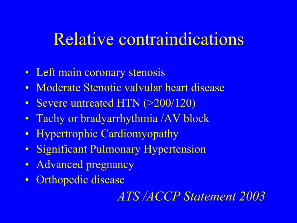
Relative contraindications
• Left main coronary stenosis• Moderate Stenotic valvular heart disease• Severe untreated HTN (>200/120)• Tachy or bradyarrhythmia /AV block• Hypertrophic Cardiomyopathy• Significant Pulmonary Hypertension• Advanced pregnancy• Orthopedic disease
ATS /ACCP Statement 2003
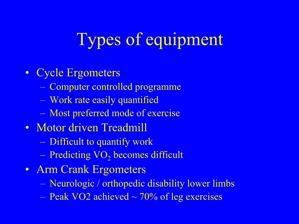
Types of equipment
• Cycle Ergometers– Computer controlled programme– Work rate easily quantified – Most preferred mode of exercise
• Motor driven Treadmill – Difficult to quantify work – Predicting VO2 becomes difficult
• Arm Crank Ergometers– Neurologic / orthopedic disability lower limbs– Peak VO2 achieved ~ 70% of leg exercises
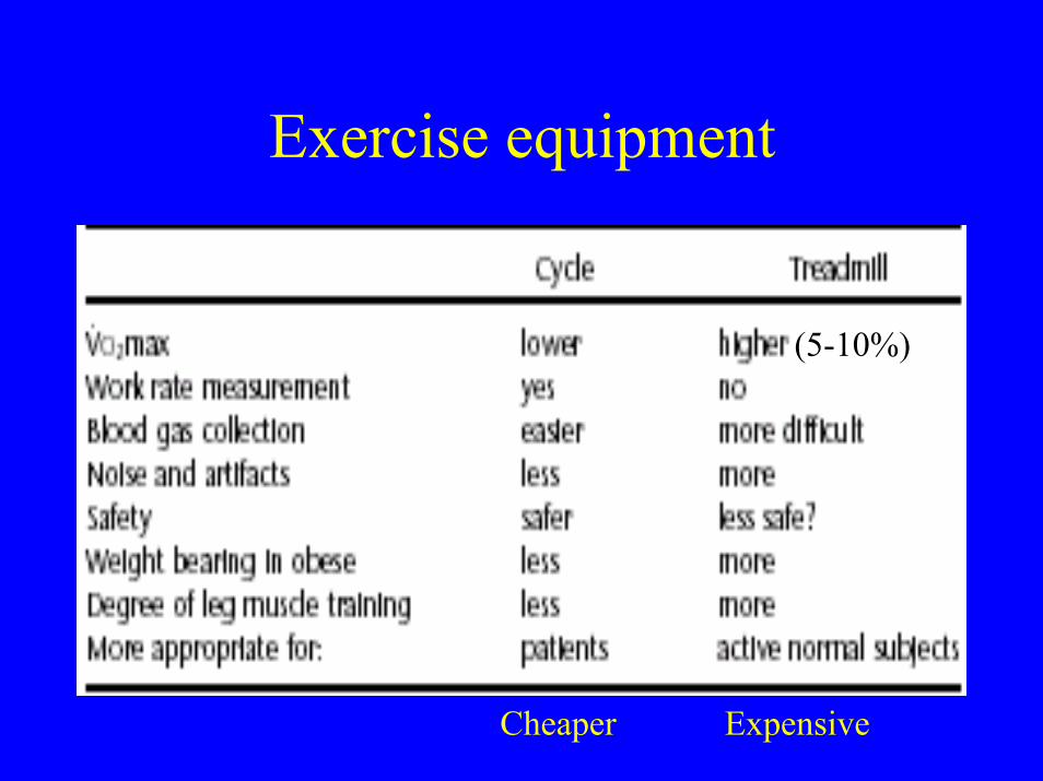
Exercise equipment
(5-10%)
Cheaper Expensive
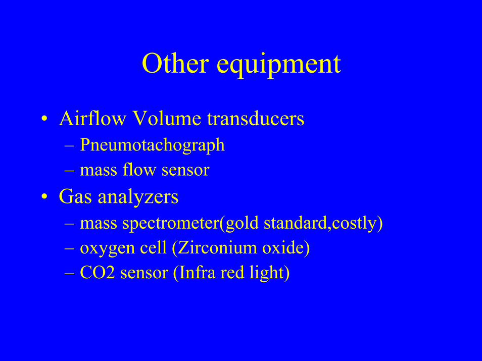
Other equipment
• Airflow Volume transducers – Pneumotachograph– mass flow sensor
• Gas analyzers– mass spectrometer(gold standard,costly)– oxygen cell (Zirconium oxide)– CO2 sensor (Infra red light)
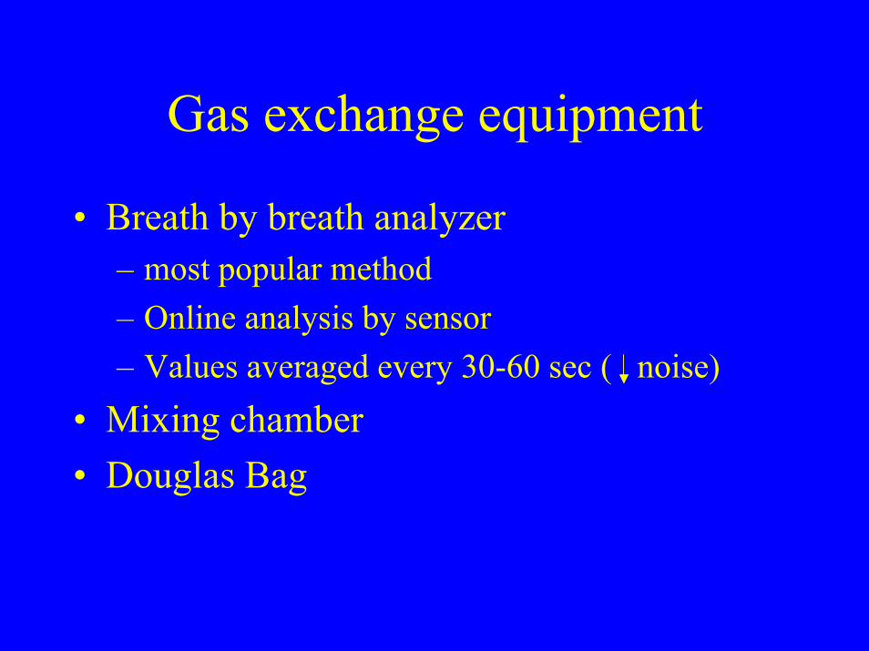
Gas exchange equipment
• Breath by breath analyzer– most popular method– Online analysis by sensor– Values averaged every 30-60 sec ( noise)
• Mixing chamber• Douglas Bag

Other data recorded
• Electrocardiography• Noninvasive blood pressure• Pulse oximetry• Arterial blood gas (if indicated)• Invasive arterial BP
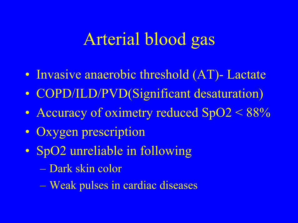
Arterial blood gas
• Invasive anaerobic threshold (AT)- Lactate• COPD/ILD/PVD(Significant desaturation) • Accuracy of oximetry reduced SpO2 < 88%• Oxygen prescription• SpO2 unreliable in following
– Dark skin color– Weak pulses in cardiac diseases

Quality control
• Supervision by cardiologist/Pulmonologist (trained in exercise physiology& testing)
• Calibrate flow transducers• Calibrate breath by breath systems• Calibrate CO2 and O2 analyzers• Noninvasive BP• Healthy member “ test” to validate the measured
VO2, VE and VCO2 with database values
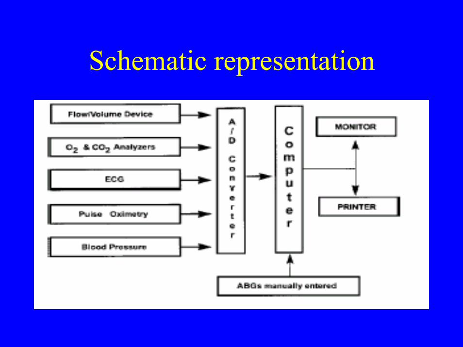
Schematic representation

Incremental Treadmill protocol
• Bruce protocol (suited for healthy,or mild diseases as high WR which increases )
• Modified Naughton protocol(low initial WRgradual build up suitable for patients)
• Balke protocol(constant speed , slope increased 1% every minute)
• Modified Balke protocol(slope increased by constant amount every min)

Incremental protocol

Constant work rate protocol
• Done at 50-70% of maximal work rate• 5-10 min achieves 70-90% VO2 max IET• For assessing response to interventions-
LVRS, LTOT , pulm rehabilitation • Analysis of Exercise FV Loops and
dynamic hyperinflation ,gas exchange kinetics

Stop exercise
• Ischemic chest pain• Ischemic ECG changes• Complex ectopy• Second or third degree heart block• Fall in systolic pressure > 20 mm Hg• Hypertension ( 250 /120 mm Hg)• Symptomatic desaturation: SpO2 <80% • Signs of respiratory failure
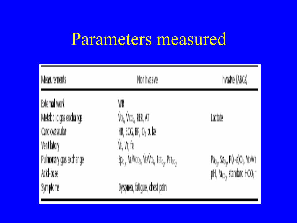
Parameters measured

Oxygen Uptake (VO2)
Factors affectingVO2• Oxygen carrying capacity(Hb%, SaO2)• Cardiac function(Cardiac output)• Distribution of blood to tissues• Extraction by tissues(capillary density,
mitochondria density& function , perfusion and diffusion)
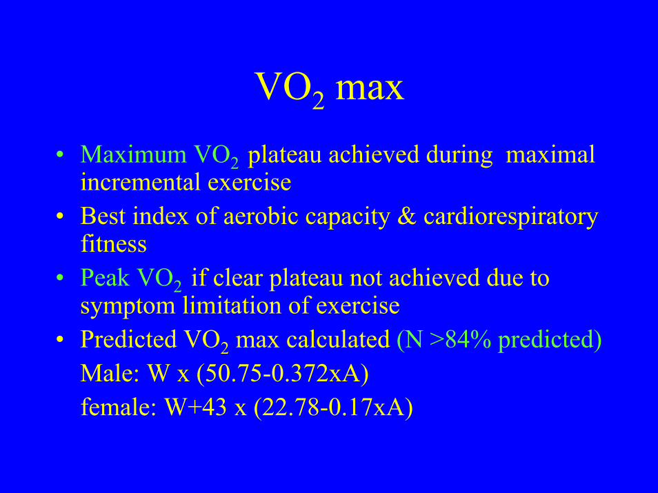
VO2 max• Maximum VO2 plateau achieved during maximal
incremental exercise• Best index of aerobic capacity & cardiorespiratory
fitness• Peak VO2 if clear plateau not achieved due to
symptom limitation of exercise • Predicted VO2 max calculated (N >84% predicted)
Male: W x (50.75-0.372xA)female: W+43 x (22.78-0.17xA)
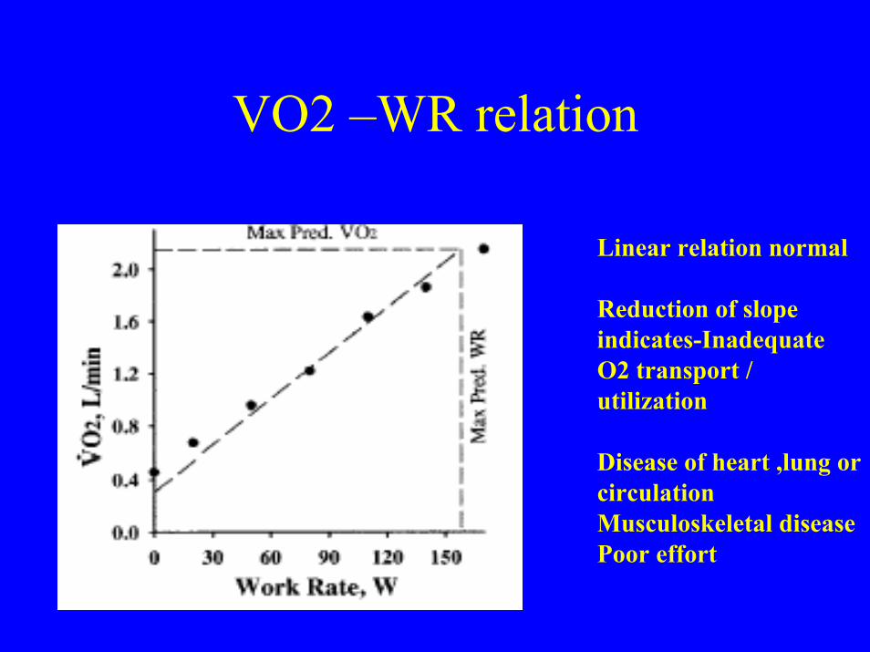
VO2 –WR relation
Linear relation normal
Reduction of slope indicates-Inadequate O2 transport / utilization
Disease of heart ,lung or circulationMusculoskeletal diseasePoor effort
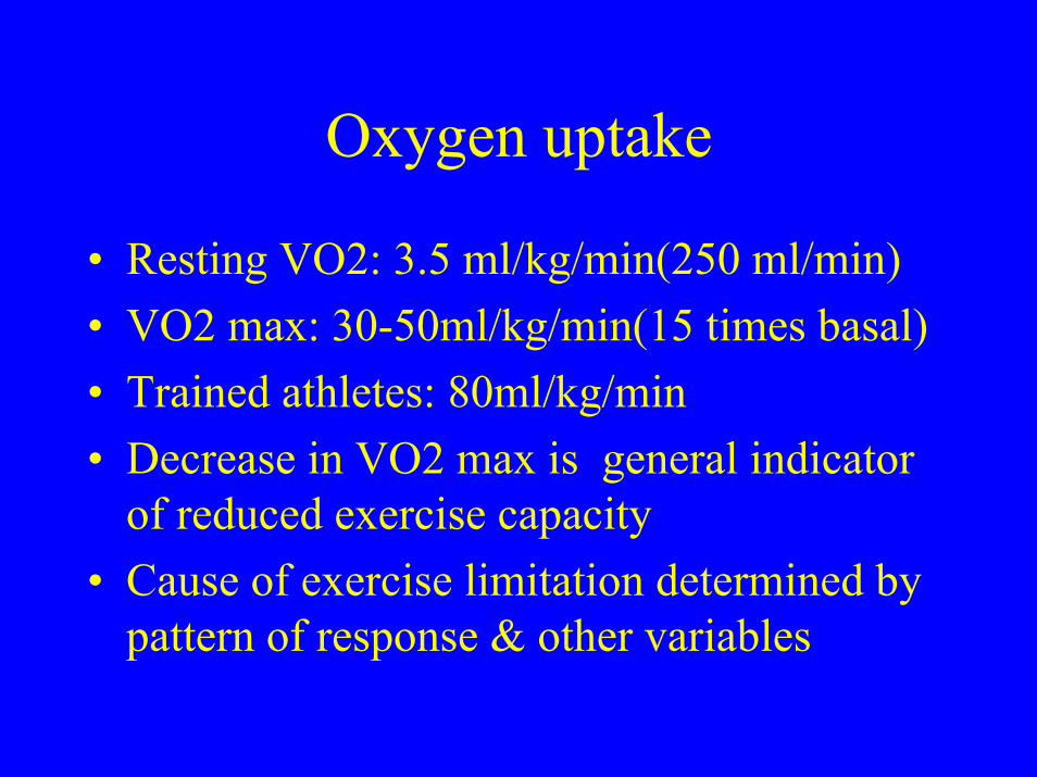
Oxygen uptake
• Resting VO2: 3.5 ml/kg/min(250 ml/min)• VO2 max: 30-50ml/kg/min(15 times basal)• Trained athletes: 80ml/kg/min• Decrease in VO2 max is general indicator
of reduced exercise capacity• Cause of exercise limitation determined by
pattern of response & other variables

Oxygen pulse
• Ratio of oxygen uptake to HR (N >80%)• Amount of oxygen extracted per heart beat• Reflects the product of stroke volume & oxygen
extraction• Indicates cardiac dysfunction(assuming O2
extraction is normal)• Low O2 pulse :
– Cardiovascular disease– Deconditioning/poor effort– Early exercise limitation(respir disease)

CO2 Output (VCO2)
• CO2 output during exercise depends on cardiac output, CO2carrying capacity and tissue exchange
• VCO2 increases nearly linear with VO2 at lower work rates, after the AT the VCO2increases steeply as lactate is buffered by bicarbonate at higher work loads.

Anaerobic Threshold - V slope method
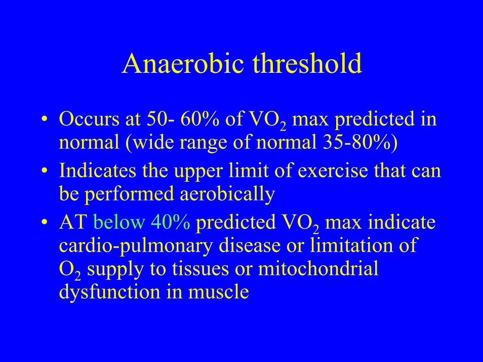
Anaerobic threshold
• Occurs at 50- 60% of VO2 max predicted in normal (wide range of normal 35-80%)
• Indicates the upper limit of exercise that can be performed aerobically
• AT below 40% predicted VO2 max indicate cardio-pulmonary disease or limitation of O2 supply to tissues or mitochondrial dysfunction in muscle
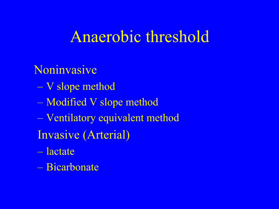
Anaerobic threshold
Noninvasive – V slope method– Modified V slope method– Ventilatory equivalent methodInvasive (Arterial)– lactate– Bicarbonate
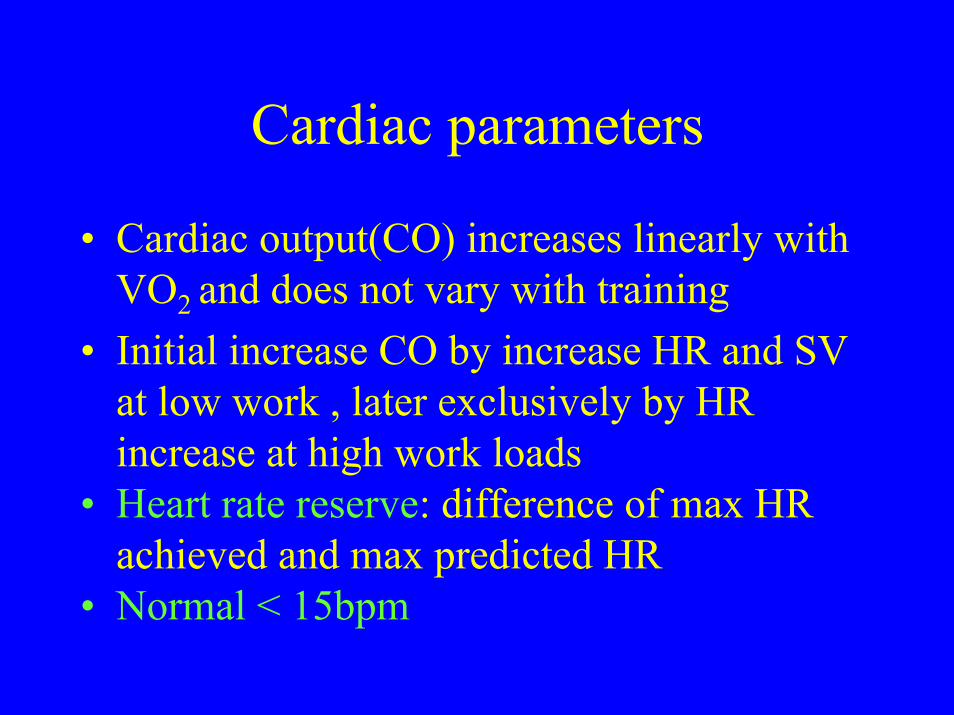
Cardiac parameters
• Cardiac output(CO) increases linearly with VO2 and does not vary with training
• Initial increase CO by increase HR and SV at low work , later exclusively by HR increase at high work loads
• Heart rate reserve: difference of max HR achieved and max predicted HR
• Normal < 15bpm
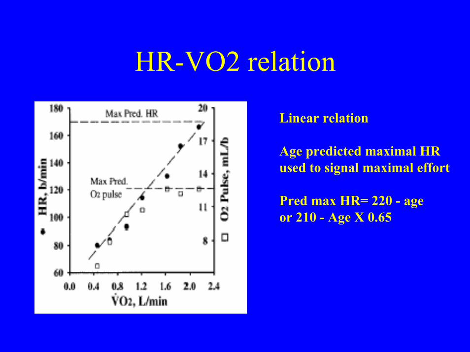
HR-VO2 relation
Linear relation
Age predicted maximal HR used to signal maximal effort
Pred max HR= 220 - ageor 210 - Age X 0.65
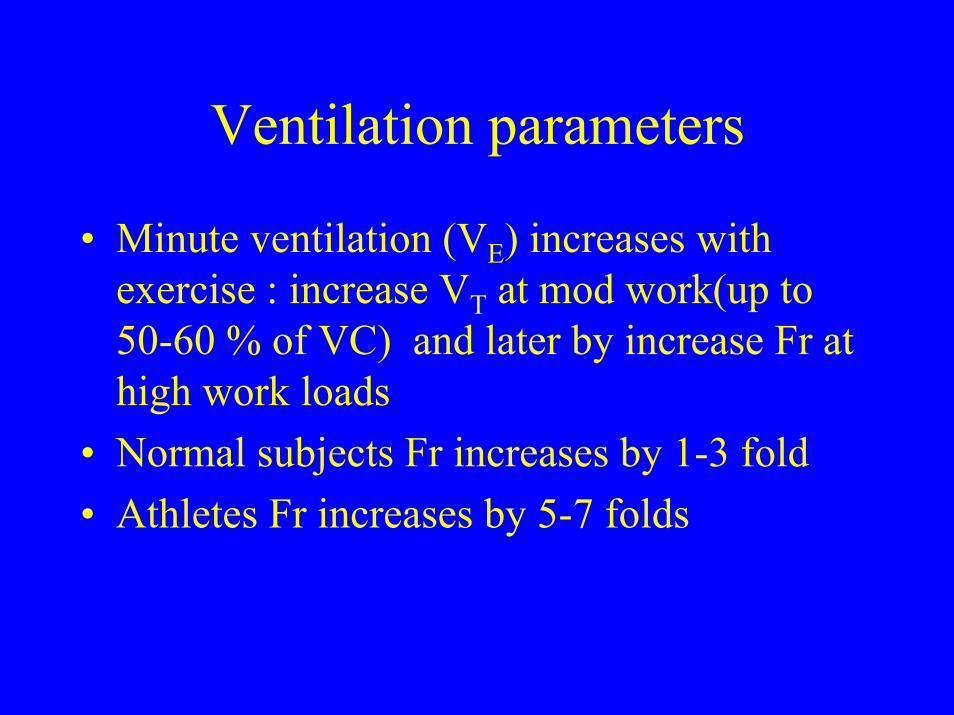
Ventilation parameters
• Minute ventilation (VE) increases with exercise : increase VT at mod work(up to 50-60 % of VC) and later by increase Fr at high work loads
• Normal subjects Fr increases by 1-3 fold• Athletes Fr increases by 5-7 folds

Ventilatory reserve
• Difference or ratio between max minute ventilation during exercise(VE max) and the maximal voluntary ventilation(MVV)
• Normal reserve > 15% of MVV (range 72+/-15%)• MVV calculated as FEV1 x 40 (approximates the
measured value)• Pulmonary diseases have reduced reserve • Cardiac diseases have normal reserve

VE and VO2
Relation complex
Usually nonlinear
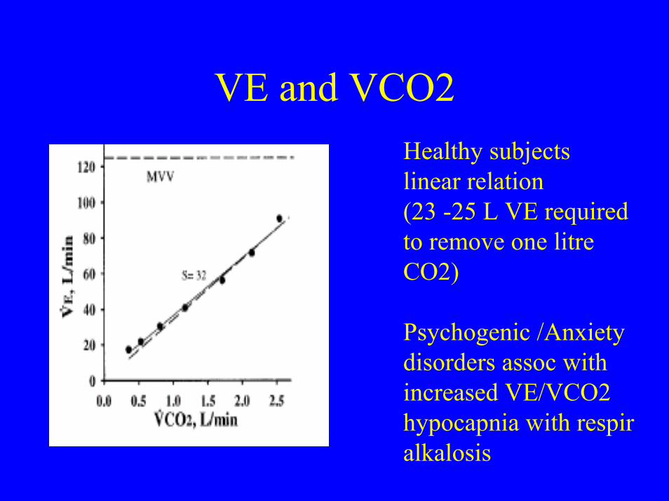
VE and VCO2Healthy subjects linear relation(23 -25 L VE required to remove one litreCO2)
Psychogenic /Anxiety disorders assoc with increased VE/VCO2 hypocapnia with respir alkalosis
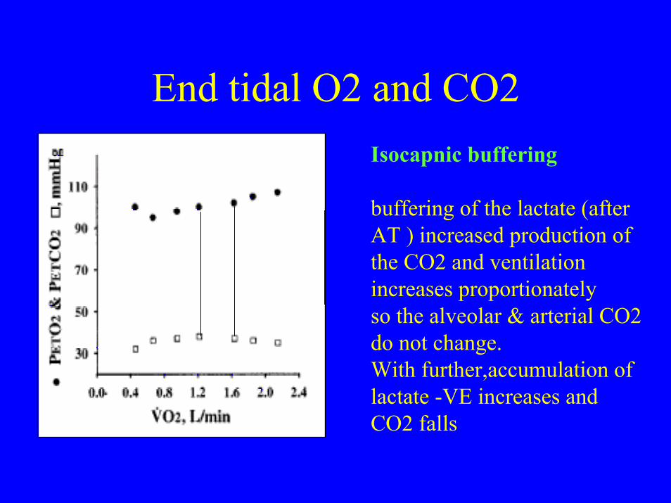
End tidal O2 and CO2Isocapnic buffering
buffering of the lactate (after AT ) increased production of the CO2 and ventilation increases proportionately so the alveolar & arterial CO2 do not change. With further,accumulation of lactate -VE increases and CO2 falls
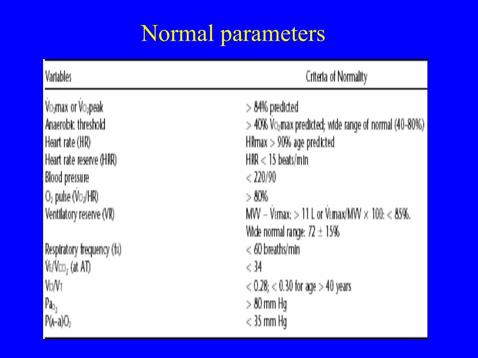
Normal parameters
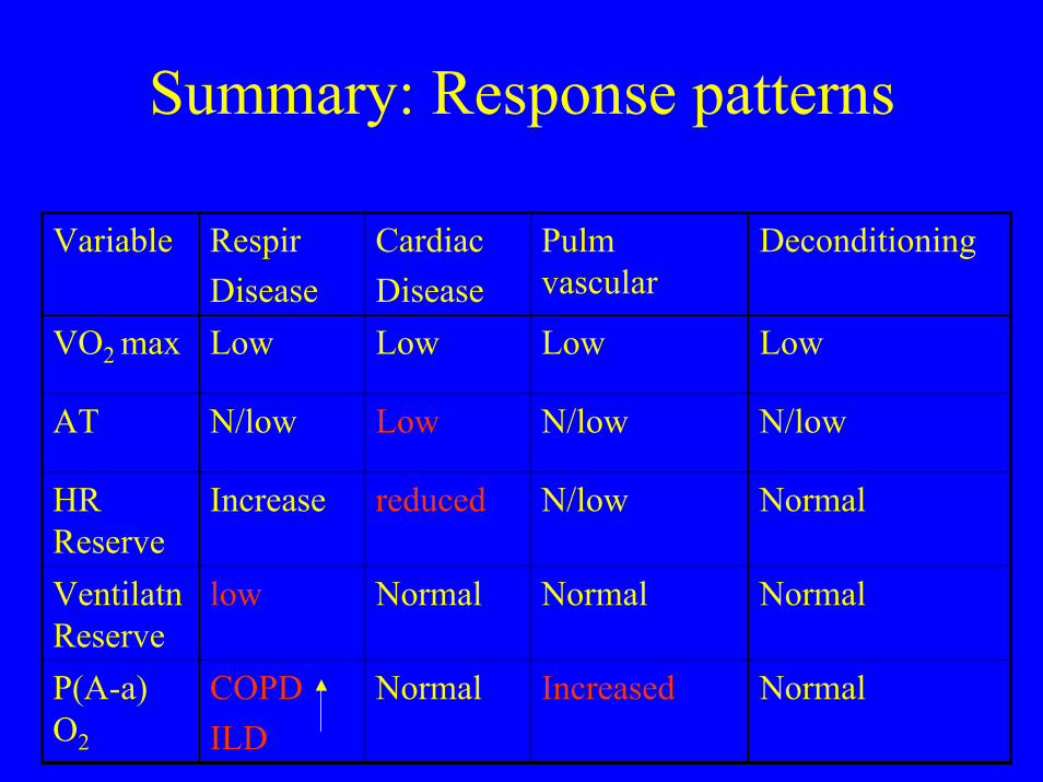
Summary: Response patterns
NormalIncreasedNormalCOPDILD
P(A-a) O2
NormalNormalNormallowVentilatn Reserve
NormalN/lowreducedIncreaseHR Reserve
N/lowN/lowLowN/lowAT
LowLowLowLowVO2 max
DeconditioningPulm vascular
CardiacDisease
RespirDisease
Variable
Interpretation
Results are rarely clear-cut ,and interpretation may be challenging,sometimes very difficult
• Review clinical and laboratory information • Identify key variables: VE max ,MVV, HR, SaO2• Compare exercise responses with appropriate
normal reference values• Evaluate cause exercise limitation• Patterns of exercise responses
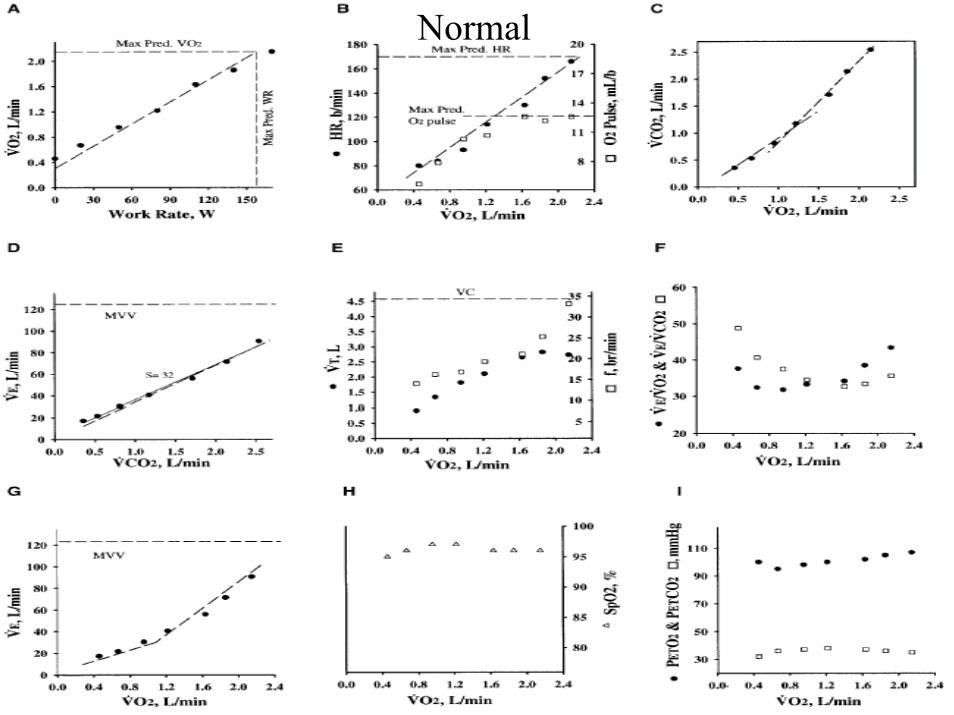
Normal
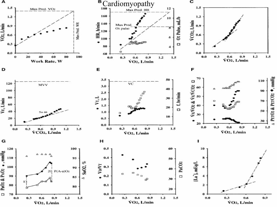
Cardiomyopathy
Cardiac disease
• Reduced peak work rate and peak VO2• Low AT(early onset metabolic acidosis)• Low oxygen pulse• High HR response ( reserve)• Ventilatory reserve normal• No desaturation
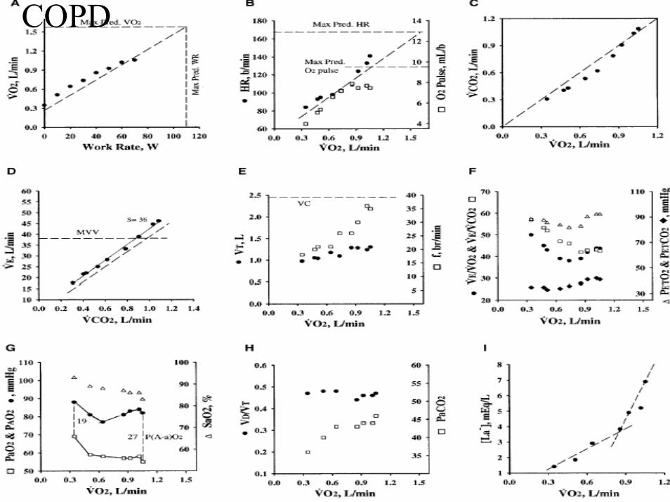
COPD

COPD
• Reduced peak work rate and peak VO2
• Noninvasive AT : ABG may avoid false positive• Reduced ventilatory reserve(>100%)• Peak HR reduced(significant HRR)• O2 pulse reduced proportionate to VO2 peak• Hypoxemia - especially in emphysema (~DLCO)• Hypercapnia(V/Q abnormalities and reduced drive
in severe cases)`

ILD

ILD
• Reduced peak work rate and peak VO2
• AT N/reduced • Reduced ventilatory reserve(>100%)• Abnormal breathing pattern(high Fr, low VT)• Significant hypoxemia (~ resting DLCO)• Wide P(A-a)O2 gradient• Low HRR- coexisting Cor pulmonale
Pulm vacular Diseases
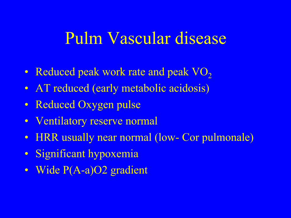
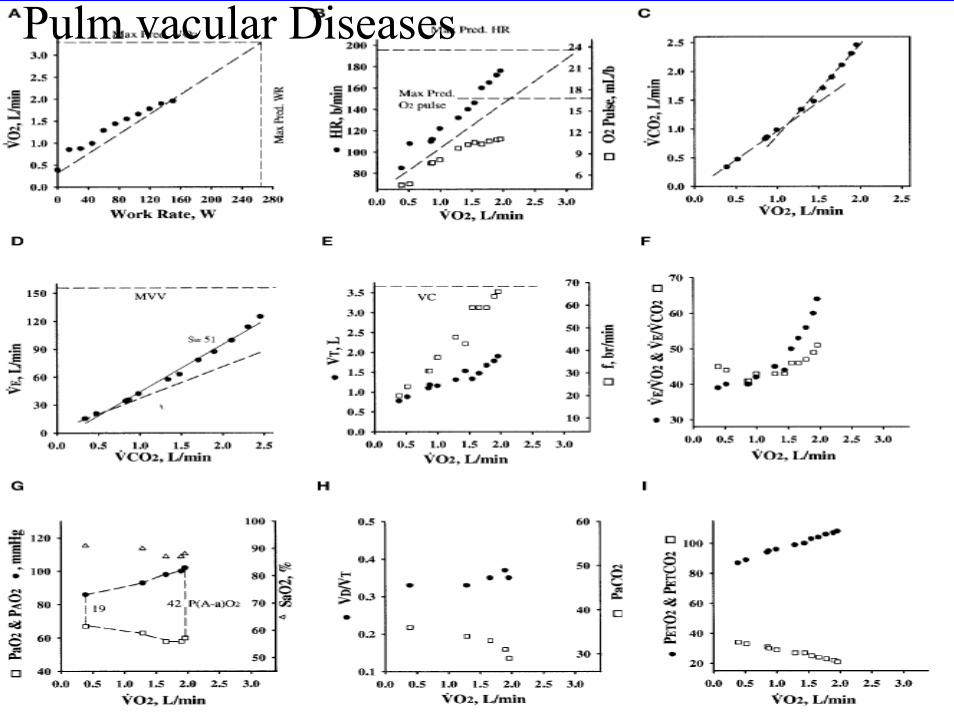
Pulm Vascular disease
• Reduced peak work rate and peak VO2
• AT reduced (early metabolic acidosis)• Reduced Oxygen pulse• Ventilatory reserve normal• HRR usually near normal (low- Cor pulmonale)• Significant hypoxemia• Wide P(A-a)O2 gradient
Deconditioning (Unfitness)
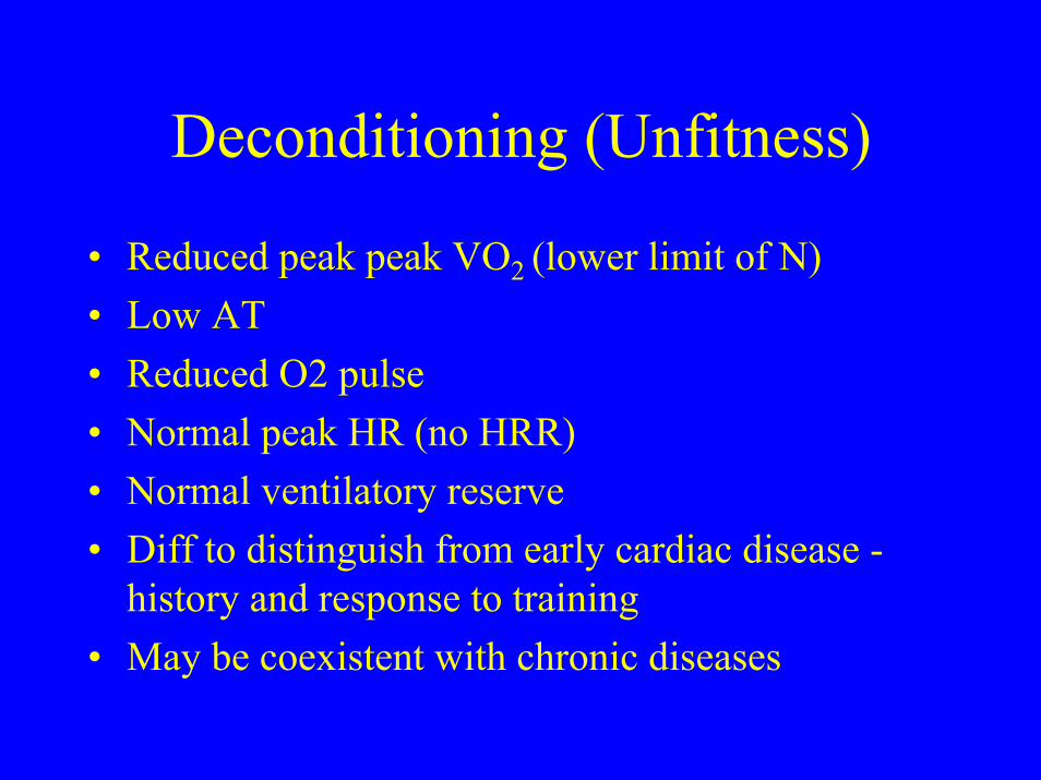
• Reduced peak peak VO2 (lower limit of N)• Low AT• Reduced O2 pulse • Normal peak HR (no HRR)• Normal ventilatory reserve• Diff to distinguish from early cardiac disease -
history and response to training• May be coexistent with chronic diseases

Interpretation of CPET
Future directions
• Reference normal values from multicenter studies required( few studies from India)
• Evidence based interpretation using standardized methodology& protocols
• Impact of pattern based analysis on clinical decision making
• Evaluate new exercise protocols(constant work rate,exponential exercise)
• Role of invasive vs noninvasive CPET