cancer diagnostics with dna microarrays (knudsen/cancer diagnostics with dna microarrays) ||...
TRANSCRIPT
19Melanoma
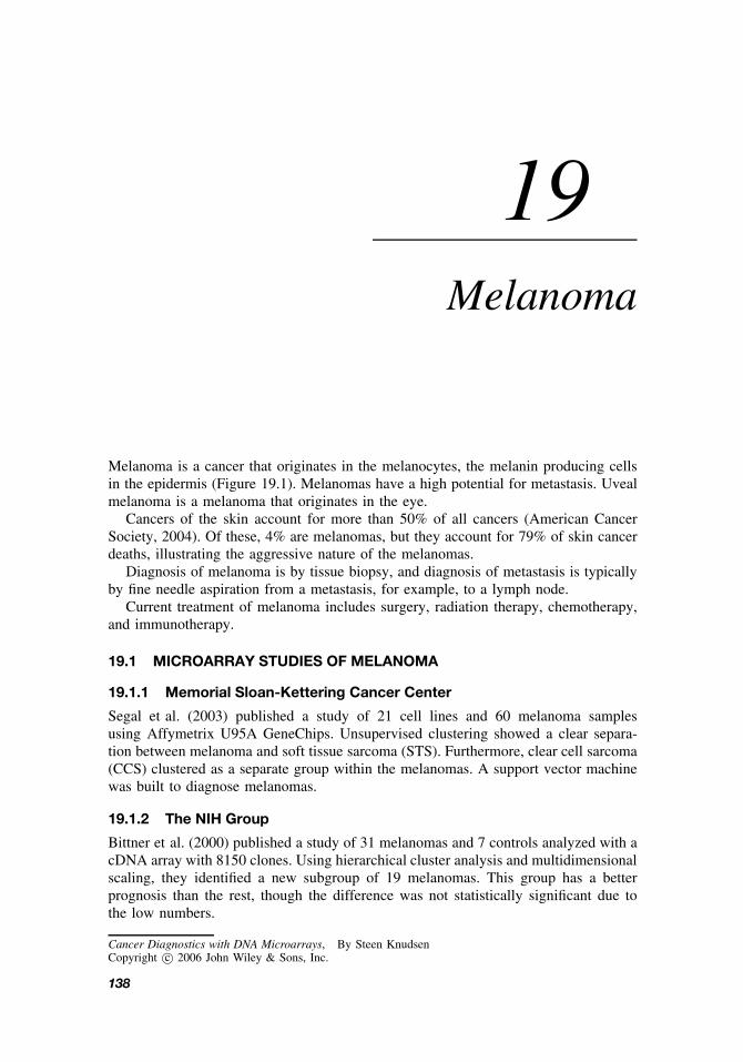
Melanoma is a cancer that originates in the melanocytes, the melanin producing cellsin the epidermis (Figure 19.1). Melanomas have a high potential for metastasis. Uvealmelanoma is a melanoma that originates in the eye.
Cancers of the skin account for more than 50% of all cancers (American CancerSociety, 2004). Of these, 4% are melanomas, but they account for 79% of skin cancerdeaths, illustrating the aggressive nature of the melanomas.
Diagnosis of melanoma is by tissue biopsy, and diagnosis of metastasis is typicallyby fine needle aspiration from a metastasis, for example, to a lymph node.
Current treatment of melanoma includes surgery, radiation therapy, chemotherapy,and immunotherapy.
19.1 MICROARRAY STUDIES OF MELANOMA
19.1.1 Memorial Sloan-Kettering Cancer Center
Segal et al. (2003) published a study of 21 cell lines and 60 melanoma samplesusing Affymetrix U95A GeneChips. Unsupervised clustering showed a clear separa-tion between melanoma and soft tissue sarcoma (STS). Furthermore, clear cell sarcoma(CCS) clustered as a separate group within the melanomas. A support vector machinewas built to diagnose melanomas.
19.1.2 The NIH Group
Bittner et al. (2000) published a study of 31 melanomas and 7 controls analyzed with acDNA array with 8150 clones. Using hierarchical cluster analysis and multidimensionalscaling, they identified a new subgroup of 19 melanomas. This group has a betterprognosis than the rest, though the difference was not statistically significant due tothe low numbers.
Cancer Diagnostics with DNA Microarrays, By Steen KnudsenCopyright c© 2006 John Wiley & Sons, Inc.
138

FURTHER READING 139
Epidermis
Melanocyte
Dermis
Figure 19.1 Melanocytes in the epidermis of the skin. (From Tortora, Principles of Human Anatomy,10th ed., 2005, p. 124. Used with permission of John Wiley & Sons, Inc.)
Wang et al. (2002) published a study of 63 fine needle aspiration samples from37 melanoma metastases from 25 patients undergoing immunotherapy. A 6108-genehuman cDNA chip was used. They identified 30 genes that correlated with clinicaloutcome (response to immunotherapy) prospectively.
19.2 SUMMARY
Most studies of melanoma have been with cell lines and mouse models looking formarkers of metastasis or targets for drug development. But a few studies have conductedtranscription profiling of larger patient cohorts and showed that microarrays can beused for diagnosis and prognosis of melanomas and their metastasis. More studies areneeded to confirm these observations.
FURTHER READING
Baldi, A., Battista, T., De, L. U., Santini, D., Rossiello, L., Baldi, F., Natali, P. G., Lombardi,D., Picardo, M., Felsani, A., and Paggi, M. G. (2003). Identification of genes down-regulatedduring melanoma progression: a cDNA array study. Exp. Dermatol. 12(2):213–218.
Baldi, A., Santini, D., De, L. U., and Paggi, M. G. (2003). cDNA array technology in melanoma:an overview. J. Cell. Physiol. 196(2):219–223 (review).
Becker, B., Roesch, A., Hafner, C., Stolz, W., Dugas, M., Landthaler, M., and Vogt, T. (2004).Discrimination of melanocytic tumors by cDNA array hybridization of tissues prepared bylaser pressure catapulting. J. Invest. Dermatol. 122(2):361–368.

140 MELANOMA
Brem, R., Hildebrandt, T., Jarsch, M., Van Muijen, G. U., and Weidle, U. H. (2001). Identificationof metastasis-associated genes by transcriptional profiling of a metastasizing versus a non-metastasizing human melanoma cell line. Anticancer Res. 21(3B):1731–1740.
Carr, K. M., Bittner, M., Trent, J. M. (2003). Gene-expression profiling in human cutaneousmelanoma. Oncogene 22(20):3076–3080 (review).
Clark, E. A., Golub, T. R., Lander, E. S., and Hynes, R. O. (2000). Genomic analysis ofmetastasis reveals an essential role for RhoC. Nature 406(6795):532–535. Erratum in: Nature2001; 411(6840):974.
Conway, R. M., Cursiefen, C., Behrens, J., Naumann, G. O., and Holbach, L. M. (2003).Biomolecular markers of malignancy in human uveal melanoma: the role of the cad-herin–catenin complex and gene expression profiling. Ophthalmologica 217(1):68-75(review).
de Wit, N. J., Burtscher, H. J., Weidle, U. H., Ruiter, D. J., and van Muijen, G. N. (2002). Dif-ferentially expressed genes identified in human melanoma cell lines with different metastaticbehaviour using high density oligonucleotide arrays. Melanoma Res. 12(1):57–69.
Dooley, T. P., Reddy, S. P., Wilborn, T. W., and Davis, R. L. (2003). Biomarkers of humancutaneous squamous cell carcinoma from tissues and cell lines identified by DNA microarraysand qRT-PCR. Biochem. Biophys. Res. Commun. 306(4):1026–1036.
Kim, C. J., Reintgen, D. S., and Yeatman, T. J. (2002). The promise of microarray technologyin melanoma care. Cancer Control 9(1):49–53 (review).
Pavey, S., Johansson, P., Packer, L., Taylor, J., Stark, M., Pollock, P. M., Walker, G. J., Boyle,G. M., Harper, U., Cozzi, S. J., Hansen, K., Yudt, L., Schmidt, C., Hersey, P., Ellem, K. A.,O’Rourke, M. G., Parsons, P. G., Meltzer, P., Ringner, M., and Hayward, N. K. (2004).Microarray expression profiling in melanoma reveals a BRAF mutation signature. Oncogene23(23):4060–4067.
McDonald, S. L., Edington, H. D., Kirkwood, J. M., and Becker, D. (2004). Expression analysisof genes identified by molecular profiling of VGP melanomas and MGP melanoma-positivelymph nodes. Cancer Biol. Ther. 3(1):110–120.
Roesch, A., Vogt, T., Stolz, W., Dugas, M., Landthaler, M., and Becker, B. (2003). Discrim-ination between gene expression patterns in the invasive margin and the tumour core ofmalignant melanomas. Melanoma Res. 13(5):503–509.
Seftor, E. A., Meltzer, P. S., Kirschmann, D. A., Pe’er, J., Maniotis, A. J., Trent, J. M., Fol-berg, R., and Hendrix, M. J. (2002). Molecular determinants of human uveal melanomainvasion and metastasis. Clin. Exp. Metastasis 19(3):233–246.
Tschentscher, F., Husing, J., Holter, T., Kruse, E., Dresen, I. G., Jockel, K. H., Anastassiou, G.,Schilling, H., Bornfeld, N., Horsthemke, B., Lohmann, D. R., and Zeschnigk, M. (2003).Tumor classification based on gene expression profiling shows that uveal melanomas withand without monosomy 3 represent two distinct entities. Cancer Res. 63(10):2578–2584.
van der Velden, P. A., Zuidervaart, W., Hurks, M. H., Pavey, S., Ksander, B. R., Krijgsman, E.,Frants, R. R., Tensen, C. P., Willemze, R., Jager, M. J., and Gruis, N. A. (2003). Expressionprofiling reveals that methylation of TIMP3 is involved in uveal melanoma development. Int.J. Cancer 106(4):472–479.

FURTHER READING 141
Zuidervaart, W., van der Velden, P. A., Hurks, M. H., van Nieuwpoort, F. A., Out-Luiting, C. J.,Singh, A. D., Frants, R. R., Jager, M. J., and Gruis, N. A. (2003). Gene expression profilingidentifies tumour markers potentially playing a role in uveal melanoma development. Br. J.Cancer 89(10):1914–1919.