c) identification species-specific antigen legionella ... · monoclonal antibody lp3iig2, and...
TRANSCRIPT

JOURNAL OF CLINICAL MICROBIOLOGY, Dec. 1984, p. 1031-1035 Vol. 20, No. 60095-1137/84/121031-05$02.00/0Copyright C) 1984, American Society for Microbiology
Identification of a Species-Specific Antigen in Legionellapneumophila by a Monoclonal Antibody
LARRY H. GOSTING,* KATHY CABRIAN, JERRILYN C. STURGE, AND LYNN C. GOLDSTEINGenetic Systems Corporation, Seattle, Washington 98121
Received 13 July 1984/Accepted 24 August 1984
A species-specific antigen in Legionella pneumophila was identified by a monoclonal antibody in enzyme-linked immunosorbent and immunofluorescence assays of serogroups 1 through 8. The species-specific antigenwas heat stable, and the molecular weight of the major band was 29,000 by immunoblot analysis. In directimmunofluorescence assays, the antigen was cryptic or only partly exposed on the surface of the cells but waseffectively exposed by treating the cells with detergent and EDTA. The monoclonal antibody was utilized indirect immunofluorescence assays to specifically identify multiple cultures of L. pneumophila serogroups.
Legionnaires disease, first characterized in 1977, is ofclinical interest as an acute human illness with a highmortality rate (5, 13, 24). In most patients, the infection ismanifested as pneumonia; however, a second clinical syn-drome, Pontiac fever, is a nonpneumonic, mild, and self-limited respiratory illness (14, 20). Although many species ofthe genus Legionella can cause legionellosis, the etiologicalagent of Legionnaires disease is Legionella pneumophila, agram-negative bacterium which consists of at least eightserogroups (2, 3, 10, 25, 26). Laboratory diagnosis of thedisease is presently demonstrated by a fourfold increase inserum indirect fluorescent antibody titer, isolation and cul-ture of Legionella spp. from clinical specimens, or directfluorescent antibody examination of clinical specimens forthe presence of organisms.The direct fluorescent antibody test is the most rapid
means of diagnosis of Legionnaires disease in laboratories,but there are several associated problems. Multiple antibodyreagents are required to identify all presently recognizedserogroups of L. pneumophila. In addition, cross-reactionshave been reported with strains of Pseudomonas fluores-cens, Pseudomonas alcaligenes, Bacteroides fragilis, andthe Flavobacterium-Xanthomonas group when conjugates ofpolyclonal antibodies are used for diagnosis (8, 27).
Serological techniques utilizing polyclonal antisera havebeen used to identify serogroup-common antigens of L.pneumophila (7, 19). In addition, monoclonal antibodiesdirected against L. pneumophila antigens have been previ-ously described (17, 18, 28). Most of the monoclonal anti-bodies reported have been serogroup specific. Optimally,the most useful diagnostic antibody would be one that reactswith a common antigen present on all isolates of L. pneumo-phila but is absent from closely related bacteria. In thisreport, we describe and characterize a species-specific anti-gen which is detected by a monoclonal antibody. We haveutilized the antibody to detect the bacterium in immunofluo-rescence assays on clinical isolates of L. pneumophila grownin culture.
MATERIALS AND METHODSBacterial strains. The bacterial strains used in this study
were received from the American Type Culture Collection,
* Corresponding author.
Rockville, Md., Centers for Disease Control, Atlanta, Ga.,or Harborview Medical Center, Seattle, Wash., and arelisted in Table 1. Legionella microorganisms were grown oncharcoal-yeast extract agar for 2 to 7 days or, in some cases,in defined medium broth (12, 29). Harvested cells used asimmunogens were either fixed in 0.5% Formalin or sonicatedon ice at 175 W with a Braun-sonic 1510 (B. Braun, Melsun-gen, Federal Republic of Germany) for a total of 7.5 min.
Production of monoclonal antibodies. Hybrid cell linesproducing monoclonal antibodies were prepared by themethod of Kohler and Milstein (21), with previously de-scribed modifications (16, 31). Spleen cells for the fusionwere obtained from a BALB/c mouse that was immunizedwith L. pneumophila serogroups 1 through 6. Methods forpropagation and stabilization of cloned cell lines and forascites production have been previously described (16, 31).Antibody assays. Anti-L. pneumophila antibodies were
detected by enzyme-linked immunosorbent assay (ELISA)on 96-well plates (Linbro Tissue Culture Multi-Well Plate;Flow Laboratories, McLean, Va.) coated with individualserogroups (1 through 6) of L. pneumophila or Pseudomonasaeruginosa (used as a negative control). Bacterial suspen-sions were added to the plates and centrifuged at 740 x g.Supernatants were aspirated, and the bacteria were fixed toplates by the addition of 95% ethanol. The plates were thenblocked with 1% bovine serum albumin in phosphate-buff-ered saline (PBS pH 7.2) for 1 h at 37°C, washed with 0.05%Tween 20 in PBS (PBS-Tween 20); and then with deionizedwater, and stored dry at 4°C until use. Plates were incubatedwith culture fluids (50 p,l per well) at 37°C for 45 min and thenwashed three times in PBS-Tween 20. Protein A-peroxidase(1:3,000 dilution in PBS-Tween 20; Zymed Laboratories,Inc., South San Francisco, Calif.) was added (50 1,u perwell), and the plates were incubated for 30 min at roomtemperature and washed as above. Substrate (0.05 M citricacid-0.1 M dibasic sodium phosphate buffer [pH 5.0] con-taining 7 mg of o-phenylenediamine and 25 ,ul of 30% H202per 50 ml) was added, and the plates were incubated for 30min at room temperature in the dark. The reaction wasstopped with 3 N H2SO4, and the colorimetric reactionswere quantified with a Microelisa Auto Reader (MR580;Dynatech Laboratories, Inc., Alexandria, Va.).For direct immunofluorescence assays, antibodies were
purified from ascites fluid with protein A-Sepharose andconjugated to fluorescein-isothiocyanate by procedures pre-viously described (11, 15). Antigen slides were prepared by
1031
on June 30, 2020 by guesthttp://jcm
.asm.org/
Dow
nloaded from

1032 GOSTING ET AL.
TABLE 1. Reactions of microorganisms tested in direct immunofluorescence assays with monoclonal antibody LP31IG2Organism No. positive/no. tested Organism No. positive/no. tested
L. pneumophila Streptococ cus pneumoniae ....... ......... 0/15Serogroup 1 ................................ 50/50 S. pyogenes.0/15Serogroup 2................................ 5/5 5. siridans group sp...................... 0/21Serogroup 3................................. 3/3 Haemophiluis influen:ae .................. 0/20Serogroup 4................................ 3/3 H. parainfluenzaee...................... 0/5Serogroup 5 ................................ 7/7 Neisseria sicca.0/1Serogroup 6................................ 6/6 N. muicosa .............................. 0/2Serogroup 7 ................................ 2/2 N. gonorrhoeae ......................... 0/3Serogroup 8 ................................ 1/1 Klebsiella pneutmoniae ................... 0/16Serogroup 9 ................................ 1/1 Staphvlococclus aurOeus..2/5"
S. epidermidis.0/3Legionella sp., non-pneumophila Flav'ohacteriumsp.0......................0/2
L. micdadei . ................................ 0/6 Pseudomonas fluorescens.0/2L. oakridgensis ............................. 0/1 P. maltophilia.0/1L. jordanis ................................. 0/1 P. alcaligenes.0/1L. longbeachae ............................. 0/6 P. aeruginosa.0/15L. sainthelensi.............................. 0/2 Bacteroides fragilis.0/2L. bozemanii ............................... 0/3 B. melaninogenicus.0/1L. dumoffii ................................. 0/3 Escherichia coli.0/15L. wadswiorthii.............................. 0/1 Salmonellasp.. 0/3L. gormanii ................................. 0/1 Enterobacter sp.0/1
Serratia sp.0/1Proteuis sp.0/5Lactobacillussp. 0/13Peptostreptococcus sp.0/1
"Due to nonimmune binding to protein A.
adding 1% Formalin-killed organisms to 10-well microscopeslides and allowing them to air dry on a 37°C slide warmer.The slides were immersed in 1% Triton X-100 in 0.15 M PBS(pH 8.5) with 100 mM EDTA solution for 20 min, washed indeionized water, and dried. Twenty-five microliters of anti-body solution (25 p.g of antibody per ml in 0.05% PBS-Tween20 with 1 mg of bovine serum albumin per ml, 0.01% NaN3,and Evans blue; optical density at 620 nm of 1.6) was addedper well, and the slides were incubated for 30 min at 37°C.The slides were washed in 0.15 M PBS and deionized water.Cover slips were mounted with 9 parts glycerol to 1 part 0.1M Tris (pH 8.5), and the slides were examined with anepiillumination fluorescence microscope and x63 oil objec-tive.
Biochemical procedures. Monoclonal antibodies were eval-uated by immunoblot methods, with modifications to previ-ously published procedures (1, 4, 22, 30, 32). Briefly, solubi-lized antigen preparations of L. pneumophila serogroups 1through 8, Legionella bozemanii, Legionella micdadei, Le-gionella dumoffli, Legionella jordanis, Legionella longbea-chae serogroups 1 and 2, and P. fluorescens were made byincubating concentrated live cultures in a solubilizationbuffer (0.3% sodium deoxycholate, 0.02 M 2-mercaptoeth-anol, 30% glycerol, 0.03 M Tris-hydrochloride [pH 7.6])overnight at 4°C or by sonicating them in 2% (wt/vol) sodiumdeoxycholate. The antigen preparations were mixed withsample buffer (2% sodium dodecyl sulfate [SDS], 5% 2-mercaptoethanol, 0.002% bromophenol blue, and 10%glycerol in 0.06 M Tris-hydrochloride [pH 6.8], final concen-trations), heated at 100°C for 10 min, and subjected to SDS-polyacrylamide gel electrophoresis in 14% slab gels. Anti-gens in the gel were then transferred to nitrocellulosemembranes (NCMs) by electrophoresis for 2 h at 27 V in 25mM sodium phosphate buffer (pH 7.0). After transfer, theNCM was blocked in PBS-Tween 20 for 1 h at roomtemperature. The NCM was incubated with antibody dilutedin PBS-Tween 20 for 1 h, washed with PBS-Tween 20, andthen incubated with protein A-horseradish peroxidase
(1:2,000 dilution in PBS-Tween 20) for 1 h at room tempera-ture. The NCM was washed and then immersed in horserad-ish peroxidase color development solution (Bio-Rad Labora-tories, Richmond, Calif.) for 20 min, and the reaction wasstopped by immersion in deionized water. The molecularweights of the antigens were determined by includingmarkers (prestained protein molecular weight standards forSDS-polyacrylamide gel electrophoresis; Bethesda ResearchLaboratories, Inc., Gaithersburg, Md.) in the SDS-poly-acrylamide gel electrophoresis. The relative migrations ver-sus the logarithms of the molecular weights of the standardswere evaluated by linear regression to derive molecularweight values for L. pneumophila antigens.
Antigens were further evaluated by susceptibility to heatand degradation by proteinase K. One percent of Formalin-killed suspensions of L. pneumophila serogroup 1 (strainPhiladelphia 1) and serogroup 4 (strain Los Angeles 1) werediluted in 0.015 M carbonate-0.035 M bicarbonate buffer(pH 9.6) to 45 ,ug of protein per ml. The cell suspensionswere immersed in a boiling water bath for 10 min and thencoated on 96-well plates overnight at 4°C. An ELISA wasperformed as described above. Solubilized antigen prepara-tions of L. pneumophila serogroups 2 and 4 were diluted to50 ,ug of protein per ml in the carbonate-bicarbonate bufferand divided into two samples. Proteinase K (Merck & Co.,Inc., Rahway, N.J.) was added at the rate of 20 p,g ofproteinase K per 50 pLg of bacterial protein. The solutionswere incubated in a 37°C water bath, and samples wereremoved at 30, 60, 90, and 120 min. The samples were coatedon 96-well plates overnight at 4°C, fixed with 95% ethanol,and blocked with 1% bovine serum albumin. The antigenswere then assayed by ELISA.
RESULTSIsolation of a hybrid cell line producing anti-L. pneumophila
monoclonal antibody. Hybridizations were performed be-tween NS-1 myeloma cells and the lymphocytes from aBALB/c mouse immunized with L. pneumophila serogroups
J. CLIN. MICROBIOL.
on June 30, 2020 by guesthttp://jcm
.asm.org/
Dow
nloaded from

SPECIES-SPECIFIC ANTIGEN OF L. PNEUMOPHILA 1033
1 through 6. Culture fluids from hybrid cells were tested foranti-L. pneumophila antibodies by an ELISA utilizing repli-cate plating techniques with individual serogroup antigenpreparations. From one fusion, 17 hybrids were produced,which secreted monoclonal antibodies reacting specificallywith various serogroups of L. pneumophila. Sixteen of theseantibodies were either specific for individual serogroups ofL. pneumophila or demonstrated limited cross-reactivityamong two or three serogroups (data not shown). However,one cell line produced a monoclonal antibody designatedLP3IIG2 which was reactive with all eight serogroups of L.pneumophila. This hybrid cell line was subsequently clonedby limiting dilution until phenotypically stable.
Characterization of the serogroup common antigen. Mono-clonal antibody LP3IIG2 was examined by immunoblotanalysis on soluble antigen preparations of L. pneumophilaserogroups 1 through 8, L. bozemanii, L. micdadei, L.dumoffii, L. jordanis, L. longbeachae serogroups 1 and 2,and P. fluorescens. Antigens were separated by SDS-poly-acrylamide gel electrophoresis and electrophoretically trans-ferred to NCM. The NCMs were incubated with normalmouse serum, L. pneumophila-immune mouse serum, ormonoclonal antibody LP3IIG2, and antigen-antibody reac-tions were detected by protein A-horseradish peroxidase.The monoclonal antibody recognized a specific antigenconsisting of a major band present in all eight serogroups ofL. pneumophila (representative serogroups are shown inFig. 1). This antigen was not detected in non-pneumophilaLegionella spp. or P. fluorescens organisms. The molecularweight of the major band, estimated by comparison with therelative migration of protein standards, was 29,000. Themonoclonal antibody also reacted with other minor bands,but these reactions varied in intensity, depending on theserogroup tested and the method of antigen extraction.The serogroup-common antigen detected by LP3IIG2 was
tested for degradation by proteinase K by incubation withthe enzyme, coating on 96-well plates, and ELISA. Therewas a substantial decrease (0.45 to 0.03) in the opticaldensity after incubation with the enzyme, which suggestedthat the antigen was a protein. The antigen was also evaluat-ed for heat stability by boiling for 10 min in carbonate-
-92.5K
-43K
-25.7K
-18.4K
-12.3K
1 2 3 4 5 6FIG. 1. Immunoblot of L. pneumophila serogroups 1 to 6 with
monoclonal antibody LP3IIG2. Lanes: 1, serogroup 1 (Philadelphia-1 strain); 2, serogroup 2 (Togus-1 strain); 3, serogroup 3 (Blooming-ton-2 strain); 4, serogroup 4 (Los Angeles-i strain); 5, serogroup 5(Dallas-lE strain); and 6, serogroup 6 (Chicago-2 strain).
bicarbonate buffer (pH 9.6). Treatment of serogroup 4 wholebacteria in this fashion before coating ELISA plates in-creased the signal by 2.7 to 3.0 times compared with theunheated control when reacted with LP3IIG2.Immunofluorescence assays on culture isolates. The mono-
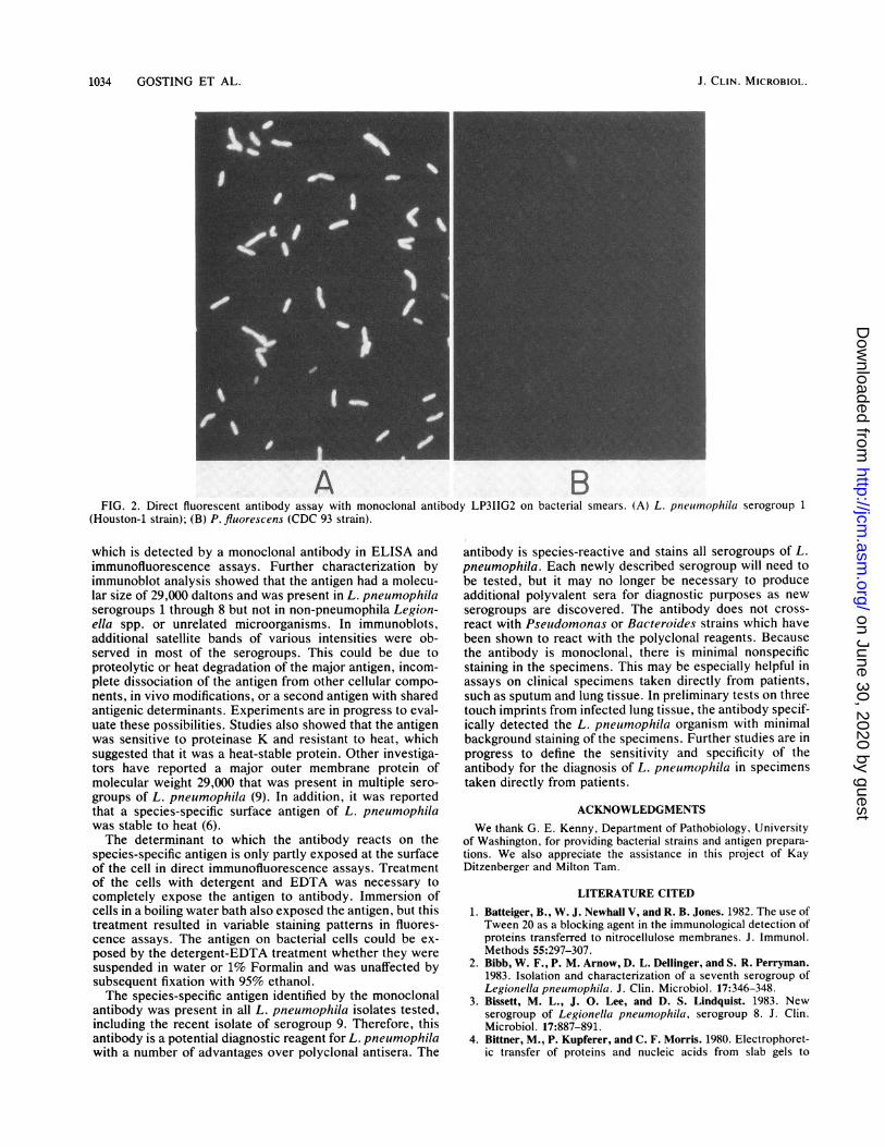
clonal antibody was conjugated to fluorescein and tested indirect immunofluorescence assays on bacteria grown inculture. In these assays, it was necessary to treat thebacteria with a detergent-EDTA solution to obtain brightsurface staining of the bacterial cells. Assays on untreatedcells or those fixed with Formalin or ethanol showed incom-plete staining of the bacteria. It therefore appeared that theantigen recognized by the monoclonal antibody was crypticor only partially exposed at the cell surface. With the directimmunofluorescence assay and detergent treatment, theantibody was evaluated on smears of bacteria after growth inculture. Representative staining of L. pneumophila as apositive control and P. fluorescens as a negative control isshown in Fig. 2. The antibody was examined on multipleculture isolates of each serogroup of L. pneumophila, non-pneumophila Legionella spp., and unrelated microorga-nisms. This study included the bacteria known to be cross-reactive with polyclonal antisera to Legionella spp., such asP. fluorescens (CDC 93), B. fragilis, and the Flavobacte-rium-Xanthomonas group. All strains of L. pneumophilawere positive, and there was no evidence of specific immunecross-reaction to non-pneumophila Legionella or non-Le-gionella bacteria. There was staining of some strains ofStaphylococcus aureus due to nonimmune binding of themouse immunoglobulin to protein A (for a review, seereference 23). The results are summarized in Table 1.
DISCUSSIONSerotypic diversity of L. pneumophila has expanded as
increased numbers of isolates are obtained from clinical andenvironmental sources. Currently, eight serogroups are rec-ognized, but this number will probably continue to increase.Already, an additional serogroup 9 has recently been pro-posed (P. H. Edelstein, W. F. Bibb, G. W. Gorman, W. L.Thacker, D. J. Brenner, H. W. Wilkinson, C. W. Moss,R. S. Buddington, C. J. Dunn, P. J. Roos, and P. L.Meenhorst, Ann. Intern. Med., in press). This poses aserious disadvantage to the direct serological diagnosis ofthe infection with polyclonal serogroup-specific reagents.Not only does the multiplicity of serogroups create difficul-ties in the production of a polyvalent serum, but there is a lagtime between the recognition of a new serogroup and pro-duction of the appropriate serogroup-specific antiserum.A far more attractive approach is to develop diagnostic
reagents against species-specific antigens. Studies have beenperformed to analyze these common antigens in multipleserogroups of L. pneumophila and a variety of Legionellaspecies. Analysis by crossed immunoelectropheresis of L.pneumophila (7, 19) has revealed numerous serogroup-common antigens (85 to 87% of total antigens). Of the L.pneumophila serogroup 1 reference system antigens (51total), 39 to 53% cross-reacted with L. bozemanii, L. dumof-fii, L. gormanii, and L. micdadei, and up to six antigenswere observed to cross-react with non-Legionella organisms(7). For species-specific antigens to be useful for the produc-tion of diagnostic reagents, they must be structurally locatedso that they are accessible to antibodies and are representedin sufficient density to provide an adequate signal in selectedassays.
In this report, we describe a species-specific antigen
VOL. 20, 1984
on June 30, 2020 by guesthttp://jcm
.asm.org/
Dow
nloaded from
1034 GOSTING ET AL.
A BFIG. 2. Direct fluorescent antibody assay with monoclonal antibody LP3IIG2 on bacterial smears. (A) L. pneuimophila serogroup 1
(Houston-1 strain); (B) P. fluorescens (CDC 93 strain).
which is detected by a monoclonal antibody in ELISA andimmunofluorescence assays. Further characterization byimmunoblot analysis showed that the antigen had a molecu-lar size of 29,000 daltons and was present in L. pneumophilaserogroups 1 through 8 but not in non-pneumophila Legion-ella spp. or unrelated microorganisms. In immunoblots,additional satellite bands of various intensities were ob-served in most of the serogroups. This could be due toproteolytic or heat degradation of the major antigen, incom-plete dissociation of the antigen from other cellular compo-nents, in vivo modifications, or a second antigen with sharedantigenic determinants. Experiments are in progress to eval-uate these possibilities. Studies also showed that the antigenwas sensitive to proteinase K and resistant to heat, whichsuggested that it was a heat-stable protein. Other investiga-tors have reported a major outer membrane protein ofmolecular weight 29,000 that was present in multiple sero-groups of L. pneumophila (9). In addition, it was reportedthat a species-specific surface antigen of L. pneumophilawas stable to heat (6).The determinant to which the antibody reacts on the
species-specific antigen is only partly exposed at the surfaceof the cell in direct immunofluorescence assays. Treatmentof the cells with detergent and EDTA was necessary tocompletely expose the antigen to antibody. Immersion ofcells in a boiling water bath also exposed the antigen, but thistreatment resulted in variable staining patterns in fluores-cence assays. The antigen on bacterial cells could be ex-posed by the detergent-EDTA treatment whether they weresuspended in water or 1% Formalin and was unaffected bysubsequent fixation with 95% ethanol.The species-specific antigen identified by the monoclonal
antibody was present in all L. pneumophila isolates tested,including the recent isolate of serogroup 9. Therefore, thisantibody is a potential diagnostic reagent for L. pneumophilawith a number of advantages over polyclonal antisera. The
antibody is species-reactive and stains all serogroups of L.pneumophila. Each newly described serogroup will need tobe tested, but it may no longer be necessary to produceadditional polyvalent sera for diagnostic purposes as newserogroups are discovered. The antibody does not cross-react with Pseudomonas or Bacteroides strains which havebeen shown to react with the polyclonal reagents. Becausethe antibody is monoclonal, there is minimal nonspecificstaining in the specimens. This may be especially helpful inassays on clinical specimens taken directly from patients,such as sputum and lung tissue. In preliminary tests on threetouch imprints from infected lung tissue, the antibody specif-ically detected the L. pneumophila organism with minimalbackground staining of the specimens. Further studies are inprogress to define the sensitivity and specificity of theantibody for the diagnosis of L. pneumophila in specimenstaken directly from patients.
ACKNOWLEDGMENTSWe thank G. E. Kenny, Department of Pathobiology, University
of Washington, for providing bacterial strains and antigen prepara-tions. We also appreciate the assistance in this project of KayDitzenberger and Milton Tam.
LITERATURE CITED1. Batteiger, B., W. J. Newhall V, and R. B. Jones. 1982. The use of
Tween 20 as a blocking agent in the immunological detection ofproteins transferred to nitrocellulose membranes. J. Immunol.Methods 55:297-307.
2. Bibb, W. F., P. M. Arnow, D. L. Dellinger, and S. R. Perryman.1983. Isolation and characterization of a seventh serogroup ofLegionella pneumophila. J. Clin. Microbiol. 17:346-348.
3. Bissett, M. L., J. 0. Lee, and D. S. Lindquist. 1983. Newserogroup of Legionella pneumophila, serogroup 8. J. Clin.Microbiol. 17:887-891.
4. Bittner, M., P. Kupferer, and C. F. Morris. 1980. Electrophoret-ic transfer of proteins and nucleic acids from slab gels to
J. CLIN. MICROBIOL.
on June 30, 2020 by guesthttp://jcm
.asm.org/
Dow
nloaded from
SPECIES-SPECIFIC ANTIGEN OF L. PNEUMOPHILA 1035
diazobenzyloxymethyl cellulose or nitrocellulose sheets. Anal.Biochem. 102:459-471.
5. Chandler, F. W., M. D. Hicklin, and J. A. Blackmon. 1977.Demonstration of the agent of Legionnaires' disease in tissue.N. Engl. J. Med. 297:1218-1220.
6. Collins, M. T., S.-N. Cho, N. H0iby, F. Espersen, L. Baek, andJ. S. Reif. 1983. Crossed immunoelectrophoretic analysis ofLegionella pneumophila serogroup 1 antigens. Infect. Immun.39:1428-1440.
7. Collins, M. T., F. Espersen, N. H0iby, S.-N. Cho, A. Friis-M0ller, and J. S. Reif. 1983. Cross-reactions between Legion-ella pneumophila (serogroup 1) and twenty-eight other bacterialspecies, including other members of the family Legionellaceae.Infect. Immun. 39:1441-1456.
8. Edelstein, P. H., R. M. McKinney, R. D. Meyer, M. A. C. Edel-stein, C. J. Krause, and S. M. Finegold. 1980. Immunologicdiagnosis of Legionnaires' disease: cross-reactions with anaero-bic and microaerophilic organisms and infections caused bythem. J. Infect. Dis. 141:652-655.
9. Ehret, W., and G. Ruckdeschel. 1983. Species specific mem-brane proteins of Legionellaceae. Zentralbl. Bakteriol. Hyg.Mikrobiol. I Abt Orig. A 255:33-38.
10. England, A. C., R. M. McKinney, R. Skaliy, and G. W. Gor-man. 1980. A fifth serogroup of Legionella pneumophila. Ann.Intern. Med. 93:58-59.
11. Ey, P. L., S. J. Prowse, and C. R. Jenkin. 1978. Isolation of pureIgG1, IgG2a and IgG2b immunoglobulins from mouse serumusing protein A-sepharose. Immunochemistry 15:429-436.
12. Feeley, J. C., R. J. Gibson, G. W. Gorman, N. C. Langford,J. K. Rasheed, D. C. Mackel, and W. B. Baine. 1979. Charcoal-yeast extract agar: primary isolation medium for Legionellapneumophila. J. Clin. Microbiol. 10:437-441.
13. Fraser, D. W., T. F. Tsai, W. Orenstein, W. E. Parkin, H. J.Beecham, R. G. Sharrar, J. Harris, G. F. Mallison, S. M. Mar-tin, J. E. McDade, C. C. Shepard, P. S. Brachman, and the FieldInvestigation Team. 1977. Legionnaires' disease: description ofan epidemic of pneumonia. N. Engl. J. Med. 297:1189-1197.
14. Glick, T. H., M. B. Gregg, B. Berman, G. Mallison, W. W.Rhodes, Jr., and I. Kassanoff. 1978. Pontiac fever. An epidemicof unknown etiology in a health department. I. Clinical andepidemiologic aspects. Am. J. Epidemiol. 107:149-160.
15. Goding, J. W. 1976. Conjugation of antibodies with fluoro-chromes: modifications to the standard methods. J. Immunol.Methods 13:215-226.
16. Goldstein, L. C., J. McDougall, R. Hackman, J. D. Meyers,E. D. Thomas, and R. C. Nowinski. 1982. Monoclonal antibod-ies to cytomegalovirus: rapid identification of clinical isolatesand preliminary use in diagnosis of cytomegalovirus pneumonia.Infect. Immun. 38:273-281.
17. Guillet, J.-G., J. Hoebeke, C. Tram, S. Marullo, and A. D.Strosberg. 1983. Characterization, serological specificity, anddiagnostic possibilities of monoclonal antibodies against Le-gionella pneumophila. J. Clin. Microbiol. 18:793-797.
18. Joly, J. R., Y.-Y. Chen, and D. Ramsay. 1983. Serogrouping andsubtyping of Legionella pneumophila with monoclonal antibod-
ies. J. Clin. Microbiol. 18:1040-1046.19. Joly, J. R., and G. E. Kenny. 1982. Antigenic analysis of
Legionella pneumophila and Tatlockia micdadei (Legionellamicdadei) by two-dimensional (crossed) immunoelectrophore-sis. Infect. Immun. 35:721-729.
20. Kaufmann, A. F., J. E. McDade, C. M. Patton, J. V. Bennett, P.Skaliy, J. C. Feeley, D. C. Anderson, M. E. Potter, V. F. New-house, M. B. Gregg, and P. S. Brackman. 1981. Pontiac fever:isolation of the etiologic agent (Legionella pneumophila) anddemonstration of its mode of transmission. Am. J. Epidemiol.114:337-347.
21. Kohler, G., and C. Milstein. 1975. Continuous cultures of fusedcells secreting antibody of predefined specificity. Nature (Lon-don) 256:495-497.
22. Laemmli, U. K. 1970. Cleavage of structural proteins during theassembly of the head of bacteriophage T4. Nature (London)227:680-685.
23. Langone, J. J. 1982. Protein A of Staphylococcus aureus andrelated immunoglobulin receptors produced by Streptococciand Pneumococci. Adv. Immunol. 32:157-252.
24. McDade, J. E., C. C. Shepard, D. W. Fraser, T. F. Tsai, M. A.Redus, W. R. Dowdle, and the Laboratory Investigation Team.1977. Legionnaires' disease: isolation of a bacterium and dem-onstration of its role in other respiratory disease. N. Engl. J.Med. 297:1197-1203.
25. McKinney, R. M., L. Thacker, P. P. Harris, K. R. Lewallen,G. A. Hebert, P. H. Edelstein, and B. M. Thomason. 1979. Fourserogroups of Legionnaires' disease bacteria defined by directimmunofluorescence. Ann. Intern. Med. 90:621-624.
26. McKinney, R. M., H. W. Wilkinson, H. M. Sommers, B. J.Fikes, K. R. Sasseville, M. M. Yungbluth, and J. S. Wolf. 1980.Legionella pneumophila serogroup six: isolation from cases oflegionellosis, identification by immunofluorescence staining,and immunological response to infection. J. Clin. Microbiol.12:395-401.
27. Orrison, L. H., W. F. Bibb, W. B. Cherry, and L. Thacker.1983. Determination of antigenic relationships among legionel-lae and non-legionellae by direct fluorescent-antibody andimmunodiffusion tests. J. Clin. Microbiol. 17:332-337.
28. Para, M. F., and J. F. Plouffe. 1983. Production of monoclonalantibodies to Legionella pneumophila serogroups 1 and 6. J.Clin. Microbiol. 18:895-900.
29. Reeves, M. W., L. Pine, S. H. Hutner, J. R. George, and W. K.Harrell. 1981. Metal requirements of Legionella pneumophila.J. Clin. Microbiol. 13:688-695.
30. Shechter, I., and K. Bloch. 1971. Solubilization and purificationof trans-farnesyl pyrophosphate-squalene synthetase. J. Biol.Chem. 246:7690-7696.
31. Tam, M. R., T. M. Buchanan, E. G. Sandstrom, K. K. Holmes,J. S. Knapp, A. W. Siadak, and R. C. Nowinski. 1982. Serologi-cal classification of Neisseria gonorrhoeae with monoclonalantibodies. Infect. Immun. 36:1042-1053.
32. Towbin, H., T. Stahelin, and J. Gordan. 1979. Electrophoretictransfer of proteins from polyacrylamide gels to nitrocellulosesheets. Proc. Natl. Acad. Sci. U.S.A. 76:4350-4354.
VOL. 20, 1984
on June 30, 2020 by guesthttp://jcm
.asm.org/
Dow
nloaded from