c copyright 2014 american college of sports medicine...
TRANSCRIPT
This is the author’s version of a work that was submitted/accepted for pub-lication in the following source:
Wearing, Scott C., Locke, Simon, Smeathers, James E., & Hooper, SueL. (2014) Tendinopathy alters cumulative transverse strain in the patellartendon postexercise. Medicine and Science in Sports and Exercise. (InPress)
This file was downloaded from: http://eprints.qut.edu.au/78116/
c© Copyright 2014 American College of Sports Medicine
This is a non-final version of an article published in final form in Medicine& Science in Sports & Exercise: Post Acceptance: June 30, 2014 doi:10.1249/MSS.0000000000000417
Notice: Changes introduced as a result of publishing processes such ascopy-editing and formatting may not be reflected in this document. For adefinitive version of this work, please refer to the published source:
http://dx.doi.org/10.1249/MSS.0000000000000417

1
Tendinopathy alters cumulative transverse strain in the patellar tendon
post exercise
Original Article
Short Title: Transverse strain in patellar tendinopathy
Scott C. Wearing,1,2 Simon Locke,2
James E. Smeathers1 Sue L. Hooper,2 1 Institute of Health and Biomedical Innovation, Queensland University of Technology, Queensland,
Australia
2 Centre of Excellence for Applied Sport Science Research, Queensland Academy of Sport, Queensland,
Australia
Conflicts of Interest and Source of Funding: This research was part funded by the Australian Institute of Sport, Queensland Academy of Sport, Queensland Government and the Australian Research Council. The authors declare no conflicts of interest, financial or otherwise. * Correspondence, proof reading, and reprint requests to: Scott C. Wearing Institute of Health and Biomedical Innovation Queensland University of Technology 60 Musk Avenue, Kelvin Grove, Qld 4059 Australia t: +61 7 3138 6444 f: +61 7 3138 60330 e: [email protected]

2
ABSTRACT
Introduction. This research evaluated the effect of tendinopathy on the cumulative
transverse strain response of the patellar tendon to a bout of resistive quadriceps exercise.
Methods. Nine adults with unilateral patellar tendinopathy (age 18.2±0.7 years, height
1.92±0.06 m and weight 76.8±6.8 kg) and ten healthy adults free of knee pain (age 17.8±0.8
years, height 1.83±0.05 m and weight 73.2±7.6 kg) underwent standardised sagittal
sonograms (7.2–14 MHz linear–array transducer) of both patellar tendons immediately prior
and following 45 repetitions of a double–leg decline–squat exercise performed against a
resistance of 145% bodyweight. Tendon thickness was determined 5–mm and 25–mm distal
to the patellar pole. Transverse Hencky strain was calculated as the natural log of the ratio of
post– to pre–exercise tendon thickness and expressed as a percentage. Measures of tendon
echogenicity were calculated within the superficial and deep aspects of each tendon site from
gray–scale profiles. Intratendinous microvessels were evaluated using power Doppler
ultrasound.
Results. The cumulative transverse strain response to exercise in symptomatic tendinopathy
was significantly lower than that of asymptomatic and healthy tendon (P<.05). There was
also a significant reduction (57%) in the area of microvascularity immediately following
exercise (P=.05), which was positively correlated (r=0.93, P<.05) with VISA-P score.
Conclusions. This study is the first to show that patellar tendinopathy is associated with an
altered morphological and mechanical response of the tendon to exercise, which is manifest
by a reduction in cumulative transverse strain and microvascularity, when present. Research
directed toward identifying factors that influence the acute microvascular and transverse
strain response of the patellar tendon to exercise in the various stages of tendinopathy is
warranted.

3
KEYWORDS
TENDON; REHABILITATION; ULTRASOUND; BIOMECHANICS; FLUID FLOW;
COLLAGEN
4
INTRODUCTION
Paragraph Number 1 Patellar tendinopathy (jumper’s knee) is a common musculoskeletal
condition in elite athletes, with a prevalence ranging from 14% in runners to 45% in elite
jumping athletes (28). Although the etiology is poorly understood, tendinopathy is generally
considered to be an “overuse” condition in which the tendon fails to adapt its material and
structural properties to prevailing loading conditions. In comparison with healthy tendon,
lower axial stiffness, elastic modulus and greater creep have been commonly observed in
Achilles tendinopathy (2). However, such differences have not been routinely reported in
patellar tendinopathy (8, 17, 23). Currently, relatively little is known about the acute adaptive
response of the patellar tendon to exercise or its interaction with tendon pathology.
Paragraph Number 2 Although most studies have observed that habitually active adults
possess a larger patellar tendon cross–sectional area (20–30%) than untrained adults (7),
longitudinal studies have shown that exercise training of three months duration (180–200
repetitions of 80% of one repetition maximum completed 3–4 times per week) either
increases (32), or has no effect on measures of patellar tendon stiffness and elastic modulus in
healthy individuals (25). While these discrepancies may in part reflect differences in
contraction type and loading impulse in the patellar tendon, acute bouts of intense cyclic
exercise in contrast, have been commonly observed to produce transient decreases in tendon
thickness (13, 15, 43-45). Equating to transverse strains of around 15%, these creep–related
effects have been widely reported by studies investigating the mechanical properties of
tendon in vitro and have been conjectured to reflect movement of interstitial fluid associated
with load–induced alignment of the solid phase of tendon matrix (16, 27). Load–induced
fluid movement and subsequent shear stress each play an important role in
mechanotransduction and tendon homeostasis (42), and reduced fluid movement has been
5
implicated in the progression of Achilles tendinopathy (15). However, to date, the effect of
tendinopathy on the transverse strain response of the patellar tendon has not been
investigated.
Paragraph Number 3 The purpose of this research, therefore, was to evaluate the effect of
tendinopathy on the sonographic characteristics, Doppler flow and acute transverse strain
response of the patellar tendon to exercise. We tested the null hypothesis that patellar
tendinopathy would have no significant effects on tendon structure, Doppler flow and
transverse tendon strain immediately following exercise.
METHODS
Paragraph Number 4 Nine young adults with unilateral patellar tendinopathy and ten healthy
control adults who competed in Australian state or reserve league volleyball competitions
participated in the study. All participants were members of the Queensland Elite
Development Volleyball Program, were non–smokers, non–medicated and participated in the
same volleyball–specific training program which incorporated a minimum of 14 hours of
physical activity (skills development training, strength and conditioning and match play) per
week based on self–report. No participant reported a medical history of diabetes,
inflammatory joint disease, or familial hypercholesterolemia. All participants with patellar
tendinopathy presented with pain and a sensation of stiffness particularly at the onset of
loading activities, focal thickening of the involved tendon and unilateral symptoms of greater
than six weeks duration. Participants with tendinopathy reported a mean Victorian Institute of
Sport Assessment for patellar tendinopathy (VISA–P) score of 70 ± 11, where a score of 0
represents worst symptoms possible and a score of 100 represents no symptoms. All
participants provided written informed consent to the procedures of the study, which received
6
approval from the institutional Ethical Committee review board. Participant numbers were
sufficient to detect a 5% difference in the immediate transverse strain response of the two
tendons (α = .05, β = .20) based on previously published data for human tendon (44).
Paragraph Number 5 All participants reported to the laboratory having abstained from
vigorous physical activity and consumption of alcohol in the previous 12 hours. Body height
was measured to the nearest millimeter using a Harpenden stadiometer (Cranlea and Co,
Birmingham, UK), and body weight was recorded to the nearest gram with clinical scales
(Tanita, Tokyo, Japan). Body mass index (BMI) was calculated by dividing body weight (kg)
by the square of body height (m).
Paragraph Number 6 Sonographic examination of the patellar tendon was undertaken by a
single operator using a 7.2–14 MHz multi–frequency linear array transducer (plt–1204ax and
Aplio XG SSA–770A/80, Toshiba, Tokyo, Japan) with standardized settings and acquisition
protocol (44). Longitudinal sonograms of the tendon were acquired perpendicular to the point
of maximum tendon width and particular care was taken to position the transducer
perpendicular to the tendon surface and parallel to the fiber direction as previously described
(34). All sonograms were acquired with participants supine and with the knee flexed at 90° to
the leg (44). The accuracy and precision of the ultrasound system was evaluated by
undertaking repeated measurements of a standard calibration phantom (040GSE, CIRS,
Norfolk, Virginia, USA), consisting of a number of Nylon monofilaments of varying
diameters (0.1–8.0 mm) and depths embedded within a tissue mimicking material
(attenuation: 0.5 ± 0.05 dB/cm-MHz). The 95% limits of agreement for repeated measures of
27 separate calibration monofilaments was ± 100 µm.
7
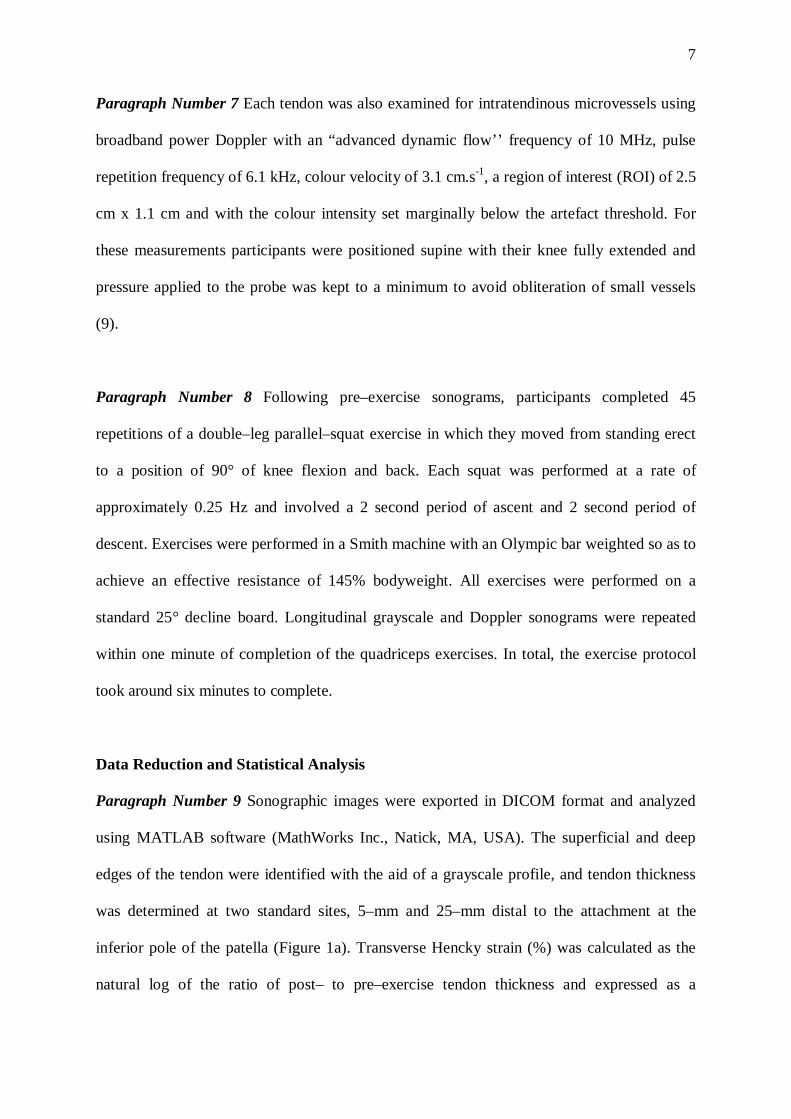
Paragraph Number 7 Each tendon was also examined for intratendinous microvessels using
broadband power Doppler with an “advanced dynamic flow’’ frequency of 10 MHz, pulse
repetition frequency of 6.1 kHz, colour velocity of 3.1 cm.s-1, a region of interest (ROI) of 2.5
cm x 1.1 cm and with the colour intensity set marginally below the artefact threshold. For
these measurements participants were positioned supine with their knee fully extended and
pressure applied to the probe was kept to a minimum to avoid obliteration of small vessels
(9).
Paragraph Number 8 Following pre–exercise sonograms, participants completed 45
repetitions of a double–leg parallel–squat exercise in which they moved from standing erect
to a position of 90° of knee flexion and back. Each squat was performed at a rate of
approximately 0.25 Hz and involved a 2 second period of ascent and 2 second period of
descent. Exercises were performed in a Smith machine with an Olympic bar weighted so as to
achieve an effective resistance of 145% bodyweight. All exercises were performed on a
standard 25° decline board. Longitudinal grayscale and Doppler sonograms were repeated
within one minute of completion of the quadriceps exercises. In total, the exercise protocol
took around six minutes to complete.
Data Reduction and Statistical Analysis
Paragraph Number 9 Sonographic images were exported in DICOM format and analyzed
using MATLAB software (MathWorks Inc., Natick, MA, USA). The superficial and deep
edges of the tendon were identified with the aid of a grayscale profile, and tendon thickness
was determined at two standard sites, 5–mm and 25–mm distal to the attachment at the
inferior pole of the patella (Figure 1a). Transverse Hencky strain (%) was calculated as the
natural log of the ratio of post– to pre–exercise tendon thickness and expressed as a

8
percentage. The within–subject coefficient of variation for repeated measurements of
transverse strain in the patellar tendon was 2.1%.
< Figure 1a & b about here>
Paragraph Number 10 Tendon echogenicity in the superficial and deep aspects of the
patellar tendon were estimated by calculating the mean grayscale value (arbitrary units [U])
within two rectilinear ROI. At each site, a rectilinear ROI was initially created in which the
length of all four sides equated to 90% of the measured tendon thickness. The ROI was
positioned equally between and centered about the digitised superficial and deep tendon
borders. The ROI at each site was subsequently bisected to form an equally sized deep (D)
and superficial (S) ROI. Thus, the superficial ROI was bound by the anterior border and
midsection of the tendon and an equivalent number of pixels proximal and distal to the
measurement site (Figure 1a). Likewise the deep ROI was bound by the midsection and
posterior border of the tendon and an equivalent number of pixels proximal and distal to the
measurement site (44). It was hypothesized that analysis of the patellar tendons from a
regional point of view may support recent work suggesting there is a difference in the strains
experienced between the posterior fibres of the tendon and those of the anterior region (10).
Grayscale values within the ROIs ranged between zero and 255, with higher values indicative
of an hyperechoic tendon. The coefficient of variation for repeated measures of tendon
echogenicity was 5.5%.
Paragraph Number 11 Intratendinous power Doppler flow (PDU) was qualitatively graded
from 0 to IV using the modified Öhberg score (9, 34). In brief, tendons were scored as 0
when no vessels were visible, and I to IV when one to four or more vessels were visible

9
within the 2.5 cm x 1.1 cm ROI. PDU images were independently scored by two examiners,
blinded to the case history of the participants, and with 100% agreement. PDU signal was
also quantitatively assessed using a modified version of a previously described method (31),
in which PDU area was calculated as the product of the number of colour pixels within the
tendon and the square of effective pixel resolution (Figure 1b,c). The limits of agreement for
repeated measurements of PDU area was ± 0.6 mm2.
Paragraph Number 12 The Statistical Package for the Social Sciences (SPSS Inc, Chicago,
IL, USA) was used for all statistical procedures. Kolmogorov–Smirnov tests were used to
evaluate data for underlying assumptions of normality. Outcome variables were determined
to be normally distributed, so means and standard deviations were used as summary statistics.
Between–group differences in body anthropometry were investigated using independent t–
tests. The effect of exercise on tendon thickness was evaluated using a repeated measures
ANOVA model within a generalized linear modeling framework, in which site (proximal/
distal) and exercise (pre/post) were treated as a within-subjects factors, while limb (control
left /control right/ asymptomatic/ symptomatic) was treated as a between-subject factor.
Between–group differences in average tendon grayscale were evaluated within a generalized
linear modeling framework using a three–way repeated measures ANOVAs in which region
(superficial/deep), site (proximal/distal) and exercise (pre/post) were treated as within–
subject factors. Where appropriate significant interactions between factors were investigated
further with separate one–way ANOVAs in combination with Tukey’s post hoc analysis.
Differences in qualitative grade of neovascularization following exercise were evaluated
using a Wilcoxon Signed Ranks Test, while differences in PDU area were assessed using a
paired t-test. Potential relationships between PDU area and symptom severity (VISA-P
10
scores) were evaluated using Pearson product-moment correlations. An alpha level of .05 was
used for all univariate tests of significance.
RESULTS
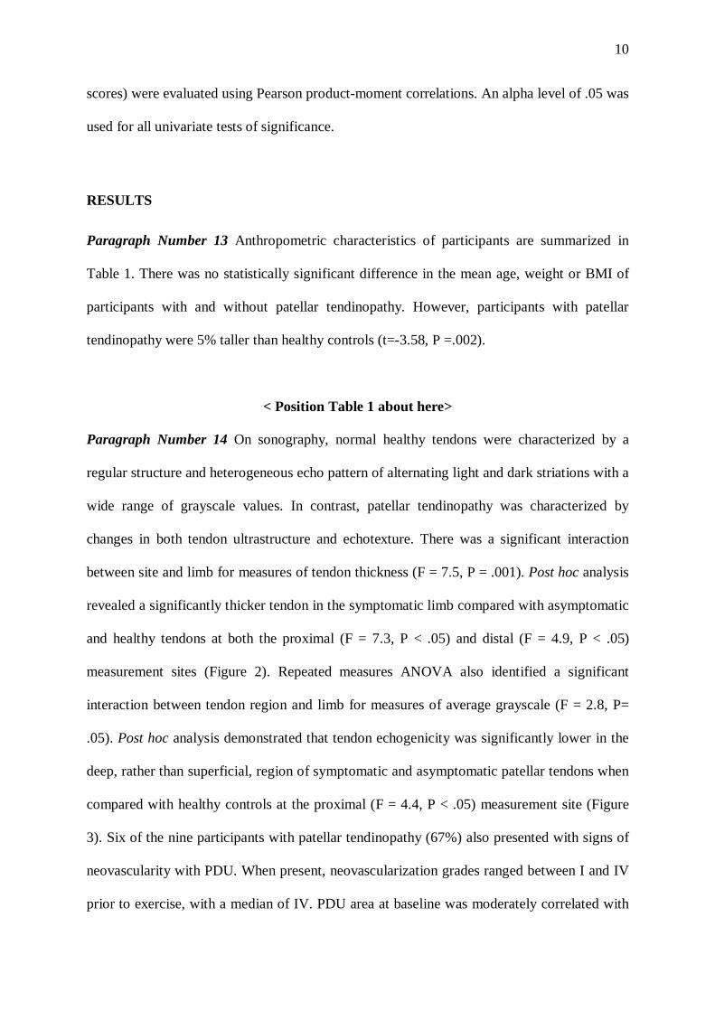
Paragraph Number 13 Anthropometric characteristics of participants are summarized in
Table 1. There was no statistically significant difference in the mean age, weight or BMI of
participants with and without patellar tendinopathy. However, participants with patellar
tendinopathy were 5% taller than healthy controls (t=-3.58, P =.002).
< Position Table 1 about here>
Paragraph Number 14 On sonography, normal healthy tendons were characterized by a
regular structure and heterogeneous echo pattern of alternating light and dark striations with a
wide range of grayscale values. In contrast, patellar tendinopathy was characterized by
changes in both tendon ultrastructure and echotexture. There was a significant interaction
between site and limb for measures of tendon thickness (F = 7.5, P = .001). Post hoc analysis
revealed a significantly thicker tendon in the symptomatic limb compared with asymptomatic
and healthy tendons at both the proximal (F = 7.3, P < .05) and distal (F = 4.9, P < .05)
measurement sites (Figure 2). Repeated measures ANOVA also identified a significant
interaction between tendon region and limb for measures of average grayscale (F = 2.8, P=
.05). Post hoc analysis demonstrated that tendon echogenicity was significantly lower in the
deep, rather than superficial, region of symptomatic and asymptomatic patellar tendons when
compared with healthy controls at the proximal (F = 4.4, P < .05) measurement site (Figure
3). Six of the nine participants with patellar tendinopathy (67%) also presented with signs of
neovascularity with PDU. When present, neovascularization grades ranged between I and IV
prior to exercise, with a median of IV. PDU area at baseline was moderately correlated with
11
VISA-P score (r = =-.58, P = .05), such that greater PDU area prior to exercise was associated
with lower (worse) symptom severity scores. Microvascularity was not present in tendons of
healthy participants.
< Position Figures 2 about here>
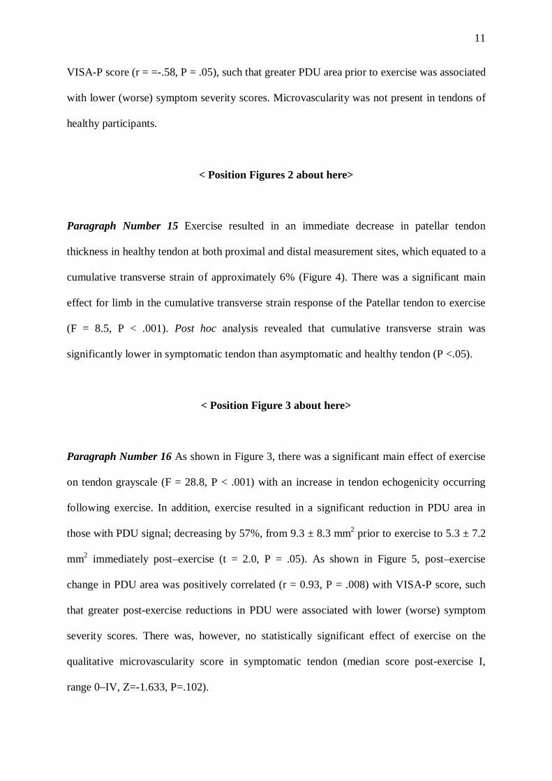
Paragraph Number 15 Exercise resulted in an immediate decrease in patellar tendon
thickness in healthy tendon at both proximal and distal measurement sites, which equated to a
cumulative transverse strain of approximately 6% (Figure 4). There was a significant main
effect for limb in the cumulative transverse strain response of the Patellar tendon to exercise
(F = 8.5, P < .001). Post hoc analysis revealed that cumulative transverse strain was
significantly lower in symptomatic tendon than asymptomatic and healthy tendon (P <.05).
< Position Figure 3 about here>
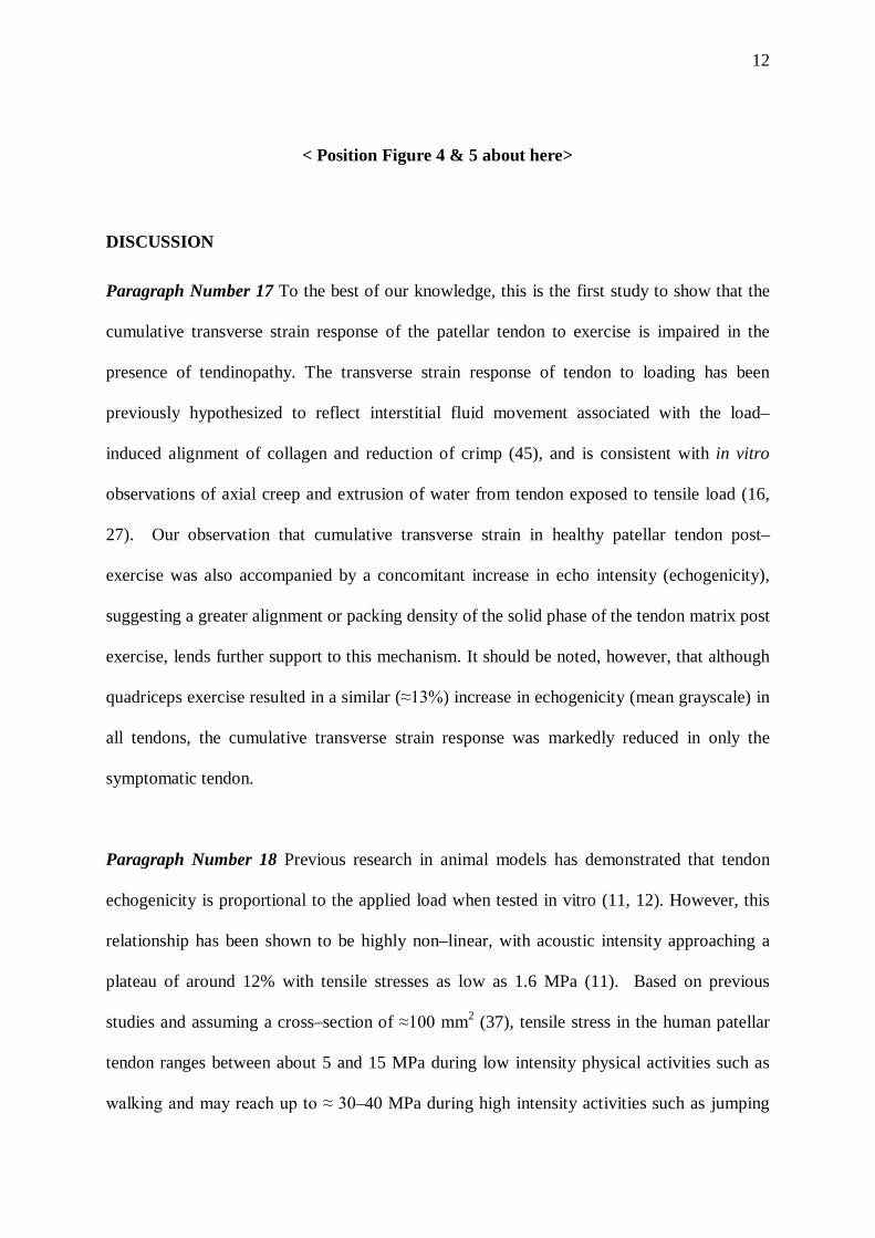
Paragraph Number 16 As shown in Figure 3, there was a significant main effect of exercise
on tendon grayscale (F = 28.8, P < .001) with an increase in tendon echogenicity occurring
following exercise. In addition, exercise resulted in a significant reduction in PDU area in
those with PDU signal; decreasing by 57%, from 9.3 ± 8.3 mm2 prior to exercise to 5.3 ± 7.2
mm2 immediately post–exercise (t = 2.0, P = .05). As shown in Figure 5, post–exercise
change in PDU area was positively correlated (r = 0.93, P = .008) with VISA-P score, such
that greater post-exercise reductions in PDU were associated with lower (worse) symptom
severity scores. There was, however, no statistically significant effect of exercise on the
qualitative microvascularity score in symptomatic tendon (median score post-exercise I,
range 0–IV, Z=-1.633, P=.102).
12
< Position Figure 4 & 5 about here>
DISCUSSION
Paragraph Number 17 To the best of our knowledge, this is the first study to show that the
cumulative transverse strain response of the patellar tendon to exercise is impaired in the
presence of tendinopathy. The transverse strain response of tendon to loading has been
previously hypothesized to reflect interstitial fluid movement associated with the load–
induced alignment of collagen and reduction of crimp (45), and is consistent with in vitro
observations of axial creep and extrusion of water from tendon exposed to tensile load (16,
27). Our observation that cumulative transverse strain in healthy patellar tendon post–
exercise was also accompanied by a concomitant increase in echo intensity (echogenicity),
suggesting a greater alignment or packing density of the solid phase of the tendon matrix post
exercise, lends further support to this mechanism. It should be noted, however, that although
quadriceps exercise resulted in a similar (≈13%) increase in echogenicity (mean grayscale) in
all tendons, the cumulative transverse strain response was markedly reduced in only the
symptomatic tendon.
Paragraph Number 18 Previous research in animal models has demonstrated that tendon
echogenicity is proportional to the applied load when tested in vitro (11, 12). However, this
relationship has been shown to be highly non–linear, with acoustic intensity approaching a
plateau of around 12% with tensile stresses as low as 1.6 MPa (11). Based on previous
studies and assuming a cross–section of ≈100 mm2 (37), tensile stress in the human patellar
tendon ranges between about 5 and 15 MPa during low intensity physical activities such as
walking and may reach up to ≈ 30–40 MPa during high intensity activities such as jumping
13
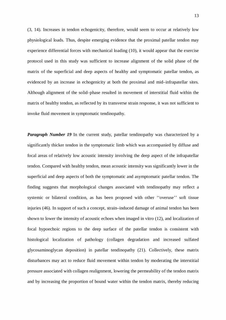
(3, 14). Increases in tendon echogenicity, therefore, would seem to occur at relatively low
physiological loads. Thus, despite emerging evidence that the proximal patellar tendon may
experience differential forces with mechanical loading (10), it would appear that the exercise
protocol used in this study was sufficient to increase alignment of the solid phase of the
matrix of the superficial and deep aspects of healthy and symptomatic patellar tendon, as
evidenced by an increase in echogenicity at both the proximal and mid–infrapatellar sites.
Although alignment of the solid–phase resulted in movement of interstitial fluid within the
matrix of healthy tendon, as reflected by its transverse strain response, it was not sufficient to
invoke fluid movement in symptomatic tendinopathy.
Paragraph Number 19 In the current study, patellar tendinopathy was characterized by a
significantly thicker tendon in the symptomatic limb which was accompanied by diffuse and
focal areas of relatively low acoustic intensity involving the deep aspect of the infrapatellar
tendon. Compared with healthy tendon, mean acoustic intensity was significantly lower in the
superficial and deep aspects of both the symptomatic and asymptomatic patellar tendon. The
finding suggests that morphological changes associated with tendinopathy may reflect a
systemic or bilateral condition, as has been proposed with other ‘‘overuse’’ soft tissue
injuries (46). In support of such a concept, strain–induced damage of animal tendon has been
shown to lower the intensity of acoustic echoes when imaged in vitro (12), and localization of
focal hypoechoic regions to the deep surface of the patellar tendon is consistent with
histological localization of pathology (collagen degradation and increased sulfated
glycosaminoglycan deposition) in patellar tendinopathy (21). Collectively, these matrix
disturbances may act to reduce fluid movement within tendon by moderating the interstitial
pressure associated with collagen realignment, lowering the permeability of the tendon matrix
and by increasing the proportion of bound water within the tendon matrix, thereby reducing

14
the available amount of unbound water (45). Although reduced permeability associated with
sulfated glycosaminoglycan deposition in tendon is thought to maintain fluid pressure and
increase compressive aggregate modulus, thereby protecting collagen fibres from further
disruption (29), hydrostatic pressure and interstitial fluid movement are known to produce a
wide range of phenomena in musculoskeletal tissues, including streaming potentials,
lubrication, nutrient transport, and mechanical signaling, which are important in tissue
differentiation and homeostasis (38). Given that interstitial fluid movement is thought to play
a key role in mechanotransduction and tendon homeostasis (16), it may be speculated that
diminished fluid movement with tendinopathy, as evidenced by a lower transverse strain
response to exercise, may contribute to the impaired capacity of the patellar tendon to adapt
to mechanical loading in tendinopathy. In support of this concept, similar changes in tendon
echotexture of the rotator cuff and an impaired transverse strain response of the Achilles
tendon to exercise have also been previously reported with clinical tendinopathy (5, 15).
Paragraph Number 20 Consistent with previous research (44), we observed an acute
decrease in tendon thickness in healthy patellar tendon in response to exercise, equating to a
cumulative transverse strain of ≈6% in healthy tendon. The magnitude of this creep response,
however, was markedly less than that previously reported for healthy Achilles tendon (≈15–
20%) following intense resistance exercise (19, 43, 45), but was comparable to that reported
for the Achilles tendon (≈5%) after 1 hour of floor–ball match play (13). Although direct
comparisons between studies is hampered by differences in study populations, the structure
of the examined tendon and loading protocols employed, the magnitude of dynamic creep in
other soft tissues, such as the intervertebral disc, is thought to depend on the quantity of
unbound fluid within the fluid–flow pathway, and marked changes in the creep behavior of
the disc have been shown with manipulation of tissue hydration (41). Thus the lower

15
transverse creep observed in healthy patellar tendon in this study may, therefore, partly
reflect differences in unbound fluid between the Achilles and patellar tendon despite evidence
that the total water content is similar between the two tendons (33). Previous research,
however, has also demonstrated that the extent of axial creep in soft tissues is dependent on
the magnitude and duration of the applied stress, with higher stresses eliciting a greater creep
response (39). Given the ratio of the cross sectional area of the Achilles tendon to the
physiological cross-sectional area of the Triceps Surae (300–500) is two to three times that of
the patellar tendon to Quadriceps Surae (100–250), it is likely that average axial stress within
the patellar tendon is typically less than that of the Achilles tendon during activities of daily
living (40). It is noteworthy that the total loading impulse of the exercise protocol used in this
study, however, is substantially lower (approximately half) than that used previously (43, 45),
with the total load and number of repetitions equating to only about 70% and 50% of that
used in common eccentric rehabilitation protocols in which 90 repetitions of unipedal loading
has been advocated (35). Nonetheless, the exercise protocol in this study was sufficient to
differentiate between individuals with and without tendinopathy and induced a similar
magnitude of transverse strain at both the proximal (5 mm) and mid–infrapatellar (25 mm)
tendon sites. This suggests that within a given tendon, the unbound fluid content, fluid
pathway, and interstitial pressure are likely comparable and that exercise–induced transverse
strains are relatively uniform across the measured tendon sites.
Paragraph Number 21 Although previous studies have shown that intratendinous Doppler
flow can be present in both healthy and symptomatic patellar tendons (18), we observed no
signs of microvascularity in healthy tendon with PDU. Thus, the cumulative transverse strain
response of tendon to exercise is unlikely to be the result of a transient reduction in
vascularity. However, six of the nine participants with patellar tendinopathy (67%) presented
16
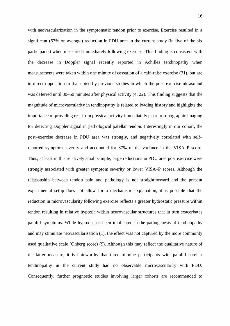
with neovascularization in the symptomatic tendon prior to exercise. Exercise resulted in a
significant (57% on average) reduction in PDU area in the current study (in five of the six
participants) when measured immediately following exercise. This finding is consistent with
the decrease in Doppler signal recently reported in Achilles tendinopathy when
measurements were taken within one minute of cessation of a calf–raise exercise (31), but are
in direct opposition to that noted by previous studies in which the post–exercise ultrasound
was deferred until 30–60 minutes after physical activity (4, 22). This finding suggests that the
magnitude of microvascularity in tendinopathy is related to loading history and highlights the
importance of providing rest from physical activity immediately prior to sonographic imaging
for detecting Doppler signal in pathological patellar tendon. Interestingly in our cohort, the
post–exercise decrease in PDU area was strongly, and negatively correlated with self–
reported symptom severity and accounted for 87% of the variance in the VISA–P score.
Thus, at least in this relatively small sample, large reductions in PDU area post exercise were
strongly associated with greater symptom severity or lower VISA–P scores. Although the
relationship between tendon pain and pathology is not straightforward and the present
experimental setup does not allow for a mechanistic explanation, it is possible that the
reduction in microvascularity following exercise reflects a greater hydrostatic pressure within
tendon resulting in relative hypoxia within neurovascular structures that in turn exacerbates
painful symptoms. While hypoxia has been implicated in the pathogenesis of tendinopathy
and may stimulate neovascularisation (1), the effect was not captured by the more commonly
used qualitative scale (Öhberg score) (9). Although this may reflect the qualitative nature of
the latter measure, it is noteworthy that three of nine participants with painful patellar
tendinopathy in the current study had no observable microvascularity with PDU.
Consequently, further prognostic studies involving larger cohorts are recommended to

17
elucidate potential relationships between the immediate post–exercise change in Doppler
flow and clinical symptoms in patellar tendinopathy.
Paragraph Number 22 This study evaluated the effect of tendinopathy on the cumulative
transverse strain response of the patellar tendon immediately following resistance exercise
that involved periodic concentric and eccentric modes of muscle contraction. While there is
evidence that both the type (eccentric/ concentric) and duration of muscle contraction may
influence blood flow and mechanical creep in tendon (24, 32), the magnitude of the acute
mechanical response observed in the current study may not be transferable to other types and
modes of exercise. A further limitation of this study is that we determined the strain response
of the patellar tendon to exercise in only one dimension. Although there is evidence that
mechanical properties of tendon are transversely isotropic (26) and that reductions in
mediolateral tendon thickness, albeit in the Achilles, have also been observed with loading
(19), it is unknown if the effects of exercise result in comparable mediolateral strains in the
patellar tendon. Similarly, we determined the response of tendon to exercise at standardized
distances (5 and 25 mm) from the inferior pole of the patella, rather than using a pre–defined
percentage of total tendon length. Evaluating transverse strains at standard distances from the
patellar pole does not account for variations in tendon length between groups. Although we
found significant differences in height between our groups and a longer patellar tendon
relative to the patella has been implicated in development of patellar tendinopathy (20),
recent research employing magnetic resonance imaging has reported no difference in tendon
length between those with and without tendinopathy (36). Moreover, it should be recognized
that patellar tendon length typically exceeds the footprint of most ultrasonic transducers and
although alternative sonographic methods, including extended field of view and surface
marker techniques have been proposed, these techniques may introduce substantial (5–35%)
18
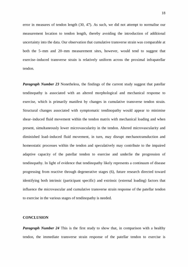
error in measures of tendon length (30, 47). As such, we did not attempt to normalise our
measurement location to tendon length, thereby avoiding the introduction of additional
uncertainty into the data. Our observation that cumulative transverse strain was comparable at
both the 5–mm and 20–mm measurement sites, however, would tend to suggest that
exercise–induced transverse strain is relatively uniform across the proximal infrapatellar
tendon.
Paragraph Number 23 Nonetheless, the findings of the current study suggest that patellar
tendinopathy is associated with an altered morphological and mechanical response to
exercise, which is primarily manifest by changes in cumulative transverse tendon strain.
Structural changes associated with symptomatic tendinopathy would appear to minimise
shear–induced fluid movement within the tendon matrix with mechanical loading and when
present, simultaneously lower microvascularity in the tendon. Altered microvascularity and
diminished load–induced fluid movement, in turn, may disrupt mechanotransduction and
homeostatic processes within the tendon and speculatively may contribute to the impaired
adaptive capacity of the patellar tendon to exercise and underlie the progression of
tendinopathy. In light of evidence that tendinopathy likely represents a continuum of disease
progressing from reactive through degenerative stages (6), future research directed toward
identifying both intrinsic (participant specific) and extrinsic (external loading) factors that
influence the microvascular and cumulative transverse strain response of the patellar tendon
to exercise in the various stages of tendinopathy is needed.
CONCLUSION
Paragraph Number 24 This is the first study to show that, in comparison with a healthy
tendon, the immediate transverse strain response of the patellar tendon to exercise is
19
diminished with tendinopathy. Given that interstitial fluid movement is thought to play a key
role in convective transport, mechanotransduction and tendon homeostasis, the acute
transverse strain response of tendon to a defined exercise protocol may reflect the progression
of patellar tendinopathy, and may provide clinicians with an index for evaluating tendon
resilience which can be used to objectively monitor the progress of tendon rehabilitation
programs. It is recommended that future research be directed toward exploring the immediate
microvascular and cumulative strain response of the patellar tendon to mechanical loading
and their potential role in the progression of patellar tendinopathy.

20
ACKNOWLEDGEMENTS
This research was part funded by the Australian Institute of Sport, Queensland Academy of
Sport, Queensland Government and the Australian Research Council. The authors would like
to thank Ms Melina Simjanovic and Ms Kristen Eales for their assistance with data
collection. The authors declare no conflicts of interest, financial or otherwise. The results of
the present study do not constitute endorsement by American College of Sports Medicine.
21
REFERENCES
1. Alfredson H, Ohberg L, and Forsgren S. Is vasculo-neural ingrowth the cause of pain in
chronic Achilles tendinosis? An investigation using ultrasonography and colour Doppler,
immunohistochemistry, and diagnostic injections. Knee Surg Sports Traumatol Arthrosc.
2003;11:334-3.
2. Arya S, and Kulig K. Tendinopathy alters mechanical and material properties of the
Achilles tendon. J Appl Physiol. 2010;108:670-5.
3. Besier TF, Fredericson M, Gold GE, Beaupre GS, and Delp SL. Knee muscle forces
during walking and running in patellofemoral pain patients and pain-free controls. J
Biomech. 2009;42:898-905.
4. Boesen AP, Boesen MI, Koenig MJ, Bliddal H, Torp-Pedersen S, and Langberg H.
Evidence of accumulated stress in Achilles and anterior knee tendons in elite badminton
players. Knee Surg Sports Traumatol Arthrosc. 2011;19:30-7.
5. Collinger JL, Fullerton B, Impink BG, Koontz AM, and Boninger ML. Validation of
greyscale-based quantitative ultrasound in manual wheelchair users: relationship to
established clinical measures of shoulder pathology. Am J Phys Med Rehabil.
2010;89(5):390-400.
6. Cook JL, and Purdam CR. Is tendon pathology a continuum? A pathology model to
explain the clinical presentation of load-induced tendinopathy. Br J Sports Med.
2009;43:409-16.
7. Couppé C, Kongsgaard M, Aagaard P, Hansen P, Bojsen-Moller J, Kjaer M, and
Magnusson SP. Habitual loading results in tendon hypertrophy and increased stiffness of
the human patellar tendon. J Appl Physiol. 2008;105(3):805-10.

22
8. Couppe C, Kongsgaard M, Aagaard P, Vinther A, Boesen M, Kjaer M, and Magnusson
SP. Differences in tendon properties in elite badminton players with or without patellar
tendinopathy. Scand J Med Sci Sports. 2013;23:e89-95.
9. de Vos RJ, Weir A, Cobben LP, and Tol JL. The value of power Doppler
ultrasonography in Achilles tendinopathy: a prospective study. Am J Sports Med.
2007;35(10):1696-701.
10. Dillon EM, Erasmus PJ, Müller JH, Scheffer C, and de Villiers RV. Differential forces
within the proximal patellar tendon as an explanation for the characteristic lesion of
patellar tendinopathy: an in vivo descriptive experimental study. Am J Sports Med.
2008;36(11):2119-27.
11. Duenwald-Kuehl S, Kobayashi H, Frisch K, Lakes R, and Vanderby Jr R. Ultrasound
echo is related to stress and strain in tendon. J Biomech. 2011;44:424-9.
12. Duenwald-Kuehl S, Lakes R, and Vanderby Jr R. Strain-induced damage reduces echo
intensity changes in tendon during loading. J Biomech. 2012;45(9):1607 - 11.
13. Fahlström M, and Alfredson H. Ultrasound and Doppler findings in the Achilles tendon
among middle-aged recreational floor-ball players in direct relation to a match. Br J
Sports Med. 2010;44 (2):140-3.
14. Finni T, Komi PV, and Lepola V. In vivo human triceps surae and quadriceps femoris
muscle function in a squat jump and counter movement jump. Eur J Appl Physiol.
2000;83:416-26.
15. Grigg NL, Wearing SC, and Smeathers JE. Achilles tendinopathy has an aberrant strain
response to eccentric exercise. Medicine & Science in Sports & Exercise. 2012;44:12-7.
16. Hannafin JA, and Arnoczky SP. Effect of cyclic and static tensile loading on water
content and solute diffusion in canine flexor tendons: an in vitro study. J Orthop Res.
1994;12:350-6.

23
17. Helland C, J. B-M, Raastad T, Seynnes O.R, Moltubakk MM, Jakobsen V, Visnes H, and
Bahr R. Mechanical properties of the patellar tendon in elite volleyball players with and
without patellar tendinopathy. Br J Sports Med. 2013;47(13):862-8.
18. Hirschmüller A, Frey V, Konstantinidis L, Baur H, Dickhuth HH, Südkamp NP, and
Helwig P. Prognostic value of Achilles tendon doppler sonography in asymptomatic
runners. Med Sci Sports Exerc. 2012;44(2):199-205.
19. Iwanuma S, Akagi R, Kurihara T, Ikegawa S, Kanehisa H, Fukunaga T, and Kawakami
Y. Longitudinal and transverse deformation of human Achilles tendon induced by
isometric plantar flexion at different intensities. J Appl Physiol. 2011;110(6):1615-21.
20. Johnson DP, Wakeley CJ, and Watt I. Magnetic resonance imaging of patellar tendonitis.
J Bone Joint Surg. 1996;78-B:452-7.
21. Khan KM, Bonar F, Desmond PM, Cook JL, Young DA, Visentini PJ, Fehrmann MW,
Kiss ZS, O'Brien PA, Harcourt PR, Dowling RJ, O'Sullivan RM, Crichton KJ, Tress BM,
and Wark JD. Patellar tendinosis (jumper's knee): findings at histopathologic
examination, US, and MR imaging. Victorian Institute of Sport Tendon Study Group.
Radiology. 1996;200:821-7.
22. Koenig MJ, Torp-Pedersen S, Boesen MI, Holm CC, and Bliddal H. Doppler
ultrasonography of the anterior knee tendons in elite badminton players: colour fraction
before and after match. Br J Sports Med. 2010;44:134-9.
23. Kongsgaard M, Qvortrup K, Larsen J, Aagaard P, Doessing S, Hansen P, Kjaer M, and
Magnusson SP. Fibril morphology and tendon mechanical properties in patellar
tendinopathy: effects of heavy slow resistance training. Am J Sports Med.
2010;38(4):749-56.

24
24. Kubo K, Ikebukuro T, Yaeshima K, and Kanehisa H. Effects of different duration
contractions on elasticity, blood volume, and oxygen saturation of human tendon in vivo.
Eur J Appl Physiol. 2009;106(3):445-55.
25. Kubo K, Ikebukuro T, Yaeshima K, Yata H, Tsunoda N, and Kanehisa H. Effects of
static and dynamic training on the stiffness and blood volume of tendon in vivo. J Appl
Physiol. 2009;106:412-7.
26. Kuo PL, Li PC, and Li ML. Elastic properties of tendon measured by two different
approaches. Ultrasound Med Biol. 2001;27(9):1275-84.
27. Lanir Y, Saland EL, and Foux A. Physico-chemical and micro-structural changes in
collagen fibre bundles following stretch in-vitro. Biorheology. 1988;25(4):591-604.
28. Lian OB, Engebretsen L, and Bahr R. Prevalence of jumper’s knee among elite athletes
from different sports: a cross-sectional study. Am J Sports Med. 2005;33:561-7.
29. Loboa EG, Wren TAL, Beaupre´ GS, and Carter DR. Mechanobiology of soft skeletal
tissue differentiation-a computational approach of a fiber-reinforced poroelastic model
based on homogeneous and isotropic simplifications. Biomech Model Mechanobiol.
2003;2:83-96.
30. Maganaris CN. Validity of procedures involved in ultrasound-based measurement of
human plantarflexor tendon elongation on contraction. J Biomech. 2005;38:9-13.
31. Malliaras P, Chan O, Simran G, Martinez de Albornoz P, Morrissey D, and Maffulli N.
Doppler ultrasound signal in Achilles tendinopathy reduces immediately after activity.
Int J Sports Med. 2012;33(6):480-4.
32. Malliaras P, Kamal B, Nowell A, Farley T, Dhamu H, Simpson V, Morrissey D,
Langberg H, Maffulli N, and Reeves ND. Patellar tendon adaptation in relation to load-
intensity and contraction type. J Biomech. 2013 46:1893-9.
25
33. Maynard JA, Pedrini VA, Pedrini-Mille A, Romanus B, and Ohlerking F. Morphological
and biochemical effects of sodium morrhuate on tendons. J Orthop Res. 1985;3:236-48.
34. Ohberg L, Lorentzon R, and Alfredson H. Neovascularisation in Achilles tendons with
painful tendinosis but not in normal tendons: an ultrasonographic investigation. Knee
Surg Sports Traumatol Arthrosc. 2001;9(4):233-8.
35. Purdam CR, Jonsson P, Alfredson H, Lorentzon R, Cook JL, and Khan KM. A pilot
study of the eccentric decline squat in the management of painful chronic patellar
tendinopathy. Br J Sports Med. 2004;38(4):395-7.
36. Shalaby M, and Almekinders LC. Patellar tendinitis: the significance of magnetic
resonance imaging findings. Am J Sports Med. 1999;27:345-9.
37. Svensson RB, Hansen P, Hassenkam T, Haraldsson BT, Aagaard P, Kovanen V,
Krogsgaard M, Kjaer M, and Magnusson SP. Mechanical properties of human patellar
tendon at the hierarchical levels of tendon and fibril. J Appl Physiol. 2012;112:419-26.
38. Swartz MA, and Fleury ME. Interstitial flow and its effects in soft tissues. Annu Rev
Biomed Eng. 2007;9:229-56.
39. Thornton GM, Shrive NG, and Frank CB. Ligament creep recruits fibres at low stresses
and can lead to modulus-reducing fibre damage at higher creep stresses: a study in rabbit
medial collateral ligament model. J Orthop Res. 2002;20:967-74.
40. Vereecke EE, and AJ. C. The role of hind limb tendons in gibbon locomotion: springs or
strings? J Exp Biol. 2013;216:3971-80.
41. Vresilovic EJ, Johannessen W, and Elliott DM. Disc mechanics with trans-endplate
partial nucleotomy are not fully restored following cyclic compressive loading and
unloaded recovery. J Biomech Eng. 2006;128(6):823-9.
26
42. Wall ME, and Banes AJ. Early responses to mechanical load in tendon: role for calcium
signaling, gap junctions and intercellular communication. J Musculoskelet Neuronal
Interact. 2005;5:70-84.
43. Wearing SC, Grigg NL, Hooper SL, and Smeathers JE. Conditioning of the Achilles
tendon via ankle exercise improves correlations between sonographic measures of tendon
thickness and body anthropometry. J Appl Physiol. 2011;110(5):1384-9.
44. Wearing SC, Hooper SL, Purdam C, Cook J, Grigg N, Locke S, and Smeathers JE. The
acute transverse strain response of the patellar tendon to quadriceps exercise. Med Sci
Sports Exerc. 2013;45(4):772-7.
45. Wearing SC, Smeathers JE, Urry SR, and Hooper SL. The time-course of acute changes
in Achilles tendon morphology following exercise. In: FK Fuss, A Subic and S Ujihashi
editors. The Impact of Technology on Sport II. Singapore: Taylor and Francis; 2008, pp.
65-8.
46. Wearing SC, Smeathers JE, Urry SR, Yates B, Sullivan PM, and Dubois P. Plantar
enthesopathy: thickening of the enthesis is correlated with energy dissipation of the
plantar fat pad during walking. Am J Sports Med. 2010;38:2522-7.
47. Weng L, Tirumalai A, Lowery C, and Nock LF. US extended-field-of-view imaging
technology. Radiology. 1997;203:877-80.

27
FIGURE LEGENDS
Figure 1. Patellar tendon thickness was measured 5 mm (L1) and 25 mm (L2) inferior to the
patella pole (P) with the aid of a grayscale profile. At each site, average grayscale in the
superficial (S1, S2) and deep (D1, D2) aspects of the tendon were determined (a). Illustration
of the intratendinous power Doppler flow (PDU) within a 2.5 cm x 1.1 cm region of interest
that was graded using the modified Öhberg score (b). The area of the same intratendinous
PDU flow was also quantified by determining the number of colour pixels within the tendon
and the square of effective pixel resolution. The bias and limits of agreement for repeated
measurements of area was -0.01 ± 0.6 mm2 (c).
Figure 2. Patellar tendon thickness in symptomatic, asymptomatic and left and right control
limbs measured 5 mm (Site 1) and 25 mm (Site 2) inferior to the patella pole. † Statistically
significant main effect for site (P < .05). * Significantly different from all other limbs at the
given site (P < .05).
Figure 3. Mean grayscale in the superficial and deep aspects of the patellar tendon prior to
(pre) and following (post) exercise in symptomatic, asymptomatic and left and right control
limbs and determined at two sites; 5–mm distal to the inferior pole of the patellar (a) and 25–
mm distal to the inferior pole of the patellar (b). Note that while average grayscale was
significantly lower in both symptomatic and asymptomatic tendon, there was a statistically
significant main effect for exercise in all tendons (P<.05).
Figure 4. Cumulative transverse strain in the patellar tendon in symptomatic, asymptomatic
and control limbs following exercise. Strain was determined 5 mm (site 1) and 25 mm (site 2)

28
distal to the inferior pole of the patella. * Significantly different from all other limbs at the
given site (P<.05).
Figure 5. The relationship between symptom severity (VISA-P) and change in PDU area in
the infrapatellar tendon immediately following exercise.