brains and nervous systems how are electrical signals passed along nerves? how are electrical...
Post on 20-Dec-2015
217 views
TRANSCRIPT

Brains and nervous systems
• How are electrical signals passed along nerves?
• How are electrical signals passed between nerves?
– and from receptors to nerves?
– and from nerves to effectors?
• Where and how does computing occur in the nervous system?
– Simple circuits
– Modulation of signals
• How is complex, structured information processed?
– Visual maps
• How is information stored and used to influence future response?
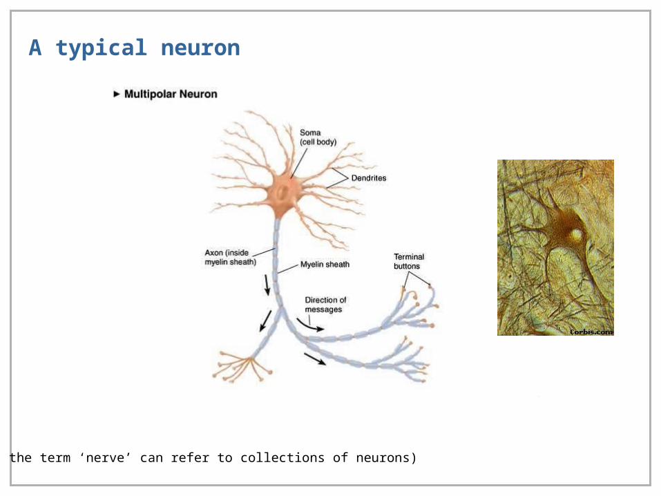
A typical neuron
(Note – the term ‘nerve’ can refer to collections of neurons)

The giant axon of squid
• Axons up to 1mm in diameter – 1000 times that of mammalian nerves
• Hodgkin and Huxley (1939) measured resting potential
• Hodgkin and Katz (1949) measured change in potential resulting from
manipulating K+ concentration
– Permeability to potassium is the primary source of the resting membrane
potential (NB permeability due to different process from gates)
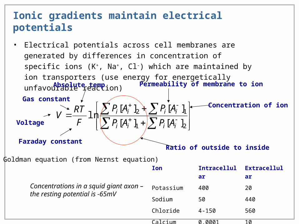
Ionic gradients maintain electrical potentials
• Electrical potentials across cell membranes are generated by differences in
concentration of specific ions (K+, Na+, Cl-) which are maintained by ion
transporters (use energy for energetically unfavourable reaction)
21
12
][][
][][ln
iiii
iiii
APAP
APAP
F
RTV
Voltage
Faraday constant
Gas constant
Absolute temp Permeability of membrane to ion
Concentration of ion
Ratio of outside to inside
Ion Intracellular Extracellular
Potassium 400 20
Sodium 50 440
Chloride 4-150 560
Calcium 0.0001 10
Concentrations in a squid giant axon – the resting potential is -65mV
Goldman equation (from Nernst equation)
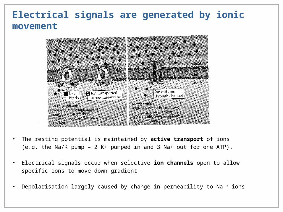
Electrical signals are generated by ionic movement
• The resting potential is maintained by active transport of ions (e.g. the Na/K pump – 2 K+
pumped in and 3 Na+ out for one ATP).
• Electrical signals occur when selective ion channels open to allow specific ions to move down
gradient
• Depolarisation largely caused by change in permeability to Na + ions
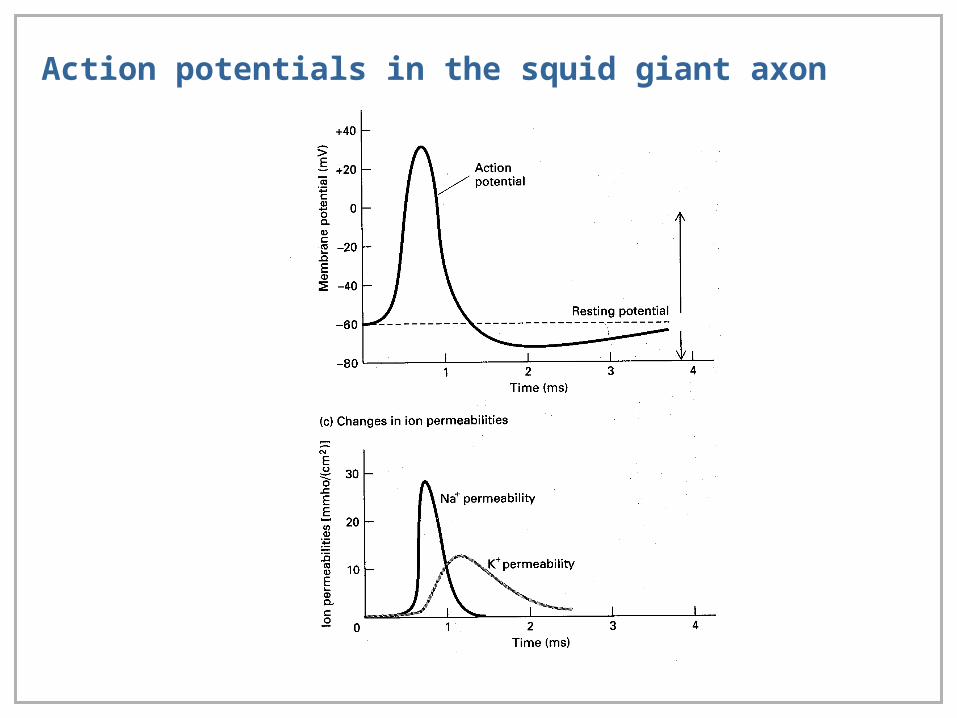
Action potentials in the squid giant axon
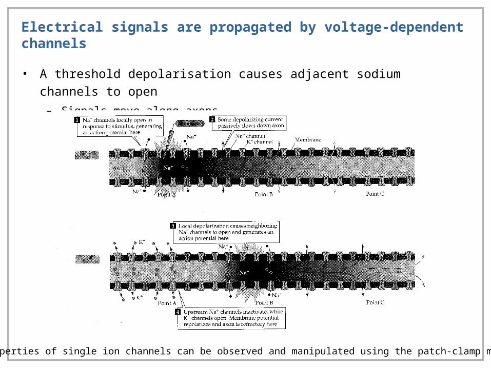
Electrical signals are propagated by voltage-dependent channels
• A threshold depolarisation causes adjacent sodium channels to open
– Signals move along axons
The properties of single ion channels can be observed and manipulated using the patch-clamp method
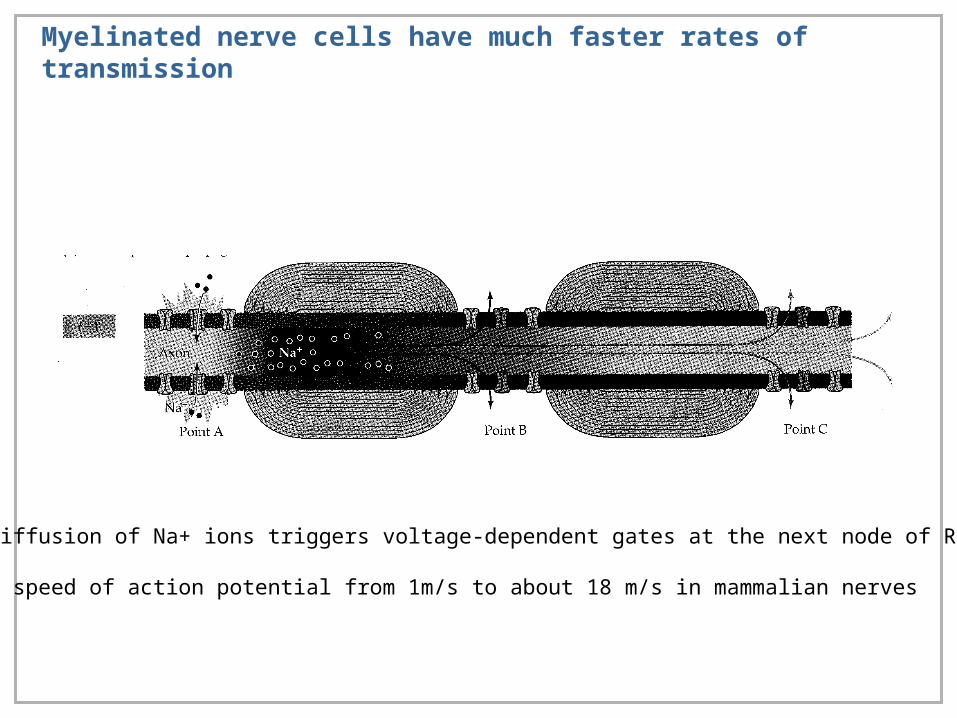
Myelinated nerve cells have much faster rates of transmission
Passive diffusion of Na+ ions triggers voltage-dependent gates at the next node of Ranvier
Increases speed of action potential from 1m/s to about 18 m/s in mammalian nerves
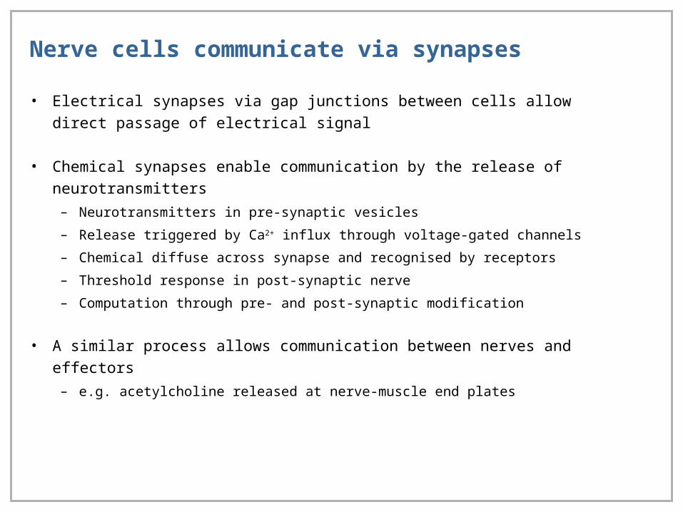
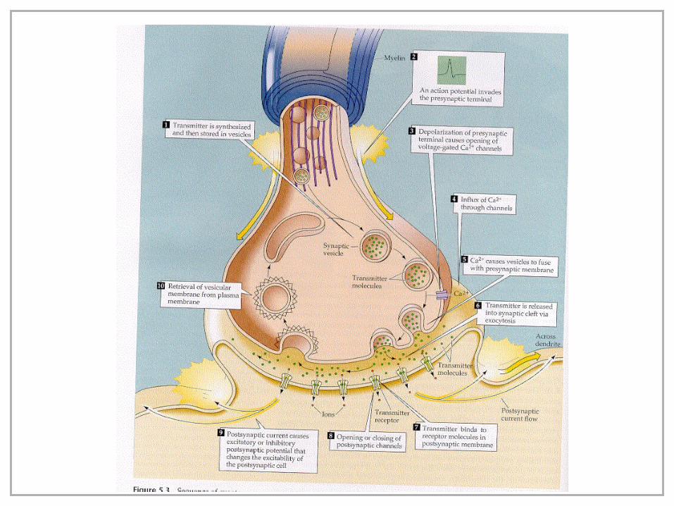
Nerve cells communicate via synapses
• Electrical synapses via gap junctions between cells allow direct passage of
electrical signal
• Chemical synapses enable communication by the release of neurotransmitters
– Neurotransmitters in pre-synaptic vesicles
– Release triggered by Ca2+ influx through voltage-gated channels
– Chemical diffuse across synapse and recognised by receptors
– Threshold response in post-synaptic nerve
– Computation through pre- and post-synaptic modification
• A similar process allows communication between nerves and effectors
– e.g. acetylcholine released at nerve-muscle end plates
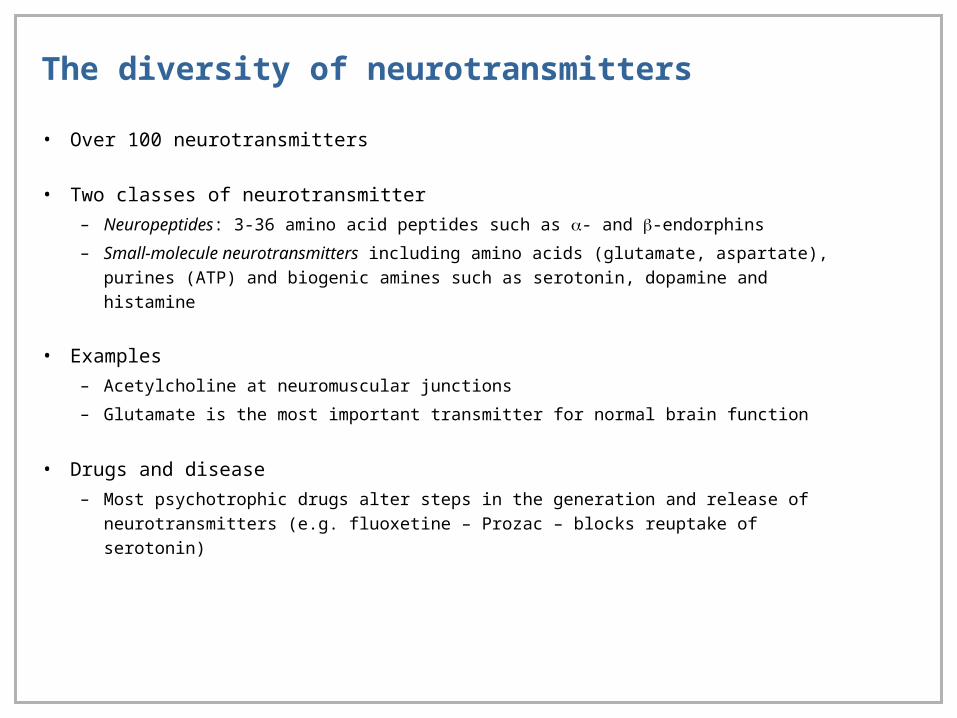
The diversity of neurotransmitters
• Over 100 neurotransmitters
• Two classes of neurotransmitter
– Neuropeptides: 3-36 amino acid peptides such as - and -endorphins
– Small-molecule neurotransmitters including amino acids (glutamate, aspartate),
purines (ATP) and biogenic amines such as serotonin, dopamine and histamine
• Examples
– Acetylcholine at neuromuscular junctions
– Glutamate is the most important transmitter for normal brain function
• Drugs and disease
– Most psychotrophic drugs alter steps in the generation and release of
neurotransmitters (e.g. fluoxetine – Prozac – blocks reuptake of serotonin)
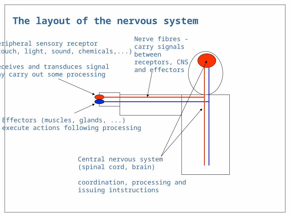
The layout of the nervous system
Peripheral sensory receptor(touch, light, sound, chemicals,...)
Receives and transduces signalMay carry out some processing
Central nervous system (spinal cord, brain)
coordination, processing andissuing intstructions
Nerve fibres - carry signals between receptors, CNS and effectors
Effectors (muscles, glands, ...)execute actions following processing
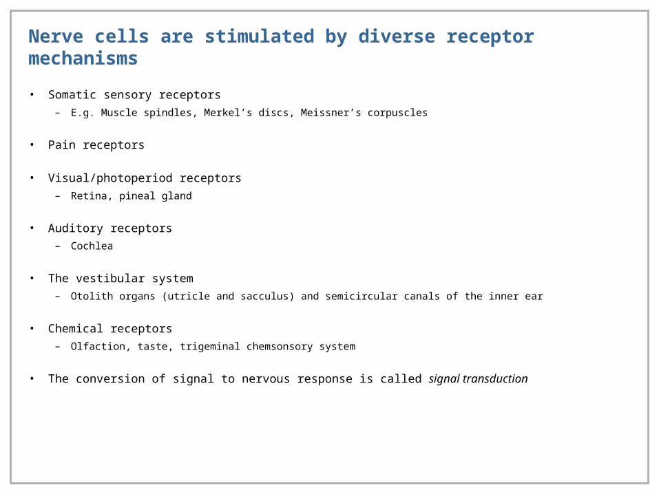
Nerve cells are stimulated by diverse receptor mechanisms
• Somatic sensory receptors
– E.g. Muscle spindles, Merkel’s discs, Meissner’s corpuscles
• Pain receptors
• Visual/photoperiod receptors
– Retina, pineal gland
• Auditory receptors
– Cochlea
• The vestibular system
– Otolith organs (utricle and sacculus) and semicircular canals of the inner ear
• Chemical receptors
– Olfaction, taste, trigeminal chemsonsory system
• The conversion of signal to nervous response is called signal transduction

Computational aspects of nerve cell communication
• Most external and internal information is analogue
• Nerve-cells typically communicate by digital responses involving threshold
responses
– Digital signals can be sent much faster and with greater ‘error’ than analogue
signals
• Several sensory systems carry out considerable processing of signals prior to
transmission to the central nervous system
– Lateral communication between receptors in the eye
– Tuning of macromechanical resonance to frequency of sound
• The spatial nature of information is maintained by maps in nerve-cell organisation

Vision
• Within the retina, light is
sensed by the rods and
cones. Rods, which
outnumber cones by about
10:1, are insensitive to
colour (unlike cones), but
more sensitive to light.
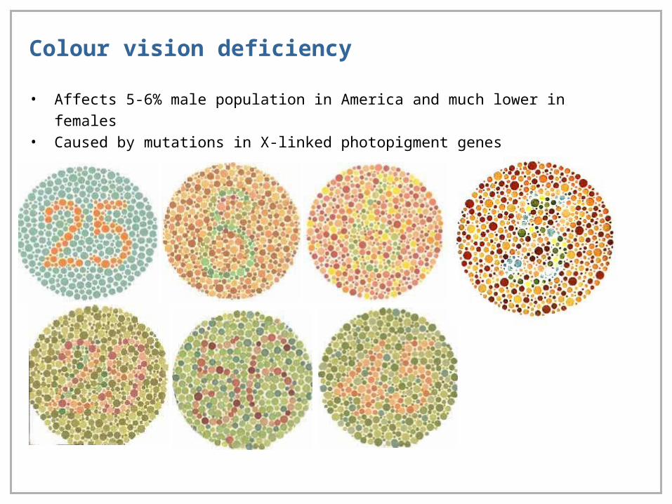
Colour vision deficiency
• Affects 5-6% male population in America and much lower in females• Caused by mutations in X-linked photopigment genes

Processing visual information in the retina
There are two classes of ganglion cells – on-centre and off-centre. On-centre are stimulated by light falling on the centre of the cell’s field, off-centre are stimulated when the light is turned off. Each change in light intensity is accompanied by a burst of activity. (Every change in luminance is accompanied by a burst of APs).
Different ganglion cells synapse with different classes of bipolar cells, which have graded responses (not action potentials)
Luminance contrast arises from antagonistic responses from the centre and periphery of a ganglion cell’s field of view


The visual map
• The physical location of ganglion cells in the retina is conserved in the arrangement of nerve-
connections with the subsequent processing units
• Subsets of ganglion cells provide information about colour contrast and moving and non-
moving objects
• The optic chasm allows information to flow between the two visual fields
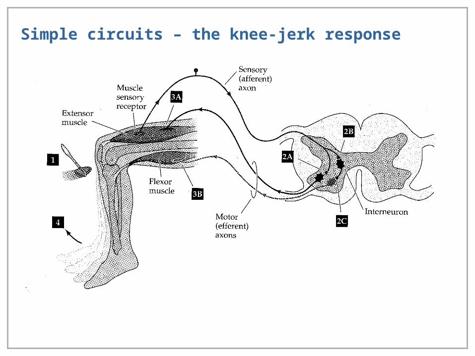
Simple circuits – the knee-jerk response
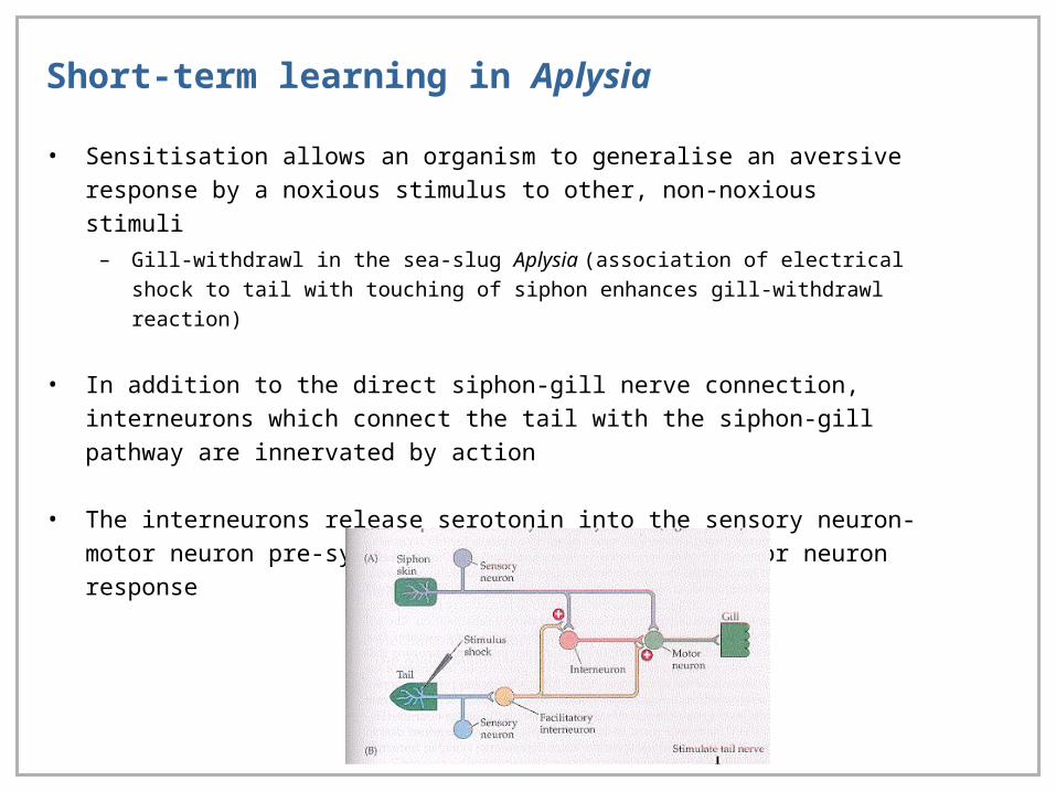
Short-term learning in Aplysia
• Sensitisation allows an organism to generalise an aversive response by a noxious
stimulus to other, non-noxious stimuli
– Gill-withdrawl in the sea-slug Aplysia (association of electrical shock to tail with
touching of siphon enhances gill-withdrawl reaction)
• In addition to the direct siphon-gill nerve connection, interneurons which connect the tail
with the siphon-gill pathway are innervated by action
• The interneurons release serotonin into the sensory neuron-motor neuron pre-synapse
which amplifies the motor neuron response
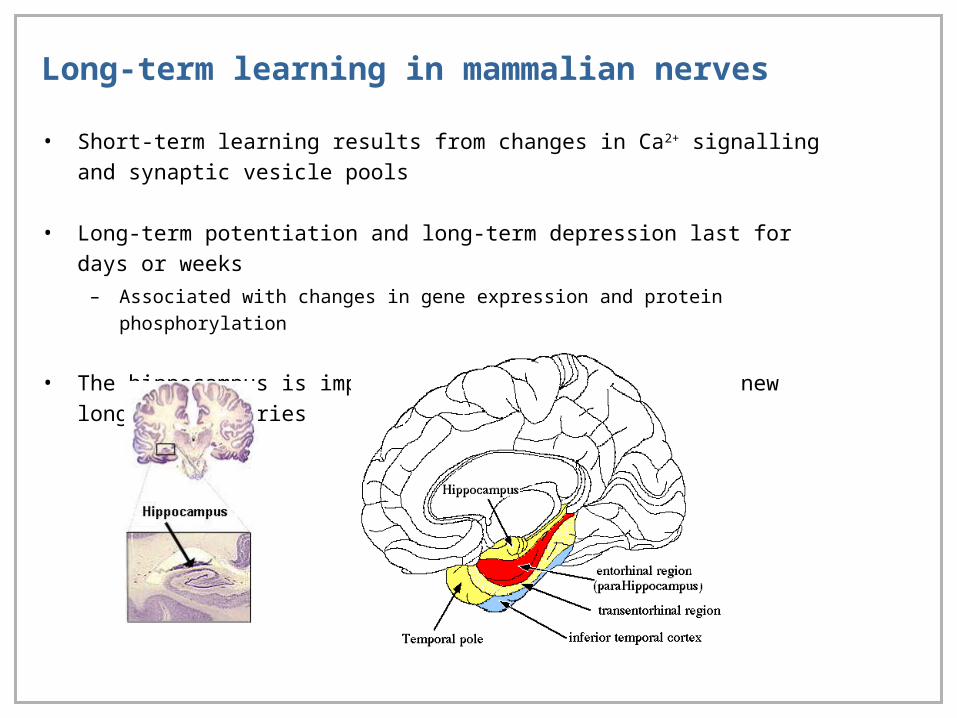
Long-term learning in mammalian nerves
• Short-term learning results from changes in Ca2+ signalling and synaptic vesicle pools
• Long-term potentiation and long-term depression last for days or weeks
– Associated with changes in gene expression and protein phosphorylation
• The hippocampus is important for the development of new long-term memories