brain distribution of cediranib is limited by active...
TRANSCRIPT

Brain Distribution of Cediranib Is Limited by Active Efflux at theBlood-Brain Barrier
Tianli Wang, Sagar Agarwal, and William F. ElmquistDepartment of Pharmaceutics and Brain Barriers Research Center, University of Minnesota, Minneapolis, Minnesota
Received November 28, 2011; accepted February 3, 2012
ABSTRACTCediranib is an orally active tyrosine kinase inhibitor that targetsthe vascular endothelial growth factor receptor family. Because ofits potent antiangiogenic and antitumor activities, cediranib hasbeen evaluated for therapy in glioma, a primary brain tumor. Thisstudy investigated the influence of two important efflux transport-ers at the blood-brain barrier, P-glycoprotein (P-gp) and breastcancer resistance protein (Bcrp), on the delivery of cediranib to thecentral nervous system. In vitro studies indicated that cediranib isa dual substrate for both P-gp and Bcrp. It is noteworthy that inspite of the in vitro data the in vivo mouse disposition studiesconclusively showed that P-gp was the dominant transporter re-stricting the brain distribution of cediranib. The brain-to-plasmapartitioning (AUCbrain/AUCplasma, where AUC is area under thecurve) and the steady-state brain-to-plasma concentration ratio ofcediranib were approximately 20-fold higher in Mdr1a/b(�/�) and
Mdr1a/b(�/�)Bcrp1(�/�) mice compared with wild-type andBcrp1(�/�) mice. Moreover, there was no significant difference inbrain distribution of cediranib between wild-type and Bcrp1(�/�)mice and between Mdr1a/b(�/�) and Mdr1a/b(�/�)Bcrp1(�/�)mice. These results show that, unlike other tyrosine kinase inhib-itors that are dual substrates for P-gp and Bcrp, Bcrp does notrestrict the distribution of cediranib across the blood-brain barrier.We also show that inhibition of P-gp using specific or nonspecificinhibitors resulted in significantly enhanced delivery of cediranib tothe brain. Concurrent administration of cediranib with chemicalmodulators of efflux transporters can be used as a strategy toenhance delivery and thus efficacy of cediranib in the brain. Thesefindings are clinically relevant to the efficacy of cediranib chemo-therapy in glioma.
IntroductionDelivery of drugs through the blood-brain barrier (BBB) is
one of the fundamental challenges in treating diseases of thecentral nervous system (CNS). The BBB is an anatomicalbarrier because of the presence of endothelial tight junctionsand a functional barrier because of the expression of activedrug efflux transporters. Together, these two aspects restrict
the passage of most small and large molecules into the brain,making the barrier effectively impermeable to many com-pounds that target diseases of the CNS. The ATP-bindingcassette (ABC) transporters P-glycoprotein (P-gp; ABCB1),and breast cancer resistance protein (Bcrp; ABCG2) are twoimportant efflux transporters that work together at the BBB,limiting the transport of several therapeutic agents into thebrain (Agarwal et al., 2011a). It has been shown that thereare overlapping substrate specificities for these two effluxpumps, and a number of drugs, including many anticancertyrosine kinase inhibitors (TKIs), are reported to be dualsubstrates for both P-gp and Bcrp (Dai et al., 2003; de Crieset al., 2007; Chen et al., 2009; Polli et al., 2009; Agarwal etal., 2010, 2011; Tang et al., 2012). Furthermore, it has beenreported that P-gp is dominant in restricting the brain pen-etration of many dual P-gp/Bcrp substrates with Bcrp being
This work was supported in part by the National Institutes of HealthNational Cancer Institute [Grant CA138437] and the Children’s Cancer Re-search Fund at the University of Minnesota (to W.F.E.). Financial support forT.W. was provided by a Ronald J. Sawchuk Fellowship in Pharmacokineticsfrom the Department of Pharmaceutics, University of Minnesota. Financialsupport for S.A. was provided by a doctoral dissertation fellowship from theUniversity of Minnesota.
Article, publication date, and citation information can be found athttp://jpet.aspetjournals.org.
http://dx.doi.org/10.1124/jpet.111.190488.
ABBREVIATIONS: BBB, blood-brain barrier; CNS, central nervous system; ABC, ATP-binding cassette; FVB, friend leukemia virus strain B; P-gp,P-glycoprotein; Bcrp, breast cancer resistance protein; TKI, tyrosine kinase inhibitor; VEGF, vascular endothelial growth factor; VEGFR, VEGF receptor;AZD2171 (cediranib), 4-[(4-fluoro-2-methyl-1H-indol-5-yl)oxy]-6-methoxy-7-[3-(pyrrolidin-1-yl)propoxy]quinazoline; LY335979 (zosuquidar), (R)-4-((1aR,6R,10bS)-1,2-difluoro-1,1a,6,10btetrahydrodibenzo-(a,e)cyclopropa(c)cycloheptan-6-yl)-�-((5-quinoloyloxy)methyl)-1-piperazine ethanol, trihydro-chloride; Ko143, (3S,6S,12aS)-1,2,3,4,6,7,12,12a-octahydro-9-methoxy-6-(2-methylpropyl)-1,4-dioxopyrazino[1�,2�:1,6]pyrido[3,4-b]indole-3-propanoicacid 1,1-dimethylethyl ester; GF120918 (elacridar), N-(4-[2-(6,7-dimethoxy-3,4-dihydro-1H-isoquinolin-2-yl)ethyl]-5-methoxy-9-oxo-10H-acridine-4-car-boxamide; HPLC, high-performance liquid chromatography; MS/MS, tandem mass spectrometry; AG1478 (tyrphostin), 4-(3-chloroanilino)-6,7-dime-thoxyquinazoline; DMSO, dimethyl sulfoxide; AUC, area under the curve; B, basolateral; A, apical; GBM, glioblastoma multiforme; MDCK, Madin-Darbycanine kidney.
1521-0103/12/3412-386–395$25.00THE JOURNAL OF PHARMACOLOGY AND EXPERIMENTAL THERAPEUTICS Vol. 341, No. 2Copyright © 2012 by The American Society for Pharmacology and Experimental Therapeutics 190488/3763367JPET 341:386–395, 2012
386
at ASPE
T Journals on Septem
ber 16, 2018jpet.aspetjournals.org
Dow
nloaded from
the less dominant transporter (Lee et al., 2005; Zhao et al.,2009; Agarwal et al., 2011). Together, these two transporterswork as a team of gatekeepers that keep substrate drugs outof the brain.
Cediranib [4-[(4-fluoro-2-methyl-1H-indol-5-yl)oxy]-6-me-thoxy-7-[3-(pyrrolidin-1-yl)propoxy]quinazoline; also knownas AZD2171 and Recentin (AstraZeneca, Wilmington, DE)] isa potent and orally active inhibitor of the vascular endothe-lial growth factor (VEGF) family of receptors (VEGFR-1,VEGFR-2, and VEGFR-3) (Batchelor et al., 2007). The role ofVEGFRs as primary mediators of angiogenesis is well estab-lished in many tumors of the CNS, especially glioblastomamultiforme (GBM) (Huang et al., 2005). In gliomas, angio-genesis is driven mainly by VEGF-A-mediated signaling viaVEGFR-2, which leads to the proliferation, migration, andsurvival of tumor endothelium and abnormal microvascularpermeability (Carmeliet and Jain, 2000). VEGFR-2 is com-monly overexpressed in tumor endothelial cells in gliomas(Hormigo et al., 2011; Sikkema et al., 2011). Therefore, tumorvasculature has been investigated as a potential target fordisrupting tumor-associated angiogenesis by inhibition ofVEGF signaling (Demeule et al., 2004). Cediranib has beenevaluated in several clinical trials for the treatment of vari-ous tumors either alone or in combination with standard orexperimental therapeutics, including recurrent GBM (Batch-elor et al., 2010), non–small-cell lung cancer (Ramalingam etal., 2010), colorectal cancer (Satoh et al., 2011), and others.Preclinical studies in glioblastoma mouse models have shownthat cediranib is effective in reducing tumor blood vesselformation and normalizing the aberrant glioma vasculature,leading to an improvement in progression-free survival.However, it is thought that this survival benefit may becaused by the alleviation of edema rather than a true anti-tumor effect (Kamoun et al., 2009). This was confirmed in arecently concluded phase II clinical study that reported thatcediranib significantly decreased tumor vessel permeabilityand diameter, as well as induced other vascular changes thatled to edema alleviation, but did not reduce tumor growth(Batchelor et al., 2010).
As mentioned earlier, the delivery of drugs across the BBBinto the brain to their intracerebral targets is a requirementfor efficacy of these drugs. Restricted delivery of chemother-apeutic agents across the BBB can significantly hamper theirefficacy against brain tumors. Cediranib has a broad struc-tural similarity with other TKIs, such as gefitinib, erlotinib,and lapatinib, which are avid substrates for P-gp/Bcrp (Polliet al., 2009; Agarwal et al., 2010; Kodaira et al., 2010). Wetherefore hypothesized that cediranib may also be a sub-strate for either P-gp or Bcrp or both. If this is true, theinteraction of cediranib with these active efflux transporterspresent at the BBB could limit its brain distribution andresult in reduced efficacy against brain tumors such as GBM.In a recent review, we discussed how efficacy of molecularlytargeted agents in GBM can be limited by several factors thataffect drug delivery to the target tumor cells in tumors suchas glioma (Agarwal et al., 2011). Glioma is a very invasivedisease, and glioma cells infiltrate normal brain areas centi-meters away from the tumor core and remain shielded be-hind an intact BBB (Kuratsu et al., 1989; Silbergeld andChicoine, 1997; Lucio-Eterovic et al., 2009). Even thoughblood vessels in or near the tumor core may show poor barrierfunction, it may still be difficult for cediranib to achieve
therapeutic concentrations in other regions of the brain en-dothelium, especially in areas that harbor the invasive tumorcells, thereby leading to its ineffectiveness in targeting theinfiltrative tumor growth. Limited delivery of cediranib to itstarget sites in the invasive glioma cells may be one of thereasons for its clinical ineffectiveness seen in GBM. Thereare no published reports investigating the distribution ofcediranib to the CNS. Likewise, the interaction of cediranibwith drug efflux transporters has not been characterized. Inthe current study, we investigated whether cediranib is asubstrate for the efflux pumps, P-gp and Bcrp, and whetherthis restricts delivery of cediranib across an intact BBB.
Materials and MethodsChemicals and Reagents
Cediranib and tyrphostin [AG1478; 4-(3-chloroanilino)-6,7-dime-thoxyquinazoline] were purchased from Selleck Chemicals LLC (Hous-ton, TX) and LC Laboratories (Woburn, MA), respectively. [3H]vinblas-tine (18.2 Ci/mmol; purity 97.4%) was purchased from MoravekBiochemicals (Brea, CA), and [3H]prazosin (70 Ci/mmol; purity 97%)was purchased from PerkinElmer Life and Analytical Sciences(Waltham, MA). Elacridar [GF120918; N-(4-[2-(6,7-dimethoxy-3,4-dihydro-1H-isoquinolin-2-yl)ethyl]-5-methoxy-9-oxo-10H-acridine-4-carboxamide] was purchased from Toronto Research Chemicals, Inc.(North York, ON, Canada). Ko143 [(3S,6S,12aS)-1,2,3,4,6,7,12,12a-octahydro-9-methoxy-6-(2-methylpropyl)-1,4-dioxopyrazino[1�,2�:1,6]pyrido[3,4-b]indole-3-propanoic acid 1,1-dimethylethyl ester] wasgenerously provided by Dr. Alfred Schinkel (The Netherlands CancerInstitute, Amsterdam, The Netherlands), and zosuquidar [LY335979;(R)-4-((1aR,6R,10bS)-1,2-difluoro-1,1a,6,10b-tetrahydrodibenzo-(a,e)cyclopropa(c)cycloheptan-6-yl)-�-((5-quinoloyloxy) methyl)-1-piperazine ethanol, trihydrochloride] was a gift from Eli Lilly & Co.(Indianapolis, IN). All other reagents and chemicals were purchasedfrom Sigma (St. Louis, MO).
In Vitro Studies
Cell Lines. In vitro studies were conducted in epithelial Madin-Darby canine kidney (MDCKII) cells that expressed either humanP-gp (MDCKII-MDR1 cell line) or murine Bcrp (MDCKII-Bcrp1 cellline). These cells were generously provided by Drs. Piet Borst andAlfred H. Schinkel (The Netherlands Cancer Institute). Cells werecultured in Dulbecco’s modified Eagle’s medium containing 10% fetalbovine serum (Sigma), penicillin (100 U/ml), streptomycin (100 �g/ml), and amphotericin B (250 ng/ml) (all Sigma) and maintained at37°C with 5% CO2 under humidifying conditions.
Intracellular Accumulation. For the intracellular accumula-tion studies, cells were grown in 12-well polystyrene plates (ThermoFisher Scientific, Waltham, MA) that were seeded at a density of 2 �105 cells/well. Growth medium was changed on alternate days untilthe cells formed confluent monolayers. On the day of the experimentcells were equilibrated for 30 min with 1 ml of growth medium withor without transporter inhibitors. After the preincubation step, theexperiment was initiated by addition of 1 ml of cediranib workingsolution (1 �M), and the plates were incubated in an orbital shakermaintained at 37°C. The experiment was terminated after a 3-haccumulation period by aspirating the drug solution from the wellsand washing the cells twice with 1 ml of ice-cold phosphate-bufferedsaline. Cells were then solubilized by addition of 0.5 ml of M-PERmammalian protein extraction reagent (Thermo Fisher Scientific) toeach well, and the protein concentration in the solubilized cell frac-tions was determined by the bicinchoninic acid protein assay(Thermo Fisher Scientific). Cediranib concentration associated witha 100-�l sample was determined by high-performance liquid chro-matography (HPLC) coupled with tandem mass spectrometry (MS/MS). The intracellular uptake of cediranib was expressed as a per-
Distribution of Cediranib to the Brain Is Limited by P-gp 387
at ASPE
T Journals on Septem
ber 16, 2018jpet.aspetjournals.org
Dow
nloaded from
centage of accumulated cediranib (nanogram per microgram ofprotein) measured in the transfected cells compared with that inwild-type cells. For inhibition studies, the cells were treated with thedual P-gp/Bcrp inhibitor GF120918 (5 �M) and the selective inhibi-tors LY335979 (1 �M) for P-gp or Ko143 (200 nM) for Bcrp duringboth the preincubation and accumulation periods. The stock solu-tions for all of the inhibitors used were prepared in dimethyl sulfox-ide and diluted by using cell growth medium to obtain workingconcentrations. The final concentration of DMSO in the workingsolutions was always less than 0.5%. [3H]vinblastine and [3H]prazo-sin were included in the accumulation studies as positive controls forP-gp and Bcrp, respectively. Radioactivity (dpm) associated with a150-�l sample was determined by liquid scintillation counting (LS-6500; Beckman Coulter, Fullerton, CA). The radioactivity in the cellfractions was normalized by the respective protein concentrations,and drug accumulation in the cells was expressed as a percentage ofaccumulated radioactivity (dpm per microgram of protein) in thetransfected cells compared with the wild-type control cells.
Directional Flux Assays. Transepithelial transport of cediranibwas assessed by using MDCKII wild-type, MDR1-transfected, andBcrp1-transfected cells. Transport assays were performed in six-wellTranswells (Corning Glassworks, Corning, NY). The cells wereseeded at a density of 2 � 105 cells/well until they formed confluentpolarized monolayers. The experiment was conducted by applying a900 ng/ml solution of cediranib (in complete growth medium) to thedonor compartment. The receiver compartment was then sampled(200-�l aliquot) at 0, 2, and 3 h after addition of cediranib to thedonor side and immediately replaced with fresh growth medium.Transport of cediranib was assessed in two directions: apical-to-basolateral (A-to-B) and basolateral-to-apical (B-to-A). The amountof cediranib transported over time was determined by measuringcediranib concentrations in the samples by HPLC-MS/MS. The ap-parent permeability (Papp) of cediranib was calculated by the follow-ing equation:
Papp �
dQdt
A � C0(1)
where dQ/dt is the rate of mass transport (determined from the slopeof the amount transported versus time plot), A is the apparentsurface area of the cell monolayer (4.67 cm2), and C0 is the initialdonor concentration. The efflux ratio, defined as the ratio of Papp inthe B-to-A direction to the Papp in the A-to-B direction, was used toestimate the magnitude of transporter-mediated efflux.
P-gp and Bcrp Inhibition Assays. The ability of cediranib toinhibit P-gp and Bcrp was evaluated by examining the intracel-lular accumulation of prototypical probe substrates, [3H]vinblas-tine for P-gp and [3H]prazosin for Bcrp in presence of varyingconcentrations of cediranib ranging from 0 to 40 �M. The accu-mulation was carried out for 2 h as described earlier, and theamount of accumulated radioactivity associated with the probesubstrates (dpm per microgram protein) was measured and plot-ted versus cediranib concentration.
In Vivo Studies
Animals. In vivo studies were conducted in wild-type, Mdr1a/b(�/�) (P-gp knockout), Bcrp1(�/�) (Bcrp knockout), and Mdr1a/b(�/�)Bcrp1(�/�) (triple knockout) mice of a FVB genetic back-ground from Taconic Farms (Germantown, NY). All animals, 8 to 10weeks old, were maintained under temperature-controlled condi-tions with a 12-h light/dark cycle and unlimited access to food andwater. Mice were handled according to the guidelines set by theNational Institutes of Health (Institute of Laboratory Animal Re-sources, 1996) and approved by the Institutional Animal Care andUse Committee of the University of Minnesota.
Plasma and Brain Pharmacokinetics after Intravenous orOral Administration. Wild-type and Mdr1a/b(�/�)Bcrp1(�/�)
mice were administered a 5 mg/kg dose of cediranib (1.0% w/v Tween80) by oral gavage. Mice were euthanized by using a CO2 chamber atthe desired time points for up to 24 h postdose (n � 4 at each timepoint), and blood and brain were harvested.
For intravenous administration of cediranib, the dosing solution wasprepared on the day of the experiment by dissolving cediranib in avehicle containing DMSO, propylene glycol, and saline (5:3:2 v/v/v) toyield a final concentration of 2 mg/ml. Wild-type, Mdr1a/b(�/�),Bcrp1(�/�), and Mdr1a/b(�/�)Bcrp1(�/�) mice received an intrave-nous dose of 4 mg/kg cediranib by tail vein injection. Blood and brainwere sampled at different time points up to 24 h postdose, n � 4 at eachtime point. Plasma was isolated from blood cells by centrifugation at3500 rpm for 10 min at 4°C. Brains were rinsed with ice-cold saline toremove extraneous blood and flash-frozen in liquid nitrogen. Plasmaand brain specimens were stored at �80°C until analysis by HPLC-MS/MS. At the time of analysis, brain tissues were homogenized in threevolumes of ice-cold 5% (w/v) bovine serum albumin in phosphate-buffered saline solution. Because the brain vascular space in mice is1.4% of the whole brain volume (Dai et al., 2003), brain concentrationwas corrected for the residual drug in the brain vasculature by thefollowing equation: Cbr, corrected � Cbr, measured � 0.014 � Cpl, whereCbr, corrected is the true cediranib concentration in the brain, Cbr, measured
is the measured cediranib concentration in the brain, and Cpl is theplasma concentration of cediranib.
Noncompartmental pharmacokinetic analyses using Phoenix Win-Nonlin 6.1 (Pharsight, Mountain View, CA) were carried out toestimate the pharmacokinetic parameters. The AUC was calculatedby using the trapezoidal rule. The maximum drug concentration(Cmax) and the time to reach peak concentration (Tmax) were deter-mined from the observed data.
Steady-State Brain Distribution of Cediranib. The partitioncoefficient of cediranib in brain was determined by measuringconcentrations in brain and plasma at steady state in wild-type,Mdr1a/b(�/�), Bcrp1(�/�), and Mdr1a/b(�/�)Bcrp1(�/�) mice.Cediranib was administered as a constant rate intraperitoneal infu-sion of 50 �g/h by using Alzet osmotic minipumps (Durect Corpora-tion, Cupertino, CA). A 50 mg/ml solution of cediranib in DMSO wasfilled in the minipumps (model 1003D), and the pumps were surgi-cally inserted into the peritoneal cavity of anesthetized mice (100mg/kg ketamine, 10 mg/kg xylazine), and the animals were allowedto recover for 1 h on a heated pad. The animals were euthanized 72 hpostsurgery, and brain and blood were collected as described earlier.Plasma and brain specimens were stored at �80°C until analysis byHPLC-MS/MS. The pumps operated at a constant flow rate of 1 �l/h,resulting in an intraperitoneal infusion rate of 50 �g/h.
The apparent plasma clearance (CLapp) after infusion to steadystate was calculated as follows:
CLapp �R0
Css(2)
where R0 is the constant rate of infusion (ng/h), and Css is thesteady-state plasma concentration (ng/ml).
Influence of P-gp and Bcrp Inhibitors on Brain Distribu-tion of Cediranib. The influence of pharmacological inhibition ofP-gp and Bcrp on brain distribution of cediranib was examined byconcurrent administration of selective and nonselective inhibitors alongwith cediranib. Wild-type mice received an intravenous dose of blankvehicle (control), 25 mg/kg LY335979, 10 mg/kg Ko143, or 10 mg/kgGF120918 30 min before a 5 mg/kg oral dose of cediranib. Brain andplasma were collected at 90 min after the cediranib dose. Likewise,brain distribution of cediranib was determined in a separate group ofwild-type, Mdr1a/b(�/�), Bcrp1(�/�), and Mdr1a/b(�/�)Bcrp1(�/�)mice that were dosed orally with 5 mg/kg cediranib. The effect ofpharmacological inhibition was compared with genetic deletion oftransporter genes at the same time point.
Quantitative Analysis of Cediranib by Liquid Chromatog-raphy-Tandem Mass Spectrometry. Quantitation of cediranib in
388 Wang et al.
at ASPE
T Journals on Septem
ber 16, 2018jpet.aspetjournals.org
Dow
nloaded from
cell lysate, cell culture media, mouse plasma, and mouse brain ho-mogenate was conducted by HPLC-MS/MS (Wang et al., 2011). Inbrief, frozen samples were thawed in a water bath at ambient tem-perature before drug extraction. Brain tissues were homogenizedwith a tissue homogenizer (Power Gen 125; Thermo Fisher Scien-tific) in three volumes of ice-cold 5% (w/v) bovine serum albumin inphosphate-buffered saline solution. A 50-�l aliquot of plasma and a100-�l aliquot of brain homogenate samples were dispensed intodisposable borosilicate glass culture tubes (13 mm � 100 mm) con-taining AG1478 (used as internal standard; 400 ng/ml in 10 �l ofmethanol) and vigorously mixed on a vortex mixer. Mouse plasmaand brain homogenate samples were extracted by using liquid-liquidextraction with acetonitrile as organic phase. The supernatant wasseparated by centrifugation and dried under nitrogen. Samples werethen reconstituted in a 75-�l mobile phase and transferred to au-tosampler vials for injection. A volume of 10 �l was injected at 10°Cby using a temperature-controlled autosampling device. HPLC anal-ysis was performed using an Agilent model 1200 separation system(Agilent Technologies, Santa Clara, CA). Separation was achieved ona ZORBAX Eclipse XDB-C18 RRHT threaded column (4.6 � 50 mm,1.8 �m; Agilent Technologies). The HPLC system was interfaced to aTSQ Quantum 1.5 triple quadrupole mass spectrometer (ThermoFisher Scientific) equipped with an electrospray ionization source.Column temperature was set to 30°C. The mobile phase was com-posed of 10 mM ammonium acetate containing 0.1% formic acid andacetonitrile (62:38 v/v). The flow rate was maintained at 0.25 ml/min,and the chromatographic run time was 9 min. The samples wereanalyzed by using an electrospray probe in the positive ionizationmode operating at an ion spray voltage of 4000 V for both cediraniband the internal standard. Selected reaction monitoring was used formass spectrometric quantitation. Data acquisition and analysis werecontrolled by the Xcalibur version 2.0.7 data system (Thermo FisherScientific). The collision gas was argon (1.5 mTorr), and the collisionenergy was set at 17 V for cediranib and 16 V for AG1478. Thetransitions monitored were m/z 451.73 112.2 for cediranib and m/z317 3 301 for the internal standard. The assay was validated for a2.5 to 2500 ng/ml concentration range for plasma and a 1 to 2000ng/ml range for brain homogenate.
Statistical Analysis
The two sample t test was used for statistical testing of two groupsby using SigmaStat, version 3.1 (Systat Software, Inc., San Jose,CA). Significance was declared at p � 0.05. Multiple groups werecompared by one-way analysis of variance with the Holm-Sidak posthoc test at a significance level of p � 0.05.
ResultsIn Vitro Studies
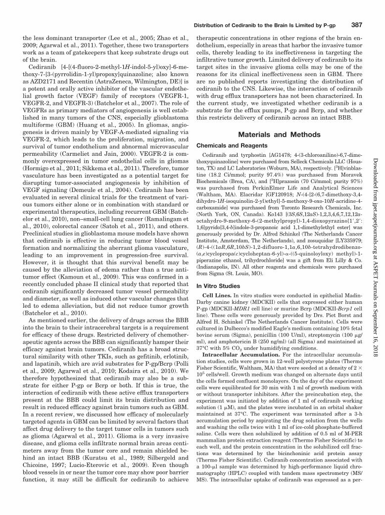
Intracellular Accumulation of Cediranib in MDCKIICells. Intracellular accumulation of cediranib in MDCKIIwild-type and MDR1- or Bcrp1-transfected cells was exam-ined. [3H]vinblastine and [3H]prazosin, prototypical sub-strates for P-gp and Bcrp, respectively, were included aspositive controls. As seen in Fig. 1A, accumulation of the P-gpsubstrate vinblastine was significantly reduced in theMDR1-transfected cells compared with wild-type control(�24% of wild type). Likewise, cediranib accumulation in theMDR1-transfected cells was significantly lower comparedwith the wild-type cells (44% of wild type; p�0.05). Cediranibaccumulation in the MDR1-transfected cells increased upontreatment with the P-gp-specific inhibitor LY335979 and theP-gp/Bcrp dual inhibitor GF120918 (Fig. 1A), such that itwas not significantly different from the accumulation in wild-type cells. This suggested that P-gp is involved in efflux ofcediranib from the cells. In the Bcrp1-transfected cells, there
was significantly lower accumulation of the Bcrp substrateprazosin compared with wild-type control (�44% of wild type;p � 0.05; Fig. 1B). Likewise, cediranib accumulation in theBcrp1-transfected cells was statistically lower than that inwild-type cells (�68% of wild type; p � 0.05). The effluxactivity of Bcrp was abolished upon treatment with the Bcrp-selective inhibitor Ko143 and the P-gp/Bcrp dual inhibitorGF120918 (Fig. 1B), which suggests that Bcrp also limitsintracellular accumulation of cediranib.
Directional Permeability of Cediranib across MDCKIICells. Transcellular transport of cediranib was determinedin MDCKII wild-type and MDR1- or Bcrp1-transfected cells.In the MDR1-transfected cells, the rate of cediranib trans-port was significantly increased in the B-to-A direction com-pared with that in the A-to-B direction (Fig. 2A; p � 0.05).The apparent permeability of cediranib in the B-to-A direc-tion was 2-fold greater than the A-to-B permeability in theMDR1-transfected cells (Table 1). The P-gp inhibitorLY339579 effectively inhibited P-gp mediated efflux of cedi-ranib in the MDR1-transfected cells, such that the B-to-Apermeability of cediranib was significantly reduced in
Fig. 1. Intracellular accumulation of cediranib in MDCKII cells. A, accu-mulation in wild-type and MDR1-transfected cells. Accumulation of thecediranib in the MDR1-transfected cells (gray bars) was significantlylower than that in the wild-type cells (black bars). B, accumulation inwild-type and Bcrp1-transfected cells. Accumulation of cediranib in theBcrp1-transfected cells (gray bars) was significantly lower than that inthe wild-type cells (black bars). Use of the P-gp selective inhibitorLY335979, Bcrp-selective inhibitor Ko143, and the dual inhibitorGF120918 abolished the difference in accumulation between the wild-type and transfected cells. Results are presented as mean S.D. (n � 4per group). �, p � 0.05.
Distribution of Cediranib to the Brain Is Limited by P-gp 389
at ASPE
T Journals on Septem
ber 16, 2018jpet.aspetjournals.org
Dow
nloaded from

LY335979-treated cells compared with untreated cells (Table1). Directional flux experiments in the Bcrp1-transfectedcells showed increased directionality in the flux of cediranib(Fig. 2B), with the apparent permeability in the B-to-A di-rection being 4-fold greater than the A-to-B permeability(Table 2). The Bcrp inhibitor Ko143 reversed the Bcrp-medi-ated efflux transport such that the B-to-A permeability ofcediranib was significantly reduced in Ko143-treated cellscompared with untreated cells (Table 2). These in vitro stud-
ies suggest that cediranib is a substrate of both P-gp andBcrp and is efficiently transported by both transporters.
Inhibition of P-gp and Bcrp by Cediranib. Figure 3shows the effect of cediranib on the accumulation of [3H]vin-blastine, a prototypical P-gp substrate, in MDR1-transfectedcells and [3H]prazosin, a prototypical Bcrp substrate, inBcrp1-transfected cells. Increasing concentrations of cedi-ranib enhanced the intracellular accumulation of vinblastinein the MDR1-transfected cells. Cytotoxicity associated withcediranib restricted the maximum usable cediranib dose to40 �M. Therefore, we were not able to estimate the IC50 ofcediranib for inhibition of P-gp. However, these resultsclearly show that cediranib inhibits the P-gp-mediated trans-port of vinblastine. It is noteworthy that cediranib treatmentdid not increase prazosin accumulation in the Bcrp1-trans-fected cells, indicating that cediranib does not inhibit Bcrp-mediated efflux, even though it is a substrate for Bcrp.
In Vivo Studies
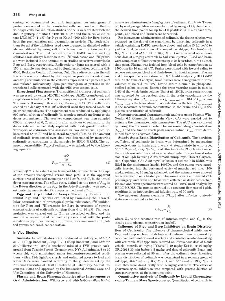
Cediranib Brain and Plasma Pharmacokinetics. Be-cause cediranib is administered orally, cediranib pharmacoki-netics in plasma and brain were first examined after oral ad-ministration in wild-type and Mdr1a/b(�/�)Bcrp1(�/�) mice.Cediranib brain concentrations in the wild-type mice were sig-nificantly lower than the plasma concentrations (p � 0.05;Fig. 4A), indicating restricted transport of cediranib across theBBB. In comparison, in the Mdr1a/b(�/�)Bcrp1(�/�) mice,brain concentrations were up to 7-fold higher than plasmaconcentrations (p � 0.05; Fig. 4B), showing the influence of P-gp
Fig. 2. Directional transport of cediranib across MDCKII cell monolayers.A, cediranib transported across wild-type (‚, A-to-B transport; Œ, B-to-Atransport) and MDR1-transfected (E, A-to-B transport; F, B-to-A trans-port) cells. B, cediranib transported across wild-type (‚, A-to-B transport;Œ, B-to-A transport) and Bcrp1-transfected (E, A-to-B transport; F, B-to-A transport) cells. Cediranib transport was significantly increased inthe B-A direction in both the MDR1- and Bcrp1-transfected cells, indi-cating P-gp- and Bcrp-mediated efflux. Results are expressed as mean S.D. (n � 3–4). �, p � 0.05.
TABLE 1Apparent permeability (Papp) of cediranib across wild-type and MDR1-transfected MDCKII cell monolayersResults are expressed as mean S.D. (n � 3–4).
Wild Type MDR1 Transfected
A-to-B B-to-A A-to-B B-to-A
�10�4cm/s
Treatment control 1.94 0.10 2.24 0.15 2.43 0.07 5.0 0.22*1 �M LY335979 1.17 0.18†
* P � 0.05 compared with A-B.† P � 0.05 compared with B-A control.
TABLE 2Apparent permeability (Papp) of cediranib across wild-type and Bcrp1-transfected MDCKII cell monolayersResults are expressed as mean S.D. (n � 3–4).
Wild Type Bcrp1 Transfected
A-to-B B-to-A A-to-B B-to-A
�10�4cm/s
Treatment control 1.25 0.13 1.22 0.11 0.93 0.02 3.59 0.03*200 nM Ko143 1.28 0.13†
* P � 0.05 compared with A-B.† Not significantly different compared with B-A control.
Fig. 3. Inhibition of P-gp and Bcrp by cediranib. Accumulation of [3H]vin-blastine in MDR1-transfected cells and [3H]prazosin in Bcrp1- trans-fected cells in the presence of increasing concentrations of cediranibranging from 0 to 40 �M is shown. Results are presented as mean S.D.(n � 4 per group; n � 2 for 40 �M cediranib point).
390 Wang et al.
at ASPE
T Journals on Septem
ber 16, 2018jpet.aspetjournals.org
Dow
nloaded from
and Bcrp on cediranib transport to the brain. There was nosignificant difference in plasma AUC(0–20 h) between the twogenotypes (Table 3), suggesting that P-gp and Bcrp do notinfluence the absorption or systemic elimination of cediranib atthese doses. The AUC in brain was 26-fold greater in theMdr1a/b(�/�)Bcrp1(�/�) mice compared with the wild type.
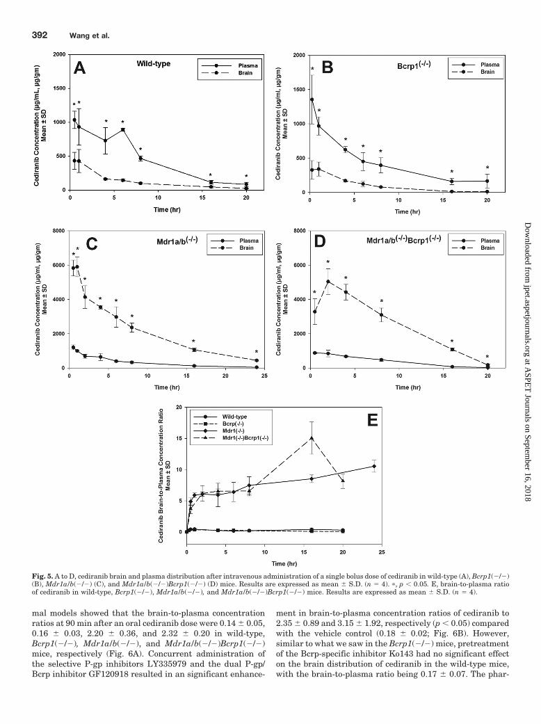
To further investigate whether the limited brain penetra-tion of cediranib is mediated by P-gp or Bcrp or both, brainand plasma pharmacokinetics of cediranib were studiedafter intravenous injection into wild-type, Mdr1a/b(�/�),
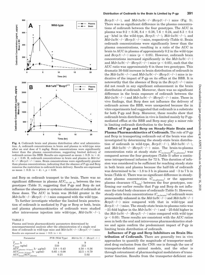
Bcrp1(�/�), and Mdr1a/b(�/�)Bcrp1(�/�) mice (Fig. 5).There was no significant difference in the plasma concentra-tions of cediranib between the four genotypes. The AUC inplasma was 9.2 0.36, 8.4 0.38, 7.6 0.24, and 8.3 0.4�g � h/ml in the wild-type, Bcrp1(�/�), Mdr1a/b(�/�), andMdr1a/b(�/�)Bcrp1(�/�) mice, respectively (Table 4). Braincediranib concentrations were significantly lower than theplasma concentrations, resulting in a ratio of the AUC inbrain to AUC in plasma of approximately 0.2 in the wild-typeand Bcrp1(�/�) mice (p � 0.05). However, cediranib brainconcentrations increased significantly in the Mdr1a/b(�/�)and Mdr1a/b(�/�)Bcrp1(�/�) mice (p � 0.05), such that theAUC ratio was approximately 6 in these two genotypes. Thisdramatic 30-fold increase in brain distribution of cediranib inthe Mdr1a/b(�/�) and Mdr1a/b(�/�)Bcrp1(�/�) mice is in-dicative of the impact of P-gp on its efflux at the BBB. It isnoteworthy that the absence of Bcrp in the Bcrp1(�/�) micedid not result in any significant enhancement in the braindistribution of cediranib. Moreover, there was no significantdifference in the brain exposure of cediranib between theMdr1a/b(�/�) and Mdr1a/b(�/�)Bcrp1(�/�) mice. These invivo findings, that Bcrp does not influence the delivery ofcediranib across the BBB, were unexpected because the invitro experiments had suggested that cediranib is a substratefor both P-gp and Bcrp. Moreover, these results show thatcediranib brain distribution in vivo is limited mainly by P-gp-mediated efflux at the BBB and Bcrp may play a minor rolein limiting cediranib distribution to the brain.
Effect of P-gp and Bcrp on Steady-State Brain andPlasma Pharmacokinetics of Cediranib. The role of P-gpand Bcrp in transporting cediranib out of the brain was alsoinvestigated by determining the steady-state brain distribu-tion of cediranib in wild-type, Bcrp1(�/�), Mdr1a/b(�/�),and Mdr1a/b(�/�)Bcrp1(�/�) mice. The brain-to-plasmaconcentration ratio at steady state (Css,brain/Css,plasma) wascompared across the four genotypes (Table 5) after a contin-uous intraperitoneal infusion for 72 h. This duration of infu-sion was considered to be sufficient for reaching steady statein both brain and plasma because the half-life of cediranibwas determined to be �1.8 to 3 h in plasma and �3 to 7 h inbrain (Table 4). There was no significant difference in steady-state plasma concentration (Css,plasma) or the apparentplasma clearance (CLapp) between the four genotypes, con-firming our earlier results that P-gp and Bcrp do not influ-ence the total body clearance of cediranib (Table 5). However,steady-state brain concentrations (Css,brain) of cediranib werepronouncedly enhanced in the Mdr1a/b(�/�) and Mdr1a/b(�/�)Bcrp1(�/�) mice compared with that in wild-type andBcrp1(�/�) mice. The steady-state brain-to-plasma ratio was�21-fold higher in the Mdr1a/b(�/�) and �25-fold higher inthe Mdr1a/b(�/�)Bcrp1(�/�) mice compared with wild type(p � 0.05). These results are consistent with the AUC ratiosseen in both the oral and intravenous pharmacokinetics stud-ies and again confirm the predominant impact of P-gp inlimiting brain distribution of cediranib.
Influence of P-gp and Bcrp Inhibitors on Brain Dis-tribution of Cediranib. There are generally two in vivoapproaches to quantify the magnitude of transporter-medi-ated drug exclusion from the CNS: one is through the use oftransporter-deficient animal models, and the other isthrough cotreatment of pharmacological modulators of trans-porter function. Results from the transporter-deficient ani-
Fig. 4. Cediranib brain and plasma distribution after oral administra-tion. A, cediranib concentrations in brain and plasma in wild-type miceafter an oral dose of 5 mg/kg. Brain concentrations were significantlylower than the plasma concentrations, suggesting limited delivery ofcediranib across the BBB. Results are expressed as mean S.D. (n � 4).�, p � 0.05. B, cediranib concentrations in brain and plasma in Mdr1a/b(�/�)Bcrp1(�/�) mice. Brain concentrations were significantly greaterthan plasma concentrations, indicating that the absence of P-gp and Bcrpremoves the restriction to cediranib brain delivery. Results are expressedas mean S.D. (n � 4). �, p � 0.05.
TABLE 3Plasma and brain pharmacokinetic parameters determined bynoncompartmental analysis after the administration of a single oraldose of cediranib in wild-type mice and Mdr1a/b(�/�) Bcrp1(�/�) miceResults are expressed as mean S.D. (n � 4).
Parameter FVB (Wild Type) Mdr1a/b(�/�)Bcrp1(�/�)
Tmax, h 4 9Cmax, ng/ml 510 313AUCplasma (0-last), h � �g/ml) 3.9 0.43 4.50 0.36AUCbrain (0-last), h � �g/ml) 0.85 0.08 22.5 0.79AUCbrain/AUCplasma 0.21 5.02
Distribution of Cediranib to the Brain Is Limited by P-gp 391
at ASPE
T Journals on Septem
ber 16, 2018jpet.aspetjournals.org
Dow
nloaded from
mal models showed that the brain-to-plasma concentrationratios at 90 min after an oral cediranib dose were 0.14 0.05,0.16 0.03, 2.20 0.36, and 2.32 0.20 in wild-type,Bcrp1(�/�), Mdr1a/b(�/�), and Mdr1a/b(�/�)Bcrp1(�/�)mice, respectively (Fig. 6A). Concurrent administration ofthe selective P-gp inhibitors LY335979 and the dual P-gp/Bcrp inhibitor GF120918 resulted in an significant enhance-
ment in brain-to-plasma concentration ratios of cediranib to2.35 0.89 and 3.15 1.92, respectively (p � 0.05) comparedwith the vehicle control (0.18 0.02; Fig. 6B). However,similar to what we saw in the Bcrp1(�/�) mice, pretreatmentof the Bcrp-specific inhibitor Ko143 had no significant effecton the brain distribution of cediranib in the wild-type mice,with the brain-to-plasma ratio being 0.17 0.07. The phar-
Fig. 5. A to D, cediranib brain and plasma distribution after intravenous administration of a single bolus dose of cediranib in wild-type (A), Bcrp1(�/�)(B), Mdr1a/b(�/�) (C), and Mdr1a/b(�/�)Bcrp1(�/�) (D) mice. Results are expressed as mean S.D. (n � 4). �, p � 0.05. E, brain-to-plasma ratioof cediranib in wild-type, Bcrp1(�/�), Mdr1a/b(�/�), and Mdr1a/b(�/�)Bcrp1(�/�) mice. Results are expressed as mean S.D. (n � 4).
392 Wang et al.
at ASPE
T Journals on Septem
ber 16, 2018jpet.aspetjournals.org
Dow
nloaded from
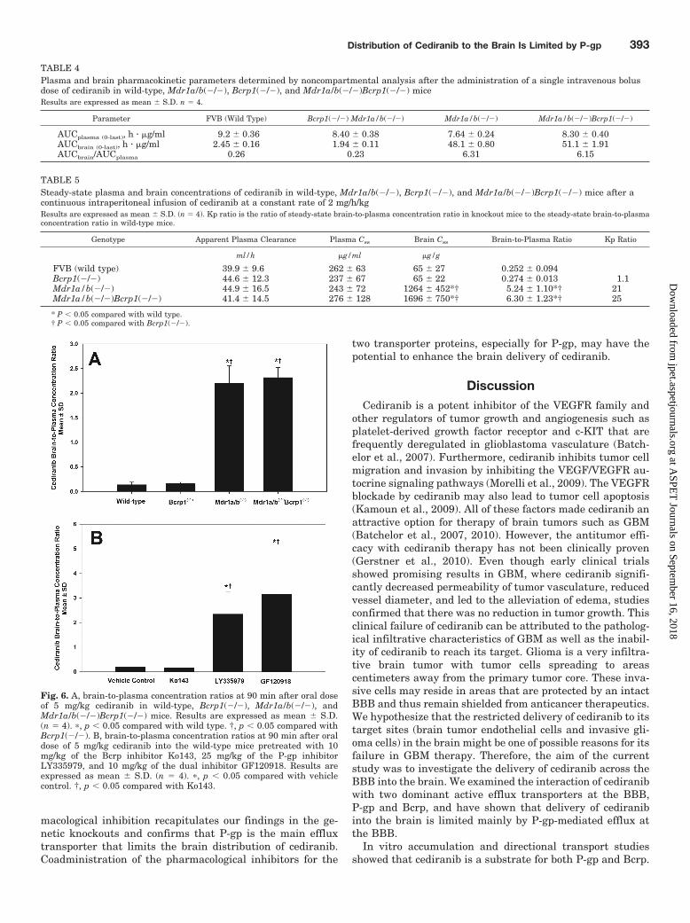
macological inhibition recapitulates our findings in the ge-netic knockouts and confirms that P-gp is the main effluxtransporter that limits the brain distribution of cediranib.Coadministration of the pharmacological inhibitors for the
two transporter proteins, especially for P-gp, may have thepotential to enhance the brain delivery of cediranib.
DiscussionCediranib is a potent inhibitor of the VEGFR family and
other regulators of tumor growth and angiogenesis such asplatelet-derived growth factor receptor and c-KIT that arefrequently deregulated in glioblastoma vasculature (Batch-elor et al., 2007). Furthermore, cediranib inhibits tumor cellmigration and invasion by inhibiting the VEGF/VEGFR au-tocrine signaling pathways (Morelli et al., 2009). The VEGFRblockade by cediranib may also lead to tumor cell apoptosis(Kamoun et al., 2009). All of these factors made cediranib anattractive option for therapy of brain tumors such as GBM(Batchelor et al., 2007, 2010). However, the antitumor effi-cacy with cediranib therapy has not been clinically proven(Gerstner et al., 2010). Even though early clinical trialsshowed promising results in GBM, where cediranib signifi-cantly decreased permeability of tumor vasculature, reducedvessel diameter, and led to the alleviation of edema, studiesconfirmed that there was no reduction in tumor growth. Thisclinical failure of cediranib can be attributed to the patholog-ical infiltrative characteristics of GBM as well as the inabil-ity of cediranib to reach its target. Glioma is a very infiltra-tive brain tumor with tumor cells spreading to areascentimeters away from the primary tumor core. These inva-sive cells may reside in areas that are protected by an intactBBB and thus remain shielded from anticancer therapeutics.We hypothesize that the restricted delivery of cediranib to itstarget sites (brain tumor endothelial cells and invasive gli-oma cells) in the brain might be one of possible reasons for itsfailure in GBM therapy. Therefore, the aim of the currentstudy was to investigate the delivery of cediranib across theBBB into the brain. We examined the interaction of cediranibwith two dominant active efflux transporters at the BBB,P-gp and Bcrp, and have shown that delivery of cediranibinto the brain is limited mainly by P-gp-mediated efflux atthe BBB.
In vitro accumulation and directional transport studiesshowed that cediranib is a substrate for both P-gp and Bcrp.
TABLE 4Plasma and brain pharmacokinetic parameters determined by noncompartmental analysis after the administration of a single intravenous bolusdose of cediranib in wild-type, Mdr1a/b(�/�), Bcrp1(�/�), and Mdr1a/b(�/�)Bcrp1(�/�) miceResults are expressed as mean S.D. n � 4.
Parameter FVB (Wild Type) Bcrp1(�/�) Mdr1a/b(�/�) Mdr1a/b(�/�) Mdr1a/b(�/�)Bcrp1(�/�)
AUCplasma (0-last), h � �g/ml 9.2 0.36 8.40 0.38 7.64 0.24 8.30 0.40AUCbrain (0-last), h � �g/ml 2.45 0.16 1.94 0.11 48.1 0.80 51.1 1.91AUCbrain/AUCplasma 0.26 0.23 6.31 6.15
TABLE 5Steady-state plasma and brain concentrations of cediranib in wild-type, Mdr1a/b(�/�), Bcrp1(�/�), and Mdr1a/b(�/�)Bcrp1(�/�) mice after acontinuous intraperitoneal infusion of cediranib at a constant rate of 2 mg/h/kgResults are expressed as mean S.D. (n � 4). Kp ratio is the ratio of steady-state brain-to-plasma concentration ratio in knockout mice to the steady-state brain-to-plasmaconcentration ratio in wild-type mice.
Genotype Apparent Plasma Clearance Plasma Css Brain Css Brain-to-Plasma Ratio Kp Ratio
ml/h �g/ml �g/g
FVB (wild type) 39.9 9.6 262 63 65 27 0.252 0.094Bcrp1(�/�) 44.6 12.3 237 67 65 22 0.274 0.013 1.1Mdr1a/b(�/�) 44.9 16.5 243 72 1264 452*† 5.24 1.10*† 21Mdr1a/b(�/�)Bcrp1(�/�) 41.4 14.5 276 128 1696 750*† 6.30 1.23*† 25
* P � 0.05 compared with wild type.† P � 0.05 compared with Bcrp1(�/�).
Fig. 6. A, brain-to-plasma concentration ratios at 90 min after oral doseof 5 mg/kg cediranib in wild-type, Bcrp1(�/�), Mdr1a/b(�/�), andMdr1a/b(�/�)Bcrp1(�/�) mice. Results are expressed as mean S.D.(n � 4). �, p � 0.05 compared with wild type. †, p � 0.05 compared withBcrp1(�/�). B, brain-to-plasma concentration ratios at 90 min after oraldose of 5 mg/kg cediranib into the wild-type mice pretreated with 10mg/kg of the Bcrp inhibitor Ko143, 25 mg/kg of the P-gp inhibitorLY335979, and 10 mg/kg of the dual inhibitor GF120918. Results areexpressed as mean S.D. (n � 4). �, p � 0.05 compared with vehiclecontrol. †, p � 0.05 compared with Ko143.
Distribution of Cediranib to the Brain Is Limited by P-gp 393
at ASPE
T Journals on Septem
ber 16, 2018jpet.aspetjournals.org
Dow
nloaded from

Inhibition of P-gp and Bcrp increased the restricted intracel-lular accumulation of cediranib in the transporter-overex-pressing cell lines to levels similar to that in the wild-typecells (Fig. 1). It is noteworthy that cediranib inhibited P-gpmediated efflux but not Bcrp (Fig. 3). These findings areclinically relevant because inhibition of P-gp by cediranibmay alter tissue pharmacokinetics of concurrently adminis-tered drugs that are substrates for P-gp. The finding thatcediranib is transported by Bcrp, but does not inhibit theBcrp-mediated efflux transport of substrates, like prazosin,is similar to the behavior of another TKI, sorafinib, as re-ported by Agarwal et al. (2011b). It is possible that cediranib,similar to sorafenib, might be transported by Bcrp by bindingto a site that is different from the prazosin binding site. Giriet al. (2008) first postulated the presence of multiple bindingsites on Bcrp. This finding further provides credence to thishypothesis and warrants further research in this area.
The in vitro studies suggested that cediranib is a substratefor both P-gp and Bcrp. We therefore studied the brain distri-bution of cediranib in FVB mice to examine the effect of thesetransporters on its transport across the BBB. Cediranib concen-trations in brain were compared among the wild-type, Mdr1a/b(�/�), Bcrp1(�/�), and Mdr1a/b(�/�)Bcrp1(�/�) mice. Uponoral administration in wild-type mice, cediranib brain concen-trations were on average 5-fold lower than plasma concentra-tions, indicating the limited partitioning of cediranib into thebrain (Fig. 4A). Absence of P-gp and Bcrp at the BBB dramat-ically enhanced cediranib brain partitioning such that concen-trations in brain were �4-fold greater than that in plasma (Fig.4B). This confirmed that cediranib was effluxed at the BBB byP-gp and/or Bcrp. Tween 80 (1%; w/v) was used as dosingvehicle for oral administration. Because all strains were dosedwith the same vehicle, comparisons based on the same dosingformulation are reasonable. To understand the impact of influ-ence individual transporter (P-gp or Bcrp) on cediranib brainpenetration, brain partitioning was determined after intrave-nous dosing in the four mouse genotypes. In the wild-type andBcrp1(�/�) mice, the AUC in brain was �4-fold lower than theAUC in plasma (Table 4). Similar brain partitioning in thesetwo mouse genotypes indicated that Bcrp does not restrictthe delivery of cediranib across the BBB. Cediranib AUC in thebrain in the Mdr1a/b(�/�) and Mdr1a/b(�/�)Bcrp1(�/�) micewas �20-fold higher than that in wild-type and Bcrp1(�/�)mice (Fig. 5; Table 4). There was no significant difference inbrain partitioning of cediranib between the Mdr1a/b(�/�) andMdr1a/b(�/�)Bcrp1(�/�) mice. The finding that that therewas no additional increase in brain partitioning of cediranib inthe Mdr1a/b(�/�)Bcrp1(�/�) mice shows that P-gp-mediatedefflux at the BBB predominantly restricts brain delivery ofcediranib and Bcrp does not play a significant role in limitingthe CNS distribution of cediranib. This is in contrast to the invitro studies that indicated cediranib was a substrate for bothP-gp and Bcrp.
To date, there are several published studies that report apossible cooperation of P-gp and Bcrp in restricting the de-livery of TKIs across the BBB. This has been shown forimatinib (Breedveld et al., 2005), dasatinib (Chen et al.,2009), gefitinib (Agarwal et al., 2010), lapatinib (Polli et al.,2009), erlotinib (Kodaira et al., 2010), and sunitinib (Tang etal., 2012). Most of these studies reported a greater thanproportional increase in brain distribution when both P-gpand Bcrp are absent in the Mdr1a/b(�/�)Bcrp1(�/�) mice.
Many of these studies also suggested that P-gp was thedominant transporter at the BBB in limiting the brain up-take of substrate drugs. Cediranib is different from otherTKIs in that the in vivo studies suggest that P-gp and Bcrp donot have a combined effect on its brain distribution. There isno difference between the brain distribution between theMdr1a/b(�/�) and Mdr1a/b(�/�)Bcrp1(�/�) mice and be-tween the wild-type and Bcrp1(�/�) mice, suggesting thatBcrp does not efflux cediranib at the BBB. We had hypothe-sized in our previous studies that a higher expression of P-gpcompared with Bcrp may be the reason behind the subduedeffect of Bcrp-mediated efflux at the BBB (Agarwal et al.,2011b). Kamiie et al. (2008) showed that there is 5-fold lowerexpression of Bcrp protein at the murine BBB than P-gp.This may in part explain the lack of Bcrp-mediated efflux ofcediranib at the BBB.
The finding that inhibition of P-gp enhanced brain pene-tration of cediranib (Fig. 6B) has significant clinical implica-tions. Concurrent use of the P-gp-specific inhibitor LY335979or the dual inhibitor GF120918 was able to increase braindistribution of cediranib to levels similar to those seen in theknockouts (Fig. 6A). This indicates that chemical modulationof efflux transporters, mainly P-gp, can be used as a promis-ing therapeutic strategy to enhance cediranib distribution tothe brain. This might improve the clinical efficacy of cedi-ranib for the treatment of brain tumors.
Glioblastoma multiforme is one of the most formidablechallenges faced by the neuro-oncology community. The abil-ity of glioma cells to migrate and promote angiogenesismakes treatment of GBM challenging and demands furtherinvestigation of novel therapies that target these processes,such as cediranib. The constraint to deliver drugs across theblood-brain barrier has prevented easy application of potent,peripherally active drugs against this tumor. Cediranib, likemany other TKIs, is effluxed by P-gp at the BBB. This is acritical finding that can influence the delivery and thus effi-cacy of cediranib in the brain tumor.
In conclusion, we have shown that cediranib is a substrateof active efflux transporters P-gp and Bcrp in vitro but onlyP-gp plays a critical role in limiting brain delivery of cedi-ranib. Efficacy of cediranib against glioma may depend onthe ability of cediranib to achieve therapeutic concentrationsin brain tumor endothelial cells and the invasive glioma cells.This study shows that use of potent inhibitors for P-gp suchas GF120918 may enhance delivery of cediranib to the brain.
Acknowledgments
We thank Jim Fisher (Clinical Pharmacology Analytical Services,University of Minnesota) for help in the development of the cediranibLC-MS/MS assay.
Authorship Contributions
Participated in research design: Wang and Elmquist.Conducted experiments: Wang and Agarwal.Contributed new reagents or analytic tools: Wang.Performed data analysis: Wang and Elmquist.Wrote or contributed to the writing of the manuscript: Wang, Agar-
wal, and Elmquist.
ReferencesAgarwal S, Sane R, Gallardo JL, Ohlfest JR, and Elmquist WF (2010) Distribution
of gefitinib to the brain is limited by P-glycoprotein (ABCB1) and breast cancerresistance protein (ABCG2)-mediated active efflux. J Pharmacol Exp Ther 334:147–155.
394 Wang et al.
at ASPE
T Journals on Septem
ber 16, 2018jpet.aspetjournals.org
Dow
nloaded from

Agarwal S, Sane R, Oberoi R, Ohlfest JR, and Elmquist WF (2011a) Delivery ofmolecularly targeted therapy to malignant glioma, a disease of the whole brain.Expert Rev Mol Med 13:e17.
Agarwal S, Sane R, Ohlfest JR, and Elmquist WF (2011b) The role of the breastcancer resistance protein (ABCG2) in the distribution of sorafenib to the brain.J Pharmacol Exp Ther 336:223–233.
Batchelor TT, Duda DG, di Tomaso E, Ancukiewicz M, Plotkin SR, Gerstner E,Eichler AF, Drappatz J, Hochberg FH, Benner T, et al. (2010) Phase II study ofcediranib, an oral pan-vascular endothelial growth factor receptor tyrosine kinaseinhibitor, in patients with recurrent glioblastoma. J Clin Oncol 28:2817–2823.
Batchelor TT, Sorensen AG, di Tomaso E, Zhang WT, Duda DG, Cohen KS, KozakKR, Cahill DP, Chen PJ, Zhu M, et al. (2007) AZD2171, a pan-VEGF receptortyrosine kinase inhibitor, normalizes tumor vasculature and alleviates edema inglioblastoma patients. Cancer Cell 11:83–95.
Breedveld P, Pluim D, Cipriani G, Wielinga P, van Tellingen O, Schinkel AH, andSchellens JH (2005) The effect of Bcrp1 (Abcg2) on the in vivo pharmacokineticsand brain penetration of imatinib mesylate (Gleevec): implications for the use ofbreast cancer resistance protein and P-glycoprotein inhibitors to enable the brainpenetration of imatinib in patients. Cancer Res 65:2577–2582.
Carmeliet P and Jain RK (2000) Angiogenesis in cancer and other diseases. Nature407:249–257.
Chen Y, Agarwal S, Shaik NM, Chen C, Yang Z, and Elmquist WF (2009) P-glyco-protein and breast cancer resistance protein influence brain distribution of dasat-inib. J Pharmacol Exp Ther 330:956–963.
Dai H, Marbach P, Lemaire M, Hayes M, and Elmquist WF (2003) Distribution ofSTI-571 to the brain is limited by P-glycoprotein-mediated efflux. J PharmacolExp Ther 304:1085–1092.
Demeule M, Regina A, Annabi B, Bertrand Y, Bojanowski MW, and Beliveau R(2004) Brain endothelial cells as pharmacological targets in brain tumors. MolNeurobiol 30:157–183.
Gerstner ER, Chen PJ, Wen PY, Jain RK, Batchelor TT, and Sorensen G (2010)Infiltrative patterns of glioblastoma spread detected via diffusion MRI after treat-ment with cediranib. Neuro Oncol 12:466–472.
Giri N, Shaik N, Pan G, Terasaki T, Mukai C, Kitagaki S, Miyakoshi N, andElmquist WF (2008) Investigation of the role of breast cancer resistance protein(Bcrp/Abcg2) on pharmacokinetics and central nervous system penetration ofabacavir and zidovudine in the mouse. Drug Metab Dispos 36:1476–1484.
Hormigo A, Ding BS, and Rafii S (2011) A target for antiangiogenic therapy: vascularendothelium derived from glioblastoma. Proc Natl Acad Sci U S A 108:4271–4272.
Huang H, Held-Feindt J, Buhl R, Mehdorn HM, and Mentlein R (2005) Expressionof VEGF and its receptors in different brain tumors. Neurol Res 27:371–377.
Institute of Laboratory Animal Resources (1996) Guide for the Care and Use ofLaboratory Animals, 7th ed. Institute of Laboratory Animal Resources, Commis-sion on Life Sciences, National Research Council, Washington, DC.
Kamiie J, Ohtsuki S, Iwase R, Ohmine K, Katsukura Y, Yanai K, Sekine Y, UchidaY, Ito S, and Terasaki T (2008) Quantitative atlas of membrane transporterproteins: development and application of a highly sensitive simultaneous LC/MS/MS method combined with novel in-silico peptide selection criteria. Pharm Res25:1469–1483.
Kamoun WS, Ley CD, Farrar CT, Duyverman AM, Lahdenranta J, Lacorre DA,Batchelor TT, di Tomaso E, Duda DG, Munn LL, et al. (2009) Edema control bycediranib, a vascular endothelial growth factor receptor-targeted kinase inhibitor,prolongs survival despite persistent brain tumor growth in mice. J Clin Oncol27:2542–2552.
Kodaira H, Kusuhara H, Ushiki J, Fuse E, and Sugiyama Y (2010) Kinetic analysisof the cooperation of P-glycoprotein (P-gp/Abcb1) and breast cancer resistance
protein (Bcrp/Abcg2) in limiting the brain and testis penetration of erlotinib,flavopiridol, and mitoxantrone. J Pharmacol Exp Ther 333:788–796.
Kuratsu J, Itoyama Y, Uemura S, and Ushio Y (1989) [Regrowth patterns of glioma–cases of glioma regrew away from the original tumor]. Gan No Rinsho 35:1255–1260.
Lee YJ, Kusuhara H, Jonker JW, Schinkel AH, and Sugiyama Y (2005) Investigationof efflux transport of dehydroepiandrosterone sulfate and mitoxantrone at themouse blood-brain barrier: a minor role of breast cancer resistance protein. J Phar-macol Exp Ther 312:44–52.
Lucio-Eterovic AK, Piao Y, and de Groot JF (2009) Mediators of glioblastoma resis-tance and invasion during antivascular endothelial growth factor therapy. ClinCancer Res 15:4589–4599.
Morelli MP, Brown AM, Pitts TM, Tentler JJ, Ciardiello F, Ryan A, JurgensmeierJM, and Eckhardt SG (2009) Targeting vascular endothelial growth factor recep-tor-1 and -3 with cediranib (AZD2171): effects on migration and invasion ofgastrointestinal cancer cell lines. Mol Cancer Ther 8:2546–2558.
Polli JW, Olson KL, Chism JP, John-Williams LS, Yeager RL, Woodard SM, Otto V,Castellino S, and Demby VE (2009) An unexpected synergist role of P-glycoproteinand breast cancer resistance protein on the central nervous system penetration ofthe tyrosine kinase inhibitor lapatinib (N-{3-chloro-4-[(3-fluorobenzyl)oxy]phenyl}-6-[5-({[2-(methylsulfonyl)ethy l]amino}methyl)-2-furyl]-4-quinazolinamine;GW572016). Drug Metab Dispos 37:439–442.
Ramalingam SS, Belani CP, Mack PC, Vokes EE, Longmate J, Govindan R, Koczy-was M, Ivy SP, and Gandara DR (2010) Phase II study of Cediranib (AZD 2171),an inhibitor of the vascular endothelial growth factor receptor, for second-linetherapy of small cell lung cancer (National Cancer Institute #7097). J ThoracOncol 5:1279–1284.
Satoh T, Yamaguchi K, Boku N, Okamoto W, Shimamura T, Yamazaki K, Shi X, andMishima H (2011) Phase I results from a two-part Phase I/II study of cediranib incombination with mFOLFOX6 in Japanese patients with metastatic colorectalcancer. Invest New Drugs http://dx.doi.org/10.1007/s10637-011-9693-6.
Sikkema AH, de Bont ES, Molema G, Dimberg A, Zwiers PJ, Diks SH, Hoving EW,Kamps WA, Peppelenbosch MP, and den Dunnen WF (2011) Vascular endothelialgrowth factor receptor 2 (VEGFR-2) signalling activity in paediatric pilocyticastrocytoma is restricted to tumour endothelial cells. Neuropathol Appl Neurobiol37:538–548.
Silbergeld DL and Chicoine MR (1997) Isolation and characterization of humanmalignant glioma cells from histologically normal brain. J Neurosurg 86:525–531.
Tang SC, Lagas JS, Lankheet NA, Poller B, Hillebrand MJ, Rosing H, Beijnen JH,and Schinkel AH (2012) Brain accumulation of sunitinib is restricted by P-glyco-protein (ABCB1) and breast cancer resistance protein (ABCG2) and can be en-hanced by oral elacridar and sunitinib coadministration. Int J Cancer 130:223–233.
Wang T, Oberoi RK, and Elmquist WF (2011) Determination of cediranib in mouseplasma and brain tissue using high-performance liquid chromatography-massspectrometry, J Chromatogr B Analyt Technol Biomed Life Sci 879:3812–3817.
Zhao R, Raub TJ, Sawada GA, Kasper SC, Bacon JA, Bridges AS, and Pollack GM(2009) Breast cancer resistance protein interacts with various compounds in vitro,but plays a minor role in substrate efflux at the blood-brain barrier. Drug MetabDispos 37:1251–1258.
Address correspondence to: William F. Elmquist, Department of Pharma-ceutics, University of Minnesota, 9-177 Weaver Densford Hall, 308 HarvardStreet SE, Minneapolis, MN 55455. E-mail: [email protected]
Distribution of Cediranib to the Brain Is Limited by P-gp 395
at ASPE
T Journals on Septem
ber 16, 2018jpet.aspetjournals.org
Dow
nloaded from