brain, behavior, and immunity - national cancer institute · s42 n.d. powell et al. / brain,...
TRANSCRIPT

Brain, Behavior, and Immunity 30 (2013) S41–S47
Contents lists available at SciVerse ScienceDirect
Brain, Behavior, and Immunity
journal homepage: www.elsevier .com/locate /ybrbi
Review
Psychosocial stress and inflammation in cancer
N.D. Powell a,⇑, A.J. Tarr a, J.F. Sheridan a,b,c
a Division of Oral Biology, College of Dentistry, The Ohio State University, Columbus, OH, USA b Institute for Behavioral Medicine Research, College of Medicine, The Ohio State University, Columbus, OH, USA c Department of Molecular Virology, Immunology, and Medical Genetics, College of Medicine, The Ohio State University, Columbus, OH, USA
a r t i c l e i n f o
Article history: Available online 9 July 2012
Keywords: Psychosocial stress IL-6 Catecholamines Glucocorticoids Immune dysregulation Cancer
a b s t r a c t
Stress-induced immune dysregulation results in significant health consequences for immune related disorders including viral infections, chronic autoimmune disease, and tumor growth and metastasis. In this mini-review we discuss the sympathetic, neuroendocrine and immunologic mechanisms by which psychosocial stress can impact cancer biology. Both human and animal studies have shown the sympathetic and neuroendocrine responses to psychosocial stress significantly impacts cancer, in part, through regulation of inflammatory mediators. Psychosocial stressors stimulate neuroendocrine, sympathetic, and immune responses that result in the activation of the hypothalamic–pituitary–adrenal (HPA)-axis, sympathetic nervous system (SNS), and the subsequent regulation of inflammatory responses by immune cells. Social disruption (SDR) stress, a murine model of psychosocial stress and repeated social defeat, provides a novel and powerful tool to probe the mechanisms leading to stress-induced alterations in inflammation, tumor growth, progression, and metastasis. In this review, we will focus on SDR as an important model of psychosocial stress in understanding neural-immune mechanisms in cancer.
© 2012 Elsevier Inc. All rights reserved.
1. Introduction
Psychosocial stressors impact many physiological and pathological disease outcomes, including cancer. In multiple clinical and epidemiological studies, tumor growth, progression, and metastasis have been correlated with reports of stress, anxiety, poor coping behaviors, depression, lack of social support, and numerous other psychological and behavioral abnormalities (Lillberg et al., 2003; Price et al., 2001; Spiegel and Giese-Davis, 2003). The majority of studies that examine the impact of stress on malignant tissue have focused on suppressed immune responses to tumors and stress-induced alterations in the tumor microenvironment. More recently, stress-mediated immune modulation of lymphoid and myeloid cells and chronic inflammation mediated by cytokines such as interleukin (IL)-6 have been implicated as predictors of cancer progression, metastasis, and recurrence (Chung and Chang, 2003; Mundy-Bosse et al., 2011; Pierce et al., 2009; Salgado et al., 2003). By using mouse models of stress, significant progress has been made in determining the mechanisms behind stress-induced alterations in inflammatory immune status. For example, studies using a mouse model of repeated social defeat, termed social disruption (SDR) stress, have shown that stress alone can trigger the
⇑ Corresponding author. Address: Section of Oral Biology, The Institute for Behavioral Medicine Research, The Ohio State University Medical Center, 460 Medical Center Drive, Room 255, Columbus, OH 43210, USA. Tel.: +1 614 366 3492; fax: +1 614 292 4820.
E-mail addresses: [email protected], [email protected] (N.D. Powell).
generation, egress, and trafficking of immature, inflammatory myeloid derived-cells that are glucocorticoid (GC) insensitive (Curry et al., 2010; Engler et al., 2004a, 2005). In addition, these GC insensitive cells produce high levels of IL-6 and other inflammatory cytokines and chemokines (Powell et al., 2009; Stark et al., 2002; Wohleb et al., 2011). As a consequence, these stress-induced changes at the cellular level translate to significant immune (enhanced inflammatory responses and immunity to microbial, viral, and allergen challenge) and behavioral (prolonged anxiety-like behavior) changes (Bailey et al., 2007, 2009b, 2009a; Dong-Newsom et al., 2010; Kinsey et al., 2007; Mays et al., 2010, 2012; Powell et al., 2011; Wohleb et al., 2011). Indicative of the important role of the SNS in stress-induced immune alteration, these changes are reversed by the blockade of sympathetic signaling prior to stressor exposure (Wohleb et al., 2011).
Studies discussed in this mini-review highlight evidence that stress and the resulting modifications in behavior, immune status, and production of stress hormones and neurotransmitters significantly influence tumor growth, progression, and metastasis. Specifically, we focus on the stress response and its mediators (i.e., GC, catecholamines, and cytokines) between the nervous, endocrine, and immune system interactions. The discussion will be extended to include the impact that stress-induced alterations in immunity and inflammation have on cancer. We discuss how psychological stressors modulate cellular immune function and tumor biology. Within the scope of this paper, we review the clinical and animal literature that focus on the effect that stress has on the im
0889-1591/$ - see front matter © 2012 Elsevier Inc. All rights reserved. http://dx.doi.org/10.1016/j.bbi.2012.06.015
S42 N.D. Powell et al. / Brain, Behavior, and Immunity 30 (2013) S41–S47
mune response, which, ultimately, can prevent or promote malignant disease. Taken together, SDR and other mouse models of stress known to impact immune function are ideal platforms that can be used to explore of the overall impact of stress and inflammation on cancer biology.
2. The stress response
The stress response results from internal or external stimuli that activate ‘‘fight or flight’’ and defeat/withdrawal responses associated with the SNS and HPA activation. It is well established that specific central nervous system (CNS) pathways act to translate social stimuli into peripheral biological signals that regulate inflammatory responses. For instance, stress activates neuroendocrine and autonomic pathways like the HPA axis, and the SNS resulting in the release of GC, catecholamines, and pro-inflammatory cytokines such as IL-1, IL-6, and TNF-a. The release of these sympathetic, neuroendocrine, and immune factors has a profound influence on immunity, behavior, and physiology in both humans and rodents and triggers peripheral biological responses that, in turn, signal back to the CNS to complete a bi-directional communication circuit. This is evident in models of repeated social defeat, like SDR, that enhance immune responses to microbial, viral, and allergic challenges and promote and prolong anxiety-like behavior in rodents (Kinsey et al., 2007; Bailey et al., 2009a,b; Mays et al., 2010). Social disruption stress-induced prolonged anxiety-like behavior coincides with a unique pattern of c-Fos activation in brain regions associated with fear and threat appraisal. For example, SDR causes increased c-Fos activation in the prefrontal cortex, amygdala, hippocampus, paraventricular nucleus, bed nucleus of the stria terminalis and the lateral septum (Wohleb et al., 2011). As a result of the activation of this circuitry and the release of neurotransmitters and hormones, compensatory physiologic changes occur that impact behavior and immunity. In humans, chronic or repeated exposure to stress is associated with increased expression of inflammatory biomarkers, worsened disease states, and affective disorders (Glaser and Kiecolt-Glaser, 2005; Gouin et al., 2012). In clinical studies, stressed individuals have reduced anti-inflammatory GC regulation and increased inflammatory nuclear factor (NF)-jB signaling (Miller et al., 2008). As such, a stressor constitutes a challenge to homeostasis that is interpreted by sensory pathways which manifest as physiologic alterations (Glaser and Kiecolt-Glaser, 2005).
3. Interactions among the nervous, endocrine, and immune systems
The stress-induced release of catecholamines and GC has been associated with modulation of immune function including impaired antigen presentation, decreased T cell proliferation, and dampened humoral and cell mediated immunity (Glaser and Kiecolt-Glaser, 2005). Bi-directional communication between the neuroendocrine and immune systems is facilitated by receptor/ligand interactions. Glucocorticoids and catecholamines are capable of binding to receptors on and within immune cells. Specifically, GC interacts with immune cells by binding to intracellular receptors. The activated GC receptor translocates to the nucleus where it binds to GC response elements in gene promoter regions. As such, activated GC receptors can inhibit the actions of transcription factors, like NF-jB, and in turn inhibit the production of inflammatory cytokines (Padgett and Glaser, 2003).
Leukocyte activation, proliferation, and migration are known to be regulated by GC. Interestingly, GC has also been shown to affect tumor cells through GC-specific suppression or activation of target genes. While GC typically induce apoptosis in leukocytes through
active or passive suppression of survival genes, recent reports in experimental models of stress have shown that GC can protect tumor cells that have been treated in vitro and in vivo with chemotherapeutic agents through GC-specific activation of survival genes (Wu et al., 2004). GC have also been shown to down-regulate tumor suppressor genes (Antonova and Mueller, 2008; Dickinson et al., 2011). In a study by Antonova et al., GC treatment of murine mammary non-tumorigenic Ras-transformed EPH4 cells induced a down-regulation of BRCA1, a tumor suppressor gene that when mutated contributes to familial breast cancer (Antonova and Mueller, 2008). GC is also known to inhibit cellular immune responses, thereby decreasing immune defenses against cancer. Thus, GC has both direct and indirect effects on tumor biology.
The catecholamines norepinephrine (NE) and epinephrine (EPI) mediate their effects on target immune cells via stimulation of two major receptor subtypes: alpha(a)- and beta(b)-adrenergic receptors (ARs) (Madden et al., 1995; Sanders, 1995). Both primary and secondary lymphoid organs are innervated by the SNS (Felten et al., 1985). Most studies suggest that splenic innervation is predominantly sympathetic, with splenic nerves being comprised of approximately 98% sympathetic fibers (Felten et al., 1985; Reilly et al., 1979; Williams et al., 1981). b-AR are G-protein coupled receptors on the surface of many cells, including immune and tumor cells. Upon interaction with its ligand(s), b-ARs signal to the nucleus to activate cyclic adenosine monophosphate (cAMP). cAMP is a second messenger that is activated upon binding of the nuclear cAMP responsive binding element (CREB), a transcription factor that is initiated by multiple signal transduction pathways in response to multiple biologic factors, including stress hormones. Activation of b2-ARs in the absence of immunological stimuli increases pro-inflammatory cytokine production by various cell types (Tomozawa et al., 1995; Tan et al., 2007). For example, activation of b2-ARs with the specific agonist salmeterol up-regulates IL-6 and IL-1b mRNA and protein levels in macrophages, which results in amplified immune responses (Tan et al., 2007). Activation of the SNS can also profoundly impact behavioral responses in a b-AR-specific manner (Wohleb et al., 2011).
While immunological and neurological changes can result as a consequence of activation of the stress response, recent compelling data argue for a critical role of the b-AR directly and indirectly influencing malignant tissue. For example, b-AR is present on breast and ovarian cells (Badino et al., 1996; Sood et al., 2006). To date, the CREB family of proteins has been implicated in various aspects of tumor biology including metastasis, growth, angiogenesis, and cell survival (Jean and Bar-Eli, 2000; Lang et al., 2004). In several studies, cAMP activation has been correlated with increased rate of tumor growth in mammary tissue. For example, a report by Sastry et al. showed that EPI protected androgen-sensitive human prostate adenocarcinoma cells (i.e., LNCaP cells) from apoptosis through a cAMP-dependent inactivation of pro-apoptotic proteins, downstream of b2-AR signaling (Sastry et al., 2007). In addition, studies have shown that NE modulates the migration of tumor cells, including breast, colon and prostate tumor cells, which can be blocked by b2-AR antagonists (Lang et al., 2004; Drell et al., 2003; Masur et al., 2001). Further, a study by Palm et al., describes that this effect can be replicated in vivo and pre-treatment with b-blockers prevents NE-driven metastasis in a model of prostate cancer in Balb/c mice (Palm et al., 2006). Coupled with the effects of GC on tumors, these studies highlight the emerging role that stress-associated factors play in tumor biology.
4. Cancer, inflammation, and immunity
There are multiple factors that impact tumor biology beyond stress and stress-associated molecules. Some are intrinsic factors
N.D. Powell et al. / Brain, Behavior, and Immunity 30 (2013) S41–S47 S43
within the tumor and the tumor microenvironment, which include increased proliferative capacity and replicative immortality, resistance to suppressive signals which impair cell growth and/or promote apoptosis, and enhanced reactivity or exposure to molecules that promote angiogenesis, tissue invasion, and metastasis (Hanahan and Weinberg, 2011). In addition, the cellular composition of the tumor microenvironment is thought to significantly influence the biology of tumors. The cell subsets comprising the tumor microenvironment include both cancer-associated cells and normal immune cells that collectively enable tumor growth and progression. Common immune cells such as inflammatory macrophages, natural killer (NK) cells, and cytotoxic T-cells (CTL) can be found in the core of primary tumors and within the invasive and metastatic tumor microenvironments (Hanahan and Weinberg, 2011).
NK cells and CTL, which are known to influence tumor progression, are also known to be regulated by stress. The importance of NK cells in cancer biology is highlighted by the unique ability to lyse tumor targets without prior sensitization. Additionally, one of the hallmarks of tumor biology is the capability of taking over the genetic machinery of major histocompatibility complex (MHC) class I genes, ultimately reducing the efficacy of cytotoxic T cell recognition. As a primary way of activation, the capabilities of NK cells to recognize infected/tumor cells by the lack of MHC class I expression becomes crucial in tumor progression and metastasis (Bubenik, 2004). Once NK cells and CTL become activated, the release of perforins, granzymes A and B, and FasL cause apoptosis of the infected cell. Additionally, activated NK cells and CTL increase anti-viral/tumor interferon (IFN) production which directly inhibits tumor growth and coordinates anti-tumor innate and adaptive immunity.
Currently, there is a growing importance placed on inhibitory receptor expression on NK cells and CTL and cancer modulation (Carena et al., 1997; Hanke et al., 1999; Yu et al., 1996). Expressed both in humans and rodents, inhibitory receptors found on NK cells and CTL regulate activation pathways that lead to anti-viral and tumor mechanisms. The use of antibodies targeting NK cell inhibitory receptors are currently being used in clinical trials lending to promising novel approaches to immune-based anti-tumor therapies (Kradin et al., 2001; Vahlne et al., 2010; Yu et al., 1996). Stress has been shown to modulate these inhibitory receptors, potentially mediating the effect of NK and CTL on cancer progression. Several studies have shown that restraint stress diminishes NK cell function, cytokine secretion, and enhances viral replication, which was shown to be partially mediated by stress-induced glucocorticoids (Hunzeker et al., 2004; Tseng et al., 2005). In a study by Andersen et al., the lytic potential of NK cells from patients with invasive breast cancer and higher stress levels was significantly reduced (Andersen et al., 1998). In addition, NK cells from cancer patients with higher reported levels of stress responded poorly to treatment with recombinant IFN-c in vitro as evidenced by a sustained decrease in lytic potential. However, stress may also cause beneficial effects on NK activity by enhancing migration and function (Schedlowski et al., 1993). Several studies have shown that subcutaneous administration of EPI or exposure to stress increases NK cell trafficking from the bone marrow, into the blood and peripheral organs (Engler et al., 2004b; Kradin et al., 2001). In addition, stress-induced increases in NK cell trafficking was blocked with propranolol or nadolol treatment, lending support to the importance of SNS signaling in stress-related immune alterations.
Similar findings were reported with CTL, which are also known to be affected by stress, and are known to target virally-infected and tumor cells. As such, the effect of stress on latent virus activation and virus-associated tumors has been explored. It has been shown that stress promotes latent virus reactivation and that there is a potential causal relationship between stress and the onset of
Epstein bar virus (EBV)-associated tumors. Moreover, stress may facilitate the onset and progression of such tumors because stress is known to impair the lytic activity and cytokine secretion of CTL and NK cells (Glaser et al., 1994; Levy et al., 1987; Sieber et al., 1992). Stress can also shift cytokine profiles of CTL and other immune cells to favor virus replication, subsequently promoting tumor formation (Glaser and Kiecolt-Glaser, 2005). It is important to note, however, that stress is also known to enhance the generation of primary and memory CTL in response to an infection, enhance inflammatory and anti-viral cytokine secretion, and enhance killing (Mays et al., 2010, 2012). The impact that stress-enhancement of CTL and NK numbers and function have on malignant disease have not been fully elucidated.
It has been established that infiltrating cells from the immune system (e.g., CTL and NK cells) can stunt tumor growth and block tumor progression. Recently, however, inflammatory immune cells have been shown to enhance those factors responsible for tumor progression at the cellular and molecular level. Interestingly, a subset of immature, undifferentiated inflammatory myeloid cells that co-express the macrophage lineage marker CD11b and the neutrophil lineage marker Gr1 in mice, or co-express CD33 and CD11b and lack lineage specific markers in humans, have been identified (Ostrand-Rosenberg and Sinha, 2009). These tumor-infiltrating myeloid cells, termed myeloid-derived suppressor cells (MDSCs), can be found in normal, inflamed, and malignant tissues alike; in addition, MDSCs can actively suppress CTL and NK cell anti-tumor mechanisms at the tumor site (Ostrand-Rosenberg and Sinha, 2009). In a randomized clinical trial by Mundy-Bosse et al., more frequent stressful life events and higher stress levels after surgery were reported to correlate with an overall increase in MDSCs (Mundy-Bosse et al., 2011). This increase was coupled with higher, but not significant levels of interleukin-1 receptor antagonist, interferon gamma-induced protein 10, granulocyte colony stimulating factor, and IL-6. In addition to changes at the cellular level, inflammatory cells in the tumor microenvironment can contribute to all phases of tumor progression through multiple mediators, including cytokines and reactive oxygen species. For example, inflammatory immune cell release of reactive oxygen species (ROS), a characteristic product of inflammatory myelomonocytic cells, can promote tumorigenic factors in neighboring cancer cells. MDSCs isolated from tumor-bearing mice produced significantly more ROS than control MDSCs. In addition, tumor cells themselves can secrete ROS that lead to enhanced angiogenesis and tumor growth (Xia et al., 2007).
Chronic inflammation has also been linked to several types of cancers, and the immune system at the cellular, molecular, and/ or genetic level has been shown to mediate the effect of stress-induced inflammation on cancer. In a study of breast cancer patients three years post-treatment, elevated levels of stress-inducible acute phase proteins correlated with an increase in morbidity and mortality in the experimental cohort (Pierce et al., 2009). Pro-inflammatory cytokines secreted as a consequence of stressful interactions (e.g., IL-6) have been described as playing an important role in the growth and progression of tumors (Kossakowska et al., 1999). Furthermore, circulating levels of IL-6 have been reported as prognostic indicator of survival and metastasis in human cancers (Chung and Chang, 2003; Salgado et al., 2003). Genetic analyses have identified IL-6 gene polymorphisms as biomarkers of breast cancer progression (DeMichele et al., 2003; Snoussi et al., 2005). In studies of mammary tumors, a single nucleotide polymorphism in the IL-6 promoter enhances gene transcription in response to b-AR signaling. At the cellular level, IL-6 gene expression is regulated by b2-AR signaling and is increased in circulation after stressor exposure. Taken together, evidence linking inflammation and stress-induced SNS activation to cancer progression provide insights into the importance of regulation of
S44 N.D. Powell et al. / Brain, Behavior, and Immunity 30 (2013) S41–S47
cancer-related genes (e.g., IL-6) that appear to modulate the effects of stress on cancer.
5. Animal models of stress and cancer
Rodent models of stress have provided significant insight into the mechanisms of neuroendocrine and sympathetic-mediated regulation of immune cells and inflammatory products involved in the biology of cancer, as well as sympathetic regulation of tumors. Animal models of stress have provided important insights into the changes in immune status that can predict regulation/promotion of tumor growth and progression. Significant changes in the effector function of immune cells that are known to regulate tumor progression, including macrophages, NK cells, and T-cells, are evident after exposure to a variety of different stressors. A study using a rodent model of cancer showed that mice that were socially isolated had decreased macrophage activity and increased tumor growth (Palermo-Neto et al., 2003). Additional studies using a mouse restraint stress model showed decreased numbers of T cells, suppressor cells, and cytokine/chemokine secretion following stress exposure in breast cancer, and skin and squamous cell carcinoma models (Saul et al., 2005; Steplewski et al., 1985). b-adrenergic agonists in rodents have been shown to influence breast cancer cell metastatic activity resulting in lung metastases (Sloan et al., 2010). In contrast, blocking the stress-induced activation of the SNS with b-antagonists ameliorates stress-associated increases in lung metastasis. Interestingly these effects were amplified when b-antagonist treatment was coupled with the anti-inflammatory drug indomethacin. In a study by Sloan et al., pharmacologic and stress-induced b2-AR signaling in macrophages and cancer cells was shown to serve as a metastatic switch in primary breast cancer (Sloan et al., 2010). In this study, b-AR signaling mediated a significant increase in the infiltration of differentiated macrophages into the primary tumor microenvironment. At the level of the tumor itself, b-AR signaling didn’t have an effect on the size of the tumor, but did increase the rate of metastasis to distant tissues. Interestingly, macrophage infiltration and tumor metastases were blocked when b-ARs were antagonized prior to stressful interactions (Sloan et al., 2010). In this study, the infiltrating macrophage itself was identified as a key intermediate in the effects of stress on tumor cell metastasis, as specifically targeting and suppressing macrophage populations in stressed mice with primary tumors reversed the stress effect on tumor metastasis. This example, as well as others, point to the importance of the intricate and complex relationship between stress, immune function, behavior, and disease.
An area largely unexplored is the effect of psychosocial stress on tumor growth, progression, and metastasis. Social disruption stress has been an effective animal model used to predict the impact of stressful interactions on disease and behavior. For example, several studies in which mice experienced repeated social defeat prior to an influenza infection indicated that repeated social defeat significantly influenced CTL priming by dendritic cells, enhanced primary and memory T cell responses, and altered lung microenvironments in response to influenza infection (Mays et al., 2010, 2012; Powell et al., 2011). Anxiety-like behavior was also shown to be pronounced in mice exposed to SDR (Wohleb et al., 2011; Kinsey et al., 2007). SDR-induced tissue remodeling lends to heightened inflammation and prolonged anxiety-like behavior (Avitsur et al., 2002; Curry et al., 2010; Engler et al., 2005; Wohleb et al., 2011). In a study by Curry et al., stress alone was able to induce significant lung inflammation in mice experiencing SDR (Curry et al., 2010). Inflammation was marked by an increase in cellular infiltrates into the lung, including monocytes and neutrophils, and an increase in inflammatory cytokines such as IL-1b. Furthermore, incre ased monocyte and neutrophil chemotactic factors, monocyte chemotactic protein 1 (MCP-1),
macrophage inflammatory protein 2 (MIP-2), and the IL-8 homologue keratinocyte chemoattractant, were reported following SDR. In addition, to cell specific factors, a more reactive endothelium was present in mice that experienced SDR (Curry et al., 2010). Resident microglia in the brains of SDR mice displayed a more reactive phenotype which coincided with an increase in infiltrating bone marrow-derived, primed myeloid cells (Wohleb et al., 2011). This activation of resident and infiltrating myeloid cells was coupled with a significant increase in gene expression of inflammatory cytokines, chemokines, and prolonged anxiety-like behavior. All of the stress-induced phenomena in the CNS were reversed when propranolol was administered prior to stressor exposure (Wohleb et al., 2011).
While these examples point to stress-induced tissue remodeling in non-immune organs, several reports have also described significant social stress-induced changes in immune organs, including the spleen and bone marrow, and in blood. Stress increases the size and the cellular composition of the spleen, primarily due to a significant increase in infiltrating CD11b+ bone marrow-derived myeloid cells (Avitsur et al., 2002; Stark et al., 2002). Like the spleen, the bone marrow and blood also show significant increases in these myeloid cell populations (Engler et al., 2004a). Along with an increase in number, SDR impacts the GC sensitivity and effector function of bone marrow-derived myeloid cells, including differentiated macrophages and dendritic cells, and immature myeloid cells alike (Bailey et al., 2007; Powell et al., 2009). The important aspect of these findings is that the experience of repeated social defeat ramps up the production of ‘‘primed’’ immature myeloid populations in the bone marrow, which egress and traffic to peripheral and central tissues. Myeloid cells derived from SDR-treated mice display increased numbers of Toll-like receptors and co-stimulatory molecules and are resistant to the anti-apoptotic effects of high levels of GC, indicative of a primed state (Bailey et al., 2007; Powell et al., 2009). As a result, primed myeloid cells trafficking to the spleen and other organs of SDR-exposed mice secrete increased amounts of pro-inflammatory cytokines when exposed to microbial and allergen challenges, and inflammatory stimuli both in vivo and in vitro (Bailey et al., 2007, 2009a, 2009b; Dong-Newsom et al., 2010; Mays et al., 2010, 2012; Powell et al., 2011; Wohleb et al., 2011). The resultant tissue remodeling has profound behavioral and immunological effects on the outcome of subsequent inflammatory and infectious challenges. The ability of these cells to traffic to sites of inflammation and infection has been documented, but the role of inflammatory myeloid cells, primed by social stress, and their regulation of the tumor microenvironment remains relatively unexplored.
The stress-induced generation and trafficking of myeloid cells to the tumor site can have significant implication for tumor progression and metastasis. For example, inflammatory cells, including myeloid cells like macrophages and neutrophils, are typically present within and along the margin of invasive and metastatic tumors. These normal immune cells present in the tumor microenvironment have been shown to promote tumors through the production of pro-angiogenic factors, pro-inflammatory cytokines, and/or pro-invasive matrix degrading enzymes like matrix metalloproteinases (MMPs) (Hanahan and Weinberg, 2011). MMPs are enzymes secreted by tumor and immune cells that allow for progressive metastasis and penetration into tissues by breaking down the extracellular matrix. The production of MMPs, which has been shown to be an integral part of both local and distant tumor spread, is enhanced by both tumor and immune cells after stressor or stress hormone exposure (Sood et al., 2006; Yang et al., 2006). In a study by Sood et al. (2006), in vitro treatment of ovarian cancer cells with stress-levels of NE increased the levels of MMP-9 and MMP-2 which lent to increased invasive potential. Interestingly, high levels of MMPs and invasiveness of the tumor
NE
Immune
Regulation
Potential Effects on Cancer BiologyInflammatory mediators/immune cells in tumor microenvironment Tumor growth Migration and invasion of tumor cells Angiogenesis/pro angiogenic cytokines (IL 6)
Immune
Regulation
Potential Effects on Cancer BiologyInflammatory mediators/immune cells in tumor microenvironment Tumor growth Migration and invasion of tumor cells Angiogenesis/pro angiogenic cytokines (IL 6)
Repeated Social
Defeat
Repeated Social
Defeat
Repeated Social
Defeat
HPHPHPAAA activactivactivation:ation:ation: GCGCGC
SNS activSNS activSNS activaaation:tion:tion: NE/EPI
Immune Dysregulation
- -- -
Potential Effects on Cancer Biology Inflammatory mediators/immune cells in tumor microenvironment Tumor growth Migration and invasion of tumor cells Angiogenesis/pro-angiogenic cytokines (IL-6)
N.D. Powell et al. / Brain, Behavior, and Immunity 30 (2013) S41–S47 S45
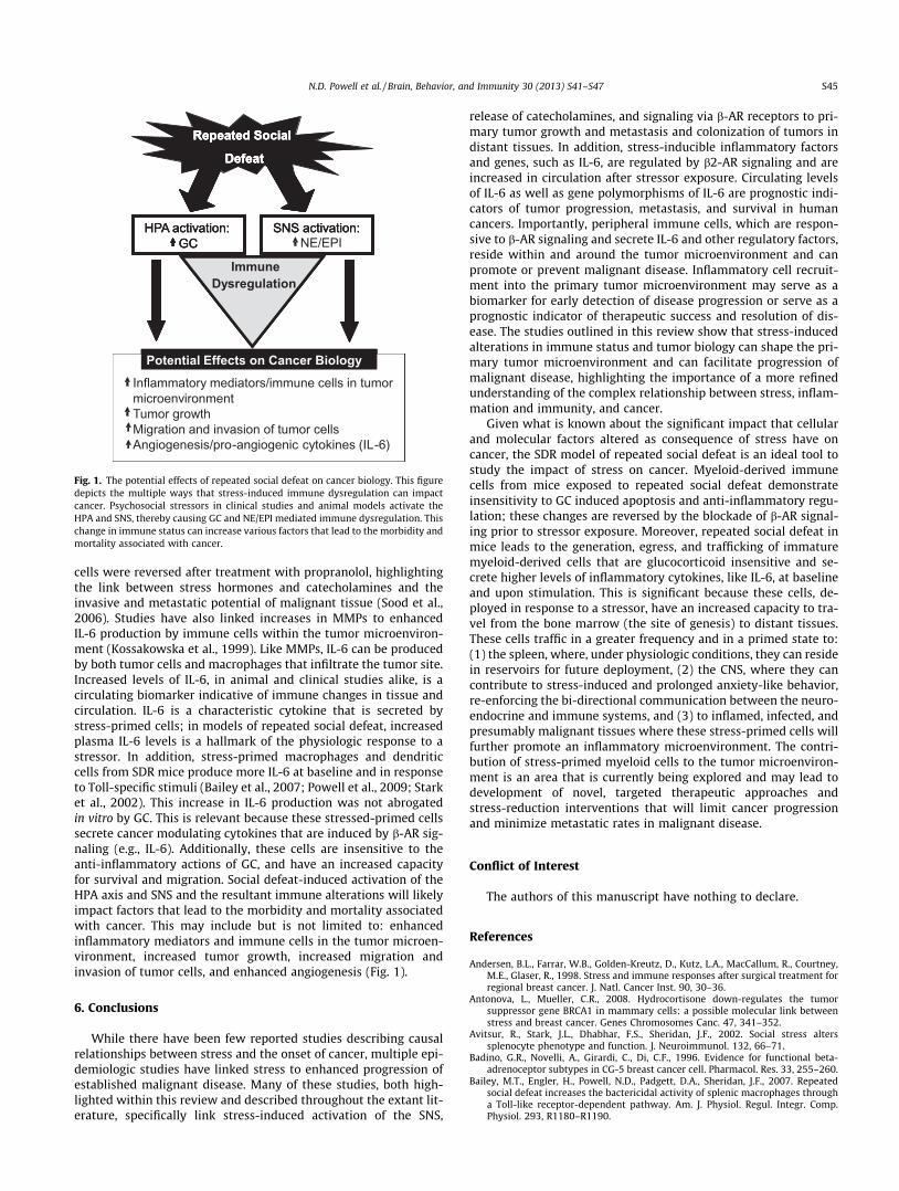
Fig. 1. The potential effects of repeated social defeat on cancer biology. This figure depicts the multiple ways that stress-induced immune dysregulation can impact cancer. Psychosocial stressors in clinical studies and animal models activate the HPA and SNS, thereby causing GC and NE/EPI mediated immune dysregulation. This change in immune status can increase various factors that lead to the morbidity and mortality associated with cancer.
cells were reversed after treatment with propranolol, highlighting the link between stress hormones and catecholamines and the invasive and metastatic potential of malignant tissue (Sood et al., 2006). Studies have also linked increases in MMPs to enhanced IL-6 production by immune cells within the tumor microenvironment (Kossakowska et al., 1999). Like MMPs, IL-6 can be produced by both tumor cells and macrophages that infiltrate the tumor site. Increased levels of IL-6, in animal and clinical studies alike, is a circulating biomarker indicative of immune changes in tissue and circulation. IL-6 is a characteristic cytokine that is secreted by stress-primed cells; in models of repeated social defeat, increased plasma IL-6 levels is a hallmark of the physiologic response to a stressor. In addition, stress-primed macrophages and dendritic cells from SDR mice produce more IL-6 at baseline and in response to Toll-specific stimuli (Bailey et al., 2007; Powell et al., 2009; Stark et al., 2002). This increase in IL-6 production was not abrogated in vitro by GC. This is relevant because these stressed-primed cells secrete cancer modulating cytokines that are induced by b-AR signaling (e.g., IL-6). Additionally, these cells are insensitive to the anti-inflammatory actions of GC, and have an increased capacity for survival and migration. Social defeat-induced activation of the HPA axis and SNS and the resultant immune alterations will likely impact factors that lead to the morbidity and mortality associated with cancer. This may include but is not limited to: enhanced inflammatory mediators and immune cells in the tumor microenvironment, increased tumor growth, increased migration and invasion of tumor cells, and enhanced angiogenesis (Fig. 1).
6. Conclusions
While there have been few reported studies describing causal relationships between stress and the onset of cancer, multiple epidemiologic studies have linked stress to enhanced progression of established malignant disease. Many of these studies, both highlighted within this review and described throughout the extant literature, specifically link stress-induced activation of the SNS,
release of catecholamines, and signaling via b-AR receptors to primary tumor growth and metastasis and colonization of tumors in distant tissues. In addition, stress-inducible inflammatory factors and genes, such as IL-6, are regulated by b2-AR signaling and are increased in circulation after stressor exposure. Circulating levels of IL-6 as well as gene polymorphisms of IL-6 are prognostic indicators of tumor progression, metastasis, and survival in human cancers. Importantly, peripheral immune cells, which are responsive to b-AR signaling and secrete IL-6 and other regulatory factors, reside within and around the tumor microenvironment and can promote or prevent malignant disease. Inflammatory cell recruitment into the primary tumor microenvironment may serve as a biomarker for early detection of disease progression or serve as a prognostic indicator of therapeutic success and resolution of disease. The studies outlined in this review show that stress-induced alterations in immune status and tumor biology can shape the primary tumor microenvironment and can facilitate progression of malignant disease, highlighting the importance of a more refined understanding of the complex relationship between stress, inflammation and immunity, and cancer.
Given what is known about the significant impact that cellular and molecular factors altered as consequence of stress have on cancer, the SDR model of repeated social defeat is an ideal tool to study the impact of stress on cancer. Myeloid-derived immune cells from mice exposed to repeated social defeat demonstrate insensitivity to GC induced apoptosis and anti-inflammatory regulation; these changes are reversed by the blockade of b-AR signaling prior to stressor exposure. Moreover, repeated social defeat in mice leads to the generation, egress, and trafficking of immature myeloid-derived cells that are glucocorticoid insensitive and secrete higher levels of inflammatory cytokines, like IL-6, at baseline and upon stimulation. This is significant because these cells, deployed in response to a stressor, have an increased capacity to travel from the bone marrow (the site of genesis) to distant tissues. These cells traffic in a greater frequency and in a primed state to: (1) the spleen, where, under physiologic conditions, they can reside in reservoirs for future deployment, (2) the CNS, where they can contribute to stress-induced and prolonged anxiety-like behavior, re-enforcing the bi-directional communication between the neuroendocrine and immune systems, and (3) to inflamed, infected, and presumably malignant tissues where these stress-primed cells will further promote an inflammatory microenvironment. The contribution of stress-primed myeloid cells to the tumor microenvironment is an area that is currently being explored and may lead to development of novel, targeted therapeutic approaches and stress-reduction interventions that will limit cancer progression and minimize metastatic rates in malignant disease.
Conflict of Interest
The authors of this manuscript have nothing to declare.
References
Andersen, B.L., Farrar, W.B., Golden-Kreutz, D., Kutz, L.A., MacCallum, R., Courtney, M.E., Glaser, R., 1998. Stress and immune responses after surgical treatment for regional breast cancer. J. Natl. Cancer Inst. 90, 30–36.
Antonova, L., Mueller, C.R., 2008. Hydrocortisone down-regulates the tumor suppressor gene BRCA1 in mammary cells: a possible molecular link between stress and breast cancer. Genes Chromosomes Canc. 47, 341–352.
Avitsur, R., Stark, J.L., Dhabhar, F.S., Sheridan, J.F., 2002. Social stress alters splenocyte phenotype and function. J. Neuroimmunol. 132, 66–71.
Badino, G.R., Novelli, A., Girardi, C., Di, C.F., 1996. Evidence for functional betaadrenoceptor subtypes in CG-5 breast cancer cell. Pharmacol. Res. 33, 255–260.
Bailey, M.T., Engler, H., Powell, N.D., Padgett, D.A., Sheridan, J.F., 2007. Repeated social defeat increases the bactericidal activity of splenic macrophages through a Toll-like receptor-dependent pathway. Am. J. Physiol. Regul. Integr. Comp. Physiol. 293, R1180–R1190.

S46 N.D. Powell et al. / Brain, Behavior, and Immunity 30 (2013) S41–S47
Bailey, M.T., Kierstein, S., Sharma, S., Spaits, M., Kinsey, S.G., Tliba, O., Amrani, Y., Sheridan, J.F., Panettieri, R.A., Haczku, A., 2009a. Social stress enhances allergen-induced airway inflammation in mice and inhibits corticosteroid responsiveness of cytokine production. J. Immunol. 182, 7888–7896.
Bailey, M.T., Kinsey, S.G., Padgett, D.A., Sheridan, J.F., Leblebicioglu, B., 2009b. Social stress enhances IL-1beta and TNF-alpha production by Porphyromonas gingivalis lipopolysaccharide-stimulated CD11b+ cells. Physiol. Behav. 98, 351–358.
Bubenik, J., 2004. MHC class I down-regulation: tumour escape from immune surveillance? (review). Int. J. Oncol. 25, 487–491.
Carena, I., Shamshiev, A., Donda, A., Colonna, M., Libero, G.D., 1997. Major histocompatibility complex class I molecules modulate activation threshold and early signaling of T cell antigen receptor-gamma/delta stimulated by nonpeptidic ligands. J. Exp. Med. 186, 1769–1774.
Chung, Y.C., Chang, Y.F., 2003. Serum interleukin-6 levels reflect the disease status of colorectal cancer. J. Surg. Oncol. 83, 222–226.
Curry, J.M., Hanke, M.L., Piper, M.G., Bailey, M.T., Bringardner, B.D., Sheridan, J.F., Marsh, C.B., 2010. Social disruption induces lung inflammation. Brain Behav. Immun. 24, 394–402.
DeMichele, A., Martin, A.M., Mick, R., Gor, P., Wray, L., Klein-Cabral, M., Athanasiadis, G., Colligan, T., Stadtmauer, E., Weber, B., 2003. Interleukin-6 -174G?C polymorphism is associated with improved outcome in high-risk breast cancer. Cancer Res. 63, 8051–8056.
Dickinson, R.E., Fegan, K.S., Ren, X., Hillier, S.G., Duncan, W.C., 2011. Glucocorticoid regulation of SLIT/ROBO tumour suppressor genes in the ovarian surface epithelium and ovarian cancer cells. PLoS One 6, e27792.
Dong-Newsom, P., Powell, N.D., Bailey, M.T., Padgett, D.A., Sheridan, J.F., 2010. Repeated social stress enhances the innate immune response to a primary HSV1 infection in the cornea and trigeminal ganglia of Balb/c mice. Brain Behav. Immun. 24, 273–280.
Drell, T.L., Joseph, J., Lang, K., Niggemann, B., Zaenker, K.S., Entschladen, F., 2003. Effects of neurotransmitters on the chemokinesis and chemotaxis of MDA-MB468 human breast carcinoma cells. Breast Cancer Res. Treat. 80, 63–70.
Engler, H., Bailey, M.T., Engler, A., Sheridan, J.F., 2004a. Effects of repeated social stress on leukocyte distribution in bone marrow, peripheral blood and spleen. J. Neuroimmunol. 148, 106–115.
Engler, H., Dawils, L., Hoves, S., Kurth, S., Stevenson, J.R., Schauenstein, K., Stefanski, V., 2004b. Effects of social stress on blood leukocyte distribution: the role of alpha- and beta-adrenergic mechanisms. J. Neuroimmunol. 156, 153–162.
Engler, H., Engler, A., Bailey, M.T., Sheridan, J.F., 2005. Tissue-specific alterations in the glucocorticoid sensitivity of immune cells following repeated social defeat in mice. J. Neuroimmunol. 163, 110–119.
Felten, D.L., Felten, S.Y., Carlson, S.L., Olschowka, J.A., Livnat, S., 1985. Noradrenergic and peptidergic innervation of lymphoid tissue. J. Immunol. 135, 755s–765s.
Glaser, R., Pearl, D.K., Kiecolt-Glaser, J.K., Malarkey, W.B., 1994. Plasma cortisol levels and reactivation of latent Epstein-Barr virus in response to examination stress. Psychoneuroendocrinology 19, 765–772.
Glaser, R., Kiecolt-Glaser, J.K., 2005. Stress-induced immune dysfunction: implications for health. Nat. Rev. Immunol. 5, 243–251.
Gouin, J.P., Glaser, R., Malarkey, W.B., Beversdorf, D., Kiecolt-Glaser, J., 2012. Chronic stress, daily stressors, and circulating inflammatory markers. Health Psychol 31 (2), 264–268, Mar.
Hanahan, D., Weinberg, R.A., 2011. Hallmarks of cancer: the next generation. Cell 144, 646–674.
Hanke, T., Takizawa, H., McMahon, C.W., Busch, D.H., Pamer, E.G., Miller, J.D., Altman, J.D., Liu, Y., Cado, D., Lemonnier, F.A., Bjorkman, P.J., Raulet, D.H., 1999. Direct assessment of MHC class I binding by seven Ly49 inhibitory NK cell receptors. Immunity 11, 67–77.
Hunzeker, J., Padgett, D.A., Sheridan, P.A., Dhabhar, F.S., Sheridan, J.F., 2004. Modulation of natural killer cell activity by restraint stress during an influenza A/PR8 infection in mice. Brain Behav. Immun. 18, 526–535.
Jean, D., Bar-Eli, M., 2000. Regulation of tumor growth and metastasis of human melanoma by the CREB transcription factor family. Mol. Cell Biochem. 212, 19– 28.
Kinsey, S.G., Bailey, M.T., Sheridan, J.F., Padgett, D.A., Avitsur, R., 2007. Repeated social defeat causes increased anxiety-like behavior and alters splenocyte function in C57BL/6 and CD-1 mice. Brain Behav. Immun. 21, 458–466.
Kossakowska, A.E., Edwards, D.R., Prusinkiewicz, C., Zhang, M.C., Guo, D., Urbanski, S.J., Grogan, T., Marquez, L.A., Janowska-Wieczorek, A., 1999. Interleukin-6 regulation of matrix metalloproteinase (MMP-2 and MMP-9) and tissue inhibitor of metalloproteinase (TIMP-1) expression in malignant nonHodgkin’s lymphomas. Blood 94, 2080–2089.
Kradin, R., Rodberg, G., Zhao, L.H., Leary, C., 2001. Epinephrine yields translocation of lymphocytes to the lung. Exp. Mol. Pathol. 70, 1–6.
Lang, K., Drell, T.L., Lindecke, A., Niggemann, B., Kaltschmidt, C., Zaenker, K.S., Entschladen, F., 2004. Induction of a metastatogenic tumor cell type by neurotransmitters and its pharmacological inhibition by established drugs. Int. J. Cancer 112, 231–238.
Levy, S., Herberman, R., Lippman, M., d’Angelo, T., 1987. Correlation of stress factors with sustained depression of natural killer cell activity and predicted prognosis in patients with breast cancer. J. Clin. Oncol. 5, 348–353.
Lillberg, K., Verkasalo, P.K., Kaprio, J., Teppo, L., Helenius, H., Koskenvuo, M., 2003. Stressful life events and risk of breast cancer in 10,808 women: a cohort study. Am. J. Epidemiol. 157, 415–423.
Madden, K.S., Sanders, V.M., Felten, D.L., 1995. Catecholamine influences and sympathetic neural modulation of immune responsiveness. Annu. Rev. Pharmacol. Toxicol. 35, 417–448.
Masur, K., Niggemann, B., Zanker, K.S., Entschladen, F., 2001. Norepinephrine-induced migration of SW 480 colon carcinoma cells is inhibited by beta-blockers. Cancer Res. 61, 2866–2869.
Mays, J.W., Bailey, M.T., Hunzeker, J.T., Powell, N.D., Papenfuss, T., Karlsson, E.A., Padgett, D.A., Sheridan, J.F., 2010. Influenza virus-specific immunological memory is enhanced by repeated social defeat. J. Immunol. 184, 2014–2025.
Mays, J.W., Powell, N.D., Hunzeker, J.T., Hanke, M.L., Bailey, M.T., Sheridan, J.F., 2012. Stress and the anti-influenza immune response: Repeated social defeat augments clonal expansion of CD8(+)T cells during primary influenza A viral infection. J. Neuroimmunol. 243, 34–42.
Miller, G.E., Chen, E., Sze, J., Marin, T., Arevalo, J.M., Doll, R., Ma, R., Cole, S.W., 2008. A functional genomic fingerprint of chronic stress in humans: blunted glucocorticoid and increased NF-kappaB signaling. Biol. Psychiatry 64, 266–272.
Mundy-Bosse, B.L., Thornton, L.M., Yang, H.C., Andersen, B.L., Carson, W.E., 2011. Psychological stress is associated with altered levels of myeloid-derived suppressor cells in breast cancer patients. Cell Immunol. 270, 80–87.
Ostrand-Rosenberg, S., Sinha, P., 2009. Myeloid-derived suppressor cells: linking inflammation and cancer. J. Immunol. 182, 4499–4506.
Padgett, D.A., Glaser, R., 2003. How stress influences the immune response? Trends Immunol. 24, 444–448.
Palermo-Neto, J., de Oliveira, M.C., Robespierre de, S.W., 2003. Effects of physical and psychological stressors on behavior, macrophage activity, and Ehrlich tumor growth. Brain Behav. Immun. 17, 43–54.
Palm, D., Lang, K., Niggemann, B., Drell, T.L., Masur, K., Zaenker, K.S., Entschladen, F., 2006. The norepinephrine-driven metastasis development of PC-3 human prostate cancer cells in BALB/c nude mice is inhibited by beta-blockers. Int. J. Cancer 118, 2744–2749.
Pierce, B.L., Ballard-Barbash, R., Bernstein, L., Baumgartner, R.N., Neuhouser, M.L., Wener, M.H., Baumgartner, K.B., Gilliland, F.D., Sorensen, B.E., McTiernan, A., Ulrich, C.M., 2009. Elevated biomarkers of inflammation are associated with reduced survival among breast cancer patients. J. Clin. Oncol. 27, 3437–3444.
Powell, N.D., Bailey, M.T., Mays, J.W., Stiner-Jones, L.M., Hanke, M.L., Padgett, D.A., Sheridan, J.F., 2009. Repeated social defeat activates dendritic cells and enhances toll-like receptor dependent cytokine secretion. Brain Behav. Immun. 23, 225–231.
Powell, N.D., Mays, J.W., Bailey, M.T., Hanke, M.L., Sheridan, J.F., 2011. Immunogenic dendritic cells primed by social defeat enhance adaptive immunity to influenza A virus. Brain Behav. Immun. 25, 46–52.
Price, M.A., Tennant, C.C., Butow, P.N., Smith, R.C., Kennedy, S.J., Kossoff, M.B., Dunn, S.M., 2001. The role of psychosocial factors in the development of breast carcinoma: part II. life event stressors, social support, defense style, and emotional control and their interactions. Cancer 91, 686–697.
Reilly, F.D., McCuskey, P.A., Miller, M.L., McCuskey, R.S., Meineke, H.A., 1979. Innervation of the periarteriolar lymphatic sheath of the spleen. Tissue Cell 11, 121–126.
Salgado, R., Junius, S., Benoy, I., Van, D.P., Vermeulen, P., Van, M.E., Huget, P., Dirix, L.Y., 2003. Circulating interleukin-6 predicts survival in patients with metastatic breast cancer. Int. J. Cancer 103, 642–646.
Sanders, V.M., 1995. The role of adrenoceptor-mediated signals in the modulation of lymphocyte function. Adv. Neuroimmunol. 5, 283–298.
Sastry, K.S., Karpova, Y., Prokopovich, S., Smith, A.J., Essau, B., Gersappe, A., Carson, J.P., Weber, M.J., Register, T.C., Chen, Y.Q., Penn, R.B., Kulik, G., 2007. Epinephrine protects cancer cells from apoptosis via activation of cAMP-dependent protein kinase and BAD phosphorylation. J. Biol. Chem. 282, 14094–14100.
Saul, A.N., Oberyszyn, T.M., Daugherty, C., Kusewitt, D., Jones, S., Jewell, S., Malarkey, W.B., Lehman, A., Lemeshow, S., Dhabhar, F.S., 2005. Chronic stress and susceptibility to skin cancer. J. Natl. Cancer Inst. 97, 1760–1767.
Schedlowski, M., Falk, A., Rohne, A., Wagner, T.O., Jacobs, R., Tewes, U., Schmidt, R.E., 1993. Catecholamines induce alterations of distribution and activity of human natural killer (NK) cells. J. Clin. Immunol. 13, 344–351.
Sieber, W.J., Rodin, J., Larson, L., Ortega, S., Cummings, N., Levy, S., Whiteside, T., Herberman, R., 1992. Modulation of human natural killer cell activity by exposure to uncontrollable stress. Brain Behav. Immun. 6, 141–156.
Sloan, E.K., Priceman, S.J., Cox, B.F., Yu, S., Pimentel, M.A., Tangkanangnukul, V., Arevalo, J.M., Morizono, K., Karanikolas, B.D., Wu, L., Sood, A.K., Cole, S.W., 2010. The sympathetic nervous system induces a metastatic switch in primary breast cancer. Cancer Res. 70, 7042–7052.
Snoussi, K., Strosberg, A.D., Bouaouina, N., Ben, A.S., Chouchane, L., 2005. Genetic variation in pro-inflammatory cytokines (interleukin-1beta, interleukin1alpha and interleukin-6) associated with the aggressive forms, survival, and relapse prediction of breast carcinoma. Eur. Cytokine Netw. 16, 253– 260.
Sood, A.K., Bhatty, R., Kamat, A.A., Landen, C.N., Han, L., Thaker, P.H., Li, Y., Gershenson, D.M., Lutgendorf, S., Cole, S.W., 2006. Stress hormone-mediated invasion of ovarian cancer cells. Clin. Cancer Res. 12, 369–375.
Spiegel, D., Giese-Davis, J., 2003. Depression and cancer: mechanisms and disease progression. Biol. Psychiatry 54, 269–282.
Stark, J.L., Avitsur, R., Hunzeker, J., Padgett, D.A., Sheridan, J.F., 2002. Interleukin-6 and the development of social disruption-induced glucocorticoid resistance. J. Neuroimmunol. 124, 9–15.
Steplewski, Z., Vogel, W.H., Ehya, H., Poropatich, C., Smith, J.M., 1985. Effects of restraint stress on inoculated tumor growth and immune response in rats. Cancer Res. 45, 5128–5133.
Tan, K.S., Nackley, A.G., Satterfield, K., Maixner, W., Diatchenko, L., Flood, P.M., 2007. Beta2 adrenergic receptor activation stimulates pro-inflammatory cytokine

N.D. Powell et al. / Brain, Behavior, and Immunity 30 (2013) S41–S47 S47
production in macrophages via PKA- and NF-kappaB-independent mechanisms. Cell Signal 19, 251–260.
Tomozawa, Y., Yabuuchi, K., Inoue, T., Satoh, M., 1995. Participation of cAMP and cAMP-dependent protein kinase in beta-adrenoceptor-mediated interleukin-1 beta mRNA induction in cultured microglia. Neurosci. Res. 22, 399–409.
Tseng, R.J., Padgett, D.A., Dhabhar, F.S., Engler, H., Sheridan, J.F., 2005. Stress-induced modulation of NK activity during influenza viral infection: role of glucocorticoids and opioids. Brain Behav. Immun. 19, 153–164.
Vahlne, G., Lindholm, K., Meier, A., Wickstrom, S., Lakshmikanth, T., Brennan, F., Wilken, M., Nielsen, R., Romagne, F., Wagtmann, N.R., Karre, K., Johansson, M.H., 2010. In vivo tumor cell rejection induced by NK cell inhibitory receptor blockade: maintained tolerance to normal cells even in the presence of IL-2. Eur. J. Immunol. 40, 813–823.
Williams, J.M., Peterson, R.G., Shea, P.A., Schmedtje, J.F., Bauer, D.C., Felten, D.L., 1981. Sympathetic innervation of murine thymus and spleen: evidence for a functional link between the nervous and immune systems. Brain Res. Bull. 6, 83–94.
Wohleb, E.S., Hanke, M.L., Corona, A.W., Powell, N.D., Stiner, L.M., Bailey, M.T., Nelson, R.J., Godbout, J.P., Sheridan, J.F., 2011. Beta-Adrenergic receptor
antagonism prevents anxiety-like behavior and microglial reactivity induced by repeated social defeat. J. Neurosci. 31, 6277–6288.
Wu, W., Chaudhuri, S., Brickley, D.R., Pang, D., Karrison, T., Conzen, S.D., 2004. Microarray analysis reveals glucocorticoid-regulated survival genes that are associated with inhibition of apoptosis in breast epithelial cells. Cancer Res. 64, 1757–1764.
Xia, C., Meng, Q., Liu, L.Z., Rojanasakul, Y., Wang, X.R., Jiang, B.H., 2007. Reactive oxygen species regulate angiogenesis and tumor growth through vascular endothelial growth factor. Cancer Res. 67, 10823–10830.
Yang, E.V., Sood, A.K., Chen, M., Li, Y., Eubank, T.D., Marsh, C.B., Jewell, S., Flavahan, N.A., Morrison, C., Yeh, P.E., Lemeshow, S., Glaser, R., 2006. Norepinephrine up-regulates the expression of vascular endothelial growth factor, matrix metalloproteinase (MMP)-2, and MMP-9 in nasopharyngeal carcinoma tumor cells. Cancer Res. 66, 10357–10364.
Yu, W.G., Yamamoto, N., Takenaka, H., Mu, J., Tai, X.G., Zou, J.P., Ogawa, M., Tsutsui, T., Wijesuriya, R., Yoshida, R., Herrmann, S., Fujiwara, H., Hamaoka, T., 1996. Molecular mechanisms underlying IFN-gamma-mediated tumor growth inhibition induced during tumor immunotherapy with rIL-12. Int. Immunol. 8, 855–865.