born to run? diverse modes of epithelial migration
TRANSCRIPT

fcell-09-704939 August 7, 2021 Time: 13:17 # 1
REVIEWpublished: 12 August 2021
doi: 10.3389/fcell.2021.704939
Edited by:Andrea Erika Münsterberg,
University of East Anglia,United Kingdom
Reviewed by:Gabriel L. Galea,
University College London,United KingdomErica Hutchins,
California Institute of Technology,United States
*Correspondence:Pengfei Lu
Specialty section:This article was submitted to
Morphogenesis and Patterning,a section of the journal
Frontiers in Cell and DevelopmentalBiology
Received: 04 May 2021Accepted: 20 July 2021
Published: 12 August 2021
Citation:Lu P and Lu Y (2021) Born
to Run? Diverse Modes of EpithelialMigration.
Front. Cell Dev. Biol. 9:704939.doi: 10.3389/fcell.2021.704939
Born to Run? Diverse Modes ofEpithelial MigrationPengfei Lu* and Yunzhe Lu
School of Life Science and Technology, ShanghaiTech University, Shanghai, China
Bundled with various kinds of adhesion molecules and anchored to the basementmembrane, the epithelium has historically been considered as an immotile tissue and, tomigrate, it first needs to undergo epithelial-mesenchymal transition (EMT). Since its initialdescription more than half a century ago, the EMT process has fascinated generationsof developmental biologists and, more recently, cancer biologists as it is believed to beessential for not only embryonic development, organ formation, but cancer metastasis.However, recent progress shows that epithelium is much more motile than previouslyrealized. Here, we examine the emerging themes in epithelial collective migration andhow this has impacted our understanding of EMT.
Keywords: cell polarity, epithelial polarity, apicobasal polarity, collective migration, EMT, extracellular matrix,mechanosensing, mesenchymal-epithelial transition
INTRODUCTION
The emergence of the epithelium is one of the earliest and most important events duringmetazoan evolution. By separating an organism from the outside world, the epithelium protectedcells within the organism from environmental insults and facilitated the rise and prosperity ofmulticellularity. Far from being a simple, protective shield or barrier, the epithelium also allowedan organism to actively exchange substances, including nutrients, wastes, gases, etc. with theenvironment, a basis on which organ systems, including the digestive, respiratory, urinary, andglandular systems emerged during evolution. These organ systems enabled the metazoans toexplore previously untapped niches and, eventually, to dominate almost every corner of the planetearth (Dickinson et al., 2011). Today, most major organs of extant animals are epithelial and mosthuman cancers are of epithelial origin, thus highlighting the importance of epithelial function inphysiology and pathology.
Historically, the epithelium is considered being immotile. This is in part because of theobservation that the epithelium is rich in myriads of cell-cell adhesion proteins, particularly tightjunctions (TJs) and adherens junctions (AJs), and focal adhesions which anchor the epitheliumto the matrix, i.e., the basement membrane. To migrate, it is believed that the epithelium mustfirst transition to a mesenchymal state via a process called epithelial-to-mesenchymal transition(EMT) (Hay, 1968, 1995). Epithelial-to-mesenchymal transition was subsequently confirmed inseveral fundamental developmental processes, including the generation of mesoderm and neuralcrest (Newgreen et al., 1979; Thiery et al., 1982). Research in the 1990s further implicated EMTwith pathologies like fibrosis and cancer (Valles et al., 1990; Miettinen et al., 1994). The notion thatEMT may be responsible for these pathologies such as cancer metastasis further catapulted EMTto one of the most intensely researched areas in not only developmental biology but in molecularbiology and cancer biology as well in the past decade (Yang et al., 2020).
Frontiers in Cell and Developmental Biology | www.frontiersin.org 1 August 2021 | Volume 9 | Article 704939

fcell-09-704939 August 7, 2021 Time: 13:17 # 2
Lu and Lu EMT and Epithelial Migration
Tremendous progress has been made since the initialdescription of EMT. We now know, for example, the epitheliumis much more motile than expected, especially during embryonicdevelopment and organ formation where large scale migrationsuch epiboly and convergent extension during gastrulation haveall been well documented. Due to space constraints, these typesof migration will not be discussed in detail in the current review.Instead, we will focus on directional movements of epithelialcells, where recent studies show that occur collectively rather thanindividually as it was previously believed (Cheung et al., 2013,2016; Wrenn et al., 2020). We first discuss recent progress inepithelial collective migration and then how it has impacted ourcurrent understanding of EMT.
THE EVOLVING CONCEPT OFEPITHELIAL-TO-MESENCHYMALTRANSITION
At the core of the current debates on EMT lie the very definitionsof what makes up an epithelial or a mesenchymal state, andwhat the most important changes are, at both the cellular andmolecular levels, that occur during the EMT process.
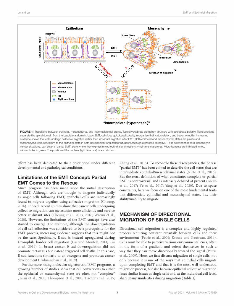
What Makes an Epithelium?Epithelium can be defined at several levels. At the functionallevel, as mentioned above, an epithelium provides tissue with aprotective barrier and substance-exchange roles. Depending ontheir cell shapes, the mature epithelium can be categorized intosimple epithelium, including squamous, columnar, or cuboidalepithelium; stratified epithelium, which contains multiple celllayers; and compound epithelium, which has a mixture ofone or more of the above features. We will not differentiateepithelial migration based on these categories of the matureepithelium as the current literature is primarily focused onthe developing epithelium. Regardless of their categories, oneof the most salient feature of epithelium is its polarity, i.e.,its plasma membrane is organized into two discrete regions:one facing the environment, called the apical domain andanother facing the inside of the tissues, called the basolateraldomain (Figure 1) (Bryant and Mostov, 2008; Mellman andNelson, 2008). With these two distinct domains, membraneproteins of various functions, for example, ion or metabolitetransporters can be differentially localized to either the apicalor the basolateral domain, and selectively regulate the exchangeof substances between the environment and the organism(St Johnston and Ahringer, 2010).
At the structural level, apicobasal polarity is made possiblein part by the unique presence and organization of variousmembrane junctional components. Specifically, epithelialpolarity is manifested by the establishment of the apicaljunctional complex (AJC), which includes the TJs in vertebrates,or septate junctions in invertebrates, and AJs. Tight junctions,which are composed of several families of transmembraneproteins, including Claudins and junctional adhesion molecules(JAMs), are organized into a tight seal that prevents the freediffusion of proteins and lipids between apical and lateral
surfaces. They also make up an important selective barrierregulating the diffusion of molecules through the paracellularspace. Interestingly, although Occludin was the first integral TJresident component identified, genetic studies have shown thatit is not essential for TJ function but it plays other importantcellular functions (Bryant and Mostov, 2008).
Basal to tight junctions in vertebrates, but apical to septatejunctions in invertebrates, are AJs, which form an adhesivebelt encircling each epithelial cell just underneath the apicalsurface. AJ transmembrane components include cadherins, mostnotably E-Cadherin, which provide cohesion between cells ofthe epithelial sheet. Although all epithelia possess apicobasalpolarity, they may differ in the kind, amount, affinity of theiradhesion molecules for other cells and matrix. They may alsodiffer in the absence or presence, and complexity of the ECM(Campbell and Casanova, 2016).
EMT in Normal Development andPathologiesEpithelial-to-mesenchymal transition (EMT) was firstdescribed by Elizabeth Hay in the 1970s while studyingchick embryogenesis using in vitro explant (Hay, 1968, 1995).It was subsequently confirmed in the context of neural crestformation (Newgreen et al., 1979; Thiery et al., 1982), heart valveformation (Markwald et al., 1977), and Mullerian duct regressionduring kidney development (Trelstad et al., 1982). Additionalclassic examples of EMT also include mesoderm formationduring gastrulation (Leptin, 1991), sclerotome formation fromthe somite (Christ and Ordahl, 1995), and wound healing(Yang et al., 2020). It is generally considered including severaldistinct steps: first, EMT signals, often extrinsic paracrine factorsand/or intrinsic transcription factor activation, stimulate targetepithelial cells; second, epithelial cells lose apicobasal polarityand downregulate E-Cadherin expression; third, epithelial cellstransition to mesenchymal cells and start to migrate.
The reverse process of EMT, known as a mesenchymal-epithelial transition (MET), also occurs during development, forexample during secondary neurulation and nephron formationof kidney development (Stark et al., 1994). Over the past twentyyears, the EMT research field has grown exponentially, thanksin part to the realization that EMT may be activated duringpathological conditions such as cancer development and tissuefibrosis (Valles et al., 1990; Miettinen et al., 1994). Indeed, itis believed that loss of epithelial polarity is a cancer hallmarkand EMT is an essential step during cancer metastasis (Humbertet al., 2003; Bilder, 2004; Lee and Vasioukhin, 2008; Hanahan andWeinberg, 2011).
Much of this research effort has focused on the molecularnature of the “EMT program”. Various paracrine factors,including TGF-beta, and transcription factors, such as Snail,Slug, Zeb1 and Zeb2, have been identified (Leptin, 1991; Yanget al., 2004; Stemmler et al., 2019). Downstream changes of theseparacrine and transcription factors during EMT have also beendescribed (Yang et al., 2004). Together, these changes have beenconsidered as a part of the EMT program and intense research
Frontiers in Cell and Developmental Biology | www.frontiersin.org 2 August 2021 | Volume 9 | Article 704939
fcell-09-704939 August 7, 2021 Time: 13:17 # 3
Lu and Lu EMT and Epithelial Migration
FIGURE 1 | Transitions between epithelial, mesenchymal, and intermediate cell states. Typical vertebrate epithelium structure with apicobasal polarity. Tight junctionsseparate the apical domain from the basolateral domain. Upon EMT, cells lose apicobasal polarity, reorganize their cytoskeleton, and become motile. Increasingevidence shows that cells undergo collective migration rather than individual migration after EMT. Both epithelial and mesenchymal states are plastic andmesenchymal cells can return to the epithelial state in both development and cancer situations through a process called MET. It is believed that cells, especially incancer situations, can enter a “partial EMT” state where they express mixed epithelial and mesenchymal gene signatures. Microfilaments are indicated in red,microtubules in green. The position of the nucleus (light blue oval) is also shown.
effort has been dedicated to their description under differentdevelopmental and pathological conditions.
Limitations of the EMT Concept: PartialEMT Comes to the RescueMuch progress has been made since the initial descriptionof EMT. Although cells are thought to migrate individuallyas single cells following EMT, epithelial cells are increasinglyfound to migrate together using collective migration (Cheung,2016). Indeed, recent studies show that cancer cells undergoingcollective migration can metastasize more efficiently and survivebetter at distant sites (Cheung et al., 2013, 2016; Wrenn et al.,2020). However, the limitations of the EMT concept have alsostarted to emerge. For example, although the downregulationof cell-cell adhesion was considered to be a prerequisite for theEMT process, increasing evidence suggests that this might notbe the case. Specifically, E-cad is instead upregulated duringDrosophila border cell migration (Cai and Montell, 2014; Caiet al., 2014). In breast cancer, E-cad downregulation did notpromote metastasis but instead triggered cell deaths. In this case,E-cad functions similarly to an oncogene and promotes cancerdevelopment (Padmanaban et al., 2019).
Furthermore, using molecular signatures of EMT programs, agrowing number of studies show that cell conversions to eitherthe epithelial or mesenchymal state are often not “complete”(Tarin et al., 2005; Thompson et al., 2005; Fischer et al., 2015;
Zheng et al., 2015). To reconcile these discrepancies, the phrase“partial EMT” has been coined to describe the cell states that areintermediate epithelial/mesenchymal states (Nieto et al., 2016).But the exact definition of what constitutes complete or partialEMT is controversial and is intensely debated at present (Aielloet al., 2017; Ye et al., 2017; Yang et al., 2020). Due to spaceconstraints, here we focus on one of the most fundamental traitsthat differentiate epithelial and mesenchymal states, i.e., theirability/inability to migrate.
MECHANISM OF DIRECTIONALMIGRATION OF SINGLE CELLS
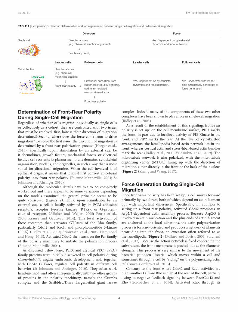
Directional cell migration is a complex and highly regulatedprocess requiring constant crosstalk between cells and theirenvironment (Petrie et al., 2009; Krause and Gautreau, 2014).Cells must be able to perceive various environmental cues, oftenin the form of a gradient, and orient themselves in such away that they can move directionally toward the signal (Petrieet al., 2009). Here, we first discuss migration of single cells, notonly because it is one of the ways that epithelial cells migrateupon completing EMT and that it is the most well-understoodmigration process, but also because epithelial collective migrationfaces similar issues as single cells and, at the individual cell level,share many similarities during migration (Table 1).
Frontiers in Cell and Developmental Biology | www.frontiersin.org 3 August 2021 | Volume 9 | Article 704939
fcell-09-704939 August 7, 2021 Time: 13:17 # 4
Lu and Lu EMT and Epithelial Migration
TABLE 1 | Comparison of direction determination and force generation between single cell migration and collective cell migration.
Direction Force
Single cell Directional cues(e.g. chemical, mechnical gradiant)
Yes. Dependent on cytoskeletaldynamics and focal adhesion.
↓
Front-rear polarity
Leader cells Follower cells Leader cells Follower cells
Cell collective Directional cues(e.g. chemical,mechnical gradiant)
↓
Front-rear polarity →
Directional cues likely fromleader cells via ERK signaling,cadherin-mediatedmechno-transduction.
Yes. Dependent on cytoskeletaldynamics and focal adhesion.
Yes. Cooperate with leadercells and actively contribute toforce generation.
↓
Front-rear polarity
Determination of Front-Rear PolarityDuring Single-Cell MigrationRegardless of whether cells migrate individually as single cellsor collectively as a cohort, they are confronted with two issuesthat must be resolved: first, how is their direction of migrationdetermined? Second, where does the force come from to drivemigration? To solve the first issue, the direction of migration isdetermined by a front-rear polarization process (Haeger et al.,2015). Specifically, upon stimulation by an external cue, beit chemokines, growth factors, mechanical forces, or electricalfields, a cell reorients its plasma membrane domains, cytoskeletalorganization, nucleus, and organelles, in such a way that is mostsuited for directional migration. When the cell involved is ofepithelial origin, it means that it must first convert apicobasalpolarity into front-rear polarity (Etienne-Manneville, 2004; StJohnston and Ahringer, 2010).
Although the molecular details have yet to be completelyworked out and there appear to be some variations dependingon the models examined, the general principle seems to bequite conserved (Figure 2). Thus, upon stimulation by anexternal cue, a cell is locally activated by its ECM adhesionreceptors, receptor tyrosine kinases (RTKs), or G-protein-coupled receptors (Affolter and Weijer, 2005; Petrie et al.,2009; Krause and Gautreau, 2014). This local activation ofthese receptors then activates GTPases of the Rho family,particularly Cdc42 and Rac1, and phosphoinositide 3-kinase(PI3K) (Ridley et al., 2003; Srinivasan et al., 2003; Hammondand Hong, 2018). Activated Cdc42 then turns on the Par familyof the polarity machinery to initiate the polarization process(Etienne-Manneville, 2004).
As discussed below, Par6, Par3, and atypical PKC (aPKC)family proteins were initially discovered in cell polarity duringCaenorhabditis elegans embryonic development and, togetherwith Cdc42 GTPases, regulate cell polarity in different cellbehavior (St Johnston and Ahringer, 2010). They often workhand-in-hand, and often antagonistically, with two other groupsof proteins in the polarity machinery, namely the Crumbscomplex and the Scribbled/Discs Large/Lethal giant larvae
complex. Indeed, many of the components of these two othercomplexes have been shown to play a role in single-cell migration(Ridley et al., 2003).
As a result of the establishment of this signaling, front-rearpolarity is set up: on the cell membrane surface, PIP3 marksthe front, in part due to localized activity of PI3 Kinase in thefront, and PIP2 marks the rear. At the level of cytoskeletonarrangements, the lamellipodia-based actin network lies in thefront, whereas cortical actin and stress-fiber-based actin bundlesmark the rear (Ridley et al., 2003; Vaidziulyte et al., 2019). Themicrotubule network is also polarized, with the microtubuleorganizing center (MTOC) lining up with the direction ofmigration either directly in the front or the back of the nucleus(Figure 2) (Zhang and Wang, 2017).
Force Generation During Single-CellMigrationOnce front-rear polarity has been set up, a cell moves forwardprimarily by two forces, both of which depend on actin filamentbut with important differences. Specifically, in addition tosetting up a front-rear polarity, activated Cdc42 promotes anArp2/3-dependent actin assembly process. Because Arp2/3 isinvolved in actin nucleation and the plus ends of actin filamentare anchored at the focal adhesions, the actin polymerizationprocess is forward-oriented and produces a network of filamentsprotruding into the front, an extension often referred to asthe lamellipodia (Figure 2) (Pollard and Borisy, 2003; Suraneniet al., 2012). Because the action network is fixed concerning thesubstratum, the front membrane is pushed out as the filamentselongate. This process is very similar to the movement of thebacterial pathogen Listeria, which moves within a cell andsometimes through a cell by “riding” on the polymerizing actintail (Bravo-Cordero et al., 2013).
Contrary to the front where Cdc42 and Rac1 activities arehigh, another GTPase Rho is high at the rear of the cell, partiallyowing to negative feedback signaling between Rac/Cdc42 andRho (Goicoechea et al., 2014). Activated Rho, through its
Frontiers in Cell and Developmental Biology | www.frontiersin.org 4 August 2021 | Volume 9 | Article 704939

fcell-09-704939 August 7, 2021 Time: 13:17 # 5
Lu and Lu EMT and Epithelial Migration
FIGURE 2 | Steps in single-cell migration. Diagram with both side and top views of a cell before and during migration to illustrate signaling events leading to polaritydetermination, microfilament dynamics, and morphological changes of the cell. For illustration purposes, a cell is theoretically “unpolarized” before being stimulatedby an external cue (0). Upon stimulation, the area with the highest Cdc42, Par6, and downstream signaling activities becomes the front, which is marked by anincrease in PIP3 levels in the membrane and focal adhesions assembly (1). As a result, lamellipodia formation starts in the front, leading to membrane forwardprotrusion, where new focal adhesions can be made (2). Concordantly, the rear of the cell undergoes a contraction process (arrowheads) involving actomyosinactivities along the cell periphery and in the stress fibers (3). The cell retracts when focal adhesions in the rear break apart due to the contraction force and, as aresult, the cell body translocates forward. The net results of the above steps are a forward movement of the cell.
main effector ROCK, promotes actomyosin activity by ROCK-mediated phosphorylation of the myosin light chain. As a result,the concerted contraction in both the stress fibers and the corticalactin leads to retraction of the rear of the cell and translocates thecell body forward (Vicente-Manzanares et al., 2009) (Figure 1).
In addition to regulating direction and force generation,there are some other aspects of cell migration that are alsoinvolved. They are the focus of many excellent reviews (Petrieet al., 2009; Krause and Gautreau, 2014) and will not bediscussed further here.
Frontiers in Cell and Developmental Biology | www.frontiersin.org 5 August 2021 | Volume 9 | Article 704939

fcell-09-704939 August 7, 2021 Time: 13:17 # 6
Lu and Lu EMT and Epithelial Migration
DETERMINATION OF FRONT-REARPOLARITY OF THE MIGRATINGCOLLECTIVE
Like migrating single cells, a cohort of cells also needs todetermine the direction and force of migration. Unlike singlecells, however, these two issues not only need to be addressedat the single-cell level but at the level of the cohort, asdiscussed below.
Leader Cells Define the Front and Drivethe Migrating CollectiveOne of the earliest morphological signs of front-rear polarityduring collective migration is the formation of actin-richfilopodia and lamellipodia at the leading edge of the migratingunit (Haeger et al., 2015). Interestingly, although every cell thatmigrates individually forms these kinds of cellular extensionspointing toward the external cues, only one or a few cells atthe migration front do so when they migrate as a collective(Friedl and Gilmour, 2009; Petrie et al., 2009). These cells areoften referred to as “leader cells” because of their location atthe leading position, whereas those in the rear are referred to as“follower cells” (Khalil and Friedl, 2010; Theveneau and Linker,2017) (Figure 3).
In all of the collective systems examined, leader cells arenot only a manifestation of directionality but also the sourceof traction forces that power the migration process (Friedland Gilmour, 2009; Petrie et al., 2009; Haeger et al., 2015).In both the Drosophila trachea during embryonic developmentand air sacs during larva stages, directional migration is drivenby leader cells in response to FGF cues and is essentialfor patterning branch (Lu et al., 2006). Likewise, leader cellsalso exist at the invasion front of other migrating collectives,including fly border cells, vertebrate neural crest, zebrafishlateral lines, etc., and are responsible for “pulling” follower cellsforward during directional migration (Friedl and Gilmour, 2009;Petrie et al., 2009) (Figure 3).
It has long been assumed that vertebrate epithelia, especiallythose from branched organs, including the lung, kidney, andmammary gland, undergo directional migration as a partof their ontogeny (Ewald et al., 2008; Lu and Werb, 2008;Lu et al., 2008). The assumption is in part based on theobservation that invertebrate and vertebrate branching systems,which are non-homologous structures, share a surprising amountof cellular and molecular events, or “deep homology” duringorgan formation (Lu et al., 2006; Lu and Werb, 2008).Interestingly, our recent work showed that leader cells inthe mammary gland are a dynamic population and movefaster and more directionally toward the FGF10 signal thanfollower cells, owing partly to their intraepithelial protrusionstoward the signal. We show that a leader cell in themigrating mammary epithelium might “pull” or “push” thecollective depending on its relative position in the collectiveand whether there are cells in front of them. It thus usesa novel mechanism during directional collective migration(Lu et al., 2020) (Figure 3).
FIGURE 3 | Representative models of collective migration. Front-rear polarityin most of these models, including the Xenopus neural crest, Drosophilaborder cells, zebrafish lateral line, and mouse mammary gland, is triggered bya chemoattractant indicated, except for the wound healing model (usinghuman skin as an example), where the external cue is thought to bemechanical forces. Leader cells are colored in green. Blue bars indicate theboundary of the apical and basolateral domains, which in vertebrates is wheretight junctions locate, and in invertebrates is the subapical complex. Theydenote the presence of apicobasal polarity and the epithelial state. Note thatneural crest cells do not have the blue bars because they are thought to haveundergone a complete EMT and have lost the apicobasal polarity. Leader cellsare colored in green.
Specification of Leader CellsMany of the aforementioned external cues, including RTKligands, are involved in specifying leader cells. In fly tracheaand air sacs, for example, mesodermal cells express Branchless(Bnl/Fgf), which causes migration and branch initiation of
Frontiers in Cell and Developmental Biology | www.frontiersin.org 6 August 2021 | Volume 9 | Article 704939

fcell-09-704939 August 7, 2021 Time: 13:17 # 7
Lu and Lu EMT and Epithelial Migration
adjacent epithelial cells expressing the receptor Breathless(Btl/Fgfr) (Ghabrial et al., 2003). Bnl/Fgf and its downstreamevents determine whether a cell becomes a leader cell, aka tip cell,or a follower cell, aka stalk cell. Although all tracheal epithelialcells express Btl/Fgfr and respond to Bnl/Fgf, they competefor the ligand, and the cell with the highest Bnl/Fgf signalingactivities becomes the leader cell (Ghabrial and Krasnow, 2006).Likewise, EGF and PVF are involved in specifying leader cellsin the fly border cell system, while CXCL12/SDF-1 is involvedin leader cell specification in neural crest and zebrafish lateralline systems (Lu et al., 2006, 2008; Friedl and Gilmour, 2009)(Figure 3). Furthermore, in a 2D-migration model, Notch1-Dll4signaling has been shown to play a role in determining leader cellfate (Riahi et al., 2015; Wang et al., 2017).
Moreover, the formation, maintenance, and subsequentmigration are under local and sometimes global regulation.In the fly trachea, the number of branches is regulated bymutual inhibition, whereby epithelial cells inhibit each other totake the leading position as they compete for branch-inducingfactors. In both the fly trachea and the vasculature, such mutualinhibition depends on Notch signaling. Once leader cells havebeen determined, they are the only cells of the primary branchthat depend on Bnl/Fgf signaling; the remaining follower cellsfollow leader cells in a Bnl/Fgf-independent manner (Ghabrialand Krasnow, 2006). Likewise, in both fly air sacs and themammary gland, FGF signaling activity is necessary for cellsto remain in the leader but not in the follower position (Luet al., 2008). Interestingly, in addition of having differentialgene expression, recent studies show that energetic regulationis distinct in leader and follower cells. Thus, in a 2D breastcancer invasion model, leader cells exhibit higher glucose uptakethan follower cells. Moreover, their energy levels, as revealedby the intracellular ATP/ADP ratio, must exceed a threshold todirectionally migrate (Zhang et al., 2019).
Recent studies have highlighted the essential roles ofmechanosensing in collective migration. Indeed, Merlin, aHippo signaling component has been shown to be anessential component of a mechano-transduction pathway guidingcollectively migrating epithelium (Das et al., 2015). Consistentwith this notion, in addition to responding to chemical gradient,a cell collective may respond to other forms of gradient,including mechanical gradient to determines its leader cells(Vishwakarma et al., 2018). Interestingly, the presence of amechanical gradient alone is sufficient to trigger directionalepithelial migration both in vitro and in vivo (Camley and Rappel,2017; Barriga et al., 2018).
In the mammary gland, leader cell formation is a multistepprocess. First, front-rear polarity is set up when front epitheliumundergoes increased cell proliferation and thickening. In thesecond step, the front epithelium becomes stratified andpartially loses apical-basal polarity, leading to the generationof leader cells. In the third step, leader cells, which area dynamic rather than a stable population and they movefaster and more directionally than rear follower cells, extendtheir intra-epithelial protrusions along the direction of theFGF10 gradient, thus generate a coordinated force to powerepithelial migration toward FGF10 signal. In the mammary
gland, epithelial geometry determines the potential branchingsites due to self-inhibition. In this case, though, self-inhibitiondepends, at least in part, on TGF-b signaling activities, suggestingthat mutual inhibition is also at work in the branchingepithelium of vertebrates. Interestingly, in mammals, physicalinteractions between epithelial and mesenchymal cells mayalso enable migration in some contexts (Labernadie et al.,2017). Whether such interactions provide directional cues toepithelial cells via chemical gradient or mechanics remainsunclear at the present.
Once determined, leader cells are generally thought todirectionally migrate using a similar set of molecular machineryas single cells. Indeed, many of the polarity genes and cytoskeletalregulators essential for single cell migration are also requiredby leader cells during collective migration (Haeger et al., 2015;Yamaguchi et al., 2015).
Front-Rear Polarity Is Regulated by anEvolutionarily Conserved MachineryEssential for Diverse Cell BehaviorComparing to studies on single-cell migration, research incollective migration is relatively recent. Emerging evidenceshows, however, that a similar set of molecules determine front-rear polarity in leader cells those in single-cell migration. Forexample, they all involve Cdc42 GTPase and interactions withthe three polarity complexes containing the Par, the Crumb, andthe Scribble complexes (Etienne-Manneville, 2004; Polgar andFogelgren, 2018).
Some of these polarity regulators, including Cdc42 and thePAR3/PAR6/aPKC complex, are best known for their role incontrolling the anterior-posterior axis during the one-cell stage ofthe C. elegans embryo (St Johnston and Ahringer, 2010; Pichaudet al., 2019). As a classic model of “mosaic development”, thefate of each cell at every stage during C. elegans embryonicdevelopment is fixed, i.e., predetermined by the presence orabsence of cell fate determinant(s). Thus, an essential rolethat these polarity proteins play is to, upon fertilization, setup an intracellular asymmetry, or cell polarity, so that fatedeterminants, be they proteins or RNAs, could be unevenlydistributed to one side of the cell and, upon cell division, oneof the two daughter cells (Figure 4A) (Macara and Mili, 2008;Roignot et al., 2013).
Subsequent studies showed that these proteins are widelyconserved and dedicated regulators of polarity in animal cells.In keeping with the notion that cell polarity is essential for fatedetermination in a wide range of metazoan cells, many of thepolarity machinery are well known for their role in as distantas the fly and mammalian neuroblasts and unicellular eukaryotessuch as yeast cells (Figure 4A) (St Johnston and Ahringer, 2010).Retrospectively, it is remarkable that this same set of polarityregulators also control the social aspect of cell behavior duringepithelial formation. In this case, cells coordinate their polaritiesin such a way that their apical domains all point toward thelumen, while basal domains to the basement membrane, thusconstituting apicobasal polarity (Figures 4B–D) (Bilder, 2004;Roignot et al., 2013).
Frontiers in Cell and Developmental Biology | www.frontiersin.org 7 August 2021 | Volume 9 | Article 704939

fcell-09-704939 August 7, 2021 Time: 13:17 # 8
Lu and Lu EMT and Epithelial Migration
FIGURE 4 | A common polarity machinery regulates distinct cell behavior. (A) Yeast cell division, anterior-posterior determination during one-cell stage embryodevelopment of C. elegans, and neuroblasts differentiation can all be viewed as different forms of cell fate determination. They all involve the asymmetric distributionof cell fate determinants (purple) upon polarity determination by regulatory machinery (green). (B) Mature immotile epithelium whose most salient feature isapicobasal polarity, as indicated by cell morphology and the presence of tight junctions (blue bars). (C) A migrating single cell, whose front is colored in green. Notethat a neuron can be viewed as a specialized migrating cell, with its growth cone being the cell front, capable of moving and sensing the environment. (D) Amigrating epithelium with a leader cell (green) and a follower cell, the latter of which still has typical apicobasal polarity. Pink arrows indicate polarity directions.
Importantly, polarity proteins are essential for AJC formationas, in the absence of any member of the PAR, CRB, or Scribcomplexes, TJ formation is defective (Bilder et al., 2000; Bilder,2004; Roignot et al., 2013; Overeem et al., 2015; Meiringet al., 2020). However, the precise hierarchy of recruitment andinterplay between polarity proteins and AJC components duringepithelial polarization remains poorly understood. Furthermore,there appear to be different levels of complexity in terms of howepithelial polarity is regulated. For example, while the loss ofScribble alone is sufficient to disrupt epithelial polarity in flyfollicular cells, it is not so in the vertebrate epithelium or the flyintestinal epithelium. It turns out that, in addition to Scribble,several other proteins, including Erbin and Lano also play asimilar function in the vertebrate epithelium (Choi et al., 2019;Schmidt and Peifer, 2020), and thus the confirmation of the roleof Scribble complex in vertebrate epithelium will have to awaitthe simultaneous removal of these other family members as well.
An important question is why a common, evolutionarilyconserved machinery regulates cell behavior as diverse asdifferentiation, migration, and epithelial organization (Figure 4).While at the first glance, one may argue that molecular
conservation of these diverse cell behavior or states indicatesan evolutionary co-option, in which the same genes are usedfor different functions. Upon further examination, however,a more likely possibility is that, despite being seeminglydiverse, these different cell behaviors all require a cell to beintrinsically asymmetric so that a cell can adjust its behaviormore readily depending on the changing environment (Nanceand Zallen, 2011). Such an asymmetry, as manifested by polarizedformation and distribution of the cytoskeleton, organelles,plasma membrane, etc. is best achieved by using the sameset of protein components, which constitutes common polaritymachinery (Polgar and Fogelgren, 2018).
FOLLOWERS, BUT NOT PASSENGERS
At present, the prevailing view is that leaders determinemigration direction and provide traction force to drive themigration process. However, increasing evidence suggests that,at least in certain models of collective migration, follower cellsare not just passive passengers; rather, they cooperate with both
Frontiers in Cell and Developmental Biology | www.frontiersin.org 8 August 2021 | Volume 9 | Article 704939

fcell-09-704939 August 7, 2021 Time: 13:17 # 9
Lu and Lu EMT and Epithelial Migration
leader cells and their neighboring follower cells, and activelyparticipate in the migration process (George et al., 2017).
Follower Cells Participate in“Supracellularity”One way follower cells may cooperate with leader cells isby working in unison with cells in the entire collective asthough they were one giant cell or a “supracell” (Friedl andMayor, 2017; Venhuizen and Zegers, 2017). This could beaccomplished by rearranging their cellular components in such away that their microfilaments, microtubules, etc. are coherentlyorganized as in a single cell (Figure 5A). For example, itwas recently reported that the periphery follower cells of themigrating neural crest form a continuous actomyosin “cable”chain that morphologically resembles the cortical actin in asingle cell (Figure 5A) (Shellard et al., 2018). This has alsobeen observed in other in vitro and in vivo settings in whichperipheral follower cells assemble supracellular actomyosincables at their outward-facing membrane domain (Jacintoet al., 2001; Hidalgo-Carcedo et al., 2011; Lucas et al., 2013;Reffay et al., 2014).
The supracellular actomyosin cable serves at least twoimportant functions during collective migration: first, itscontraction can generate force to directly power the forwardmovement of the collective. During neural crest migration, forexample, a disruption of the supracell actin cable abrogatesmigration, whereas its ectopic activation is sufficient to powercollective migration without activation of and input from leadercells. This is not unlike the contraction of cortical actin thatpowers the retraction of the rear of the cell during single-cellmigration that we mentioned earlier (Figure 2) (Shellard et al.,2018). Second, the actin cable also prevents ectopic leader cellformation. In its absence, for example, ectopic leader cells formin fly border cells and, as a result, border cells fail to migrate totheir destination (Ramel et al., 2013). Ablating this cable leadsto recoil, indicating that it is under tension, and causes theformation of lamellipodia from peripheral follower cells (Ramelet al., 2013; Reffay et al., 2014). Finally, it is important to notethat, in addition to unite and engage follower cells, supracellularactomyosin cables also exist in leader cells and play an importantrole in certain contexts of wound healing and epithelial closure(Vedula et al., 2015). It is thus an essential mechanism to unite allof the cells, both leader cells and follower cells, in the collective toform a supracell.
At present, the mechanism by which a supracell forms theactin cable remains largely unclear. However, recent evidencesuggests that it may be similar to what regulates the formation ofcortical actin and stress fibers in single cells, both of which dependon Rho-ROCK signaling but are inhibited by cell-cell adhesions,including E-Cad and Discoidin domain receptor 1 (DDR1)-basedsignaling (Wennerberg et al., 2003; Hidalgo-Carcedo et al., 2011).This potential mechanism explains why supracellular actin cableis formed only at the outside, rather than inside or other corticaldomains of the periphery follower cells; it also explains why it isabsent from inside follower cells as well (Friedl and Mayor, 2017;Venhuizen and Zegers, 2017).
Evidence That Follower Cells MayGenerate ForceAs mentioned above, the main school of thought in the field ofcollective migration currently holds that leader cells both providethe driving force and determine the direction of migration(Trepat et al., 2009; Kim et al., 2013). Emerging evidence suggeststhat, at least in some model systems, follower cells can alsogenerate force and thus actively drive the forward movement.Indeed, Farooqui and Fenteany showed in the Madin-DarbyCanine Kidney cells wound-healing assays that follower cells,rows behind leader cells, also form lamellipodia toward thedirection of migration. Unlike those formed by leader cells, thesefollower cells’ lamellipodia are “hidden” under the cells in frontof them and thus have been referred to as “cryptic” lamellipodia(Farooqui and Fenteany, 2005) (Figure 5B).
Similar cryptic lamellipodia have also been observed infollower cells of the zebrafish lateral line migration model(Lecaudey et al., 2008; Dalle Nogare et al., 2014), thus suggestingthat it might be a more general feature that people have previouslyrealized. Importantly, however, we recently showed that thelamellipodia that mammary gland leader cells form are all hidden,cryptic ones; they are different from the typical lamellipodia sentinto the matrix as observed in all of the other collective systemsdocumented so far (Lu et al., 2020). Thus, “cryptic lamellipodia”may simply be a common feature of the cellular extensionsformed by the cells that do not have a free edge. They are mostlikely the same as those typical lamellipodia and are based on theArp2/3-mediated actin network.
Finally, while in most developmental settings, forces aregenerated on the basal side of the cells, it important to notethat exceptions do exist. For example, during gastrulation ofthe red flour beetle, the apical domain of the blastodermengages with the vitelline envelope, and the force herebygenerated is essential for the morphological process to take place(Munster et al., 2019).
Polarity Issues in Follower CellsThere are two issues, both concerning polarity in follower cellsif we accept that they can generate a driving force for themigrating collective. First, how might front-rear polarity be set upin follower cells? Given our earlier discussions on how externalcues specify leader cell fate and set up a polarity, it is safeto assume that a different polarity cue should be at work forfollower cells. One way this could be accomplished is by usingself-generated chemotactic gradients (Dona et al., 2013; DalleNogare et al., 2014; Muinonen-Martin et al., 2014). For example,leader cells in the zebrafish lateral line, which are stimulated byCXCL12/SDF-1, secret FGF which can themselves function aschemoattractants (Dona et al., 2013; Dalle Nogare et al., 2014).Thus, in theory, at least, FGF secreted by leader cells may functionas a secondary gradient to set up follower cells’ front-rear polarityin the lateral line.
Another way to set up front-rear polarity in follower cellsis by mechanical force-sensing. Although direct evidence isstill lacking, recent studies based on 2D wound-healing assaysshow that cadherin-based cell-cell adhesion can mediate force
Frontiers in Cell and Developmental Biology | www.frontiersin.org 9 August 2021 | Volume 9 | Article 704939

fcell-09-704939 August 7, 2021 Time: 13:17 # 10
Lu and Lu EMT and Epithelial Migration
FIGURE 5 | Roles of follower cells during collective migration. (A) Periphery follower cells in some models, including neural crest, form actin “cable” whosecontraction could propel the forward movement of the collective. Note that actin cable could be a misnomer as it might not represent what the actin-polymer lookslike in 3D. (B) Follower cells in some models have been shown to form cryptic lamellipodia, which can generate force and promote collective migration. However, asshown in the side view, an essential question is how the follower solves the potential conflict of having two polarities, namely apicobasal and front-rear polaritiessimultaneously in the same cell when they are mutually exclusive in all other models examined.
propagation to set up polarity in follower cells (Cai et al.,2014; Plutoni et al., 2016). For example, Plutoni et al. showedthat P-cadherin plays an essential role in mouse myoblasts-based 2D models to set up front-rear polarity during collectivemigration (Plutoni et al., 2016). Moreover, during endothelialsheet migration, front-rear polarity in leader cells and followercells is determined by an FGF gradient and cadherin-associatedproteins, respectively (Vitorino and Meyer, 2008). These findingsthus correlate with the crucial roles of cadherins in followercell polarity and with earlier studies showing that follower cellscan actively migrate using similar mechanisms as leader cells,including dependence on Rac activation (Fenteany et al., 2000;Farooqui and Fenteany, 2005).
However, a second, and more important issue is how followercells may have two polarities, namely apicobasal polarity andfront-rear polarity, in the same cells. Up to this point, thesetwo kinds of polarities are mutually exclusive in our discussion.Indeed, in all of the epithelial and epithelial-derived modelsthat have currently been studied, a cell either has an apicobasalpolarity, which makes it epithelial, or front-rear polarity, whichmakes it mesenchymal. This is understandable because theestablishment and maintenance of both forms of polarity requirethe same set of regulators, which is the basis of why these two
polarities are mutually exclusive in the systems we have examinedthus far (Figure 5B).
Before any mechanistic insight can be provided, an importanttask toward understanding this interesting question is to validatein future studies that follower cells indeed have both apicobasaland front-rear polarities. This could be accomplished by acareful characterization of their organelles, cytoskeleton, andmembrane proteins and lipids that are known to be differentiallydistributed in polarized cells. This should then be followed bydetermination of the subcellular localizations of components ofthe polarity machinery. The implication is that certain polaritycomponents that mostly locate in only one domain under mostconditions should now be found at, for example, both the apicaland the front of follower cells. However, an alternative, andpotentially more likely and more intriguing possibility is that adifferent set of polarity components may be employed in thisunique situation.
Furthermore, an important question is how direction may berelayed from leader cells to follower cells, i.e., how is directionaldetermination coupled between leader cells and follower cells.Interestingly, recent studies show that a wave of ERK signalingactivation exists during collective migration. Its propagationfrom leader cells to follower cells appear to be essential for
Frontiers in Cell and Developmental Biology | www.frontiersin.org 10 August 2021 | Volume 9 | Article 704939

fcell-09-704939 August 7, 2021 Time: 13:17 # 11
Lu and Lu EMT and Epithelial Migration
setting up directional information in the latter cells (Aoki et al.,2017; Hino et al., 2020). Moreover, cell-cell adhesion molecules,including both cadherins and catenins, may also play a role inthis process, presumably via setting up a mechano-transductionsignaling event (Bazellieres et al., 2015; Colak-Champollion et al.,2019; Khalil and de Rooij, 2019; Ozawa et al., 2020).
DIVERSE MODES OF EPITHELIALMIGRATION
Based on the above discussion, it is clear that epithelial cells aremuch more migratory than historically recognized. Moreover,neither epithelial or mesenchymal state is static; rather, theyare highly dynamic and can convert into one another giventhe right stimuli.
Migration Is a Fundamental Cell BehaviorModern technological advances have changed the way biologyis studied. Long-term live imaging, for example, has given usan unprecedented view of the lives of epithelial cells that haveeluded scientists until just a decade ago. The notion is nowobsolete that epithelial cells, irrespective of whether they arefrom a mature epithelial tissue during postnatal homeostasis, butespecially during embryonic development, are a static, immotilepopulation of cells. Consistent with this notion, tight junctions,once thought to be stable structures that prevent epithelial cellsfrom movements, are very dynamic and are under constantmodulation and remodeling (Takeichi, 2014).
Epithelial motility is especially astounding during earlyembryonic development and organ formation. Indeed, long-range, large-scale migration of epithelial cells are evident duringepiboly, in which ectodermal cells envelop the entire embryo,gastrulation, in which the previously separated ectoderm andendoderm are brought in contact with each other to generatemesoderm, convergent extension, in which mesodermal cellsmigrate toward each other to narrow one of their 3D axes whileextending another, so on and so forth (Andrew and Ewald, 2009).In this sense, it is not an exaggeration to say that epithelial cellsare born to run.
However, not all of the above modes of epithelial migrationare directional, which is the focus of the current review. As such,they do not all involve a polarity change from apicobasal to front-rear polarity. Take convergent extension, for example, epithelialcells do not acquire front-rear polarity and their migration isinstead regulated by components of planar cell polarity, the axisthat organizes cells in the plane of the tissue (Andrew and Ewald,2009). Despite a lack of direction as a collective during theirmigration, these epithelial morphological processes share manysimilarities with directional migration. For example, in almostall of these various processes, there is a tremendous amount ofcell shape changes and movements or migration of individualcells within a given cell cluster (McShane et al., 2015; Diaz-de-la-Loza et al., 2018). Moreover, arrangements of both theapical and basal domains are also integrated into the overallmorphological processes.
EMT-Dependent and -IndependentModes of MigrationAlthough it was proposed to explain the puzzling observationswhere migrating cells expressing a mixture of both epithelialand mesenchymal signatures, based on the above discussions theterm “partial EMT” suffers from the following issues. First, it wascoined to explain how various epithelial collectives, as detailedin the current review, migrate even though they do not followthe traditional description of the EMT process. Despite this goodintention, the scientists who coined the term were unaware at thetime of an important characteristic of collective migration, i.e.,most migrating epithelial collectives are a heterogeneous, ratherthan a homogeneous population as previously believed. Asidefrom leader cells, which have lost apicobasal polarity, followercells, which consist of the majority of the cell population, arestill epithelial, having apicobasal polarity, tight junctions, and allother epithelial-related features.
Interestingly, one of the recent breakthroughs in cancerbiology is the recognition, thanks to the advancement in single-cell biology, that cancer cells are not homogenous as peoplepreviously believed, and cancer cell heterogeneity is essentialfor understanding every aspect of cancer biology, includingdrug resistance, and the development of novel therapeuticinterventions (Vasan et al., 2019). Similarly, the use of “partialEMT” ignores, though unintentionally, the observation thatmigrating collectives are a mosaic and heterogeneous populationand prevents a more thorough understanding of its mechanism.
A second issue with “partial EMT” is that it assumes thatan EMT program can be best or accurately described, usinga set of molecular signatures, at the level of gene expression.However, during collective migration, regardless whether EMTis involved, changes of gene expression levels, as emphasized by“partial EMT,” often do not matter nearly as much as how theessential regulators are changed in their subcellular locations,phosphorylation status, etc. Specifically, as discussed above, bothepithelial and mesenchymal states show cell polarities, and bothof which are regulated by the same set of regulators. Manyof these regulators are either kinases or their functions changedepending on their phosphorylation status (St Johnston andAhringer, 2010). Thus, when epithelial and mesenchymal statesconvert, it is the intracellular locations and protein modificationstatus of these polarity regulators and their targets that change,rather than their gene expression levels.
Finally, one of the basic differentiating factors of the epithelialor mesenchymal state is their migratory ability. As discussedabove, this is mainly regulated at the level of cytoskeletal, andespecially actin dynamics, which cannot be accurately describedby changes in gene expression (Pollard and Borisy, 2003; Petrieet al., 2009; Vicente-Manzanares et al., 2009; Suraneni et al., 2012;Bravo-Cordero et al., 2013; Krause and Gautreau, 2014). Thus,while we recognize the historical roles that the EMT concept hasplayed in our understanding of epithelial migration, we mustalso acknowledge that directional migration of the epitheliumis dynamic and diverse, containing both EMT-dependent and -independent modes of migration. Together, we argue that theterm “partial EMT” is a misnomer and its usage, especially
Frontiers in Cell and Developmental Biology | www.frontiersin.org 11 August 2021 | Volume 9 | Article 704939

fcell-09-704939 August 7, 2021 Time: 13:17 # 12
Lu and Lu EMT and Epithelial Migration
regarding it being a mechanism by which collective epithelialmigration occurs, should be discontinued.
CONCLUSION
The discovery of EMT is a landmark event in the history ofboth developmental biology and cancer biology. It uncovereda recurring mode of epithelial migration that is central for theunderstanding of various key processes in these two disciplines.However, as a historical concept, it is not entirely congruent withrecent progress in epithelial collective migration. In the currentreview, we discussed the root causes of the inconsistencies andcontroversies involving EMT and epithelial collective migration.We argue that the epithelial state is dynamic and there are diversemodes of epithelial migration, some are dependent on EMTwhereas others are not.
We have come a long way in understanding epithelialmotility, from our initial belief that an epithelium was astatic, immotile tissue to one that is highly plastic, dynamic,and being able to engage in multiple modes of the migrationprocess. While much of the advances can be attributed tothe progress that has been made in our understanding ofepithelial collective migration, many important questions remainunanswered. For example, although cell polarity in invertebratesystems is relatively well studied, it appears to be governed bya complex and previously unappreciated redundant mechanismin the vertebrate epithelium (Choi et al., 2019; Schmidt andPeifer, 2020). Moreover, consistent with being a part of thesocial collective, follower cells are increasingly thought to play anactive role in collective migration (Venhuizen and Zegers, 2017).
The exact mechanism, however, regarding how follower cellscoordinate with leader cells has remained largely unclear.
The last decade has witnessed an incredible numberof technological advances. CRISPR-mediated genome editingtechniques have allowed us to study gene functions muchmore easily than before, while the development of a largearray of biosensors, including those for GTPases, mechanicalforces, and cytoskeletal dynamics, etc. have made it possible tocapture key events during epithelial migration. We await withgreat anticipation the next phase of tremendous progress inthis exciting area.
AUTHOR CONTRIBUTIONS
Both authors conceived topics and wrote the manuscriptstogether.
FUNDING
This work was supported by grants from the Ministry of Scienceand Technology of China (2017YFA0103502), the NationalNatural Science Foundation of China (31671494), and a startupfund from ShanghaiTech University (to PL).
ACKNOWLEDGMENTS
We thank members of the Lu laboratory for the critical readingof the manuscript.
REFERENCESAffolter, M., and Weijer, C. J. (2005). Signaling to cytoskeletal dynamics during
chemotaxis. Dev. Cell 9, 19–34. doi: 10.1016/j.devcel.2005.06.003Aiello, N. M., Brabletz, T., Kang, Y., Nieto, M. A., Weinberg, R. A., and Stanger,
B. Z. (2017). Upholding a role for EMT in pancreatic cancer metastasis. Nature547, E7–E8.
Andrew, D. J., and Ewald, A. J. (2009). Morphogenesis of epithelial tubes: insightsinto tube formation, maintenance and elaboration. Dev. Biol. 341, 34–55. doi:10.1016/j.ydbio.2009.09.024
Aoki, K., Kondo, Y., Naoki, H., Hiratsuka, T., Itoh, R. E., and Matsuda, M. (2017).Propagating wave of ERK activation orients collective cell migration. Dev. Cell43, 305–317.e5.
Barriga, E. H., Franze, K., Charras, G., and Mayor, R. (2018). Tissue stiffeningcoordinates morphogenesis by triggering collective cell migration in vivo.Nature 554, 523–527. doi: 10.1038/nature25742
Bazellieres, E., Conte, V., Elosegui-Artola, A., Serra-Picamal, X., Bintanel-Morcillo,M., Roca-Cusachs, P., et al. (2015). Control of cell-cell forces and collectivecell dynamics by the intercellular adhesome. Nat. Cell Biol. 17, 409–420. doi:10.1038/ncb3135
Bilder, D. (2004). Epithelial polarity and proliferation control: links from theDrosophila neoplastic tumor suppressors. Genes Dev. 18, 1909–1925. doi: 10.1101/gad.1211604
Bilder, D., Li, M., and Perrimon, N. (2000). Cooperative regulation of cell polarityand growth by Drosophila tumor suppressors. Science 289, 113–116. doi: 10.1126/science.289.5476.113
Bravo-Cordero, J. J., Magalhaes, M. A., Eddy, R. J., Hodgson, L., and Condeelis, J.(2013). Functions of cofilin in cell locomotion and invasion. Nat. Rev. Mol. CellBiol. 14, 405–415. doi: 10.1038/nrm3609
Bryant, D. M., and Mostov, K. E. (2008). From cells to organs: building polarizedtissue. Nat. Rev. Mol. Cell Biol. 9, 887–901. doi: 10.1038/nrm2523
Cai, D., Chen, S. C., Prasad, M., He, L., Wang, X., Choesmel-Cadamuro, V., et al.(2014). Mechanical feedback through E-cadherin promotes direction sensingduring collective cell migration. Cell 157, 1146–1159. doi: 10.1016/j.cell.2014.03.045
Cai, D., and Montell, D. J. (2014). Diverse and dynamic sources and sinks ingradient formation and directed migration. Curr. Opin. Cell Biol. 30, 91–98.doi: 10.1016/j.ceb.2014.06.009
Camley, B. A., and Rappel, W. J. (2017). Cell-to-cell variation sets a tissue-rheology-dependent bound on collective gradient sensing. Proc. Natl. Acad. Sci.U.S.A. 114, E10074–E10082.
Campbell, K., and Casanova, J. (2016). A common framework for EMT andcollective cell migration. Development 143, 4291–4300. doi: 10.1242/dev.139071
Cheung, K. J. (2016). A collective route to metastasis: seeding by tumor cell clusters.Science 352, 167–169. doi: 10.1126/science.aaf6546
Cheung, K. J., Gabrielson, E., Werb, Z., and Ewald, A. J. (2013). Collectiveinvasion in breast cancer requires a conserved basal epithelial program. Cell155, 1639–1651. doi: 10.1016/j.cell.2013.11.029
Cheung, K. J., Padmanaban, V., Silvestri, V., Schipper, K., Cohen, J. D., Fairchild,A. N., et al. (2016). Polyclonal breast cancer metastases arise from collectivedissemination of keratin 14-expressing tumor cell clusters. Proc. Natl. Acad. Sci.U.S.A. 113, E854–E863.
Choi, J., Troyanovsky, R. B., Indra, I., Mitchell, B. J., and Troyanovsky, S. M. (2019).Scribble, Erbin, and Lano redundantly regulate epithelial polarity and apicaladhesion complex. J. Cell Biol. 218, 2277–2293. doi: 10.1083/jcb.201804201
Christ, B., and Ordahl, C. P. (1995). Early stages of chick somite development.Anat. Embryol (Berl.) 191, 381–396. doi: 10.1007/bf00304424
Frontiers in Cell and Developmental Biology | www.frontiersin.org 12 August 2021 | Volume 9 | Article 704939

fcell-09-704939 August 7, 2021 Time: 13:17 # 13
Lu and Lu EMT and Epithelial Migration
Colak-Champollion, T., Lan, L., Jadhav, A. R., Yamaguchi, N., Venkiteswaran, G.,Patel, H., et al. (2019). Cadherin-mediated cell coupling coordinates chemokinesensing across collectively migrating cells. Curr. Biol. 29, 2570–2579.e7.
Dalle Nogare, D., Somers, K., Rao, S., Matsuda, M., Reichman-Fried, M., Raz, E.,et al. (2014). Leading and trailing cells cooperate in collective migration ofthe zebrafish posterior lateral line primordium. Development 141, 3188–3196.doi: 10.1242/dev.106690
Das, T., Safferling, K., Rausch, S., Grabe, N., Boehm, H., and Spatz, J. P. (2015).A molecular mechanotransduction pathway regulates collective migration ofepithelial cells. Nat. Cell Biol. 17, 276–287. doi: 10.1038/ncb3115
Diaz-de-la-Loza, M. D., Ray, R. P., Ganguly, P. S., Alt, S., Davis, J. R., Hoppe,A., et al. (2018). Apical and basal matrix remodeling control epithelialmorphogenesis. Dev. Cel. 46, 23–39.e5.
Dickinson, D. J., Nelson, W. J., and Weis, W. I. (2011). A polarized epitheliumorganized by beta- and alpha-catenin predates cadherin and metazoan origins.Science 331, 1336–1339. doi: 10.1126/science.1199633
Dona, E., Barry, J. D., Valentin, G., Quirin, C., Khmelinskii, A., Kunze, A.,et al. (2013). Directional tissue migration through a self-generated chemokinegradient. Nature 503, 285–289. doi: 10.1038/nature12635
Etienne-Manneville, S. (2004). Cdc42–the centre of polarity. J. Cell Sci. 117,1291–1300. doi: 10.1242/jcs.01115
Ewald, A. J., Brenot, A., Duong, M., Chan, B. S., and Werb, Z. (2008). Collectiveepithelial migration and cell rearrangements drive mammary branchingmorphogenesis. Dev. Cell 14, 570–581. doi: 10.1016/j.devcel.2008.03.003
Farooqui, R., and Fenteany, G. (2005). Multiple rows of cells behind anepithelial wound edge extend cryptic lamellipodia to collectively drive cell-sheetmovement. J. Cell Sci. 118, 51–63. doi: 10.1242/jcs.01577
Fenteany, G., Janmey, P. A., and Stossel, T. P. (2000). Signaling pathways and cellmechanics involved in wound closure by epithelial cell sheets. Curr. Biol. 10,831–838. doi: 10.1016/s0960-9822(00)00579-0
Fischer, K. R., Durrans, A., Lee, S., Sheng, J., Li, F., Wong, S. T., et al.(2015). Epithelial-to-mesenchymal transition is not required for lung metastasisbut contributes to chemoresistance. Nature 527, 472–476. doi: 10.1038/nature15748
Friedl, P., and Gilmour, D. (2009). Collective cell migration in morphogenesis,regeneration and cancer. Nat. Rev. Mol. Cell Biol. 10, 445–457. doi: 10.1038/nrm2720
Friedl, P., and Mayor, R. (2017). Tuning collective cell migration by cell-celljunction regulation. Cold Spring Harb. Perspect. Biol. 9:a029199. doi: 10.1101/cshperspect.a029199
George, M., Bullo, F., and Campas, O. (2017). Connecting individual to collectivecell migration. Sci. Rep. 7:9720.
Ghabrial, A., and Krasnow, M. A. (2006). Social Interactions among epithelial cellsduring tracheal branching morphogenesis. Nature 441, 746–749. doi: 10.1038/nature04829
Ghabrial, A., Luschnig, S., Metzstein, M. M., and Krasnow, M. A. (2003). Branchingmorphogenesis of the Drosophila tracheal system. Annu. Rev. Cell Dev. Biol. 19,623–647.
Goicoechea, S. M., Awadia, S., and Garcia-Mata, R. (2014). I’m coming to GEFyou: regulation of RhoGEFs during cell migration. Cell Adh. Migr. 8, 535–549.doi: 10.4161/cam.28721
Haeger, A., Wolf, K., Zegers, M. M., and Friedl, P. (2015). Collective cell migration:guidance principles and hierarchies. Trends Cell Biol. 25, 556–566. doi: 10.1016/j.tcb.2015.06.003
Hammond, G. R., and Hong, Y. (2018). Phosphoinositides and membranetargeting in cell polarity. Cold Spring Harb. Perspect. Biol. 10:a027938. doi:10.1101/cshperspect.a027938
Hanahan, D., and Weinberg, R. A. (2011). Hallmarks of cancer: the next generation.Cell 144, 646–674. doi: 10.1016/j.cell.2011.02.013
Hay, E. D. (1968). “Epithelial–mesenchymal interactions,” in Proceedings of the18th Hahnemann Symposium, eds R. Fleischmajer and R. E. Billingham(Philadelphia, PA: Williams and Wilkins), 31–35.
Hay, E. D. (1995). An overview of epithelio–mesenchymal transformation. ActaAnat. 154, 8–20. doi: 10.1159/000147748
Hidalgo-Carcedo, C., Hooper, S., Chaudhry, S. I., Williamson, P., Harrington,K., Leitinger, B., et al. (2011). Collective cell migration requires suppressionof actomyosin at cell-cell contacts mediated by DDR1 and the cell polarityregulators Par3 and Par6. Nat. Cell Biol. 13, 49–58. doi: 10.1038/ncb2133
Hino, N., Rossetti, L., Marin-Llaurado, A., Aoki, K., Trepat, X., Matsuda, M.,et al. (2020). ERK-mediated mechanochemical waves direct collective cellpolarization. Dev. Cell 53, 646–660.e8.
Humbert, P., Russell, S., and Richardson, H. (2003). Dlg, scribble and Lgl in cellpolarity, cell proliferation and cancer. Bioessays 25, 542–553. doi: 10.1002/bies.10286
Jacinto, A., Martinez-Arias, A., and Martin, P. (2001). Mechanisms of epithelialfusion and repair. Nat. Cell Biol. 3, E117–E123.
Khalil, A. A., and de Rooij, J. (2019). Cadherin mechanotransduction in leader-follower cell specification during collective migration. Exp. Cell Res. 376, 86–91.doi: 10.1016/j.yexcr.2019.01.006
Khalil, A. A., and Friedl, P. (2010). Determinants of leader cells in collective cellmigration. Integr. Biol. (Camb.) 2, 568–574. doi: 10.1039/c0ib00052c
Kim, J. H., Serra-Picamal, X., Tambe, D. T., Zhou, E. H., Park, C. Y., Sadati, M.,et al. (2013). Propulsion and navigation within the advancing monolayer sheet.Nat. Mater. 12, 856–863. doi: 10.1038/nmat3689
Krause, M., and Gautreau, A. (2014). Steering cell migration: lamellipodiumdynamics and the regulation of directional persistence. Nat. Rev. Mol. Cell Biol.15, 577–590. doi: 10.1038/nrm3861
Labernadie, A., Kato, T., Brugues, A., Serra-Picamal, X., Derzsi, S., Arwert, E., et al.(2017). A mechanically active heterotypic E-cadherin/N-cadherin adhesionenables fibroblasts to drive cancer cell invasion. Nat. Cell Biol. 19, 224–237.doi: 10.1038/ncb3478
Lecaudey, V., Cakan-Akdogan, G., Norton, W. H., and Gilmour, D. (2008).Dynamic Fgf signaling couples morphogenesis and migration in the zebrafishlateral line primordium.Development 135, 2695–2705. doi: 10.1242/dev.025981
Lee, M., and Vasioukhin, V. (2008). Cell polarity and cancer–cell and tissue polarityas a non-canonical tumor suppressor. J. Cell Sci. 121, 1141–1150. doi: 10.1242/jcs.016634
Leptin, M. (1991). Twist and snail as positive and negative regulators duringDrosophila mesoderm development. Genes Dev. 5, 1568–1576. doi: 10.1101/gad.5.9.1568
Lu, P., Ewald, A. J., Martin, G. R., and Werb, Z. (2008). Genetic mosaic analysisreveals FGF receptor 2 function in terminal end buds during mammary glandbranching morphogenesis. Dev. Biol. 321, 77–87. doi: 10.1016/j.ydbio.2008.06.005
Lu, P., Sternlicht, M. D., and Werb, Z. (2006). Comparative mechanismsof branching morphogenesis in diverse systems. J. Mammary Gland Biol.Neoplasia 11, 213–228. doi: 10.1007/s10911-006-9027-z
Lu, P., and Werb, Z. (2008). Patterning mechanisms of branched organs. Science322, 1506–1509. doi: 10.1126/science.1162783
Lu, Y., Deng, R., You, H., Xu, Y., Antos, C., Sun, J., et al. (2020). Asymmetricstratification-induced polarity loss and coordinated individual cell movementsdrive directional migration of vertebrate epithelium. Cell Rep. 33:108246. doi:10.1016/j.celrep.2020.108246
Lucas, E. P., Khanal, I., Gaspar, P., Fletcher, G. C., Polesello, C., Tapon, N., et al.(2013). The Hippo pathway polarizes the actin cytoskeleton during collectivemigration of Drosophila border cells. J. Cell Biol. 201, 875–885. doi: 10.1083/jcb.201210073
Macara, I. G., and Mili, S. (2008). Polarity and differential inheritance–universalattributes of life? Cell 135, 801–812. doi: 10.1016/j.cell.2008.11.006
Markwald, R. R., Fitzharris, T. P., and Manasek, F. J. (1977). Structuraldevelopment of endocardial cushions. Am. J. Anat. 148, 85–119. doi: 10.1002/aja.1001480108
McShane, S. G., Mole, M. A., Savery, D., Greene, N. D., Tam, P. P., andCopp, A. J. (2015). Cellular basis of neuroepithelial bending during mousespinal neural tube closure. Dev. Biol. 404, 113–124. doi: 10.1016/j.ydbio.2015.06.003
Meiring, J. C. M., Shneyer, B. I., and Akhmanova, A. (2020). Generation andregulation of microtubule network asymmetry to drive cell polarity. Curr. Opin.Cell Biol. 62, 86–95. doi: 10.1016/j.ceb.2019.10.004
Mellman, I., and Nelson, W. J. (2008). Coordinated protein sorting, targetingand distribution in polarized cells. Nat. Rev. Mol. Cell Biol. 9, 833–845. doi:10.1038/nrm2525
Miettinen, P. J., Ebner, R., Lopez, A. R., and Derynck, R. (1994). TGF-betainduced transdifferentiation of mammary epithelial cells to mesenchymal cells:involvement of type I receptors. J. Cell Biol. 127, 2021–2036. doi: 10.1083/jcb.127.6.2021
Frontiers in Cell and Developmental Biology | www.frontiersin.org 13 August 2021 | Volume 9 | Article 704939

fcell-09-704939 August 7, 2021 Time: 13:17 # 14
Lu and Lu EMT and Epithelial Migration
Muinonen-Martin, A. J., Susanto, O., Zhang, Q., Smethurst, E., Faller, W. J.,Veltman, D. M., et al. (2014). Melanoma cells break down LPA to establishlocal gradients that drive chemotactic dispersal. PLoS Biol. 12:e1001966. doi:10.1371/journal.pbio.1001966
Munster, S., Jain, A., Mietke, A., Pavlopoulos, A., Grill, S. W., and Tomancak,P. (2019). Attachment of the blastoderm to the vitelline envelope affectsgastrulation of insects. Nature 568, 395–399. doi: 10.1038/s41586-019-1044-3
Nance, J., and Zallen, J. A. (2011). Elaborating polarity: PAR proteins and thecytoskeleton. Development 138, 799–809. doi: 10.1242/dev.053538
Newgreen, D. F., Ritterman, M., and Peters, E. A. (1979). Morphology andbehaviour of neural crest cells of chick embryo in vitro. Cell Tissue Res. 203,115–140.
Nieto, M. A., Huang, R. Y., Jackson, R. A., and Thiery, J. P. (2016). Emt: 2016. Cell166, 21–45.
Overeem, A. W., Bryant, D. M., and van, I. S. C. (2015). Mechanisms of apical-basalaxis orientation and epithelial lumen positioning. Trends Cell Biol. 25, 476–485.doi: 10.1016/j.tcb.2015.04.002
Ozawa, M., Hiver, S., Yamamoto, T., Shibata, T., Upadhyayula, S., Mimori-Kiyosue,Y., et al. (2020). Adherens junction regulates cryptic lamellipodia formation forepithelial cell migration. J. Cell Biol. 219:e202006196.
Padmanaban, V., Krol, I., Suhail, Y., Szczerba, B. M., Aceto, N., Bader, J. S.,et al. (2019). E-cadherin is required for metastasis in multiple models of breastcancer. Nature 573, 439–444. doi: 10.1038/s41586-019-1526-3
Petrie, R. J., Doyle, A. D., and Yamada, K. M. (2009). Random versus directionallypersistent cell migration. Nat. Rev. Mol. Cell Biol. 10, 538–549. doi: 10.1038/nrm2729
Pichaud, F., Walther, R. F., and Nunes de Almeida, F. (2019). Regulation of Cdc42and its effectors in epithelial morphogenesis. J. Cell Sci. 132:jcs217869.
Plutoni, C., Bazellieres, E., Le Borgne-Rochet, M., Comunale, F., Brugues, A.,Seveno, M., et al. (2016). P-cadherin promotes collective cell migration viaa Cdc42-mediated increase in mechanical forces. J. Cell Biol. 212, 199–217.doi: 10.1083/jcb.201505105
Polgar, N., and Fogelgren, B. (2018). Regulation of cell polarity by exocyst-mediated trafficking. Cold Spring Harb. Perspect. Biol. 10:a031401. doi: 10.1101/cshperspect.a031401
Pollard, T. D., and Borisy, G. G. (2003). Cellular motility driven by assembly anddisassembly of actin filaments. Cell 112, 453–465. doi: 10.1016/s0092-8674(03)00120-x
Ramel, D., Wang, X., Laflamme, C., Montell, D. J., and Emery, G. (2013). Rab11regulates cell-cell communication during collective cell movements. Nat. CellBiol. 15, 317–324. doi: 10.1038/ncb2681
Reffay, M., Parrini, M. C., Cochet-Escartin, O., Ladoux, B., Buguin, A., Coscoy,S., et al. (2014). Interplay of RhoA and mechanical forces in collective cellmigration driven by leader cells. Nat. Cell Biol. 16, 217–223. doi: 10.1038/ncb2917
Riahi, R., Sun, J., Wang, S., Long, M., Zhang, D. D., and Wong, P. K. (2015).Notch1-Dll4 signalling and mechanical force regulate leader cell formationduring collective cell migration. Nat. Commun. 6:6556.
Ridley, A. J., Schwartz, M. A., Burridge, K., Firtel, R. A., Ginsberg, M. H., Borisy,G., et al. (2003). Cell migration: integrating signals from front to back. Science302, 1704–1709. doi: 10.1126/science.1092053
Roignot, J., Peng, X., and Mostov, K. (2013). Polarity in mammalian epithelialmorphogenesis. Cold Spring Harb. Perspect. Biol. 5:a013789. doi: 10.1101/cshperspect.a013789
Schmidt, A., and Peifer, M. (2020). Scribble and Dlg organize a protection racketto ensure apical-basal polarity. Proc. Natl. Acad. Sci. U.S.A. 117, 13188–13190.doi: 10.1073/pnas.2007739117
Shellard, A., Szabo, A., Trepat, X., and Mayor, R. (2018). Supracellular contractionat the rear of neural crest cell groups drives collective chemotaxis. Science 362,339–343. doi: 10.1126/science.aau3301
Srinivasan, S., Wang, F., Glavas, S., Ott, A., Hofmann, F., Aktories, K., et al. (2003).Rac and Cdc42 play distinct roles in regulating PI(3,4,5)P3 and polarity duringneutrophil chemotaxis. J. Cell Biol. 160, 375–385. doi: 10.1083/jcb.200208179
St Johnston, D., and Ahringer, J. (2010). Cell polarity in eggs and epithelia: parallelsand diversity. Cell 141, 757–774. doi: 10.1016/j.cell.2010.05.011
Stark, K., Vainio, S., Vassileva, G., and McMahon, A. P. (1994). Epithelialtransformation of metanephric mesenchyme in the developing kidney regulatedby Wnt-4. Nature 372, 679–683. doi: 10.1038/372679a0
Stemmler, M. P., Eccles, R. L., Brabletz, S., and Brabletz, T. (2019). Non-redundantfunctions of EMT transcription factors. Nat. Cell Biol. 21, 102–112. doi: 10.1038/s41556-018-0196-y
Suraneni, P., Rubinstein, B., Unruh, J. R., Durnin, M., Hanein, D., and Li, R. (2012).The Arp2/3 complex is required for lamellipodia extension and directionalfibroblast cell migration. J. Cell Biol. 197, 239–251. doi: 10.1083/jcb.201112113
Takeichi, M. (2014). Dynamic contacts: rearranging adherens junctions to driveepithelial remodelling. Nat. Rev. Mol. Cell Biol. 15, 397–410. doi: 10.1038/nrm3802
Tarin, D., Thompson, E. W., and Newgreen, D. F. (2005). The fallacy of epithelialmesenchymal transition in neoplasia. Cancer Res. 65, 5996–6000; discussion6000–1.
Theveneau, E., and Linker, C. (2017). Leaders in collective migration: are frontcells really endowed with a particular set of skills? F1000Res. 6:1899. doi:10.12688/f1000research.11889.1
Thiery, J. P., Duband, J. L., Rutishauser, U., and Edelman, G. M. (1982). Celladhesion molecules in early chicken embryogenesis. Proc. Natl. Acad. Sci. U.S.A.79, 6737–6741. doi: 10.1073/pnas.79.21.6737
Thompson, E. W., Newgreen, D. F., and Tarin, D. (2005). Carcinoma invasionand metastasis: a role for epithelial-mesenchymal transition? Cancer Res. 65,5991–5; discussion 5995. 5995,
Trelstad, R. L., Hayashi, A., Hayashi, K., and Donahoe, P. K. (1982). The epithelial-mesenchymal interface of the male rate Mullerian duct: loss of basementmembrane integrity and ductal regression. Dev. Biol. 92, 27–40. doi: 10.1016/0012-1606(82)90147-6
Trepat, X., Wasserman, M. R., Angelini, T. E., Millet, E., Weitz, D. A., Butler,J. P., et al. (2009). Physical forces during collective cell migration. Nat. Phys.5, 426–430.
Vaidziulyte, K., Coppey, M., and Schauer, K. (2019). Intracellular organization incell polarity - placing organelles into the polarity loop. J. Cell Sci. 132:jcs230995.
Valles, A. M., Boyer, B., Badet, J., Tucker, G. C., Barritault, D., and Thiery, J. P.(1990). Acidic fibroblast growth factor is a modulator of epithelial plasticity ina rat bladder carcinoma cell line. Proc. Natl. Acad. Sci. U.S.A. 87, 1124–1128.doi: 10.1073/pnas.87.3.1124
Vasan, N., Baselga, J., and Hyman, D. M. (2019). A view on drug resistance incancer. Nature 575, 299–309.
Vedula, S. R., Peyret, G., Cheddadi, I., Chen, T., Brugues, A., Hirata, H., et al.(2015). Mechanics of epithelial closure over non-adherent environments. Nat.Commun. 6:6111.
Venhuizen, J. H., and Zegers, M. M. (2017). Making heads or tails of it: cell-celladhesion in cellular and supracellular polarity in collective migration. ColdSpring Harb. Perspect. Biol. 9:a027854. doi: 10.1101/cshperspect.a027854
Vicente-Manzanares, M., Ma, X., Adelstein, R. S., and Horwitz, A. R. (2009). Non-muscle myosin II takes centre stage in cell adhesion and migration. Nat. Rev.Mol. Cell Biol. 10, 778–790. doi: 10.1038/nrm2786
Vishwakarma, M., Di Russo, J., Probst, D., Schwarz, U. S., Das, T., and Spatz, J. P.(2018). Mechanical interactions among followers determine the emergence ofleaders in migrating epithelial cell collectives. Nat. Commun. 9:3469.
Vitorino, P., and Meyer, T. (2008). Modular control of endothelial sheet migration.Genes Dev. 22, 3268–3281. doi: 10.1101/gad.1725808
Wang, S., Sun, J., Xiao, Y., Lu, Y., Zhang, D. D., and Wong, P. K. (2017).Intercellular tension negatively regulates angiogenic sprouting of endothelialtip cells via Notch1-Dll4 signaling. Adv. Biosyst. 1:1600019. doi: 10.1002/adbi.201600019
Wennerberg, K., Forget, M. A., Ellerbroek, S. M., Arthur, W. T., Burridge, K.,Settleman, J., et al. (2003). Rnd proteins function as RhoA antagonists byactivating p190 RhoGAP. Curr. Biol. 13, 1106–1115. doi: 10.1016/s0960-9822(03)00418-4
Wrenn, E. D., Yamamoto, A., Moore, B. M., Huang, Y., McBirney, M., Thomas,A. J., et al. (2020). Regulation of collective metastasis by nanolumenal signaling.Cell 183:e19.
Yamaguchi, N., Mizutani, T., Kawabata, K., and Haga, H. (2015). Leader cellsregulate collective cell migration via Rac activation in the downstream signalingof integrin beta1 and PI3K. Sci. Rep. 5:7656.
Yang, J., Antin, P., Berx, G., Blanpain, C., Brabletz, T., Bronner, M., et al. (2020).Guidelines and definitions for research on epithelial-mesenchymal transition.Nat. Rev. Mol. Cell Biol. 21, 341–352.
Frontiers in Cell and Developmental Biology | www.frontiersin.org 14 August 2021 | Volume 9 | Article 704939

fcell-09-704939 August 7, 2021 Time: 13:17 # 15
Lu and Lu EMT and Epithelial Migration
Yang, J., Mani, S. A., Donaher, J. L., Ramaswamy, S., Itzykson, R. A., Come, C.,et al. (2004). Twist, a master regulator of morphogenesis, plays an essential rolein tumor metastasis. Cell 117, 927–939. doi: 10.1016/j.cell.2004.06.006
Ye, X., Brabletz, T., Kang, Y., Longmore, G. D., Nieto, M. A., Stanger, B. Z., et al.(2017). Upholding a role for EMT in breast cancer metastasis. Nature 547,E1–E3.
Zhang, J., Goliwas, K. F., Wang, W., Taufalele, P. V., Bordeleau, F., andReinhart-King, C. A. (2019). Energetic regulation of coordinated leader-follower dynamics during collective invasion of breast cancer cells. Proc. Natl.Acad. Sci. U.S.A. 116, 7867–7872. doi: 10.1073/pnas.1809964116
Zhang, J., and Wang, Y. L. (2017). Centrosome defines the rear of cells duringmesenchymal migration. Mol. Biol. Cell 28, 3240–3251. doi: 10.1091/mbc.e17-06-0366
Zheng, X., Carstens, J. L., Kim, J., Scheible, M., Kaye, J., Sugimoto, H., et al.(2015). Epithelial-to-mesenchymal transition is dispensable for metastasis butinduces chemoresistance in pancreatic cancer. Nature 527, 525–530. doi: 10.1038/nature16064
Conflict of Interest: The authors declare that the research was conducted in theabsence of any commercial or financial relationships that could be construed as apotential conflict of interest.
Publisher’s Note: All claims expressed in this article are solely those of the authorsand do not necessarily represent those of their affiliated organizations, or those ofthe publisher, the editors and the reviewers. Any product that may be evaluated inthis article, or claim that may be made by its manufacturer, is not guaranteed orendorsed by the publisher.
Copyright © 2021 Lu and Lu. This is an open-access article distributed underthe terms of the Creative Commons Attribution License (CC BY). The use,distribution or reproduction in other forums is permitted, provided the originalauthor(s) and the copyright owner(s) are credited and that the original publicationin this journal is cited, in accordance with accepted academic practice. Nouse, distribution or reproduction is permitted which does not comply withthese terms.
Frontiers in Cell and Developmental Biology | www.frontiersin.org 15 August 2021 | Volume 9 | Article 704939