original article epithelial-mesenchymal transition and mesenchymal-epithelial ... · 2018-08-31 ·...
TRANSCRIPT
Int J Clin Exp Med 2015;8(8):12076-12085www.ijcem.com /ISSN:1940-5901/IJCEM0010256
Original ArticleEpithelial-mesenchymal transition and mesenchymal-epithelial transition response during differentiation of growth-plate chondrocytes in endochondral ossification
Shasha Zhou1*, Yihang Shen2*, Linlin Wang3,4, Pin Li1
1Department of Endocrinology, Shanghai Children’s Hospital, Shanghai Jiao Tong University, Shanghai 200040, People’s Republic of China; 2Divisions of Cancer Biology, University of Hawaii Cancer Center, University of Hawaii, Honolulu, HI, 96813, USA; 3Department of Oncology, Jinan Central Hospital Affiliated to Shandong University, Jinan 250013, Shandong, People’s Republic of China; 4Department of Radiation Oncology, Shandong Cancer Hospital and Institute, Jinan 250117, Shandong, People’s Republic of China. *Equal contributors.
Received May 15, 2015; Accepted July 22, 2015; Epub August 15, 2015; Published August 30, 2015
Abstract: For linear longitudinal bone elongation, the stem-like progenitor chondrocytes distributed in resting zone (RZ) of growth plate have a capacity to differentiate towards the spindle chondrocytes in proliferative zone (PZ), then towards the columnar and tightly adjacent chondrocytes in hypertrophic zone (HZ). We hypothesized this process of endochondral ossification with cells morphological change was occurred along with the inter-conversion between epithelial to mesenchymal cell types. Consistent with this hypothesis, our study demonstrated the chondrocytes highly expressed mesenchymal-like biomarkers and loss of epithelial surface markers in PZ, while converse in RZ and HZ of the growth plate in mice distal tibia in vivo. To further determine these process and correlation regulatory pathway, the 4-week old male and female mice were treated with estradiol cypionate or oxandrolone, then investi-gated the response of epithelial- and mesenchymal biomarkers, and demonstrated that estrogen blocked the EMT process from RZ to PZ while androgen promoted MET from PZ to HZ. Our observations supported the hypotheses that the growth plate firstly go through EMT from RZ to PZ, then MET process from PZ to HZ during the epiphyseal fusion. Our results could interpret the different roles of estrogen and androgen in growth plate cartilage when en-dochondral ossification.
Keywords: Growth plate, cartilage, EMT, MET, estrogen, androgen
Introduction
Multiple tissues differentiation and organs for-mation in embryonic development arise from a series of conversion from epithelial to mesen-chymal cells, through epithelial to mesenchy-mal transition (EMT) or mesenchymal to epithe-lial transition (MET). In primary EMT process, the primitive epithelia lose their characteriza-tion of rounded shape, sequential arrangement and compact junctions to convert a population of spindle, loosely organized but motile mesen-chymal cells for gastrulation formation and neural crest migration. Then, after a transient epithelial structure condensation through MET, these population in notochord, somites, soma- topleure and splanchnopleure derived from
mesoderm generate mesenchymal cells which have ability to differentiate into specific cells types of diverse tissues via the secondary EMT [1, 2].
With regard to chondrogenesis and osteogene-sis during embryonic development, the neural crest cells migrate to somites of mesoderm fol-lowing stereotyped pathways and undergo a secondary EMT to generate mesenchymal con-densation. These mesenchymal cells differenti-ate into osteoprogenitors for intramembranous ossification and chondrocytes for endochondral ossification. The radial elongation of longitudi-nal bone occurs via endochondral ossification at the growth plate cartilage [3]. The growth plate consists of three histologically and func-
Epithelial-mesenchymal transition in endochondral ossification
12077 Int J Clin Exp Med 2015;8(8):12076-12085
tionally multilayer, resting zone (RZ), containing stem-like cells capable of differentiating into proliferative chondrocytes [4]; proliferative zone (PZ), a population of spindle, loosened arranged and rapidly dividing chondrocytes [5]; and hypertrophic zone (HZ), the columnar dis-tributed and tightly adjacent chondrocytes with stromal vesicles having a controversy that pro-gram to terminally differentiation or preceding osteogenesis [3].
The growth plate undergoes a program from differentiation with the developing stage, prolif-eration to calcification, ultimately causing epiphyseal fusion and bone elongation termina-tion. The whole process is governed locally or indirectly by one complicated network of endo-crine signals or extracellular matrix, while the complete regulatory mechanism is still obscure. However, the alteration of cell morphology occurs in growth plate is reminiscent of the fea-ture of EMT and MET conversion. Given the his-toembryology and development biology, EMT is broadly recognized as the differentiation pro-cess, while MET in contrast, comes along with the somatic cell reprogramming [6]. Pluripotent stem cells exhibit epithelial characteristics, down-regulate the epithelial markers such as Cdh1, Cldn6, Epcam and enhance the mesen-
chymal markers including Snai1/2, Zeb1, CtnnbIP1 [7-11] regulated by TGF-beta, BMP signaling pathways which were also determined to play a key role in chondrocytes differentia-tion and epiphyseal fusion [12, 13]. Thus, we attempt to demonstrate the hypothesis that the endochondral ossification with morphological change of cells was occurred along with EMT or MET, thereby to gain insight into the regulatory mechanism by which hormones effects in growth plate and screening the new drug tar-gets for epiphyseal premature fusion when pre-cocious puberty. Herein, the wide type mice were used for this study to validate cell type conversion of the growth plate in vivo and to investigate the endocrine regulatory roles that estrogen and androgen play in when puberty initiation.
Materials and methods
Animal study and chondrocytes isolation
All the procedure was approved by the Institutional Animal Care and Use Committee of Shanghai, China. The outbred ICR mice (JAX laboratory) were used in this study. Animals were fed with food and water ad libitum freely. Protocols were conducted to minimize pain and
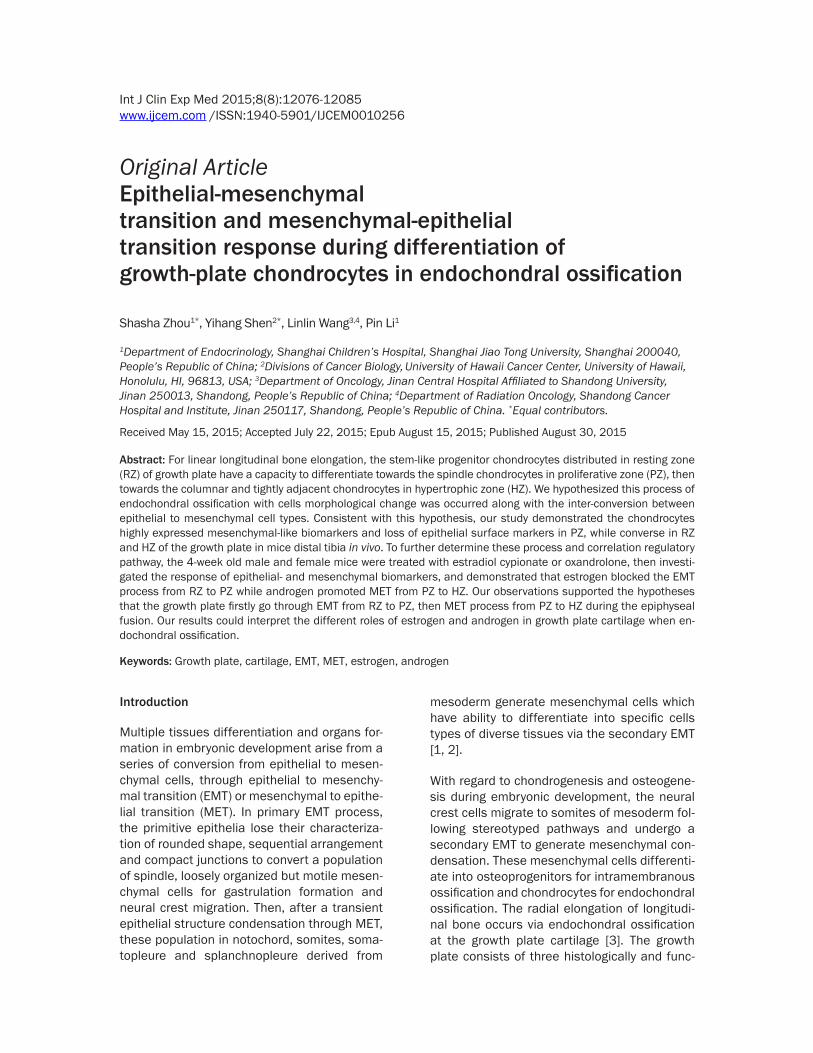
Figure 1. X-ray detection of mouse skeleton. The left growth plate at the distal tibia (C-F) with the 4-(A) and 16-(B) week old. The red arrows showing the gap between knee and tibia or femur means cartilage of growth plate.
Epithelial-mesenchymal transition in endochondral ossification
12078 Int J Clin Exp Med 2015;8(8):12076-12085
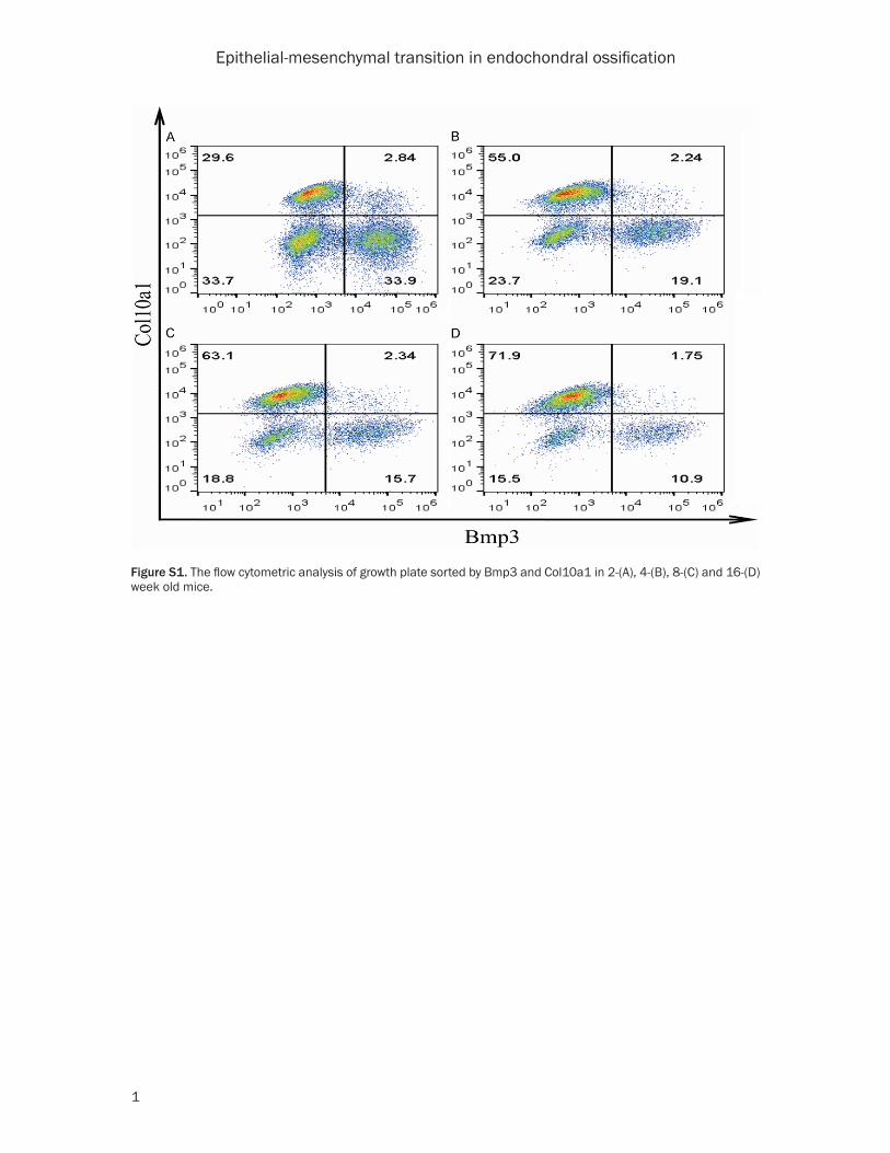
discomfort to the animals. 6 mice in one group randomly according to gender and age and weekly intraperitoneal injected 70 ug/kg estra-diol cypionate [14], 15 mg/kg oxandrolone [15] and 2.5 mg/kg SIS3 [16] respectively for 4 weeks. The mice were sedated to assess the status of skeleton using X-ray assay (Philips Digital Diagnost, 50 kV, 2 mA·s for the whole body and 45 kV, 1.5 mA·s for the feature on leg), then sacrificed by cervical dislocation. The growth plates between proximal femur and dis-tal tibia were separated and cut into pieces of 1 mm×1 mm in cold PBS with 1% antibiotic under integrated microscope, and digested by 0.25% trypsin for 10 min and 0.1% type II col-lagenase overnight in 37°C, then sieved 70-μm filter [17]. The cells pellet was fixed by 1% PFA for 30 min at room temperature, then 90% methanol for 30 min at 4°C, after washing by PBS twice, incubated with 10 μl Col10a1 and 15 μl Bmp3 (Santa Cruz, USA) antibodies [18] diluted by PBS with 0.5% BSA and 0.1% Triton X-100 for 1 h at room temperature, washed by PBS 3 times, spin down with 800 rpm, then
incubated with 1:2000 Alexa Fluor 488 nm anti-goat IgG and 1:2000 Alexa Fluor 594 nm anti-rabbit IgG (Invitrogen, USA) in dark place for 30 min in 37°C, washed by PBS 3 times and spin down with 800 rpm again. The cells pellet was resuspended by 300 μl PBS for fluorescent sorting using BD FACSAria III. The flow cytomet-ric analysis showed the chondrocytes were divided into 3 groups, in which cell population of Bmp3+ and Col10a1+ represent RZ and HZ respectively, and the double negative is PZ (Figure S1).
HE staining
The fresh proximal femur and distal tibia washed by PBS, was fixed within 4% formalin 1 h at room temperature, decalcified in 10% HCl 72 h, orderly dehydrated in the ethanol of 50%, 75%, 85%, 95%, 100% and 100%, displaced the ethanol out using xylene of 50%, 100% and 100%, incubated in paraffin of 50%, 100% and 100% at 50°C to remove the additional xylene, then embedded within the 60°C wax box 1 h,
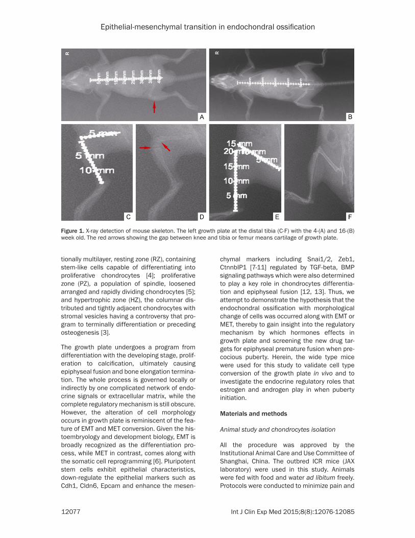
Figure 2. HE-staining of the growth plate with 2-(A), 4-(B), 8-(C), 16-(D) week old mice.
Epithelial-mesenchymal transition in endochondral ossification
12079 Int J Clin Exp Med 2015;8(8):12076-12085
cooled down in water 4 h, and sectioned into 7-10 μm paralleled the tibia. The slices were dewaxed followed the reversed steps of previ-ous paraffin preparation, then incubated in the hematine 15 min and 1% hydrochloric acid alcohol 10 s and washed 30 min, and in 0.5% eosin 15 min, then followed the reversed proto-col of dehydration, and enveloped for observation.
Real-time PCR
The total RNA of cells isolated was extracted using trizol. cDNA was produced from 100 ng of total RNA by Transcriptor First Strand cDNA Synthesis Kit (Roche, Switzerland) according to the manufacturer’s protocol. Quantitative real-time PCR was performed using the SYBR Premix ExTaq Kit (Roche, Switzerland). The PCR protocol consisted of 95°C for 30 s, 40 cycles of 5 s at 95°C, 30 s at 60°C. Mouse Gapdh transcript served as an internal reference gene. The values were analyzed using the compara-
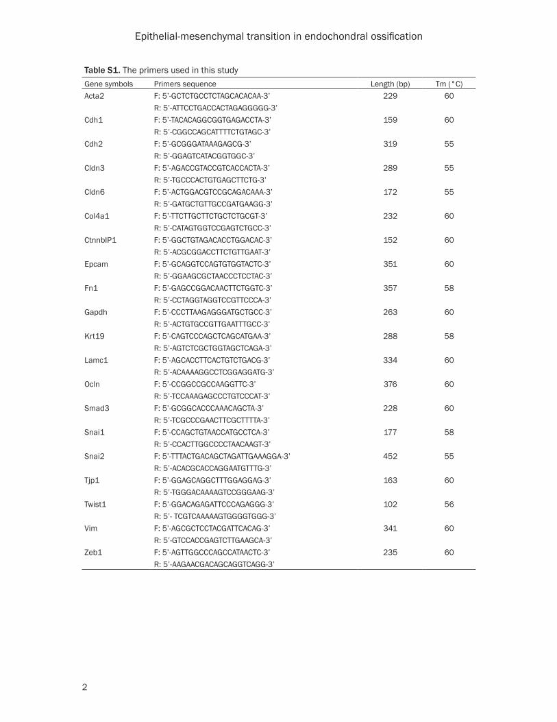
tive CT method. The primers used in this study were listed in Table S1.
Western blot
In brief, the proteome was harvested using RIPA buffer, denatured with 1× SDS buffer at 95°C for 10 min. 30 μg protein was separated by 8% polyacrylamide gel subjected to 90 V constant voltage SDS electrophoresis, and transferred onto NC membrane subjected to 100 V constant voltage, then blocked by 5% nonfat milk, and incubated with the following antibodies AR, Col4a1, ER-alpha, ER-beta, Gapdh, Krt19, Smad3, Tgf-beta1, Tgf-beta2, anti-mouse, anti-rabbit, anti-goat (Santa Cruz, USA) and finally determined by ECL enhancing solution of chemiluminescence agents.
Statistical analysis
The Student’s t-test was used to analyze values of measurement data. All analyses were pro-
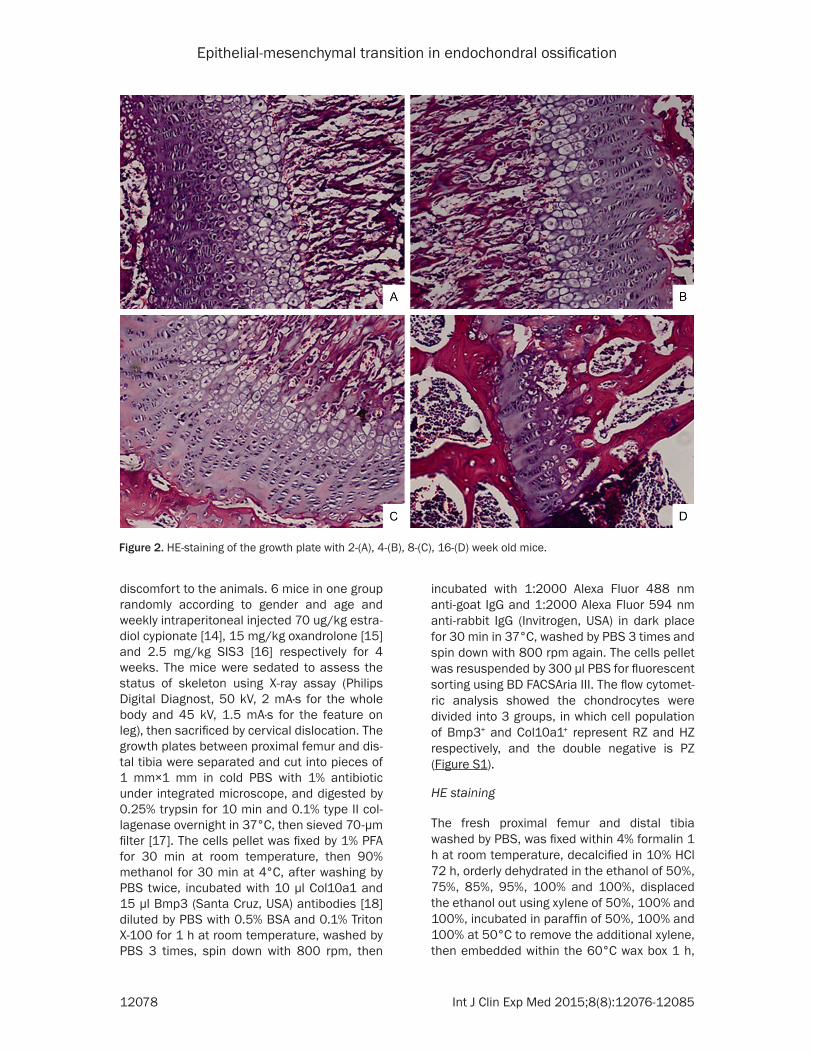
Figure 3. The mRNA expression of epithelial-(A) and mesenchymal-(B) like genes within each zone of the growth plate in 8-week old mouse.

Epithelial-mesenchymal transition in endochondral ossification
12080 Int J Clin Exp Med 2015;8(8):12076-12085
cessed by SPSS 20 software. Values of P<0.05 were regarded as statistical significance.
Results
To verify the histological and morphological alteration of endochondral ossification during longitudinal bone development, the growth plate in distal tibia harvested from 2, 4, 8 and 16-week old ICR mice were detected using X-ray and HE staining assay. A notable pres-ence of growth plate was observed in distal tibia of the 4-week but not in 16-week old mice (Figure 1). Moreover, the cells in RZ and PZ were randomly distributed and displayed vague boundary in 2- and 4-week old mice; and then emerged the distinct difference in 8-week old mice, which the linear arrangement was found in the cells in PZ; and finally were largely
exhausted in 16-week old mice. Whereas, the HZ gradually spread across the growth plate and tightly stacked from 2- to 8-week old and then disappear in 16-week old mice (Figure 2). Consistent with the X-ray assay, the total size of growth plate declined during the time course of bone elongation. Taken together, the observa-tion of histological patterns of growth plate in mice confirmed the differentiation process of cells within growth plate cartilage when epiphy-seal fusion and determined the significant time points for further study.
Since this cell morphological alteration was coincident with a conversion between epithelial and mesenchymal cell types, thus the expres-sion of epithelial- and mesenchymal-like bio-markers in different zones harvested from 8-week old mice were further investigated to
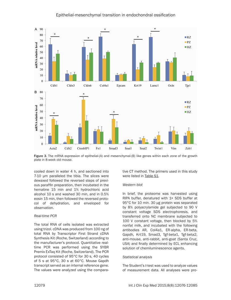
Figure 4. The length assay of spine (A, D), femur (B, E) and tibia (C, F) in male and female mice treated by estrogen and androgen. Con, EC, OX represent untreatment, estradiol cypionate and oxandrolone resectively.
Epithelial-mesenchymal transition in endochondral ossification
12081 Int J Clin Exp Med 2015;8(8):12076-12085
validate the hypothesis. The mRNA level of epi-thelial markers including Cdh1, Cldn6, Col4a1, Krt19, Lamc1 expressed in RZ and HZ were sig-nificantly higher than in PZ while the mesenchy-mal markers such as Acta2, Ctnnb1, Smad3, displayed the converse tendency (Figure 3). The results suggested that a process of EMT occurred in the programming of RZ towards PZ and MET in PZ towards HZ.
To further determine the EMT or MET process and the potential regulatory mechanism, 4-week old mice were weekly treated with 70 ug/kg estradiol cypionate and 15 mg/kg oxan-drolone respectively for 4 weeks. The length of spine, femur and tibia was investigated using X-ray assay to show that estrogen inhibited while androgen enhanced the skeleton radial growth both in male and female mice (Figure 4). And the level of epithelial- and mesenchy-mal-like biomarkers expressed in different
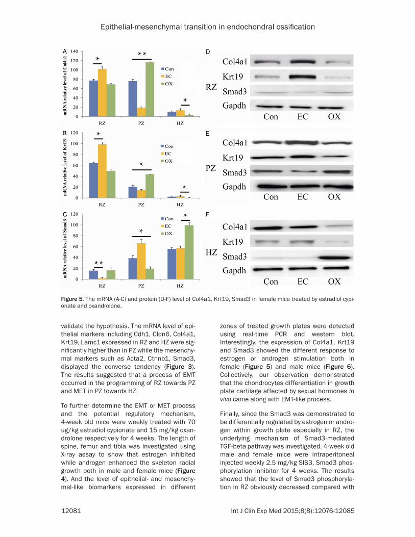
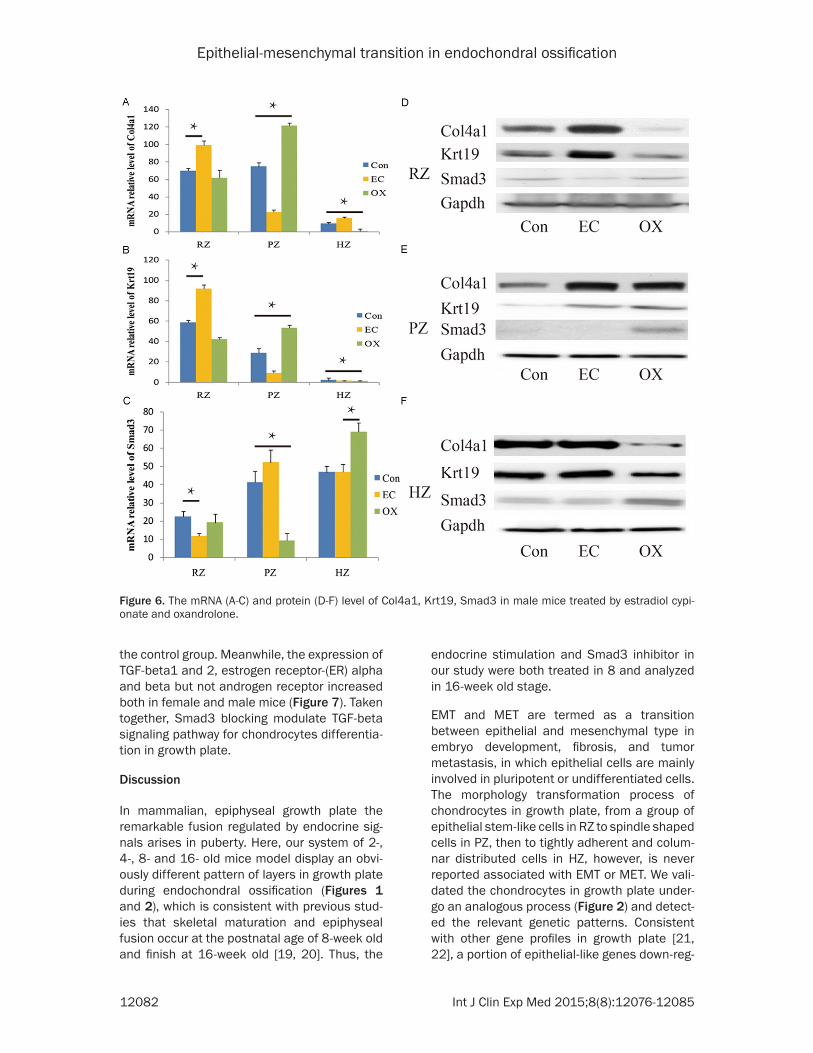
zones of treated growth plates were detected using real-time PCR and western blot. Interestingly, the expression of Col4a1, Krt19 and Smad3 showed the different response to estrogen or androgen stimulation both in female (Figure 5) and male mice (Figure 6). Collectively, our observation demonstrated that the chondrocytes differentiation in growth plate cartilage affected by sexual hormones in vivo came along with EMT-like process.
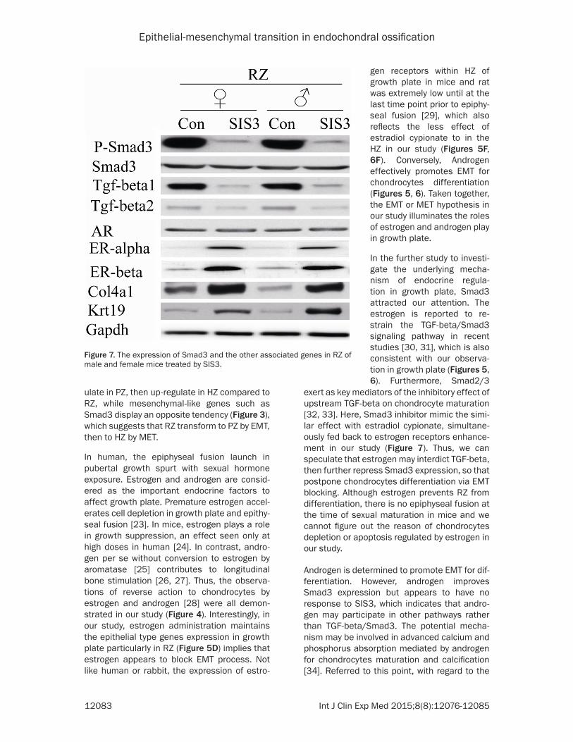
Finally, since the Smad3 was demonstrated to be differentially regulated by estrogen or andro-gen within growth plate especially in RZ, the underlying mechanism of Smad3-mediated TGF-beta pathway was investigated. 4-week old male and female mice were intraperitoneal injected weekly 2.5 mg/kg SIS3, Smad3 phos-phorylation inhibitor for 4 weeks. The results showed that the level of Smad3 phosphoryla-tion in RZ obviously decreased compared with
Figure 5. The mRNA (A-C) and protein (D-F) level of Col4a1, Krt19, Smad3 in female mice treated by estradiol cypi-onate and oxandrolone.
Epithelial-mesenchymal transition in endochondral ossification
12082 Int J Clin Exp Med 2015;8(8):12076-12085
the control group. Meanwhile, the expression of TGF-beta1 and 2, estrogen receptor-(ER) alpha and beta but not androgen receptor increased both in female and male mice (Figure 7). Taken together, Smad3 blocking modulate TGF-beta signaling pathway for chondrocytes differentia-tion in growth plate.
Discussion
In mammalian, epiphyseal growth plate the remarkable fusion regulated by endocrine sig-nals arises in puberty. Here, our system of 2-, 4-, 8- and 16- old mice model display an obvi-ously different pattern of layers in growth plate during endochondral ossification (Figures 1 and 2), which is consistent with previous stud-ies that skeletal maturation and epiphyseal fusion occur at the postnatal age of 8-week old and finish at 16-week old [19, 20]. Thus, the
endocrine stimulation and Smad3 inhibitor in our study were both treated in 8 and analyzed in 16-week old stage.
EMT and MET are termed as a transition between epithelial and mesenchymal type in embryo development, fibrosis, and tumor metastasis, in which epithelial cells are mainly involved in pluripotent or undifferentiated cells. The morphology transformation process of chondrocytes in growth plate, from a group of epithelial stem-like cells in RZ to spindle shaped cells in PZ, then to tightly adherent and colum-nar distributed cells in HZ, however, is never reported associated with EMT or MET. We vali-dated the chondrocytes in growth plate under-go an analogous process (Figure 2) and detect-ed the relevant genetic patterns. Consistent with other gene profiles in growth plate [21, 22], a portion of epithelial-like genes down-reg-
Figure 6. The mRNA (A-C) and protein (D-F) level of Col4a1, Krt19, Smad3 in male mice treated by estradiol cypi-onate and oxandrolone.
Epithelial-mesenchymal transition in endochondral ossification
12083 Int J Clin Exp Med 2015;8(8):12076-12085
ulate in PZ, then up-regulate in HZ compared to RZ, while mesenchymal-like genes such as Smad3 display an opposite tendency (Figure 3), which suggests that RZ transform to PZ by EMT, then to HZ by MET.
In human, the epiphyseal fusion launch in pubertal growth spurt with sexual hormone exposure. Estrogen and androgen are consid-ered as the important endocrine factors to affect growth plate. Premature estrogen accel-erates cell depletion in growth plate and epithy-seal fusion [23]. In mice, estrogen plays a role in growth suppression, an effect seen only at high doses in human [24]. In contrast, andro-gen per se without conversion to estrogen by aromatase [25] contributes to longitudinal bone stimulation [26, 27]. Thus, the observa-tions of reverse action to chondrocytes by estrogen and androgen [28] were all demon-strated in our study (Figure 4). Interestingly, in our study, estrogen administration maintains the epithelial type genes expression in growth plate particularly in RZ (Figure 5D) implies that estrogen appears to block EMT process. Not like human or rabbit, the expression of estro-
exert as key mediators of the inhibitory effect of upstream TGF-beta on chondrocyte maturation [32, 33]. Here, Smad3 inhibitor mimic the simi-lar effect with estradiol cypionate, simultane-ously fed back to estrogen receptors enhance-ment in our study (Figure 7). Thus, we can speculate that estrogen may interdict TGF-beta, then further repress Smad3 expression, so that postpone chondrocytes differentiation via EMT blocking. Although estrogen prevents RZ from differentiation, there is no epiphyseal fusion at the time of sexual maturation in mice and we cannot figure out the reason of chondrocytes depletion or apoptosis regulated by estrogen in our study.
Androgen is determined to promote EMT for dif-ferentiation. However, androgen improves Smad3 expression but appears to have no response to SIS3, which indicates that andro-gen may participate in other pathways rather than TGF-beta/Smad3. The potential mecha-nism may be involved in advanced calcium and phosphorus absorption mediated by androgen for chondrocytes maturation and calcification [34]. Referred to this point, with regard to the
Figure 7. The expression of Smad3 and the other associated genes in RZ of male and female mice treated by SIS3.
gen receptors within HZ of growth plate in mice and rat was extremely low until at the last time point prior to epiphy-seal fusion [29], which also reflects the less effect of estradiol cypionate to in the HZ in our study (Figures 5F, 6F). Conversely, Androgen effectively promotes EMT for chondrocytes differentiation (Figures 5, 6). Taken together, the EMT or MET hypothesis in our study illuminates the roles of estrogen and androgen play in growth plate.
In the further study to investi-gate the underlying mecha-nism of endocrine regula- tion in growth plate, Smad3 attracted our attention. The estrogen is reported to re- strain the TGF-beta/Smad3 signaling pathway in recent studies [30, 31], which is also consistent with our observa-tion in growth plate (Figures 5, 6). Furthermore, Smad2/3
Epithelial-mesenchymal transition in endochondral ossification
12084 Int J Clin Exp Med 2015;8(8):12076-12085
controversial fate of hypertrophic chondro-cytes, the type 2 EMT, a physiological response for organ fibrosis [2] may be an analogous pro-cess occurring when HZ finally turn to the calci-fied cartilage zone.
Collectively, our results demonstrated EMT and MET conduct the chondrocytes differentiation and cartilage epiphyseal fusion in growth plate, and revealed the different roles that estrogen and androgen played in growth plate cartilage via the transition between epithelial and mes-enchymal types.
Acknowledgements
This work was supported by National Natural Science Foundation of China (81370686), Shanghai Science and Technology Committee (12411950403, 12411950400) and Shanghai Municipal Commission of Health and Family planning (20134Y086).
Disclosure of conflict of interest
None.
Address correspondence to: Pin Li, Department of Endocrinology, Shanghai Children’s Hospital, Shang- hai Jiao Tong University, Shanghai 200040, People’s Republic of China. E-mail: [email protected]
References
[1] Thiery JP, Acloque H, Huang RY, Nieto MA. Epithelial-mesenchymal transitions in develop-ment and disease. Cell 2009; 139: 871-890.
[2] Zeisberg M, Neilson EG. Biomarkers for epithe-lial-mesenchymal transitions. J Clin Invest 2009; 119: 1429-1437.
[3] Tsang KY, Chan D, Cheah KS. Fate of growth plate hypertrophic chondrocytes: Death or lin-eage extension? Dev Growth Differ 2015; 57: 179-192.
[4] Abad V, Meyers JL, Weise M, Gafni RI, Barnes KM, Nilsson O, Bacher JD, Baron J. The role of the resting zone in growth plate chondrogene-sis. Endocrinology 2002; 143: 1851-1857.
[5] Spath SS, Andrade AC, Chau M, Nilsson O. Local regulation of growth plate cartilage. Endocr Dev 2011; 21: 12-22.
[6] Chen J, Han Q, Pei D. EMT and MET as para-digms for cell fate switching. J Mol Cell Biol 2012; 4: 66-69.
[7] Li R, Liang J, Ni S, Zhou T, Qing X, Li H, He W, Chen J, Li F, Zhuang Q, Qin B, Xu J, Li W, Yang J, Gan Y, Qin D, Feng S, Song H, Yang D, Zhang B, Zeng L, Lai L, Esteban MA, Pei D. A mesenchy-mal-to-epithelial transition initiates and is re-
quired for the nuclear reprogramming of mouse fibroblasts. Cell Stem Cell 2010; 7: 51-63.
[8] Samavarchi-Tehrani, P, Golipour A, David L, Sung HK, Beyer TA, Datti A, Woltjen K, Nagy A, Wrana JL. Functional genomics reveals a BMP-driven mesenchymal-to-epithelial transition in the initiation of somatic cell reprogramming. Cell Stem Cell 2010; 7: 64-77.
[9] Redmer T, Diecke S, Grigoryan T, Quiroga-Negreira A, Birchmeier W, Besser D. E-cadherin is crucial for embryonic stem cell pluripotency and can replace OCT4 during somatic cell re-programming. EMBO Rep 2011; 12: 720-726.
[10] Wang L, Xue Y, Shen Y, Li W, Cheng Y, Yan X, Shi W, Wang J, Gong Z, Yang G, Guo C, Zhou Y, Wang X, Zhou Q, Zeng F. Claudin 6: a novel sur-face marker for characterizing mouse pluripo-tent stem cells. Cell Res 2012; 22: 1082-1085.
[11] Gonzalez B, Denzel S, Mack B, Conrad M, Gires O. EpCAM is involved in maintenance of the murine embryonic stem cell phenotype. Stem Cells 2009; 27: 1782-1791.
[12] Wang W, Rigueur D, Lyons KM. TGFbeta signal-ing in cartilage development and mainte-nance. Birth Defects Res C Embryo Today 2014; 102: 37-51.
[13] Fischerauer EE, Manninger M, Seles M, Janezic G, Pichler K, Ebner B, Weinberg AM. BMP-6 and BMPR-1a are up-regulated in the growth plate of the fractured tibia. J Orthop Res 2013; 31: 357-363.
[14] Nilsson O, Weise M, Landman EB, Meyers JL, Barnes KM, Baron J. Evidence that estrogen hastens epiphyseal fusion and cessation of longitudinal bone growth by irreversibly deplet-ing the number of resting zone progenitor cells in female rabbits. Endocrinology 2014; 155: 2892-2899.
[15] Perry RJ, Gault EJ, Paterson WF, Dunger DB, Donaldson MD. Effect of oxandrolone and tim-ing of oral ethinylestradiol initiation on puber-tal progression, height velocity and bone matu-ration in the UK Turner study. Horm Res Paediatr 2014; 81: 298-308.
[16] Li J, Qu X, Yao J, Caruana G, Ricardo SD, Yamamoto Y, Yamamoto H, Bertram JF. Blockade of endothelial-mesenchymal transi-tion by a Smad3 inhibitor delays the early de-velopment of streptozotocin-induced diabetic nephropathy. Diabetes 2010; 59: 2612-2624.
[17] Xu T, Yang K, You H, Chen A, Wang J, Xu K, Gong C, Shao J, Ma Z, Guo F, Qi J. Regulation of PTHrP expression by cyclic mechanical strain in postnatal growth plate chondrocytes. Bone 2013; 56: 304-311.
[18] Chau M, Lui JC, Landman EB, Späth SS, Vortkamp A, Baron J, Nilsson O. Gene expres-sion profiling reveals similarities between the
Epithelial-mesenchymal transition in endochondral ossification
12085 Int J Clin Exp Med 2015;8(8):12076-12085
spatial architectures of postnatal articular and growth plate cartilage. PLoS One 2014; 9: e103061.
[19] Briggs MD, Bell PA, Pirog KA. The utility of mouse models to provide information regard-ing the pathomolecular mechanisms in human genetic skeletal diseases: The emerging role of endoplasmic reticulum stress (Review). Int J Mol Med 2015; 35: 1483-92.
[20] Ascenzi M, Du X, Harding JI, Beylerian EN, de Silva BM, Gross BJ, Kastein HK, Wang W, Lyons KM, Schaeffer H. Automated Cell Detection and Morphometry on Growth Plate Images of Mouse Bone. Appl Math (Irvine) 2014; 5: 2866-2880.
[21] Hutchison MR, Bassett MH, White PC. SCF, BDNF, and Gas6 are regulators of growth plate chondrocyte proliferation and differentiation. Mol Endocrinol 2010; 24: 193-203.
[22] Belluoccio D, Bernardo BC, Rowley L, Bateman JF. A microarray approach for comparative ex-pression profiling of the discrete maturation zones of mouse growth plate cartilage. Biochim Biophys Acta 2008; 1779: 330-340.
[23] Yao X, Chen H, Ohtake N, Shoumura S. Morphological alterations in the growth plate cartilage of ovariectomized mice. Med Mol Morphol 2006; 39: 193-197.
[24] Nilsson O, Marino R, De Luca F, Phillip M, Baron J. Endocrine regulation of the growth plate. Horm Res 2005; 64: 157-165.
[25] Oz OK, Millsaps R, Welch R, Birch J, Zerwekh JE. Expression of aromatase in the human growth plate. J Mol Endocrinol 2001; 27: 249-253.
[26] Nilsson O, Chrysis D, Pajulo O, Boman A, Holst M, Rubinstein J, Martin Ritzén E, Sävendahl L. Localization of estrogen receptors-alpha and -beta and androgen receptor in the human growth plate at different pubertal stages. J Endocrinol 2003; 177: 319-326.
[27] Irie T, Aizawa T, Kokubun S. The role of sex hor-mones in the kinetics of chondrocytes in the growth plate. A study in the rabbit. J Bone Joint Surg Br 2005; 87: 1278-1284.
[28] Lindberg MK, Vandenput L, Movèrare Skrtic S, Vanderschueren D, Boonen S, Bouillon R, Ohlsson C. Androgens and the skeleton. Minerva Endocrinol 2005; 30: 15-25.
[29] Nilsson O Abad V, Chrysis D, Ritzén EM, Sävendahl L, Baron J. Estrogen receptor-alpha and -beta are expressed throughout postnatal development in the rat and rabbit growth plate. J Endocrinol 2002; 173: 407-414.
[30] Goto N, Hiyoshi H, Ito I, Iida K, Nakajima Y, Nagasawa K, Yanagisawa J. Identification of a Novel Compound That Suppresses Breast Cancer Invasiveness by Inhibiting Transforming Growth Factor-beta Signaling via Estrogen Receptor alpha. J Cancer 2014; 5: 336-343.
[31] Li YC, Ding XS, Li HM, Zhang Y, Bao J. Role of G protein-coupled estrogen receptor 1 in modu-lating transforming growth factor-beta stimu-lated mesangial cell extracellular matrix syn-thesis and migration. Mol Cell Endocrinol 2014; 391: 50-59.
[32] Ferguson CM, Schwarz EM, Reynolds PR, Puzas JE, Rosier RN, O’Keefe RJ. Smad2 and 3 mediate transforming growth factor-beta1-in-duced inhibition of chondrocyte maturation. Endocrinology 2000; 141: 4728-4735.
[33] Pedrozo HA, Schwartz Z, Gomez R, Ornoy A, Xin-Sheng W, Dallas SL, Bonewald LF, Dean DD, Boyan BD. Growth plate chondrocytes store latent transforming growth factor (TGF)-beta 1 in their matrix through latent TGF-beta 1 binding protein-1. J Cell Physiol 1998; 177: 343-354.
[34] Schwartz Z, Nasatzky E, Ornoy A, Brooks BP, Soskolne WA, Boyan BD. Gender-specific, mat-uration-dependent effects of testosterone on chondrocytes in culture. Endocrinology 1994; 134: 1640-1647.
Epithelial-mesenchymal transition in endochondral ossification
1
Figure S1. The flow cytometric analysis of growth plate sorted by Bmp3 and Col10a1 in 2-(A), 4-(B), 8-(C) and 16-(D) week old mice.
Epithelial-mesenchymal transition in endochondral ossification
2
Table S1. The primers used in this studyGene symbols Primers sequence Length (bp) Tm (°C)Acta2 F: 5’-GCTCTGCCTCTAGCACACAA-3’ 229 60
R: 5’-ATTCCTGACCACTAGAGGGGG-3’Cdh1 F: 5’-TACACAGGCGGTGAGACCTA-3’ 159 60
R: 5’-CGGCCAGCATTTTCTGTAGC-3’Cdh2 F: 5’-GCGGGATAAAGAGCG-3’ 319 55
R: 5’-GGAGTCATACGGTGGC-3’Cldn3 F: 5’-AGACCGTACCGTCACCACTA-3’ 289 55
R: 5’-TGCCCACTGTGAGCTTCTG-3’Cldn6 F: 5’-ACTGGACGTCCGCAGACAAA-3’ 172 55
R: 5’-GATGCTGTTGCCGATGAAGG-3’Col4a1 F: 5’-TTCTTGCTTCTGCTCTGCGT-3’ 232 60
R: 5’-CATAGTGGTCCGAGTCTGCC-3’CtnnbIP1 F: 5’-GGCTGTAGACACCTGGACAC-3’ 152 60
R: 5’-ACGCGGACCTTCTGTTGAAT-3’Epcam F: 5’-GCAGGTCCAGTGTGGTACTC-3’ 351 60
R: 5’-GGAAGCGCTAACCCTCCTAC-3’Fn1 F: 5’-GAGCCGGACAACTTCTGGTC-3’ 357 58
R: 5’-CCTAGGTAGGTCCGTTCCCA-3’Gapdh F: 5’-CCCTTAAGAGGGATGCTGCC-3’ 263 60
R: 5’-ACTGTGCCGTTGAATTTGCC-3’Krt19 F: 5’-CAGTCCCAGCTCAGCATGAA-3’ 288 58
R: 5’-AGTCTCGCTGGTAGCTCAGA-3’Lamc1 F: 5’-AGCACCTTCACTGTCTGACG-3’ 334 60
R: 5’-ACAAAAGGCCTCGGAGGATG-3’Ocln F: 5’-CCGGCCGCCAAGGTTC-3’ 376 60
R: 5’-TCCAAAGAGCCCTGTCCCAT-3’Smad3 F: 5’-GCGGCACCCAAACAGCTA-3’ 228 60
R: 5’-TCGCCCGAACTTCGCTTTTA-3’Snai1 F: 5’-CCAGCTGTAACCATGCCTCA-3’ 177 58
R: 5’-CCACTTGGCCCCTAACAAGT-3’Snai2 F: 5’-TTTACTGACAGCTAGATTGAAAGGA-3’ 452 55
R: 5’-ACACGCACCAGGAATGTTTG-3’Tjp1 F: 5’-GGAGCAGGCTTTGGAGGAG-3’ 163 60
R: 5’-TGGGACAAAAGTCCGGGAAG-3’Twist1 F: 5’-GGACAGAGATTCCCAGAGGG-3’ 102 56
R: 5’- TCGTCAAAAAGTGGGGTGGG-3’Vim F: 5’-AGCGCTCCTACGATTCACAG-3’ 341 60
R: 5’-GTCCACCGAGTCTTGAAGCA-3’Zeb1 F: 5’-AGTTGGCCCAGCCATAACTC-3’ 235 60
R: 5’-AAGAACGACAGCAGGTCAGG-3’