bones of the foot (1) 26 bones phalanges = 14 numbered 1-5 (big toe = hallux = #1) distal...
TRANSCRIPT

Bones of the Foot (1)
26 bones Phalanges = 14
Numbered 1-5 (big toe = Hallux = #1) distal Interphalangeal joint (DIP) proximal interphalangeal joint (PIP) Metatarsal phalangeal joint (MP)
Metatarsals (numbered 1-5)

Bone of the foot (2)
Tarsals- Fig 12-2 Calcaneous (heel bone) Talus (main weight bearing) Navicular- medial 3 cuneiforms Cuboid-lateral

The arches Fig 12-10
Function to support and distribute body weight
Three arches: Fig 12-10 Medial longitudinal (higher)
supported by calcaneal-navicular ligament (spring)
Lateral longitudinal Transverse
Plantar Fascia Supports longitudinal arch

The Ankle Fig 12-1/12-3
Ankle joint = talocrural joint Three bones
Tibia (medial malleolus)-major weight bearing Fibula (lateral malleolus) Talus
Hinge joint Dorsi-Flexion Plantar Flexion

Subtalar Joint
The joint between the talus and the calcaneous
Shifts during weight bearing (WB) Supination/inversion Pronation/eversion

Tibiofibular Joint -Fig 12-3
Composed of Tibia and Fibula Ligaments/Membrane
Anterior Tibofibular Lig Posterior Tibofibular Lig Interosseious membrane- connects the
tibia and fibula; runs the entire diaphysis of both bones

Ankle ligaments (1) – Fig 12-3
Medial Deltoid ligament -4 parts Triangular shape (very strong)
Lateral Anterior talofibular (ATF) Calcaneofibular (CF) Posterior talofibular (PTF)
Ankle and foot are composed of numerous ligaments; where ever two bones meet

Muscles of the Lower Leg Thick sheaths of fascia divide muscles into 4
compartments Anterior Compartment
Dorsiflexion (DF), Toe Extension (EXT), Inversion (INV)
Lateral Compartment Eversion (EV)
Deep Posterior Compartment Toe Flexion (flex), Inversion (INV)
Superficial Posterior (Plantar flexion (PF))

Nerves and Blood Supply
Nerves Sciatic nerve branches into the peroneal
(ant/lat) and tibial nerves (post) Blood Supply
Femoral Artery →Popliteal artery → Anterior and Posterior Tibial artery
Anterior Tibial becomes the dorsalis pedis artery →dorsal pedal on the dorsum of foot
Posterior Tibial is located behind medial malleolus.
ROM
DF-tibialis anterior, extensor digitorum PF- gastroc and soleus INV- tibialis anterior and posteror EV- peroneals Toe Ext.-extensor digitorum and hallucis Toe Flexion flexor digitorum and hallucis

Review
http://www.csuchico.edu/~sbarker/shock/Anklequiz.html
http://www.rad.washington.edu/atlas2/ http://www.medicalmultimediagroup.com/pate
d/foot/achilles/achilles.html

Prevention of Injury
Stretch achilles tendon tight achilles increases risk of plantar fasciitis,
achilles tendonitis, and ankle sprains Strengthen anterior leg muscles
important for shin splints Strengthen lateral/medial leg muscles Strengthen intrinsic foot muscles Good shoes; change shoes, correct type of
shoes for playing surface

Injury information
Precursors = something that may predispose an athlete to that injury
All injuries should be treated for symptoms thus RICE. This will not be listed with each injury but should be remembered
HOPS includes information typically seen or heard during the HOPS assessment. Most injuries include swelling, discoloration etc in area, this is not included in slides

Lateral Ankle Sprain
MOI: PF and/or Inv More common than medial ankle sprains due
to (make up about 90% of ankle sprains): differing length of malleoli (lateral is longer) Stronger deltoid ligament
Precursors: tight achilles, improper shoes, previous ankle injury
HOPS and Tx See field strategy 12.2

Medial Ankle Sprain
Less common then lateral ankle sprains MOI: eversion Sometimes accompanied by a fracture HOPS
point tenderness over deltoid and anterior/ posterior joint line
Swelling not as obvious Takes almost twice as long to recover in some
cases

Achilles tendonitis
Precursors: achilles tendon tightness, change in shoes, running surfaces, workout changes
HOPS chronic injury pn during and after activity Thickening of the tendon Crepitation Pn with Resistive PF, Passive DF
Tx: stretch achilles, heel lift, tape, ultrasound
Achilles Tendon rupture
Precursors: athletes between 30 and 40, power sports (BB); recreational athletes
HOPT MOI: push off with knee extending sharp pain, feels snap or pop “kicked in the back of the leg” visible defect/palpable defect positive Thompson test Excessive passive DF
Tx: refer to physician

Medial Tibial Stress Syndrome (1)
“shin splints” Precursors: achilles tendon tightness, change
in shoes, running surfaces, workout changes, arch problems
HOPS sometimes bilateral; pn along distal 1/3 of
medial tibial border initially: pn at start of activity that decreases
with activity, then recurring after activity Later: pain before during and after activity

Medial Tibial Stress Syndrome (2)
HOPS (cont) Pn increased with AROM PF, INV Usually responds well to treatment
Tx Cryotherapy stretching of achilles strengthen deep posterior muscle strengthen anterior muscles

Plantar Fasciitis
Precursors: obesity, achilles tendon tightness, overuse, shoes
HOPT chronic injury pn first thing in the morning point tenderness over the medial calcaneal
tubercle Pn with toe extension and ankle DF TX- Hot and Cold Modalities, stretching, rest,
orthodics, change in shoes, heel lift, tape, roll foot over soda can

Compartment Syndrome (1)“Volkman's Ischemic Contracture”
Two types: Exertional (MOI:previous injury in leg, chronic
onset); Read; **Exertional CS can lead to Acute CS
Acute (MOI: blow to front of the leg) Acute-HOPT
Increasing pain in the front of the leg firm tight skin in front of shin loss of sensation between 1st and 2nd toes

Compartment Syn (2)
diminished pulse at dorsalis pedis artery Inability to DF ankle, or extend toes
(progressive) The 5 Ps (ie, pain, pallor, paresthesias,
paralysis, pulselessness) Tx
Ice and Immobilize Get to physician (MEDICAL EMERGENCY) Abnormalities can occur within 30 minutes;
irreversible damage can occur within 12-24 hrs

“Turf” Toe
Precursors: hard surfaces, lightweight, flexible shoes, artificial turf
HOPT MOI-jamming of hallux, hyperextension of toe sport position requiring hyperextension Pn, point tnederness over 1st MP joint Push off phase of running is painful Pn with passive extension of the great toe
Treatment (TX) taping, metatarsal pad, stiff soled shoes,
manage symptoms

Ingrown toenail
Precursors improper cutting of toenails, too small shoes,
contant sliding of foot in shoes HOPT
nail grown into the surrounding skin signs of infection around the nail bed
Tx See field strategy 12.5
Motron’s Neuroma
Precursors: tight fitting shoes, HOPT
pn on the plantar side of the foot, usually between the 3rd and 4th metatarsal
Pn and numbness radiates to the 3rd & 4th toes Pain relieved by Non weight bearing (NWB) Pn caused by squeezing the foot
Treatment (TX) taping, metatarsal pad, wider shoes, cortisone
shots, surgery

Stress Fractures Precursors: female athletes with menstrual
irregularities (amenorrhea) increase in training regimen, old shoes
common sides: tibia, fibula, neck of 2nd metatarsal
HOPS pn on WB, relieved by NWB localized pn (often unilateral)
Tx: complete rest 4-12 weeks, referral for bone scan

Jones Fracture
Avulsion fracture of the peroneus brevis tendon where it attaches to the base of the 5th metatarsal
Common with severe inversion ankle injuries HOPS:
pn over base of 5th metatarsal MOI: severe, forceful inversion

Bunions- Hallux Valgus
Medial aspect of 1st MP joint HOPT
C/S- Shoes, congenital, lig. laxity, prolonged pronation of foot
Angular deformity of the great toe Pain around the first MP joint (inflammation)
Treatment (TX) taping, wider shoes, surgery last option
Tests
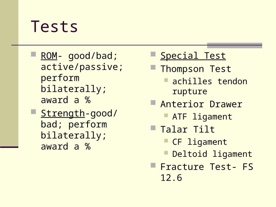
ROM- good/bad; active/passive; perform bilaterally; award a %
Strength-good/bad; perform bilaterally; award a %
Special Test Thompson Test
achilles tendon rupture Anterior Drawer
ATF ligament Talar Tilt
CF ligament Deltoid ligament
Fracture Test- FS 12.6

Test
Functional Test (p.235-236)- heel raises, walking, balancing, squatting, running, jumping, (progression is the key)

Specialized Rehab
Towel crunches Theraband Exercises all ROM Picking up objects (marbles) BAPS (wobble board) Stability Trainers http://www.promedproducts.com/Merchant2/
merchant.mv?Screen=CTGY&Store_Code=PP&Category_Code=BB
Achilles Stretch straight = gastroc bent = soleus