bones and joints of the neck. learning outcomes after working through these notes and associated...
TRANSCRIPT
Bones and Joints of the Neck

Learning OutcomesAfter working through these notes and associated reading, you should be able to…
• Identify the main features of both typical and atypical cervical vertebrae
• Describe the structure and function (movements) of the joints of the cervical spine
• Describe the ligaments and how they contribute to the stability of the neck

CERVICAL SPINE: OSTEOLOGY

Cervical Spine: Left Lateral ViewRef: Gilroy, MacPherson & Ross (2008) Atlas of Anatomy

Atlas, Axis and Typical Cervical VertebraeRef: Gilroy, MacPherson & Ross (2008) Atlas of Anatomy
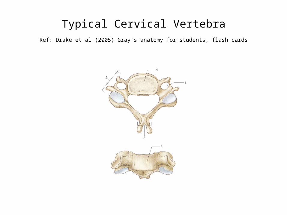
Typical Cervical VertebraRef: Drake et al (2005) Gray’s anatomy for students, flash cards
Typical Cervical Vertebra: The Body
– Small and broad
– Anterior surface is convex and slightly overhangs the disc below
– Superior surface is concave in the transverse plane with a prominent lip at each side, called the uncinate processes. These articulate with the lateral edges of the vertebra above at the Joints of Luschka. These small synovial joints develop at about 9 – 10 years of age.

Typical Cervical Vertebra: Pedicles, Laminae & Vertebral Foramen
• The PEDICLES are short and project posterolaterally
• The LAMINAE then curve posteromedially
• The VERTEBRAL FORAMEN is roughly triangular.
• On the inner surface of the laminae, ridges can be seen which are caused by ligamentous pull. Osteophytes may develop at this point.
Q: What ligament attaches here?
Typical Cervical Vertebra: SPs & TPs
• The SPINOUS PROCESS is short and bifid. It ends at two tubercles, often unequal in size
Q: What ligament attaches at these tubercles?
• The TRANSVERSE PROCESSES begin at the junction of pedicles and laminae, as dorsal and ventral roots or bars. These are located on either side of the transverse foramen
Q: What passes through the transverse foramen?
• Lateral to the foramen, the dorsal and ventral bars are joined together by the intertubercular lamella. (This is sometimes called the costotransverse bar, but this is not really an appropriate term). The two bars then continue laterally, ending as the posterior and anterior tubercles. These are important sites of muscle attachment.

Typical Cervical Vertebra: Articular Processes and Articular Pillar
The SUPERIOR and INFERIOR ARTICULAR PROCESSES are located at the junction of the laminae and pedicles. The inferior processes are directly below the superior, so, in the articulated spine, they form a continuous column of bone: the articular pillar. This important landmark is palpable posterior to the transverse processes.

Atlas and AxisRef: Marieb & Hoehn , Human Anatomy & Physiology (7th edn)

C1: The Atlas
• Easily recognised as it forms a ring of bone, with no vertebral body and no true spinous process
• It consists of two lateral masses, connected by a short anterior arch and a longer posterior arch.

C1: Anterior & Posterior Arches
• The ANTERIOR ARCH is slightly convex anteriorly, with a small anterior tubercle in the midline
• The POSTERIOR ARCH forms about two fifths of the ring. In the midline, a small posterior tubercle can be seen. This is a rudimentary spinous process
• On either side, just posterior to the lateral masses, there is a wide groove. The lateral mass overhangs this groove, sometimes actually forming a tunnel
Q: What runs in this groove?

C1: Lateral Masses
• Located between the anterior and posterior arches, the LATERAL MASSES are the strongest part of the vertebra
• On the superior aspect of each lateral mass, there is a large, concave, kidney-shaped facet
Q: What do these facet articulate with?
• On the inferior aspect, there is a smaller, almost circular facet which is flat or slightly concave
Q: What do these facet articulate with?
• On the medial aspect of each lateral mass, a small tubercle can be seen. The transverse ligament attaches here.

C1: TPs
• The TRANSVERSE PROCESSES (TPs) are long, making C1 the widest of the cervical vertebrae except C7
• The tips of the TPs of C1 are palpable just anterior to the mastoid process
• The TPs are similar to those of the typical cervical vertebrae in that they have two roots surrounding the transverse foramen, but the apex does not divide into two tubercles
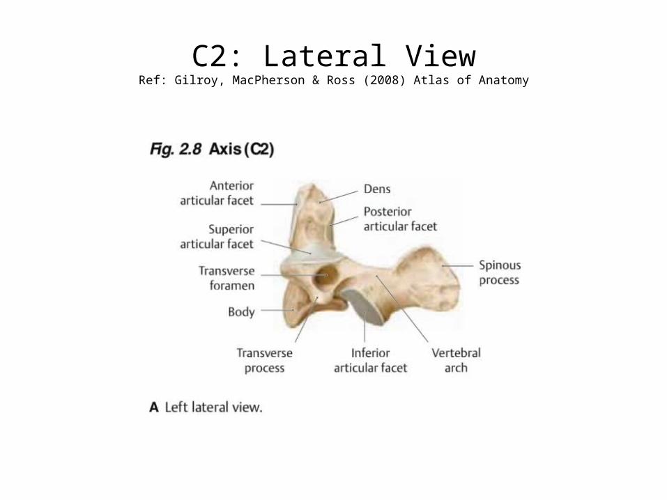
C2: Lateral ViewRef: Gilroy, MacPherson & Ross (2008) Atlas of Anatomy

C2: The Axis: The Dens
• The most prominent feature is the DENS or ODONTOID PROCESS. This is a strong, cone-shaped projection from the superior aspect of the body. On its anterior aspect, a small facet can be seen.
Q: What articulates with this facet?
• On its posterior aspect, the dens is grooved by the transverse ligament of the atlas. Above this groove, the dens is flattened on each side – this is where the alar ligaments attach. The apical ligament attaches to the pointed apex.

C2: Body & Articular Surfaces
• The BODY of C2 is obscured superiorly by the dens and the superior articular facets
• The lower border of the body projects inferiorly to overhang the disc, as with the typical cervical vertebrae
• The SUPERIOR ARTICULAR FACETS are large, flat and oval. They extend from the body partly onto the transverse processes
• The INFERIOR ARTICULAR FACETS are located posterior to the superior ones, so they do not contribute to the articular pillar

C2: TPs, Transverse Foraminae & SPs
• The TRANSVERSE PROCESSES are small, with blunt tips which are comparable to the posterior tubercle on the TPs of typical cervical vertebrae.
• The TRANSVERSE FORAMINAE deviate superolaterally. This directs the vertebral artery laterally in order to reach the more widely spaced foramina of C1
• The SPINOUS PROCESS of C2 is short and bifid, as with typical cervical vertebrae.

C7: Vertebra Prominens
• The SPINOUS PROCESS is long and prominent (hence its name). It is thick and it projects almost horizontally, ending in a slight tubercle, to which the ligamentum nuchae attaches. It is palpable at the lower end of the nuchal furrow, but is not necessarily the most prominent SP
• The TRANSVERSE PROCESSES are large, especially the posterior root. There is just one tubercle at the tip. The smaller anterior root is sometimes separated, forming a cervical rib. The TRANSVERSE FORAMEN is small and contains only an accessory vertebral vein
JOINTS OF THE NECK
Joints of the Upper Cervical Complex
• C0/C1: Atlanto-occipital joints– Synovial, condylar joints
• C1/2: Atlanto-axial joints– Median A-A joint (synovial, pivot joint)– Lateral A-A joints (synovial, plane joints)

Atlanto-Axial (A/A) JointRef: Gilroy, MacPherson & Ross (2008) Atlas of Anatomy

Upper Cervical Complex: The Ligaments
• Apical• Alar• Transverse• Cruciform– Superior longitudinal band– Inferior longitudinal band
• Tectorial membrane continuous with PLL
• Posterior atlanto-occipital membrane continuous with lig. flavum

Upper Cervical Complex: The LigamentsRef: Gilroy, MacPherson & Ross (2008) Atlas of Anatomy
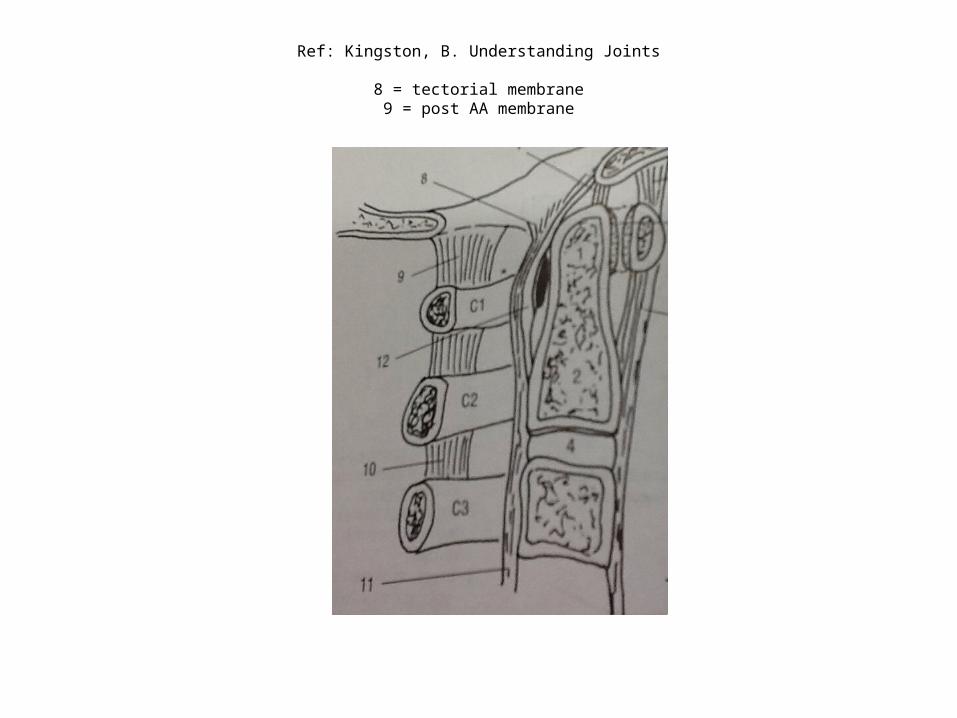
Ref: Kingston, B. Understanding Joints
8 = tectorial membrane9 = post AA membrane

C3-7: The Joints
• IVDs– Symphysis
• Apophyseal (facet) joints– Synovial plane
• Uncovertebral joints (joints of Luschka)– Synovial, plane

Uncovertebral JointsRef: Gilroy, MacPherson & Ross (2008) Atlas of Anatomy

C3-7: The Ligaments
• Ligamentum nuchae
• Ligamentum flavum
• ALL
• PLL