bmp and tgf-β pathway mediators are critical upstream regulators of wnt signaling during midbrain...
TRANSCRIPT
Developmental Biology 376 (2013) 62–73
Contents lists available at SciVerse ScienceDirect
Developmental Biology
0012-16
http://d
n Corr
E-m
journal homepage: www.elsevier.com/locate/developmentalbiology
BMP and TGF-b pathway mediators are critical upstream regulators of Wntsignaling during midbrain dopamine differentiation in human pluripotentstem cells
Jingli Cai, Stephanie Schleidt, Joshua Pelta-Heller, Danielle Hutchings,Gregory Cannarsa, Lorraine Iacovitti n
Department of Neuroscience, Farber Institute for Neurosciences, Thomas Jefferson University, 900 Walnut Street, Philadelphia, PA. 19107, USA
a r t i c l e i n f o
Article history:
Received 12 December 2012
Received in revised form
8 January 2013
Accepted 9 January 2013Available online 23 January 2013
Keywords:
Human pluripotent stem cells
Midbrain dopaminergic differentiation
BMP
TGF-beta
SMAD
Wnt
SHH
06/$ - see front matter & 2013 Elsevier Inc. A
x.doi.org/10.1016/j.ydbio.2013.01.012
esponding author.
ail address: [email protected] (L
a b s t r a c t
Although many laboratories currently use small molecule inhibitors of the BMP (Dorsomorphin/DM)
and TGF-b (SB431542/SB) signaling pathways in protocols to generate midbrain dopamine (mDA)
neurons from hES and hiPS cells, until now, these substances have not been thought to play a role in the
mDA differentiation process. We report here that the transient inhibition of constitutive BMP (pSMADs
1, 5, 8) signaling, either alone or in combination with TGF-b inhibition (pSMADs 2, 3), is critically
important in the upstream regulation of Wnt1-Lmx1a signaling in mDA progenitors. We postulate that
the mechanism via which DM or DM/SB mediates these effects involves the up-regulation in SMAD-
interacting protein 1 (SIP1), which results in greater repression of the Wnt antagonist, secreted frizzled
related protein 1 (Sfrp1) in stem cells. Accordingly, knockdown of SIP1 reverses the inductive effects of
DM/SB on mDA differentiation while Sfrp1 knockdown/inhibition mimics DM/SB. The rise in
Wnt1-Lmx1a levels in SMAD-inhibited cultures is, however, accompanied by a reciprocal down-
regulation in SHH-Foxa2 levels leading to the generation of few THþ neurons that co-express Foxa2. If
however, exogenous SHH/FGF8 is added along with SMAD inhibitors, equilibrium in these two
important pathways is achieved such that authentic (Lmx1aþFoxa2þTHþ) mDA neuron differentia-
tion is promoted while alternate cell fates are suppressed in stem cell cultures. These data indicate that
activators/inhibitors of BMP and TGF-b signaling play a critical upstream regulatory role in the mDA
differentiation process in human pluripotent stem cells.
& 2013 Elsevier Inc. All rights reserved.
Introduction
Cell replacement therapy remains a potentially importanttreatment strategy to replace the dead or dying midbrain dopa-mine (mDA) neurons that underlie Parkinson’s Disease (PD). Thesuccess of this approach, however, greatly depends upon thediscovery of an abundant source of cells capable of mDAergicfunction in the brain. Currently, pluripotent stem cells, eitherhuman embryonic stem cells (hES cells) or human inducedpluripotent stem cells (hiPS cells) remain the most promisingsource of cells capable of differentiating into mDA neurons (Kimet al., 2002; Ben-Hur et al., 2004; Yang et al., 2004; Arenas, 2005;Hedlund et al., 2008; Cai et al., 2009, 2010; Friling et al., 2009; Leeet al., 2010). Understanding the mechanism underlying dopami-nergic differentiation from pluripotent stem cells is key to
ll rights reserved.
. Iacovitti).
successfully obtaining large numbers of transplantable cells forPD cell replacement therapy.
This endeavor has been greatly facilitated by studies examin-ing similar mDA differentiation processes in the developingmouse midbrain (Ye et al., 1998; Arenas, 2002; Simon andBhatt, 2003; Andersson et al., 2006; Prakash and Wurst, 2006;Prakash et al., 2006; Pollard et al., 2008; Joksimovic et al., 2009;Nakatani et al., 2010; Zhang and Zhang, 2010). In brief, develop-ment of mouse mDA neurons depends upon spatial and temporaldifferentiation cues derived from two key brain centers, the mid-hindbrain isthmus and the midbrain floor plate (Roussa andKrieglstein, 2004). The glycoprotein Sonic hedgehog (SHH) whichis secreted by floor plate cells is thought to regulate dorsal–ventral patterning (Ye et al., 1998; Blaess et al., 2006) along withFGF8 while positioning along the anterior–posterior axis ismediated by the proto-oncoprotein Wnt1 derived from isthmuscells (Prakash and Wurst, 2006; Prakash et al., 2006). Thesesecreted factors act by inducing expression of complex inter-related transcriptional cascades which are thought to specify anmDA fate in midbrain neuroepithelial cells (Chung et al., 2009;

J. Cai et al. / Developmental Biology 376 (2013) 62–73 63
Lin et al., 2009). Key among these is the gene for LIM homeoboxtranscription factor 1 alpha (Lmx1a) which lies downstream ofWnt (Andersson et al., 2006; Cai et al., 2009, 2010; Chung et al.,2009; Friling et al., 2009). The transcriptional repressor or homeo-box protein Msx1 and bicoid-like protein Otx2, promoting neu-ronal differentiation (via transcription factor Ngn2) and directlyregulating the mDA transcription factors Nurr1 and Pitx3 whilesuppressing alternative cell fates (Andersson et al., 2006; Kittappaet al., 2007). Working coordinately with the floor plate forkheadtranscription factors (Foxa1/2) which lie downstream of SHH,Lmx1 is thought to commit mouse floor plate cells to an mDA fate(Kittappa et al., 2007; Chung et al., 2009; Lin et al., 2009; Lee et al.,2010; Nakatani et al., 2010).
Over the last decade, significant strides have been made indeveloping tissue culture protocols that recapitulate the mDAdifferentiation process in hES and hiPS cell cultures (Cai et al.,2009, 2010; Chung et al., 2009; Friling et al., 2009; Cooper et al.,2010; Fasano et al., 2010; Nakatani et al., 2010). Most of theseemploy a 5-stage protocol that moves cells from the undifferen-tiated state, through pseudo-gastrulation in the embryoid body (EB)to mDA committed neural progenitors (hNPs) and finally into mDAneurons. However, recently, many labs studying hES and hiPS cellshave moved away from EBs to monolayer cultures which use smallmolecule inhibitors of BMP/TGF-b signaling in their media formula-tions to enhance generation of neural progenitors and neurons byinhibiting mesenchymal differentiation (Chambers et al., 2009;Denham and Dottori, 2009). While a number of labs report thedifferentiation of mDA neurons in these monolayer cultures (Jaegeret al., 2011; Kim et al., 2011; Kriks et al., 2011; Vogt et al., 2011;Lipchina et al., 2012; Nefzger et al., 2012; Xi et al., 2012), there havebeen no systematic studies of the effects of BMP/TGF-b inhibitorsspecifically on the mDA differentiation process.
In general, the superfamily of TGF-b ligands (BMPs, GDFs, activin,nodal, etc.) are thought to mediate their effects by binding specificreceptors which phosphorylate SMADs and co-SMADs to formcomplexes that move to the nucleus where they bind transcriptionfactor promoters. Inhibitors such as DM bind BMP type I receptorsALK2, ALK3 and ALK6 to block phosphorylation of SMADs 1,5,8 (Yuet al., 2008). SB inhibits the activin type I receptor ALK5, the TGFbR1receptor ALK4 and the nodal type I receptor ALK7 which phosphor-ylate SMADs 2 and 3 (Inman and Hill, 2002). Whether SMADinhibition by small molecule BMP and/or TGF-b inhibitors altersmDA differentiation and whether it does so by affecting specifictranscription factors etc. remains to be established.
In this paper, we will show evidence that transient inhibitionof the constitutive BMP pathway, either alone or in combinationwith TGF-b inhibition, is critical to the upstream regulation of theSMAD-interacting protein 1 (SIP1) and its downstream effectorsecreted frizzled related protein 1 (Sfrp1) and their reciprocalregulation of Wnt1-Lmx1a and Shh-FoxA2 signaling during mDAdifferentiation in stem cell cultures. However, generating authen-tic mDA neurons is achieved only when a proper balance betweenWnt and SHH pathways is attained which requires both SMADinhibition and exogenous SHH/FGF8.
Results
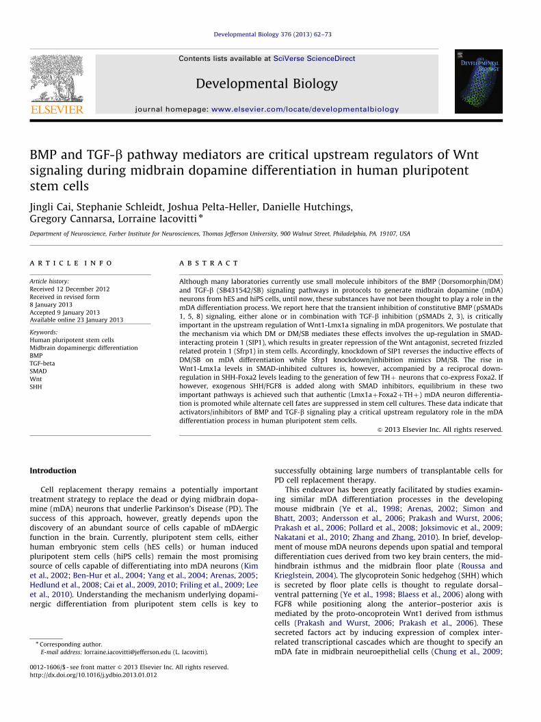
In the last year, in addition to our usual EB method ofdifferentiating mDA neurons from hES/hiPS cells, cells were alsodifferentiated using a simplified monolayer method (Fig. 1A).This was in part made possible by the discovery that therho-associated kinase (ROCK) inhibitor, Y-27632, markedlydiminishes dissociation-induced apoptosis in stem cells(Watanabe et al., 2007), allowing us to proceed directly from
undifferentiated cell colonies to mDA differentiation while omit-ting the EB step.
Curiously, when hNPs were generated in monolayer cultures,we found that some hNPs expressed Lmx1a but rarely went on todevelop into tyrosine hydroxylase (TH)-expressing neurons atstage 5. Instead, they appeared to be ‘trapped’ at the mDAspecification step (Fig. 1B). However, when both the BMP inhi-bitor, Dorsomorphin (DM) and the TGF-b inhibitor, SB431542 (SB)were added, we found a dramatic rise in Lmx1a expression in NPsas well as a marked amplification in the number of THþ mDAneurons, disproportionate to the increase in nestinþ progenitorsand b-III tubulinþ (b-III tub) neurons observed in the samecultures (Fig. 1B,C). Likewise, when EB cultures were treated withDM/SB, there was a significant increase in Lmx1a and TH overnestin and b-III tub expression (Fig. 1B,C). Taken together, theseresults suggested that while DM/SB modestly increases NP andneuron production in monolayer cultures, it greatly increases theproportion of those cells that are mDA-specified and that go on tobecome THþ neurons.
We next investigated the mechanism via which BMP/TGF-binhibitors of specific receptor SMADs exerted their effects on mDAdifferentiation. Western analysis of hES cells maintained in basalgrowth media (control cultures) exhibited moderate levels ofpSMADs 1, 5, 8 and pSMADs 2, 3 (Fig. 2A, C). However, constitu-tive BMP signaling was nearly totally blocked after treatment(stage 2) with highly specific BMP pathway inhibitor, DM (Fig. 2A,C). In contrast to DM, 10 mM SB was a relatively ineffectualinhibitor of the TGF-b pathway, only partially blocking theformation of pSMADs 2, 3 in stage 2 (Fig. 2A, C). After removalof SMAD inhibitors, phosphorylation of all SMADs was restored tonear normal levels in stage 3.
To identify potential downstream molecular targets of BMP/TGF-b inhibitors, we used human PCR arrays (Qiagen PAHS-047Z— stem cell signaling) or (Qiagen PAHS-035Z — BMP/TGF-bsignaling pathway) to compare control and DM/SB-treated mono-layer cultures. While a number of genes were induced by DM/SBtreatment, only those that were increased at least 5-fold upontreatment were verified by qPCR (Suppl. Fig. 1). Of that group, wefound that inhibition of SMAD signaling in both EB and monolayercultures caused a dramatic rise in the levels of the transcriptionfactor, SMAD-interacting protein 1 (SIP1, also known as Zincfinger E-box-binding homeobox 2 or ZEB2). Interestingly, SIP1levels were also elevated in untreated EB cultures compared tountreated monolayers, suggesting that the same factors may havebeen involved in mediating mDA differentiation in EB cultureseven in the absence of DM/SB supplementation, possibly as aresult of endogenous BMP/TGF-b inhibitors (ie. noggin)(Chambers et al., 2009; Krause et al., 2011).
An important confirmation of SIP1’s role in mDA specificationand differentiation was provided by SIP1 knockdown experi-ments. In these studies, SIP1 shRNA and control (empty andscramble) vectors were transfected into undifferentiated stemcells. Following puromycin selection and subsequent differentia-tion, qPCR analysis revealed significant knockdown in SIP1transcripts, and importantly, a reduction in Lmx1a in stage 4 hNPsand TH in stage 4/5 neurons, without a change in nestin or b-IIItub expression (Fig. 3A). Cleaved caspase 3 protein was notincreased in SIP1 knockdown cultures (Fig. 3B), indicating thatthe decrease in Lmx1a and TH was not due to enhanced toxicity/cell death from genetic engineering. These data demonstrate thatSIP1 knockdown results in decreased mDA specification anddifferentiation without altering neurogenesis, suggesting thatthe two developmental processes are likely mediated by differentpathways acting downstream of DM/SB. Moreover, these datafurther suggest that constitutive SIP1 levels normally hold incheck Wnt1-Lmx1a-TH expression in stem cells, and that by
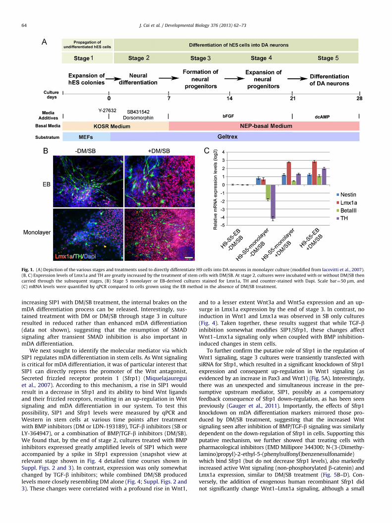
Fig. 1. (A) Depiction of the various stages and treatments used to directly differentiate H9 cells into DA neurons in monolayer culture (modified from Iacovitti et al., 2007).
(B, C) Expression levels of Lmx1a and TH are greatly increased by the treatment of stem cells with DM/SB. At stage 2, cultures were incubated with or without DM/SB then
carried through the subsequent stages, (B) Stage 5 monolayer or EB-derived cultures stained for Lmx1a, TH and counter-stained with Dapi. Scale bar¼50 mm, and
(C) mRNA levels were quantified by qPCR compared to cells grown using the EB method in the absence of DM/SB treatment.
J. Cai et al. / Developmental Biology 376 (2013) 62–7364
increasing SIP1 with DM/SB treatment, the internal brakes on themDA differentiation process can be released. Interestingly, sus-tained treatment with DM or DM/SB through stage 3 in cultureresulted in reduced rather than enhanced mDA differentiation(data not shown), suggesting that the resumption of SMADsignaling after transient SMAD inhibition is also important inmDA differentiation.
We next sought to identify the molecular mediator via whichSIP1 regulates mDA differentiation in stem cells. As Wnt signalingis critical for mDA differentiation, it was of particular interest thatSIP1 can directly repress the promoter of the Wnt antagonist,Secreted frizzled receptor protein 1 (Sfrp1) (Miquelajaureguiet al., 2007). According to this mechanism, a rise in SIP1 wouldresult in a decrease in Sfrp1 and its ability to bind Wnt ligandsand their frizzled receptors, resulting in an up-regulation in Wntsignaling and mDA differentiation in our system. To test thispossibility, SIP1 and Sfrp1 levels were measured by qPCR andWestern in stem cells at various time points after treatmentwith BMP inhibitors (DM or LDN-193189), TGF-b inhibitors (SB orLY-364947), or a combination of BMP/TGF-b inhibitors (DM/SB).We found that, by the end of stage 2, cultures treated with BMPinhibitors expressed greatly amplified levels of SIP1 which wereaccompanied by a spike in Sfrp1 expression (snapshot view atrelevant stage shown in Fig. 4 detailed time courses shown inSuppl. Figs. 2 and 3). In contrast, expression was only somewhatchanged by TGF-b inhibitors; while combined DM/SB producedlevels more closely resembling DM alone (Fig. 4; Suppl. Figs. 2 and3). These changes were correlated with a profound rise in Wnt1,
and to a lesser extent Wnt3a and Wnt5a expression and an up-surge in Lmx1a expression by the end of stage 3. In contrast, noinduction in Wnt1 and Lmx1a was observed in SB only cultures(Fig. 4). Taken together, these results suggest that while TGF-binhibition somewhat modifies SIP1/Sfrp1, these changes affectWnt1–Lmx1a signaling only when coupled with BMP inhibition-induced changes in stem cells.
To further confirm the putative role of Sfrp1 in the regulation ofWnt1 signaling, stage 3 cultures were transiently transfected withsiRNA for Sfrp1, which resulted in a significant knockdown of Sfrp1expression and consequent up-regulation in Wnt1 signaling (asevidenced by an increase in Pax3 and Wnt1) (Fig. 5A). Interestingly,there was an unexpected and simultaneous increase in the pre-sumptive upstream mediator, SIP1, possibly as a compensatoryfeedback consequence of Sfrp1 down-regulation, as has been seenpreviously (Gauger et al., 2011). Importantly, the effects of Sfrp1knockdown on mDA differentiation markers mirrored those pro-duced by DM/SB treatment, suggesting that the increased Wntsignaling seen after inhibition of BMP/TGF-b signaling was similarlydependent on the down-regulation of Sfrp1 in cells. Supporting thisputative mechanism, we further showed that treating cells withpharmacological inhibitors (EMD Millipore 344300; N-(3-(Dimethy-lamino)propyl)-2-ethyl-5-(phenylsulfonyl)benzenesulfonamide)which bind Sfrp1 (but do not decrease Sfrp1 levels), also markedlyincreased active Wnt signaling (non-phosphorylated b-catenin) andLmx1a expression, similar to DM/SB treatment (Fig. 5B–D). Con-versely, the addition of exogenous human recombinant Sfrp1 didnot significantly change Wnt1–Lmx1a signaling, although a small
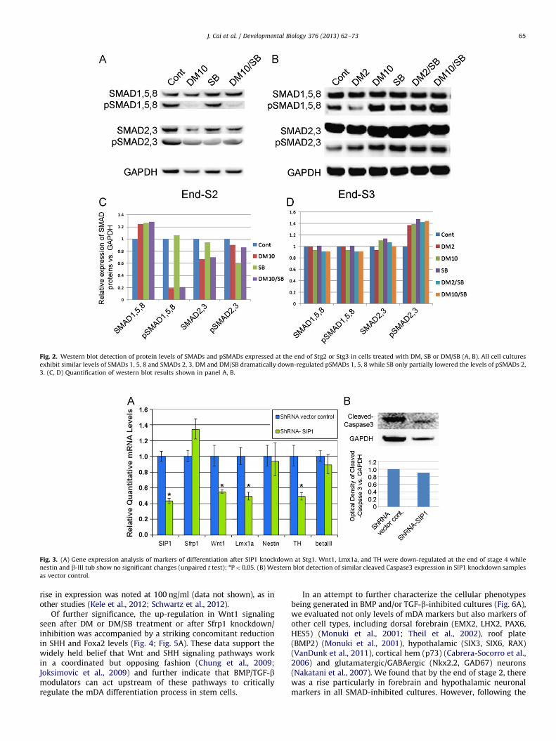
Fig. 3. (A) Gene expression analysis of markers of differentiation after SIP1 knockdown at Stg1. Wnt1, Lmx1a, and TH were down-regulated at the end of stage 4 while
nestin and b-III tub show no significant changes (unpaired t test): nPo0.05. (B) Western blot detection of similar cleaved Caspase3 expression in SIP1 knockdown samples
as vector control.
Fig. 2. Western blot detection of protein levels of SMADs and pSMADs expressed at the end of Stg2 or Stg3 in cells treated with DM, SB or DM/SB (A, B). All cell cultures
exhibit similar levels of SMADs 1, 5, 8 and SMADs 2, 3. DM and DM/SB dramatically down-regulated pSMADs 1, 5, 8 while SB only partially lowered the levels of pSMADs 2,
3. (C, D) Quantification of western blot results shown in panel A, B.
J. Cai et al. / Developmental Biology 376 (2013) 62–73 65
rise in expression was noted at 100 ng/ml (data not shown), as inother studies (Kele et al., 2012; Schwartz et al., 2012).
Of further significance, the up-regulation in Wnt1 signalingseen after DM or DM/SB treatment or after Sfrp1 knockdown/inhibition was accompanied by a striking concomitant reductionin SHH and Foxa2 levels (Fig. 4; Fig. 5A). These data support thewidely held belief that Wnt and SHH signaling pathways workin a coordinated but opposing fashion (Chung et al., 2009;Joksimovic et al., 2009) and further indicate that BMP/TGF-bmodulators can act upstream of these pathways to criticallyregulate the mDA differentiation process in stem cells.
In an attempt to further characterize the cellular phenotypesbeing generated in BMP and/or TGF-b-inhibited cultures (Fig. 6A),we evaluated not only levels of mDA markers but also markers ofother cell types, including dorsal forebrain (EMX2, LHX2, PAX6,HES5) (Monuki et al., 2001; Theil et al., 2002), roof plate(BMP2) (Monuki et al., 2001), hypothalamic (SIX3, SIX6, RAX)(VanDunk et al., 2011), cortical hem (p73) (Cabrera-Socorro et al.,2006) and glutamatergic/GABAergic (Nkx2.2, GAD67) neurons(Nakatani et al., 2007). We found that by the end of stage 2, therewas a rise particularly in forebrain and hypothalamic neuronalmarkers in all SMAD-inhibited cultures. However, following the
Fig. 4. mRNA levels (A) and protein levels (B) of mDA markers examined at different stages after treatment of hES (H9 line) cells with DM, SB or DM/SB. At the end of Stg2,
both SIP1 and Sfrp1 expression levels were increased after DM and DM/SB treatment. By mid-Stg3, Sfrp1 expression levels fell dramatically with DM and DM/SB treatment.
At the end of Stg3, DM and DM/SB treatment greatly increased the expression of Wnt1 and Lmx1a (and somewhat increased Wnt3a and Wnt5a) while SHH expression
decreased. At the end of Stg5, TH expression levels were increased with DM, SB and DM/SB treatment. (C) Quantification of Western blot results shown in panel B.
J. Cai et al. / Developmental Biology 376 (2013) 62–7366
removal of BMP or TGF-b inhibitors from the media, expression ofthese markers fell to near control levels (with the exception ofEMX2) as mDA phenotypic markers (Wnt1, Lmx1a) increaseddramatically in stage 3 cultures (Suppl. Fig. 4). Indeed, when sistercultures were immunocytochemically stained, we found manyLmx1aþ NPs in DM and DM/SB-treated stage 3 cultures ascompared to control or SB cultures (data not shown). Importantly,however, these Lmx1aþ NPs did not co-label with Foxa2although the culture did contain many brightly fluorescentFoxa2þ cells (Fig. 6B).
At the end of differentiation (stage 5), all cultures were stainedimmunocytochemically for TH. Somewhat unexpectedly, weobserved flattened neurite-free THþ cells in control cultureswhich increased in number after SB treatment (Suppl. Fig. 5A).These THþ cells did not stain for nestin or b-III tub and did not
incorporate BrdU (Suppl. Fig. 5B–D), indicating that they were notdividing neural progenitors or postmitotic neurons. Importantly,this non-neural THþ cell type was not routinely seen in DM orDM/SB-treated cultures where TH staining was observed only inprocess-bearing cells that co-labeled for b-III tub (data notshown). However, despite their mature appearance, these neu-rons did not co-label for Foxa2 (although many Foxa2þ cells werepresent) (Fig. 6C). These data, taken together with the qPCR andWestern results (Fig. 4), suggest that TGF-b-inhibition aloneyields a non-neural THþ cell type in culture. In contrast, culturestreated with BMP inhibitors or combined BMP/TGF-b inhibitorsare initially induced to become dorsal forebrain and hypothalamicneurons. Upon removal of these inhibitors from the media, NPslose expression of these phenotypic markers and partially differ-entiate down the mDA pathway to express the mDA fate gene
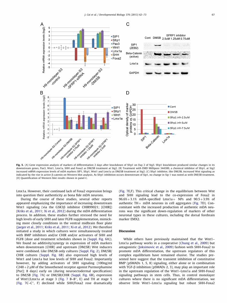
Fig. 5. (A) Gene expression analysis of markers of differentiation 2 days after knockdown of Sfrp1 on Day 2 of Stg3. Sfrp1 knockdown produced similar changes in its
downstream genes, Pax3, Wnt1, Lmx1a, SHH and Foxa2 as DM/SB treatment at Stg2. (B) Treatment with EMD Millipore 344300, a chemical inhibitor of Sfrp1, at Stg2
increased mRNA expression levels of mDA markers SIP1, Sfrp1, Wnt1 and Lmx1a as DM/SB treatment at Stg2. (C) Sfrp1 inhibitor, like DM/SB, increased Wnt signaling as
indicated by the rise in active b-catenin on Western blot analysis. As Sfrp1 inhibition occurs downstream of Sip1, no change in Sip 1 was noted as with DM/SB treatment.
(D) Quantification of Western blot results shown in panel C.
J. Cai et al. / Developmental Biology 376 (2013) 62–73 67
Lmx1a. However, their continued lack of Foxa2 expression bringsinto question their authenticity as bona fide mDA neurons.
During the course of these studies, several other reportsappeared emphasizing the importance of increasing downstreamWnt1 signaling (via the GSK3b inhibitor CHIR99021; [CHIR])(Kriks et al., 2011; Xi et al., 2012) during the mDA differentiationprocess. In addition, these studies further stressed the need forhigh levels of early SHH and later FGF8 supplementation, mimick-ing more closely conditions in the ventral midbrain floor plate(Jaeger et al., 2011; Kriks et al., 2011; Xi et al., 2012). We thereforeinitiated a study in which cultures were simultaneously treatedwith BMP inhibitors and/or CHIR and/or activators of SHH andFGF8 (dose and treatment schedules shown in (Suppl. Fig. 6A)).We found no additivity/synergy in expression of mDA markerswhen downstream (CHIR) and upstream (DM/SB) Wnt inducerswere combined. Like DM/SB only cultures (Suppl. Fig. 2), DM/SB/CHIR cultures (Suppl. Fig. 6B) also expressed high levels ofWnt1 and Lmx1a but low levels of SHH and Foxa2. Importantlyhowever, by adding activators of SHH signaling (100ng/mlC24IIþ2 mM of the Smoothened receptor agonist Purmorphamine[Pur]; 8 days) early on (during neuroectodermal specification)to DM/SB (Fig. 7A) or DM/SB/CHIR (Suppl. Fig. 6B), expressionof Wnt1/Lmx1a at stage 3 (Fig. 7 B–B0 0, E) and TH at stage 5(Fig. 7C–C0 0, F) declined while SHH/Foxa2 rose dramatically
(Fig. 7E,F). This critical change in the equilibrium between Wntand SHH signaling lead to the co-expression of Foxa2 in96.6%þ3.1% mDA-specified Lmx1aþ NPs and 90.5þ3.9% ofauthentic THþ mDA neurons in cell aggregates (Fig. 7D). Con-comitant with the increased production of authentic mDA neu-rons was the significant down-regulation of markers of otherneuronal types in these cultures, including the dorsal forebrainmarker EMX2.
Discussion
While others have previously maintained that the Wnt1–Lmx1a pathway works in a cooperative (Chung et al., 2009) butantagonistic (Joksimovic et al., 2009) fashion with SHH-Foxa2 topromote mDA differentiation, the upstream regulators of thiscomplex equilibrium have remained elusive. The studies pre-sented here suggest that the transient inhibition of constitutiveBMP (pSMADs 1, 5, 8) signaling, either alone or in combinationwith TGF-b inhibition (pSMADs 2, 3), may play an important rolein the upstream regulation of the Wnt1–Lmx1a and SHH-Foxa2signaling pathways in stem cells. Thus, in control monolayercultures where there is no significant mDA differentiation, weobserve little Wnt1–Lmx1a signaling but robust SHH-Foxa2
Fig. 6. (A) Experiment paradigm of DM/SB protocol used to directly differentiate
H9 cells into DA neurons in monolayer culture. (B, C) Immunocytochemical
analysis of differentiated DA NPs and neurons. At the end of Stg3, many Lmx1aþ
NPs were found in DM/SB-treated cultures. These Lmx1aþ NPs did not overlap
with brightly fluorescent Foxa2þ cells (B). At the end of Stg5, mature DA neurons
were detected as THþ cells, most of which were Foxa2- in DM/SB-treated cultures
(C, C0). Scale bars¼50 mm.
J. Cai et al. / Developmental Biology 376 (2013) 62–7368
signaling. However, this equilibrium is reversed when cells aretransiently exposed to BMP inhibitors or BMP/TFG-b inhibitorsearly on in differentiation, leading to a marked amplification inWnt1, Lmx1a and TH expression at subsequent stages and aconcomitant decline in SHH and Foxa2. Gene knockdown experi-ments further implicate the SMAD-interacting transcription factorSIP1 and its downstream target gene Sfrp1 as important media-tors of these effects, linking upstream BMP/TGF-b pathways anddownstream Wnt1–Lmx1a and SHH-Foxa2 pathways. Togetherthese results have led us to postulate a novel putative pathway forregulation of this complex signaling during mDA differentiation instem cells (Fig. 8).
According to this pathway, pSMADs together with SIP1 act toco-repress the Wnt antagonist Sfrp1 in stem cells as in other cellsystems (Postigo et al., 2003; Miquelajauregui et al., 2007). Withless Sfrp1 available to compete for frizzled receptors (Molenaaret al., 1996; Peifer, 1997; Van de Wetering et al., 1997; Uren et al.,2000), Wnt ligands are able to promote Lmx1a expression andmDA differentiation. This then begs the question of how SMADinhibition enhances this process. We would postulate that this isachieved through a series of critical molecular steps. Undernormal basal culture conditions, stem cells express Sfrp1 despiteconstitutive SMAD signaling, possibly as a result of low levels ofSIP1 co-repressors. With the addition of BMP inhibitors (DM,LDN) or combined BMP/TGF-ß inhibitors (DM/SB) that blockpSMADs 1, 5, 8 and/or pSMADs 2, 3, SIP1-mediated repressionof Sfrp1 is even further diminished, resulting in a spike in Sfrp1levels during stage 2. These elevated levels of Sfrp1 furtherantagonize Wnt signaling, working against the differentiation ofan mDA phenotype in stem cells and in favor of alternate cellfates. As such, we find an induction in dorsal forebrain andhypothalamic markers LHX2, EMX2, SIX3, etc. in stage 2 afterSMAD inhibition. Consistent with these results, other studies havealso reported that dorsal forebrain markers LHX2 (Monuki et al.,2001) and EMX2 (Theil et al., 2002) are highly expressed with low(but not high) BMP signaling in stem cells.
However, another major consequence of BMP or BMP/TGF-binhibition in stem cells is the dramatic rise in SIP1 levels duringstage 2–3, possibly as a rebound response to the early upsurge inSfrp1 levels. We posit that it is this elevation in SIP1 that allowsSfrp1 expression to be dramatically repressed once DM/SB isremoved from the media and SMAD signaling is restored. Thus,both the rise in SIP1 co-repressors during transient BMP/TGF-b
inhibition and the subsequent restoration of SMAD co-repressorsafter cessation of treatment may be necessary steps in ultimatelydriving down Sfrp1 levels and driving forward mDA differentia-tion. The importance of SMAD/SIP1 regulation in CNS develop-ment is not limited to the mDA differentiation process but isthought to also be involved in SVZ gliogenesis and myelination(Nityanandam et al., 2012; Weng et al., 2012).
Concomitant with the reduction in Sfrp1 in NPs is a shift in theequilibrium towards Wnt signaling, as evidenced by an increasein Wnt1/Pax3/b-catenin, and to a lesser extent Wnt3a and Wnt5a.Although in rare instances, low concentrations of Sfrp1 have beenshown to increase rather than decrease mDA differentiation instem cell cultures (Kele et al., 2012; Schwartz et al., 2012), ourresults after treatment with human recombinant Sfrp1, Sfrp1antagonists or Sfrp1 siRNA, suggests that it is the decline,not the spike, in Sfrp1 which induces Wnt signaling in hEScell cultures. As a result of the rise in Wnt signaling in DM orDM/SB-treated stage 3 cultures, the vast majority of NPs go on toexpress Lmx1a while expression of other forebrain markersdeclines.
Of particular significance is the fact that increased Wnt1signaling in DM and DM/SB-treated cultures results in a reciprocalreduction in SHH and Foxa2 levels. Precisely how downstreammediators of SMAD inhibition regulate SHH-Foxa2 signalingremains unclear. In the literature, no direct modulatory effect ofSfrp1 on the SHH promoter has been reported, although theconverse has been widely observed (Ingram et al., 2002; Heet al., 2006; Yauch et al., 2008; Katoh and Katoh, 2009; Shahiet al., 2011). Thus, the regulation of SHH-Foxa2 by Sfrp1 mayoccur through an indirect compensatory feedback pathway.Supporting this possibility is the previous demonstration in othersystems that SHH, working through TGF-b signaling, can alterSIP1 and Sfrp1 levels to inhibit the Wnt pathway (He et al., 2006;Katoh and Katoh, 2009). Taken together, these data raise thepossibility that the widely recognized cross talk that occursbetween the SHH and Wnt signaling pathways during mDAdifferentiation (Chung et al., 2009; Joksimovic et al., 2009) maytranspire at points (ie. SIP1 and Sfrp1) further upstream thanformerly appreciated.
At the completion of differentiation when the phenotype ofcells treated with SMAD inhibitors was assessed, we weresurprised to find TH staining in cells with vastly differentmorphologies. Thus, control monolayer cultures containednon-dividing non-neural THþ cells which increased in numberwith SB treatment. The identity of these cells remains unknown.However, it is a well-established fact that Alk4 receptor signalingpromotes the formation of mesoendoderm over ectoderm (Chnget al., 2010). Since in control cultures, and even after 10 mM SB,Alk4/SMAD 2, 3 signaling remains high (470% of control), it ispossible that THþ non-neural cells are mesoendodermal in origin(Kunisada et al., 2012). Alternatively, SB has also been used todifferentiate trophectoderm in hES cell cultures (Chambers et al.,2009) and therefore derivatives of this germ layer may also bepresent in our cultures. Distinguishing between these possibilitieswill require continued investigation. Presumably, however, it isthis critical difference in cellular composition that accounts forthe fact that SB-induced changes in SIP1 and Sfrp1 do not amplifyWnt1–Lmx1a signaling or mDA differentiation in these cultures.
Taken together, these data raise questions as to the benefitsof routinely adding SB to monolayer cultures as is now donein many labs (He et al., 2011; Kim et al., 2011; Kriks et al., 2011;Vogt et al., 2011; Lipchina et al., 2012; Nefzger et al., 2012).Arguing in favor of the continued use of SB, we found that whencombined with DM, SB induced changes in SIP1/Sfrp1 furtherenhanced Wnt1–Lmx1a signaling in NPs. Also, other labs reportthe efficient production of mDA neurons in monolayer cultures
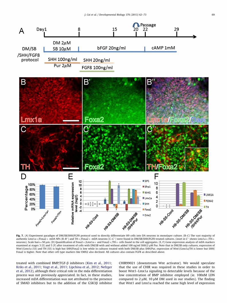
Fig. 7. (A) Experiment paradigm of DM/SB/SHH/FGF8 protocol used to directly differentiate H9 cells into DA neurons in monolayer culture. (B–C) The vast majority of
authentic Lmx1aþ/Foxa2þ mDA NPs (B–B0 0) and THþ/Foxa2þ mDA neurons (C–C0 0) were found in DM/SB/SHH/FGF8 treated cultures. (inset in C0 0 shows Lmx1aþ/THþ
neurons). Scale bars¼50 mm. (D) Quantification of Foxa2þ/Lmx1aþ and Foxa2þ/THþ cells found in the cell aggregates. (E, F) Gene expression analysis of mDA markers
examined at stages 3 (E) and 5 (F) after treatment of cells with DM/SB with and without added 100 ng/ml SHH/2 mM Pur. Note that in DM/SB only cultures, expression of
Wnt1/Lmx1a (S3) and TH (S5) is high but SHH/Foxa2 is low while in cultures treated with both DM/SB plus SHH/Pur, expression of Wnt1/Lmx1a/TH is lower but SHH/
Foxa2 is higher. Note that other cell type markers like EMX2 also declined. All cultures also contain FGF8 as described above.
J. Cai et al. / Developmental Biology 376 (2013) 62–73 69
treated with combined BMP/TGF-b inhibitors (Kim et al., 2011;Kriks et al., 2011; Vogt et al., 2011; Lipchina et al., 2012; Nefzgeret al., 2012), although their critical role in the mda differentiationprocess was not previously appreciated. In fact, in these studies,increased mDA differentiation was not attributed to the presenceof SMAD inhibitors but to the addition of the GSK3b inhibitor
CHIR99021 (downstream Wnt activator). We would speculatethat the use of CHIR was required in these studies in order toboost Wnt1–Lmx1a signaling to detectable levels because of thelow concentration of BMP inhibitor employed (ie. 100nM LDNcompared to 2 mM, 10 mM DM used in our studies). The findingthat Wnt1 and Lmx1a reached the same high level of expression

Fig. 8. Diagram of putative mDA differentiation pathway based on this and other studies.
J. Cai et al. / Developmental Biology 376 (2013) 62–7370
after DM/SB or DM/SB/CHIR treatment of our cultures suggeststhat additivity/synergy between upstream and downstream Wntactivators is in fact unnecessary if BMP inhibitors are used atsufficiently high concentration.
Despite the enhanced production of Lmx1aþ NPs in DM/SBand DM/SB/CHIR-treated cultures, unexpectedly, these cells didnot express the floor plate marker Foxa2. Moreover, at laterstages, these progenitors did not give rise to THþ neurons thatco-expressed Foxa2, bringing into question their authenticity asmDA neurons. Thus, increasing Wnt signaling via BMP/TGF-b/GSK3b inhibition, while desirable, is not sufficient to fully drivemDA differentiation in stem cells. This is likely a result of theconcomitant decline in SHH signaling and the forkhead floor platemarker Foxa2 that accompanies increased Wnt signaling in cells.Since authentic mDA neurons derive from NPs that express bothLmx1a and Foxa2 in vivo, it seems likely that it is necessary todrive both SHH and Wnt signaling in stem cell cultures in order tostrike the proper balance in these opposing mDA differentiationpathways. Indeed, when potent activators of SHH signaling(C24IIþSmoothened agonist Purmorphamine) were added alongwith Wnt activators DM/SB/CHIR and were continued at low dosefor an extended period along with FGF8, we found a dramatic risein SHH signaling and in the number of mDA-specified Lmx1aþNPs and THþ neurons which co-labeled for Foxa2, similar to theresults reported previously using variations of the same protocol(Jaeger et al., 2011; Kriks et al., 2011; Xi et al., 2012). Consistentwith the enhancement in mDA differentiation in these cultures,markers of other neuronal phenotypes, such as, hypothalamic(SIX3, SIX6, RAX and Nkx2.1), dorsal forebrain (HES5, EMX2,PAX6), roof plate (BMP2) and cortical (GABA) neurons weredown-regulated. We conclude that the development of bona fidemDA neurons from stem cells requires sufficiently high SHH–Foxa2 signaling (from potent SHH agonists and FGF8) to balancethe robust Wnt signaling seen after treatment with SMADinhibitors.
Whether upstream (BMP/TGFb) and downstream (Wnt/SHH)regulatory events occur in the same or different cell populationsin our heterogeneous hES cell cultures remains unclear. SinceSfrp1 is a secreted molecule, it is indeed possible that changes inSMAD signaling/SIP1/Sfrp1 in one group of cells (ie. non-mDAspecified NPs) could impact, in a paracrine fashion, Wnt/SHHsignaling in another stem cell group (ie. prospective mDA-specified NPs). Alternatively, secreted Sfrp1 could act in autocrinefashion to drive mDA differentiation in those NPs which produceit. Drawing these distinctions will depend upon the futuredevelopment of reliable antibodies able to detect small quantita-tive changes in SIP1 and Sfrp1, thus allowing for single cellanalysis by immuncytochemistry. Regardless of cellular location,however, our data indicate that changes in BMP/TGFb signaling
critically influence Wnt/SHH levels and ultimately the degree ofmDA differentiation observed in stem cell cultures. WhetherSMADs similarly regulate the Wnt-Lmx1a and SHH-Foxa2 path-ways during the development of mDA neurons in vivo remains tobe investigated.
In summary, the findings of this paper indicate that thetransient inhibition of the constitutive BMP pathway is neededto increase SIP1 such that Sfrp1 can be co-repressed by pSMADS 1,5, 8, after BMP inhibitor removal. This decline in the Wntantagonist Sfrp1 results in an up-regulation in Wnt1 signalingand Lmx1a expression in mDA specified NPs. Importantly, theaddition of TGF-b inhibitors of SMADs 2, 3 can further amplifythis effect but only when combined with BMP inhibitors whichhave already neuralized stem cells. However, a major conse-quence of increased Wnt signaling in these cells is the simulta-neous and reciprocal down-regulation in SHH-Foxa2 signaling,resulting in the generation of Lmx1aþ NPs and THþ neuronswhich lack Foxa2 expression, an impediment which is rectified byco-treating SMAD-inhibited cultures with SHH and FGF8 early onduring cell specification. We conclude that inhibitors of BMP andTGF-b signaling play a critical upstream regulatory role in themDA differentiation process, driving Wnt1–Lmx1a signaling instem cells but that the generation of authentic mDA neuronsrequires additional factors (SHH, FGF8) to properly balance theequilibrium between Wnt–Lmx1a and SHH-Foxa2 mDA path-ways. We would further postulate that it is the regulation ofthese critical mDA pathways by SMAD inhibitors which isresponsible for the high efficiency production of authentic mDAneurons seen in the present study and in studies publishedpreviously (Kriks et al., 2011; Xi et al., 2012). Establishing themechanisms via which authentic mDA neurons are produced inlarge quantity from human embryonic stem cells and humaninduced pluripotent stem cells (our unpublished data, Mak et al.,2012) is key to the successful translation of this technology for PDcell replacement therapy.
Materials and methods
Tissue culture
hES cells (H9 cells, Passage 35–50) were purchased fromWicell Research Institute and maintained according to the sup-plier’s instructions. Briefly, cells were grown on a monolayer ofprimary mouse fibroblasts (MEFs; Millipore) in DMEM/F12 media(invitrogen) supplemented with 20% Knockout Serum ReplacerTM
(KOSR; invitrogen), 1% Non-Essential Amino Acids (invitrogen),1 mM L-glutamine (invitrogen), 0.1 mM 2-mercaptoethanol, and4 ng/ml bFGF (R&D systems). Cell propagation was achieved
J. Cai et al. / Developmental Biology 376 (2013) 62–73 71
through manual dissection and transfer of cell colonies once perweek. The differentiation process was initiated by passaging themon Geltrex (Invitrogen 1:100)-coated tissue culture plates withtwo TGF/BMP inhibitors SB431542 (SB, Tocris, 10 mM) and Dor-somorphin (DM, Tocris, 2 mM) for 1 week. Additional smallchemicals and growth factors, such as LDN-193189 (LDN, Stem-gent, 2 mM), LY-364947 (LY, Tocris, 10 mM), CHIR 99021 (CHIR,Tocris, 0.4 mM), SHH (C24II) (SHH, R&D systems, 100 ng/ml),Purmorphamine (Pur, Stemgent, 2 mM), FGF8 (R&D systems,100 ng/ml) and Sfrp1 inhibitor (Millipore) were also used at Stage2. Then neural progenitors (NPs) were generated in N2/B27 NEP-basal medium. Rosettes were then expanded in NEP-basal med-ium supplemented with 20 ng/ml bFGF (R&D system) every otherday. For further differentiation down the DA pathway, cells wereincubating for 1 week in NEP-basal medium supplemented with1 mM dibutyryl cAMP (dbcAMP, Sigma) (Fig.1A).
Immunocytochemistry
Cultures were fixed with 4% paraformaldehyde for 30min at4 1C and stained with primary antibodies (Suppl. Table 1) at 4 1Covernight. All secondary antibodies were Alexa Fluor antibodiesfrom Invitrogen used at 1:200 for 30 min at room temperature.Cultures were also counter stained with Hoechst 33258 (Invitro-gen) at 1:1000. Slides were covered with ProLong Gold antifadereagent (Invitrogen). Single and double labeled cells were countedin all fields of ES cell aggregates in triplicate cultures andaveragedþSEM using an Olympus IX81 Image Analysis System.
RNA isolation and cDNA synthesis
Total RNA was isolated directly from freshly collected differenttreatments and different stages of H9 cells with TRIzol (Invitro-gen), a modification of the guanidine isothiocyanate–phenol–chloroform extraction method. cDNA was synthesized by using1 mg total RNA in a 20 mL reaction with Superscript III (Invitrogen)and oligo (dT)12–18 (Invitrogen). One microliter of RNase H(Invitrogen) was added to each reaction tube, and the tubes wereincubated for 20 min at 37 1C before proceeding to PCR.
Real-time PCR analysis
Real-time PCR was carried out by 7500 Real-Time PCR Systemusing SYBR green PCR master mix (both from Applied Biosys-tems). GAPDH was used as an internal control. All PCR productswere checked by running an agarose gel for the first time and bydoing dissociation assay every time to exclude the possibility ofmultiple products. PCR analyses were conducted in triplicate foreach sample. Primers are listed as Suppl. Table 2. Statisticalanalysis was performed using the unpaired t test.
RT2ProfilerTM qPCR array
TGF-beta/BMP signaling and stem cell signaling qPCR arrayswere obtained from Qiagen. Five mg of total RNA was reversetranscribed in a final reaction mix of 20 mL using RT2 First StrandKit (Qiagen) according to the manufacturer’s instructions. cDNAwas diluted by adding Nuclease-free water (Ambion). The PCR wascarried out using the 7500 Real-Time PCR System (Applied Biosys-tem) according to the protocol provided by Qiagen. For one 96-well-plate of the PCR array, 2550 mL of PCR master mix containing2x SuperArray RT2 qPCR Master Mix and 102 mL of diluted cDNAwas mixed and aliquots of 25 mL were added to each well.Universal cycling conditions (10 min at 95 1C, 15 s at 95 1C, 1 min60 1C for 40 cycles) were used. After the PCR is done, raw datawas saved and submitted to Qiagen’s website http://pcrdata-
analysis.sabiosciences.com/pcr/arrayanalysis.php for fold changecalculating.
shRNA and siRNA
ShRNA vectors of Sip1 and siRNA oligos of Sfrp1 were purchasedfrom Origene. Sip1 shRNA vectors and scrambled or empty controlvectors were nucleofected into feeder-free H9 cells on a nucleofectordevice (Lonza). Two days after transfection, cells were selected withPuromycin (1 mg/ml) for about 14 days when single cell coloniesappeared. Then cells were further differentiated before collection atthe end of Stg4 and processed with ICC and Real-time PCR. Threepairs of verified siRNAs for human Sfrp1 and scrambled negativecontrol siRNA were transfected into H9 dissociated Day2–Stg3 cellsusing Lipofectamine 2000 (Invitrogen). Transfection efficiency wasmonitored by co-transfecting a Trilencer-27 Fluorescent-labeledtransfection control siRNA duplex (Origene). Total RNA was isolatedtwo days later and further analyzed by real-time RT-PCR.
Western blot
Cultured cells were rinsed quickly with PBS and lysed in 1mlTrizol (Invitrogen). The protein phase was collected and rinsed, anddissolved in 1% SDS containing protease and phosphatase inhibitors(Roche). All cell lysates were stored at �20 1C. Prior to electrophor-esis, proteins were quantified to ensure equal gel loading using theLowry Assay. Samples were prepared for western blotting with LDSsample buffer and DTT (Invitrogen), heated at 100 1C for 15 min, andloaded into individual wells of a 4–12% Bis-Tris gel (20 ml/well,Invitrogen), resolved for 60 min at 120 mA. Gels were transferred tonitrocellulose membrane for blocking in either 5% milk/0.1% PBST(Phosphate-Buffered Saline/0.1% Tween-20) or 1% BSA/0.1% PBST, andblots labeled overnight with primary antibody at 4 1C in blockingsolution. Primary antibodies were listed in Suppl. Table 3. Blots werewashed with PBST for 15 min and labeled for one hour at roomtemperature with goat-a-rabbit-HRP secondary antibody (Santa CruzBiotechnology, 1:5000 in blocking solution). Blots were washed againfor 15 min with 0.1% PBST, and exposed to peroxide substrate (Pierce)for chemiluminescent imaging. Protein signals on western blots weremeasured and quantified using densitometry algorithms by Image Jsoftware (NIH.gov).
Acknowledgments
This work was generously supported by NIH NS075839, theParkinson’s Council and The Newell DeValpine Foundation.
Appendix A. Supporting information
Supplementary information associated with this article can befound in the online version at http://dx.doi.org/10.1016/j.ydbio.2013.01.012
References
Andersson, E., Tryggvason, U., Deng, Q., Friling, S., Alekseenko, Z., Robert, B.,Perlmann, T., Ericson, J., 2006. Identification of intrinsic determinants ofmidbrain dopamine neurons. Cell 124, 393–405.
Arenas, E., 2002. Stem cells in the treatment of Parkinson’s disease. Brain Res. Bull.57, 795–808.
Arenas, E., 2005. Engineering a dopaminergic phenotype in stem/precursorcells: role of Nurr1, glia-derived signals, and Wnts. Ann. N Y Acad. Sci. 1049,51–66.
Ben-Hur, T., Idelson, M., Khaner, H., Pera, M., Reinhartz, E., Itzik, A., Reubinoff, B.E.,2004. Transplantation of human embryonic stem cell-derived neural
J. Cai et al. / Developmental Biology 376 (2013) 62–7372
progenitors improves behavioral deficit in Parkinsonian rats. Stem Cells 22,1246–1255.
Blaess, S., Corrales, J.D., Joyner, A.L., 2006. Sonic hedgehog regulates Gli activatorand repressor functions with spatial and temporal precision in the mid/hindbrain region. Development 133, 1799–1809.
Cabrera-Socorro, A., Pueyo Morlans, M., Suarez Sola, M.L., Gonzalez Delgado, F.J.,Castaneyra-Perdomo, A., Marin, M.C., Meyer, G., 2006. Multiple isoforms of thetumor protein p73 are expressed in the adult human telencephalon and choroidplexus and present in the cerebrospinal fluid. Eur. J. Neurosci. 23, 2109–2118.
Cai, J., Donaldson, A., Yang, M., German, M.S., Enikolopov, G., Iacovitti, L., 2009. Therole of Lmx1a in the differentiation of human embryonic stem cells intomidbrain dopamine neurons in culture and after transplantation into aParkinson’s disease model. Stem Cells 27, 220–229.
Cai, J., Yang, M., Poremsky, E., Kidd, S., Schneider, J.S., Iacovitti, L., 2010. Dopami-nergic neurons derived from human induced pluripotent stem cells survive andintegrate into 6-OHDA-lesioned rats. Stem Cells Dev. 19, 1017–1023.
Chambers, S.M., Fasano, C.A., Papapetrou, E.P., Tomishima, M., Sadelain, M., Studer,L., 2009. Highly efficient neural conversion of human ES and iPS cells by dualinhibition of SMAD signaling. Nat. Biotechnol. 27, 275–280.
Chng, Z., Teo, A., Pedersen, R.A., Vallier, L., 2010. SIP1 mediates cell-fate decisionsbetween neuroectoderm and mesendoderm in human pluripotent stem cells.Cell Stem Cell 6, 59–70.
Chung, S., Leung, A., Han, B.-S., Chang, M.-Y., Moon, J.-I., Kim, C.-H., Hong, S.,Pruszak, J., Isacson, O., Kim, K.-S., 2009. Wnt1-lmx1a forms a novel auto-regulatory loop and controls midbrain dopaminergic differentiation synergis-tically with the SHH-FoxA2 pathway. Cell Stem Cell 5, 646–658.
Cooper, O., Hargus, G., Deleidi, M., Blak, A., Osborn, T., Marlow, E., Lee, K., Levy, A.,Perez-Torres, E., Yow, A., Isacson, O., 2010. Differentiation of human ES andParkinson’s disease iPS cells into ventral midbrain dopaminergic neuronsrequires a high activity form of SHH, FGF8a and specific regionalization byretinoic acid. Mol. Cell. Neurosci. 45, 258–266.
Denham, M., Dottori, M., 2009. Signals involved in neural differentiation of humanembryonic stem cells. Neurosignals 17, 234–241.
Fasano, C., Kortleven, C., Trudeau, L.-E., 2010. Chronic activation of the D2autoreceptor inhibits both glutamate and dopamine synapse formation andalters the intrinsic properties of mesencephalic dopamine neurons in vitro.Eur. J. Neurosci. 32, 1433–1441.
Friling, S., Andersson, E., Thompson, L.H., Jonsson, M.E., Hebsgaard, J.B., Nanou, E.,Alekseenko, Z., Marklund, U., Kjellander, S., Volakakis, N., Hovatta, O., ElManira, A., Bjorklund, A., Perlmann, T., Ericson, J., 2009. Efficient productionof mesencephalic dopamine neurons by Lmx1a expression in embryonic stemcells. Proc. Natl. Acad. Sci. USA 106, 7613–7618.
Gauger, K.J., Chenausky, K.L., Murray, M.E., Schneider, S.S., 2011. SFRP1 reductionresults in an increased sensitivity to TGF-b signaling. BMC Cancer 11, 59.
He, J., Sheng, T., Stelter, A.A., Li, C., Zhang, X., Sinha, M., Luxon, B.A., Xie, J., 2006.Suppressing Wnt signaling by the hedgehog pathway through sFRP-1. J. Biol.Chem. 281, 35598–35602.
He, X.-B., Yi, S.-H., Rhee, Y.-H., Kim, H., Han, Y.-M., Lee, S.-H.S.-H., Lee, H., Park, C.-H.,Lee, Y.-S., Richardson, E., Kim, B.-W., 2011. Prolonged membrane depolarizationenhances midbrain dopamine neuron differentiation via epigenetic histonemodifications. Stem Cells 29, 1861–1873.
Hedlund, E., Pruszak, J., Lardaro, T., Ludwig, W., Vinuela, A., Kim, K.-S., Isacson, O.,2008. Embryonic stem cell-derived Pitx3-enhanced green fluorescent proteinmidbrain dopamine neurons survive enrichment by fluorescence-activatedcell sorting and function in an animal model of Parkinson’s disease. Stem Cells26, 1526–1536.
Iacovitti, L., Donaldson, A.E., Marshall, C.E., Suon, S., Yang, M., 2007. A protocol forthe differentiation of human embryonic stem cells into dopaminergic neuronsusing only chemically defined human additives: studies in vitro and in vivo.Brain Res. 1127, 19–25.
Ingram, W.J., Wicking, C.A., Grimmond, S.M., Forrest, A.R., Wainwright, B.J., 2002.Novel genes regulated by Sonic Hedgehog in pluripotent mesenchymal cells.Oncogene 21, 8196–8205.
Inman, G.J., Hill, C.S., 2002. Stoichiometry of active smad-transcription factorcomplexes on DNA. Journal Biol. Chem. 277, 51008–51016.
Jaeger, I., Arber, C., Risner-Janiczek, J.R., Kuechler, J., Pritzsche, D., Chen, I.-C.,Naveenan, T., Ungless, M.A., Li, M., 2011. Temporally controlled modulation ofFGF/ERK signaling directs midbrain dopaminergic neural progenitor fate inmouse and human pluripotent stem cells. Development 4374, 4363–4374.
Joksimovic, M., Yun, B.A., Kittappa, R., Anderegg, A.M., Chang, W.W., Taketo, M.M.,McKay, R.D.G., Awatramani, R.B., 2009. Wnt antagonism of Shh facilitatesmidbrain floor plate neurogenesis. Nat. Neurosci. 12, 125–131.
Katoh, M., Katoh, M., 2009. Integrative genomic analyses of ZEB2: transcriptionalregulation of ZEB2 based on SMADs, ETS1, HIF1alpha, POU/OCT, and NF-kappaB. Int. J. Oncol. 34, 1737–1742.
Kele, J., Andersson, E.R., Villaescusa, J.C., Cajanek, L., Parish, C.L., Bonilla, S., Toledo,E.M., Bryja, V., Rubin, J.S., Shimono, A., Arenas, E., 2012. SFRP1 and 2 dose-dependently regulate midbrain dopamine neuron development in vivo and inembryonic stem cells. Stem Cells 30, 865–875.
Kim, H., Lee, G., Ganat, Y., Papapetrou, E.P., Lipchina, I., Socci, N.D., Sadelain, M.,Studer, L., 2011. miR-371-3 expression predicts neural differentiation propen-sity in human pluripotent stem cells. Cell Stem Cell 8, 695–706.
Kim, J.-H., Auerbach, J.M., Rodrıguez-Gomez, J.A., Velasco, I., Gavin, D., Lumelsky,N., Lee, S.-H., Nguyen, J., Sanchez-Pernaute, R., Bankiewicz, K., McKay, R., 2002.Dopamine neurons derived from embryonic stem cells function in an animalmodel of Parkinson’s disease. Nature 418, 50–56.
Kittappa, R., Chang, W.W., Awatramani, R.B., McKay, R.D.G., 2007. The foxa2 genecontrols the birth and spontaneous degeneration of dopamine neurons in oldage. PLoS Biol. 5, e325.
Krause, C., Guzman, A., Knaus, P., 2011. Noggin. Int. J. Biochem. Cell Biol. 43,478–481.
Kriks, S., Shim, J.-W., Piao, J., Ganat, Y.M., Wakeman, D.R., Xie, Z., Carrillo-Reid, L.,Auyeung, G., Antonacci, C., Buch, A., Yang, L., Beal, M.F., Surmeier, D.J.,Kordower, J.H., Tabar, V., Studer, L., 2011. Dopamine neurons derived fromhuman ES cells efficiently engraft in animal models of Parkinson’s disease.Nature 480, 547–551.
Kunisada, Y., Tsubooka-Yamazoe, N., Shoji, M., Hosoya, M., 2012. Small moleculesinduce efficient differentiation into insulin-producing cells from humaninduced pluripotent stem cells. Stem Cell Res. 8, 274–284.
Lee, H.-S., Bae, E.-J., Yi, S.-H., Shim, J.-W., Jo, A.-Y., Kang, J.-S., Yoon, E.-H., Rhee, Y.-H., Park, C.-H., Koh, H.-C., Kim, H.-J., Choi, H.-S., Han, J.-W., Lee, Y.-S., Kim, J., Li,J.-Y., Brundin, P., Lee, S.-H., 2010. Foxa2 and Nurr1 synergistically yield A9nigral dopamine neurons exhibiting improved differentiation, function, andcell survival. Stem Cells 28, 501–512.
Lin, W., Metzakopian, E., Mavromatakis, Y.E., Gao, N., Balaskas, N., Sasaki, H.,Briscoe, J., Whitsett, J.A., Goulding, M., Kaestner, K.H., Ang, S.-L., 2009. Foxa1and Foxa2 function both upstream of and cooperatively with Lmx1a andLmx1b in a feedforward loop promoting mesodiencephalic dopaminergicneuron development. Dev. Biol. 333, 386–396.
Lipchina, I., Studer, L., Betel, D., 2012. The expanding role of miR-302-367 inpluripotency and reprogramming. Cell Cycle 11, 1517–1523.
Mak, S.K., Huang, Y.A., Iranmanesh, S., Vangipuram, M., Sundararajan, R., Nguyen,L., Langston, J.W., Schule, B., 2012. Small molecules greatly improve conversionof human-induced pluripotent stem cells to the neuronal lineage. Stem CellsInt. 2012, 140427.
Miquelajauregui, A., Van de Putte, T., Polyakov, A., Nityanandam, A., Boppana, S.,Seuntjens, E., Karabinos, A., Higashi, Y., Huylebroeck, D., Tarabykin, V., 2007.Smad-interacting protein-1 (Zfhx1b) acts upstream of Wnt signaling in themouse hippocampus and controls its formation. Proc. Natl. Acad. Sci. USA 104,12919–12924.
Molenaar, M., Van de Wetering, M., Oosterwegel, M., Peterson-Maduro, J., Godsave,S., Korinek, V., Roose, J., Destree, O., Clevers, H., 1996. XTcf-3 transcriptionfactor mediates beta-catenin-induced axis formation in Xenopus embryos. Cell86, 391–399.
Monuki, E.S., Porter, F.D., Walsh, C.A., 2001. Patterning of the dorsal telence-phalon and cerebral cortex by a roof plate-Lhx2 pathway. Neuron 32,591–604.
Nakatani, T., Kumai, M., Mizuhara, E., Minaki, Y., Ono, Y., 2010. Lmx1a and Lmx1bcooperate with Foxa2 to coordinate the specification of dopaminergic neuronsand control of floor plate cell differentiation in the developing mesencephalon.Dev. Biol. 339, 101–113.
Nakatani, T., Minaki, Y., Kumai, M., Ono, Y., 2007. Helt determines GABAergic overglutamatergic neuronal fate by repressing Ngn genes in the developingmesencephalon. Development 134, 2783–2793.
Nefzger, C.M., Su, C.T., Fabb, S.A., Hartley, B.J., Beh, S.J., Zeng, W.R., Haynes, J.M.,Pouton, C.W., 2012. Lmx1a allows context-specific isolation of progenitors ofGABAergic or dopaminergic neurons during neural differentiation of embryo-nic stem cells. Stem Cells 30, 1349–1361.
Nityanandam, A., Parthasarathy, S., Tarabykin, V., 2012. Postnatal subventricularzone of the neocortex contributes GFAPþ cells to the rostral migratory streamunder the control of Sip1. Dev. Biol. 366, 341–356.
Peifer, M., 1997. Beta-catenin as oncogene: the smoking gun. Science 275,1752–1753.
Pollard, S.M., Wallbank, R., Tomlinson, S., Grotewold, L., Smith, A., 2008. Fibroblastgrowth factor induces a neural stem cell phenotype in foetal forebrainprogenitors and during embryonic stem cell differentiation. Mol. Cell. Neu-rosci. 38, 393–403.
Postigo, A.A., Depp, J.L., Taylor, J.J., Kroll, K.L., 2003. Regulation of Smad signalingthrough a differential recruitment of coactivators and corepressors by ZEBproteins. EMBO J. 22, 2453–2462.
Prakash, N., Brodski, C., Naserke, T., Puelles, E., Gogoi, R., Hall, A., Panhuysen, M.,Echevarria, D., Sussel, L., Weisenhorn, D.M.V., Martinez, S., Arenas, E., Simeone,A., Wurst, W., 2006. A Wnt1-regulated genetic network controls the identityand fate of midbrain-dopaminergic progenitors in vivo. Development 133,89–98.
Prakash, N., Wurst, W., 2006. Development of dopaminergic neurons in themammalian brain. Cell. Mol. Life Sci.: CMLS 63, 187–206.
Roussa, E., Krieglstein, K., 2004. Induction and specification of midbraindopaminergic cells: focus on SHH, FGF8, and TGF-beta. Cell Tissue Res. 318,23–33.
Schwartz, C.M., Tavakoli, T., Jamias, C., Park, S.-S., Maudsley, S., Martin, B., Phillips,T.M., Yao, P.J., Itoh, K., Ma, W., Rao, M.S., Arenas, E., Mattson, M.P., 2012.Stromal factors SDF1a, sFRP1, and VEGFD induce dopaminergic neurondifferentiation of human pluripotent stem cells. J. Neurosci. Res. 90,1367–1381.
Shahi, M.H., Schiapparelli, P., Afzal, M., Sinha, S., Rey, J.A., Castresana, J.S., 2011.Expression and epigenetic modulation of sonic hedgehog-GLI1 pathway genesin neuroblastoma cell lines and tumors. Tumour Biol.: J. Int. Soc. Oncodev. Biol.Med. 32, 113–127.
Simon, H.H., Bhatt, L., Gherbassi, D., Sgado, P., Alberı, L., 2003. Midbrain dopami-nergic neurons: determination of their developmental fate by transcriptionfactors. Ann. N. Y. Acad. Sci. 991, 36–47.
J. Cai et al. / Developmental Biology 376 (2013) 62–73 73
Theil, T., Aydin, S., Koch, S., Grotewold, L., Ruther, U., 2002. Wnt and Bmp signallingcooperatively regulate graded Emx2 expression in the dorsal telencephalon.Development 129, 3045–3054.
Uren, A., Reichsman, F., Anest, V., Taylor, W.G., Muraiso, K., Bottaro, D.P.,Cumberledge, S., Rubin, J.S., 2000. Secreted frizzled-related protein-1 bindsdirectly to Wingless and is a biphasic modulator of Wnt signaling. J. Biol.Chem. 275, 4374–4382.
Van de Wetering, M., Cavallo, R., Dooijes, D., Van Beest, M., Van Es, J., Loureiro, J.,Ypma, A., Hursh, D., Jones, T., Bejsovec, A., Peifer, M., Mortin, M., Clevers, H.,1997. Armadillo coactivates transcription driven by the product of theDrosophila segment polarity gene dTCF. Cell 88, 789–799.
VanDunk, C., Hunter, L.A., Gray, P.A., 2011. Development, maturation, andnecessity of transcription factors in the mouse suprachiasmatic nucleus.J. Neurosci.: Off. J. Soc. Neurosci. 31, 6457–6467.
Vogt, J., Traynor, R., Sapkota, G.P., 2011. The specificities of small moleculeinhibitors of the TGFß and BMP pathways. Cell. Signal. 23, 1831–1842.
Watanabe, K., Ueno, M., Kamiya, D., Nishiyama, A., Matsumura, M., Wataya, T.,Takahashi, J.B., Nishikawa, S., Nishikawa, S., Muguruma, K., Sasai, Y., 2007. AROCK inhibitor permits survival of dissociated human embryonic stem cells.Nat. Biotechnol. 25, 681–686.
Weng, Q., Chen, Y., Wang, H., Xu, X., Yang, B., He, Q., Shou, W., Chen, Y., Higashi, Y.,Van den Berghe, V., Seuntjens, E., Kernie, S.G., Bukshpun, P., Sherr, E.H.,
Huylebroeck, Q.R., Lu, Q.R., 2012. Dual-mode modulation of Smad signalingby Smad-interacting protein Sip1 is required for myelination in the centralnervous system. Neuron 73, 713–728.
Xi, J., Liu, Y., Liu, H., Chen, H., Emborg, M.E., Zhang, S.-C., 2012. Specification ofmidbrain dopamine neurons from primate pluripotent stem cells. Stem Cells30, 1655–1663.
Yang, M., Donaldson, A.E., Marshall, C.E., Shen, J., Iacovitti, L., 2004. Studies on thedifferentiation of dopaminergic traits in human neural progenitor cells in vitroand in vivo. Cell Transplant. 13, 535–547.
Yauch, R.L., Gould, S.E., Scales, S.J., Tang, T., Tian, H., Ahn, C.P., Marshall, D., Fu, L.,Januario, T., Kallop, D., Nannini-Pepe, M., Kotkow, K., Marsters, J.C., Rubin, L.L.,De Sauvage, F.J., 2008. A paracrine requirement for hedgehog signalling incancer. Nature 455, 406–410.
Ye, W., Shimamura, K., Rubenstein, J.L., Hynes, M.A., Rosenthal, A., 1998. FGF andShh signals control dopaminergic and serotonergic cell fate in the anteriorneural plate. Cell 93, 755–766.
Yu, P.B., Hong, C.C., Sachidanandan, C., Babitt, J.L., Deng, D.Y., Hoyng, S.A., Lin, H.Y.,Bloch, K.D., Peterson, R.T., 2008. Dorsomorphin inhibits BMP signals requiredfor embryogenesis and iron metabolism. Nat. Chem. Biol. 4, 33–41.
Zhang, X.-Q., Zhang, S.-C., 2010. Differentiation of neural precursors and dopami-nergic neurons from human embryonic stem cells. Methods Mol. Biol. 584,355–366.