bme department gas embolotherapy: vascular microbubbles for cancer treatment joseph l. bull, brijesh...
TRANSCRIPT

BME Department
Gas Embolotherapy: Vascular Microbubbles for Cancer Treatment
Joseph L. Bull, Brijesh Eshpuniyani, Andres J. Calderon, Tao Ye, and J. Brian Fowlkes
Department of Biomedical EngineeringThe University of Michigan
[email protected]://www.umich.edu/~joebull

BME Department
Introduction•Embolotherapy involves using emboli to starve tumors by occluding the blood flow to them
•Previous work has focused on using solid emboli
•Embolotherapy is well-suited for treatment of renal and hepatocellular carcinoma—these don’t respond well to chemotherapy and surgical resection is difficult
•Currently embolotherapy for tumors is primarily used as a last resort after conventional treatment modalities have failed
•Infarction of healthy tissue is a concern in using these methods

BME Department
Introduction
•We are developing a gas embolotherapy technique that will allow selective delivery of emboli to tumors
•The bubbles originate as 6 μm-diameter liquid droplets of dodecafluoropentane (DDFP, C5F12) mixed in saline and albumin, and are injected into the vascular system
•The boiling point of DDFP is 29 C at atmospheric pressure
•The droplets are small enough to pass through capillary beds, allowing them to circulate until the next stage of the treatment

BME Department
Acoustic Droplet Vaporization•The droplets may be non-invasively vaporized at a strategic location, increasing their volume ~125 times•Example driving: 3.5 MHz and 33 cycle tone burst at 1.5 to 10 MPa•Pressure threshold decreases with increasing frequency

BME Department
Transport of Emboli
•Emboli are transported by blood flow until they become lodged
•Life of static DDFP is O(days) in blood
•Goal of treatment is to occlude flow to most of tumor
•Successful treatment has been observed with 78% necrosis (De Signi 1997)
For more details, see: Bull J.L. Critical Reviews in Biomedical Engineering 33(4): 299-346, 2005.

BME Department
Embolotherapy Research Topics•Droplet vaporization, bubble expansion, and potential to rupture/damage vessels •Bubble transport
•Gravitational effects•Single and multiple bifurcations
•Bubble sticking•Surface tension effects•Adhesion involving surface active molecules
•Homogeneity of occlusion, multiple bifurcations, and effect of multiple doses•Interaction of bubbles with endothelium and blood-borne proteins and phospholipids•Drug delivery and functionalization in addition to occlusion
Methods•Animal experiments•Bench top experiments•Computational and theoretical models

BME Department
Bubble Vaporization/Expansion in a Rigid Tube
• Motivation: potential bio-effects, such as vessel rupture and endothelial injury, could be induced by flow stresses on the vessel wall
• Assumptions: Axisymmetric, isothermal flow; Rigid, impermeable tube wall; Viscous, incompressible liquid; Ideal gas inside bubble, initially high pressure; Constant surface tension
• T. Ye and J.L. Bull. Direct Numerical Simulations of Micro-Bubble Expansion in Gas Embolotherapy. Journal of Biomechanical Engineering 126(6): 745-759, 2004.
Liquid
P P
L
T ube W all
T ube W all
D
r
z
W all Boundar y
S ymmet r ic Boundar y
0

BME Department
Physical Scales
• Tube dimension and initial bubble diameter
– Tube diameter, D: 36 μm
– Tube length, L: 36 μm × 32 = 1.152 mm
– Initial bubble diameter, di: 0.1, 0.3, 0.5, 0.7, and 0.9 times tube diameter
• 3.6 μm, 10.8 μm, 18 μm, 25.2 μm, 32.4 μm
• Velocity scale, U, range: 1.628 m/s to 3.440 m/s
• Time scale, T, range: 10.5 × 10⁻⁶ s to 22.1 × 10⁻⁶ s
• Stress scale range: 2540 to 11340 Pa

BME Department
Initial Bubble Diameter = 0.5
• Re = 428, We = 6.93, St = 10.5, Pinitial = 176
– Time interval is 0.25 dimensionless unit (10.5 × 10⁻⁶ s / unit)
z
r
0 0.5 1 1.5-0.5
0
0.5Growth

BME Department
Initial Bubble Diameter = 0.5
• Pressure and shear stress along the top wall at various times
– Stress : 11338.8 N/m2 per dimensionless unitz
Pre
ssu
re
0 4 8 12 160
60
120
180
240time = 0.025
1.252.53.755.06.25
zS
he
ar
Str
ess
0 1 2 3 4-0.1
0
0.1
0.2
0.3
0.4
0.5time = 0.025
1.252.53.755.06.25
BME Department
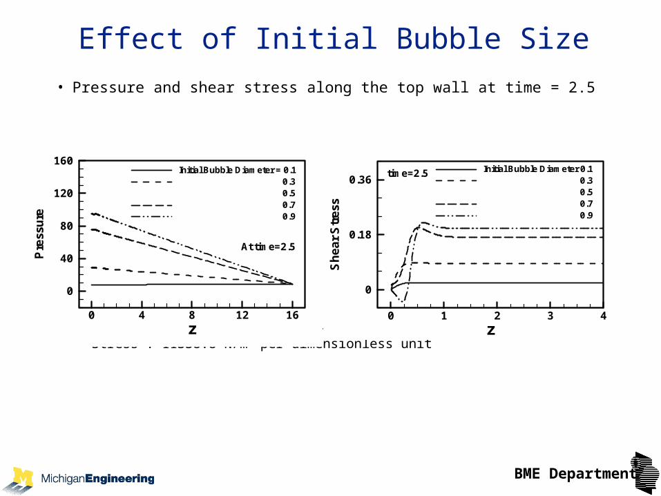
Effect of Initial Bubble Size
• Pressure and shear stress along the top wall at time = 2.5
– Time: 10.5 × 10⁻⁶ s per dimensionless unit
– Stress : 11338.8 N/m2 per dimensionless unit
z
Pre
ssu
re
0 4 8 12 16
0
40
80
120
160Initial Bubble Diameter = 0.1
0.30.50.70.9
At time=2.5
zS
he
ar
Str
ess
0 1 2 3 4
0
0.18
0.36Initial Bubble Diameter 0.1
0.30.50.70.9
time=2.5

BME Department
Rigid Tube Conclusions
• Wall pressure peaks near the beginning of expansion, proportional to initial bubble pressure
• Larger initial bubbles result in higher pressure and shear stress on the wall
• The peak shear stress usually occurs when the bubble moves close to the wall, lagging behind peak pressure in time
• Higher viscosity and surface tension reduce the peak shear stress, as does lower initial pressure

BME Department
Bubble Expansion in a Flexible Tube
• Flexible wall with stiffness and tension components
w
w
spk
liquid
bubble
z
r
• T. Ye and J.L. Bull Microbubble expansion in a flexible tube. Journal of Biomechanical Engineering, 128(4): 554-563, 2006.
BME Department
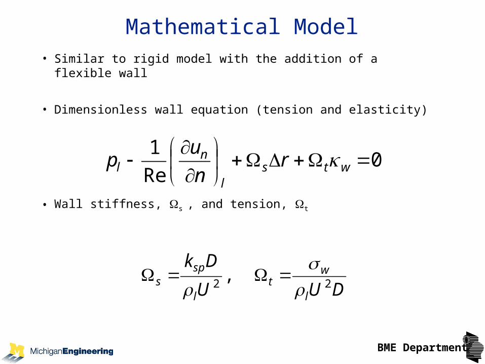
Mathematical Model
• Similar to rigid model with the addition of a flexible wall
• Dimensionless wall equation (tension and elasticity)
• Wall stiffness, s , and tension, t
0Re
1
wtsl
nl r
n
up
DUU
Dk
l
wt
l
sps 22
,
BME Department
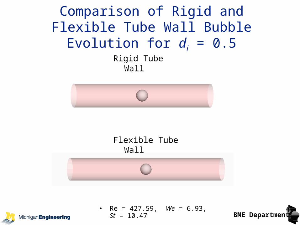
Comparison of Rigid and Flexible Tube Wall Bubble Evolution for di = 0.5
Rigid Tube Wall
Flexible Tube Wall
• Re = 427.59, We = 6.93, St = 10.47

BME Department
Streamlines for di = 0.3
• Streamline snapshotsz/Dt
r/D
t
0.0 0.5 1.0 1.5 2.0 2.5 3.0 3.5
-0.5
0.0
0.5
z/Dt
r/D
t
0.0 0.5 1.0 1.5 2.0 2.5 3.0 3.5
-0.5
0.0
0.5
z/Dt
r/D
t
0.0 0.5 1.0 1.5 2.0 2.5 3.0 3.5
-0.5
0.0
0.5
z/Dt
r/D
t
0.0 0.5 1.0 1.5 2.0 2.5 3.0 3.5
-0.5
0.0
0.5
z/Dt
r/D
t
0.0 0.5 1.0 1.5 2.0 2.5 3.0 3.5
-0.5
0.0
0.5
z/Dt
r/D
t
0.0 0.5 1.0 1.5 2.0 2.5 3.0 3.5
-0.5
0.0
0.5
t (dimensionless) = 0, 0.4, 0.8, 1.2, 1.6, 2, time scale = 10.5 μs

BME Department
Flexible Tube Conclusions
• The bubble expansion results in an increase of local tube volume, which generates liquid in flow at the open ends of the tube
• In flow and flow due to expanding bubble result in complex flow patterns and stagnation points
• Maximum wall pressure occurs near the bubble during the early stage of expansion
• High shear stresses concentrate at the center of the tube close to the bubble and near the open ends of the tube
• Larger initial bubble diameters result in higher shear stress

BME Department
Emboli Transport
•A bubble may be initially carried along by the flow without contacting the vessel walls
•The bubble then dries the walls of the vessel, but may still be swept along by the flow
•If the driving pressure is insufficient to over come the surface tension forces on the bubble, the bubble will become lodged
•Understanding the dynamics of bubble sticking is essential to knowing how to achieve a homogeneous distribution of bubbles and subsequently a uniform necrosis of the tumor

BME Department
Bubble Splitting in a Single Bifurcation
Objective: determine effects of flow rate, bubble size, and gravity on splitting ratio of the bubble in a single bifurcation
Calderón A.J., Fowlkes J.B., and Bull J.L. Bubble splitting in bifurcating tubes: a model study of cardiovascular gas emboli transport. Journal of Applied Physiology, 99: 479-487, 2005.
BME Department
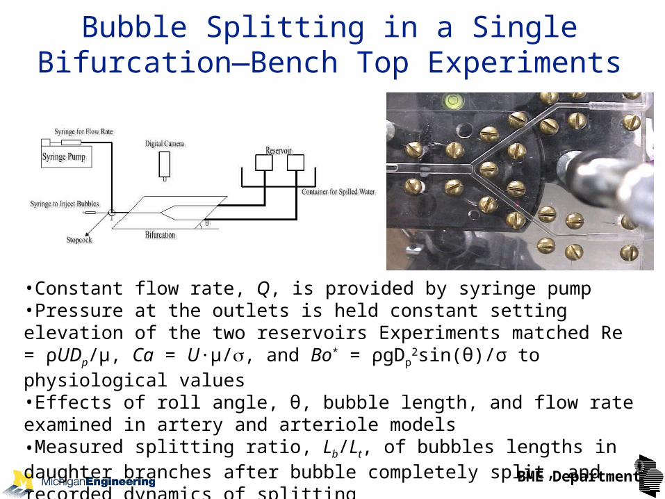
Bubble Splitting in a Single Bifurcation—Bench Top Experiments
•Constant flow rate, Q, is provided by syringe pump•Pressure at the outlets is held constant setting elevation of the two reservoirs Experiments matched Re = ρUDp/μ, Ca = U∙µ/, and Bo* = ρgDp
2sin(θ)/σ to physiological values•Effects of roll angle, θ, bubble length, and flow rate examined in artery and arteriole models•Measured splitting ratio, Lb/Lt, of bubbles lengths in daughter branches after bubble completely split, and recorded dynamics of splitting

BME Department
Bubble Splitting in a Single Bifurcation—Theory
1( )t
Q
P e z e z eP e
1
2
1( = 0 )t
L
L
P b
2( )t
•Considered one dimensional flow•Poiseuille flow ahead of bubble•“Bretherton problem” approach to model the motion of long bubbles•Governing equations
•Conservation of mass•Conservation of momentum
•Interfacial stress and kinematic boundary conditions

BME Department
Splitting Ratio vs. Capillary Number
•Capillary number, Ca = U∙µ/, indicates flow rate•Splitting ratio = Lb/Lt, is 1 if splitting is even
Artery Model Arteriole Model
Experiments θ = 0° □, θ = 15° Δ, θ = 30° ○, θ = 45° ◊, θ = 60° , θ = 90° . Theory θ = 0° ―――, θ = 15° ―― ――, θ = 30° ―― – ―― , θ = 45° …, θ = 60° ――― ―――, θ = 90° ―― – – ――.
Experiments θ = 0° □, θ = 5° Δ, θ = 10° , θ = 15° , θ = 20° ◊, θ = 30° . Theory θ = 0° ―――, θ = 5° ―― ――, θ = 10° ―― – ―― , θ = 15° …, θ = 20° ――― ―――, θ = 30° ―― – – ――.
θ
θ

BME Department
Bubble Reversal
TheoryExperiment

BME Department
Single Bifurcation Bubble Splitting Conclusions
• Higher flow rates will improve homogeneity and there is a critical flow rate below which bubbles will not split
• Bubble size relative to vessel size affects splitting behavior• Theory captures behavior of experiments• Resolved apparent paradox between Chang et al. and
Souders et al. studies• Acoustic droplet vaporization in small vessels could
potentially lead to even distribution of microbubbles• Inertial, viscous, and surface tension forces are important
—gravity doesn’t tell the whole story!

BME Department
Bubble Lodging: Experiments and Theory
•Investigated bubble lodging a microfluidics bifurcation (PDMS)
•Impose driving pressure and determine critical pressure for lodging and dislodging
Calderón A.J., Heo Y.S., Huh D., Futai N., Takayama S., Fowlkes J.B., and Bull J.L. A microfluidic model of bubble lodging in microvessel bifurcations. Applied Physics Letters, 89(24): Art. No. 244103, 2006.
235 m
120 m
200 m
91 m
Reservoir 1
Syringe
Bifurcation
Reservoir 2
Parent Channel Cross-Section Daughter Channel Cross-Section

BME Department
Theoretical Analysis
• A long bubble will lodge in a bifurcation of decreasing diameter if the pressure difference can be supported by a stationary bubble.
PD
pd s
4 41
co s co s
Sca les
Dd
DP D
I
*
*
co s
1
4
11
DI*

BME Department
Bubble Lodging States
• Two states (A & B) identified
• State A occurred at a lower pressure
• Bubble reversal often lead to bubble passing through one branch when dislodged (C)
C
C
BME Department
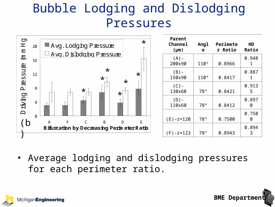
Bubble Lodging and Dislodging Pressures
• Average lodging and dislodging pressures for each perimeter ratio.
0
4
8
12
16
20
A F C B D E
Bifurcation by Decreasing Perimeter Ratio
Dri
vin
g P
ress
ure
(m
mH
g)
Avg. Lodging Pressure
Avg. Dislodging Pressure
(b)
**
**
*
*
*Parent
Channel (µm) Angle
Perimeter Ratio
HD Ratio
(A)-200x90 110º 0.8966 0.9481
(B)-150x90 110º 0.8417 0.8871
(C)-130x60 78º 0.8421 0.9135
(D)-110x60 78º 0.8412 0.8970
(E)-r=120 78º 0.7500 0.7500
(F)-r=123 78º 0.8943 0.8943

BME Department
Dimensionless Bubble Lodging Pressure vs. Diameter Ratio
• Theoretical curves and experimental data
• Smaller hydraulic diameter ratio (daughter channel diameter : parent channel diameter) results in higher lodging pressure

BME Department
•Expect microbubbles in gas embolotherapy to lodge in the microcirculation in vessels with diameters of 20 μm and less
•Once lodged, bubbles tend to remain lodged
•Bubbles tend to lodge at bifurcations, but can lodge in straight channels
•Lodged bubbles occluded transport of microspheres—suggesting they would occlude transport of red blood cells
•Follow up doses of microbubbles go to the un-occluded regions of the vasculature, indicating a strategy for occluding the entire tumor
Bubble Lodging Conclusions
BME Department
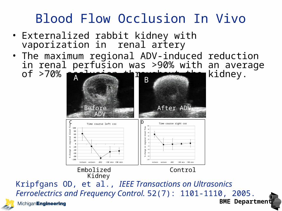
Blood Flow Occlusion In Vivo• Externalized rabbit kidney with vaporization in renal artery• The maximum regional ADV-induced reduction in renal
perfusion was >90% with an average of >70% occlusion throughout the kidney.
Time course right cortex
-100
-80
-60
-40
-20
0
20
40
60
80
100
intern extern ADV +30 min +90 min
%-c
hange in r
egio
nal blo
od fl
ow
Time course left cortex
-100
-80
-60
-40
-20
0
20
40
60
80
100
intern extern ADV +30 min +90 min
%-c
hange in r
egio
nal blo
od fl
ow
A B
C D
Before ADV After ADV
Embolized Kidney Control
Kripfgans OD, et al., IEEE Transactions on Ultrasonics Ferroelectrics and Frequency Control. 52(7): 1101-1110, 2005.

BME Department
Acknowledgements
• Collaborators (Department of Radiology, UMHS)– J. Brian Fowlkes, Oliver Kripfgans, Jon Rubin, David Williams
• Students and postdoctoral fellows (present and past)– Work shown here: Andres Calderon, Brijesh Eshpuniyani, Tao Ye
– Others in group: Khalil Khanafer, David Li, Yu-chun Lin, Molly O’Loughlin, Stan Samuel, Marty Schlicht, Robinson Seda, Balaji Srinivasan, Brad Steele, James Stephen, Doug Valassis, Zheng Zheng Wong
• Funding– NIH R01EB006476
– NIH EB003541
– NSF BES-0301278
– Whitaker Foundation RG-03-0017