bme 201 project liver bioreactor
TRANSCRIPT

Executive Summary Tissue engineering and organ regeneration are relatively new concepts in the field of biomedical engineering. The liver is the second largest internal organ and plays a vital role in human survival, yet there are many problems with the functionality of the liver. There is no concrete
solution other than a transplant that can keep a patient with liver failure alive. While many options have been analyzed, nothing has been deemed successful. We felt that a remedy to this
problem would be to create a bioreactor that combines pre-existing concepts. Our hope is that such a device will be able to produce bioartificial livers that can adequately fulfill all the duties of a natural liver and allow patients suffering from liver failure to live longer and healthier lives.
The Task Bioreactors encompass a lot of emerging technology with a vast set of applications in the fields of medicine and biomedical engineering. Being able to grow artificial organs that are fully
functioning within a bioreactor has the potential to save thousands of lives. There is always a higher demand for organs than there are organs that are able to be transplanted. The Hollow fiber-cylindrical combination bioreactor design is simple enough to be implemented and tested
because it does not involve any new technology - but rather the combination and collaboration of existing technologies. Our model has the potential to create a fully functioning liver.
The Problem Statement Select appropriate scaffolds and design a bioreactor to grow a tissue engineered liver or kidney
for organ transplantation or for life support.
The Process As a group, we discussed the subject matter and realized that our first task was to choose if we wanted to create a bioreactor that could grow a kidney or a liver. We looked into the anatomy of
each organ and problems that are well known. After discussing this material we decided to create a bioreactor that would be able to grow a fully functioning liver. We then realized that our
knowledge in regards to bioreactors was limited. We had a lot of questions that needed answers. Some of the areas that we had questions in were current technologies, problems in growing a liver in vitro, how bioreactors function, and the best way to create a durable scaffold. We divided
up the subject matter and everyone had a different area of research. Once we all had a better understanding of bioreactor systems we were able to establish project goals and start coming up
with our own ideas. Next we discussed all the current technology and answered everyone’s questions. Then we started to compare each existing bioreactor and scaffold keeping in mind the pros and cons of each. We considered the pros of each model and chose what factors we wanted
to input in our design. Once we had an overall idea, we discussed the problems that we would encounter if we were to actually carry this project out. The team then elaborated on the process
we would use to validate our results. A plan of development was created to keep the team on track for finishing the final report.

The Modifications/ Solutions As a team we researched existing bioreactor technologies and looked into problems that people
have experienced up to date with creating a liver in vitro. We brainstormed numerous potential solutions. During the initial brainstorming period we were receptive of all ideas. Each of our
solutions were similar to existing bioreactors. The bioreactors that we reviewed include: the hollow fiber bioreactor, the cylindrical bioreactor, the rotary bioreactor, and the single use bag bioreactor. After the initial brainstorming phase was complete, we then weighed the pros and
cons of each alternative and decided that our model would combine existing bioreactor and scaffold.
Conclusion We ultimately decided that combining the key attributes of the hollow fiber bioreactor and the cylindrical bioreactor would best fulfill the task at hand. This solution in combination with a unique scaffold has the greatest likelihood of creating a fully functioning liver in vitro. With the
implementation of these modifications, bioengineers will be one step closer to fully understanding all the functionalities of the liver and being able to produce an artificial liver that
can serve as a viable option for patient transplant.
Background
As the largest internal organ, the liver performs some of the most diverse functions in the
human body. Besides the vascular system, the liver is composed of two lobes and a complex
biliary system. Functionality ranges from detoxification, to the creation of essential biological
components (amino acids, bile, proteins, vitamins, etc.), to storage of energy.
The liver is undoubtedly vital to sustaining life, so when it impaired a solution must be
implemented. There are several ways a liver can be damaged. This includes viral infections
(Hepatitis strains), toxin damages, and various genetic diseases. While the liver can regenerate
itself from as low as 25% of its initial volume, this natural solution is not always enough. Other
solutions include liver dialysis and, more recently, disposable bioreactors that function through
tissue engineered liver tissue. Both solutions ultimately fall short of completing the wide array
of functions a natural, healthy liver performs. Transplants exist as an option to replace a faulty

liver, but this requires a donor. However, another approach exists in tissue engineering. This
focus of biomedical engineering has the potential to cure damaged and diseased livers, allowing
patients to return to normalcy.
Problems Cause by Liver Failure
Without a fully functioning liver, many organs within the body will degenerate and incite
permanent illnesses. The major effects of liver failure are highlighted including jaundice, liver
cancer, portal hypertension, and hepatitis.
Jaundice results from having too much bilirubin within the blood. Bilirubin forms when
hemoglobin in red blood cells breaks down when recycling an old or damaged red blood cell. It
flows in the bloodstream, continues to disintegrate in the liver, and is disposed of through urine.
If the liver cannot break down the bilirubin within the bile ducts quick enough, it streams
through the blood again and is disposed of on the epidermis. This produces a yellow pigment on
the skin or whites of a person’s eyes. This can become an extreme problem among infants
because it can cause serious brain damage called kernicterus.
Liver cancer usually results from cancer cells found within another part of the body.
Since the liver filters blood from all parts of the body, cancer cells usually originate elsewhere
and lodge inside the liver. Two types of tumors can form. Benign tumors are relatively
harmless. Hemangioma is the most common benign tumor which causes abnormal growth of
blood vessels beginning in the fetus. It has no symptoms and requires no treatment. Malignant
tumors are capable of spreading from the liver to the rest of the body and have serious side
effects. The most common form of primary liver cancer is hepatocellular carcinoma. It affects
liver cells and has many different growth patterns that affect different parts of the body. Most

symptoms of liver cancer are seen in weight loss, loss of appetite, abdominal pain, jaundice, and
abdominal fluid.
Portal hypertension causes abnormally high blood pressure in the portal vein, a large vein
that brings blood from the intestine to the liver, and its branches. This is most likely from
cirrhosis, scarring that damages the liver performance, and can lead to swollen in the abdomen
and bleeding from the digestive tract. Two factors that contribute to portal hypertension are an
increase in volume of blood flowing through the vessels and increased resistance to blood flow
through the liver. The body will try to instinctively fix this problem by creating new collateral
vessels that bypass the liver and redirect blood into general circulation. This could lead to more
problems because toxins will not be broken down within the liver. In addition, an increase of
blood pressure in the spleen will decrease the total count of white blood cells and platelets.
Hepatitis is inflammation in the liver caused by a virus. There are three forms of
Hepatitis. Hepatitis A spreads when a person comes in contact with an infected person’s stool.
Many time symptoms will not show but patients feel flu-like symptoms and jaundice. It usually
dissipates on its own with no serious complications. Hepatitis A is usually acute and newly
occurring infections typically do not become chronic. Hepatitis B (HBV) is caused by the liver
swelling. If a mother carrying HBV has a baby, the baby is at risk of developing chronic HBV
which can lead to cirrhosis, liver cancer, and liver failure. Most people do not experience
symptoms but possible symptoms include tiredness, fever, loss of appetite, headache, muscle
soreness, pain near the liver and jaundice. Hepatitis C (HCV) spreads by contact with an
infected person’s blood. HCV is a lifelong liver disease as there is no vaccine currently
available. What start off looking like cold symptoms could result in permanent scars on the

liver. If patients are diagnosed with a chronic case of Hepatitis C, they usually develop cirrhosis
and liver cancer.
Bioreactors
A new method developed to tackle solutions for liver damage introduces bioreactors that
cultivate cells to perform certain tasks. Bioreactors allow cells or tissues to be grown in a closed
culture environment in vitro, with the intention of being used in vivo. A bioreactor stimulates
cells to proliferate and differentiate by mechanical stimulation, which ultimately encourages the
production of extracellular matrix (ECM). With careful consideration of the in vivo environment
and types a scaffolds used, bioreactors can support all types of tissue and eventually sustain
whole organs.
To grow properly, engineers must control variables in an in vitro setting. Some factors
that must be looked into are temperature, pH, stress, forces on the cell, and concentration of
certain compounds like oxygen. One of the main challenges is creating an environment in which
cells can grow and differentiate into tissue once implanted. The bioreactor must be also able to
generate different cell types and be biocompatible with the selected scaffold.
There are three situations in which tissue engineers would use bioreactors. One case
utilizes bioreactors to grow cells in a closed controlled environment that can later be used for
transplantation. In a second case, bioreactors can house a 3D tissue structure in vitro to be
implanted into an in vivo. Mainly bioreactors become organ support systems, aiding waning
organs like the liver or kidney.
To design a bioreactor, one must first access the desired cells and grow them in a well
controlled culture that keeps all of the variables, stated before, as if inside the body. It is

important that cells create cell lines with primary and progenitor cells so that the growth and
differentiation is maintained. It is also necessary for the culture to distinguish between specific
cell types and prevent microorganisms from invade intervening. The tissue parameters are not
constant and must be monitored at all times to keep the cells contained in an in vivo-like
environment. Mechanical forces and stresses on the tissues and cells must also be taken into
account.
Scaffolds
Scaffolds are used to create the physical form of the organ by holding cells in the desired
shape. There are two principal procedures in which cells are integrated onto the scaffold - cell
seeding and continue growth. A scaffold can be seeded either by transferring differentiated cells
from an adjacent bioreactor or by directly pumping cells onto the scaffold located inside the
bioreactor. The cell seeding process allows for maximum cell growth, creating a stronger tissue
structure. One of the major challenges in creating a good bioreactor is substrate rigidity. “In vivo,
tissues are on average no further than 100 µm from a capillary providing nutrients” (Chaudhuri
et al., 2005). This small area makes tissue engineers “consider a cube with sides of 100 µm as
being the fundamental microenvironment for tissues” (Chaudhuri et al., 2005). Tissues of this
size usually have 500 to 1000 mixed types of cell, being proliferated and differentiated
(Chaudhuri et al., 2005). This is very hard for a man-made bioreactor to achieve because of all
the components that the specific tissue needs. If the bioreactor is 100 micrometers thick the
scaffold would have to be a few millimeters from the thickness of the shape. This results in the
bioreactor being farther away from the nutrient source capillaries and might lead the in vivo
bioreactor to die. To overcome this problem, a thin porous scaffold must be customized for each

tissue to allow the maximum amount of nutrient transport to each cell. The porous scaffold also
allows permits fluid and molecular mass transfer while exposing waste out of the bioreactor,
mimicking regular flow rate in the body. To see what the mass transfer concentration is for a
specific organ, start testing at the average starting point. “Overall in vivo rates of oxygen intake
are 25 to 250 moll O2/cm3/h and perfusion rates are 0.07 mL/cm3/min, based on an average
cellularity of 500 million cells per cm3” (Chaudhuri et al., 2005).
An aspiring idea for many tissue engineers is to make more complex tissues by growing
multiple cell types in the same bioreactor. Many complex tissues are made of several types of
cells that work interdependently. The challenge is constructing a scaffold that allows for a co-
culture accommodating cells with different requirements for constituents needed to differentiate
each cell. Current ideas involve growing individual cell types in independent cell cultures for a
period of time, then merging the matured cells into a common growth culture at the end.
More research must be conducted towards computing the correct constraints for each
given scaffold. No matter what scaffold is used, it must be biocompatible and exude appropriate
surface properties, substrate rigidity, pore structure, mechanical strength, and degradation rate.
The scaffold is biocompatible if it does not trigger an immune or inflammatory response, is
sterilizable, and degrade without breaking into components that are cytotoxic, inflammatory, or
immunogenic. If a scaffold has proper surface properties, it is hydrophilic and exhibits qualities
regarding roughness, crystallinity, charge, and functionality comparable to that tissue.
Furthermore cell cytoskeletal shape and the associated cellular functions are determined by the
rigidity of the stiffness of the scaffold (Albert, Lewis, et al., 2007). Scaffold stiffness similar to
native tissue will yield a cellular phenotype similar to that of native tissue. Cell proliferation and
degradation occurs within this set space, and tissue induction and growth ensures throughout the
scaffold once the tissue operates in vivo. Therapeutic agents such as growth factors and nutrients
must also be delivered through the scaffold. The structure integrity induces tissue-specific
mechanical forces to the cells for appropriate cell behavior.
Selecting the proper cells to incorporate into the scaffold is an important factor. The liver
is mostly composed of parenchymal cells, of which there are many different types. Hepatocytes,
hepatocyte precursor cells, stellate cells, kuppfer cells, epithelial cells, sinusoidal epithelial cells,
biliary epithelial cells, and fibroblasts all are cells that the liver is composed of. Hepatocytes
make up seventy percent of the cellular portion of the liver. These cells perform major metabolic
functions such as plasma protein synthesis and transport, xenobiotic metabolism, glucose
homeostasis, urea synthesis, and ketogenesis (Kazemnejad, 2014).
Rationale for Design
Hollow Fiber-Cylindrical Combination Bioreactor
The liver is a very complex organ and it is very unlikely that a single in vitro liver will be
able to perform every individual as well as integrated function of the liver. In order for a
successful model to be created it is important to understand that the bioreactor model must be
able to “include approaches that involve a designed or programmed fluid flow as an integral part
of the culture format, where flow is used to enhance molecular transport, provide mechanical
stimulation, control addition of drugs or biological regulators on a diurnal or other temporal
basis, or to otherwise influence cell function and/or assay performance in ways that are not
readily accessible in static culture formats” (Ebrahimkhani,1).

As a team we felt that the hollow tube bioreactor and the cylindrical bioreactor had the
most potential and were understood the best by the experimenting community. Hollow fiber
bioreactors have been used to create an environment that is isolated from outside influences.
This type of environment allows cells to grow naturally. Throughout the isolated cell growth
process the cells in culture will be introduced to a medium from the central reservoir that will
continuously circulate various nutrients, growth factors, and drugs. The culture can be
monitored to see how the cells function independently of one another as well as to see how
resistant strains are formed (Advantages,2). Hollow fiber bioreactors are usually used to create a
cell that has been conditioned to complete a single specific task as well as to monitor and
understand the development of resistant cell strains. The isolated environment in combination
with close observation allows for bacterial loads to remain constant in the hopes of modeling
accurate absorption and elimination curves for each cell type( Caddwell,4).
Hollow fibers are small-tube like fibers about 200 microns in diameter whose molecular
weight can be cut-off between 5 kd and 0.1 µm. Hydrophilic polysulfone fibers will be used in
the cartridge shell. Based on the information collected by FiberCell® Systems, hydrophilic
polysulfone fibers result in good culture performance. At FiberCell® Systems they offer three
different molecular weight cut-off (MWCO): 5 kd, 20 kd, and 0.1 micron and three types of
fibers: polysulfone, cellulosic, and PS+. Both their 5 kd MWCO and 20 kd MWCO have ten
times the gross filtration rate than equivalent cellulosic fibers for rapid nutrient and waste
exchange. This means better cell growth and higher fidelity of protein production not found in
cellulosic fibers. “Any cell type that will attach to plastic or grow in suspension should grow
well in the polysulfone cartridge” (Advantages,3). The fibers will be cast with little waves,
resulting in an even distribution of fiber package within the cartridge shell. The fibers provide

an immense amount of surface area in a small volume. Cells grow on and around the fibers at
densities of greater than 1 × 10⁸ per mL. The cartridge used depends on the size of secreted
product and amount collected. Modeled off of FiberCell® Systems’ medium sized C2011 model
with surface area of 3,000 𝑐𝑚2 and volume of 20 mL, the bioreactor would produce 10 L bag
type bioreactor.
In our design, separate vessels containing the cell culture medium, growth factors, and
nutrients and oxygen will be pumped into one end of the hollow fiber cartridge shell at a
controlled rate. The extracapillary space between the hollow fibers is where bone marrow stem
cells will receive nutrients from the culture medium and grow independently. Waste from the
cells permeate through the fibers and are dissipated into a waste chamber at the subsequent end
of the shell. After a period of time, bone marrow stromal cells mature into hepatic progenitor
cells and are circulated into the scaffold.
The cells chosen to be cultured in the hollow fiber bioreactor are stem cells obtained from
the bone marrow of adults. Bone marrow stem cells that have differentiated into hepatic
progenitor cells are also possible to use. Hepatic progenitor cells can differentiate into many
functional cells of the liver, such as hepatocytes or bile duct cells. Primary mature hepatocytes
are not useful because they do not replicate sufficiently in vitro. Many growth factors and
signaling proteins are needed to cause proper differentiation of the bone marrow stem cells into
hepatocytes and progenitor cells. Some of these necessary proteins and growth factors are
activin, fibroblastic growth factor (FGF), bone morphogenesis protein (BMP), hepatocyte growth
factor (HGF), and Oncostatin M (OSM). Hepatocyte growth factor mediates the growth,
proliferation, angiogenesis, and cell motility of these cells; it is one of the most important factors.
Further growth and differentiation of hepatocytes is controlled by epidermal growth factor

(EGF), FGF, Interleukin-6 (IL-6), transforming Growth Factor, and insulin- like Growth Factor
(IGF). Function and differentiation of the hepatocytes is stimulated by corticosteroids, amino
acids, OSM, nicotanimide, and dimethyl sulfide (DMSO). The proper combination of all of these
growth factors is necessary to obtain the proper amounts of each type of cell needed to form a
functional tissue-engineered liver (Kazemnejad, 2014). Dulbecco’s Modified Eagle’s Medium
Modified is our chosen cell medium. This medium is useful for applications with stem cells. It
is in “powder form with L-glutamine and 1000 mg/L glucose, without sodium bicarbonate,
formulated at 10.0 grams of powder per liter of medium, and has been cell culture tested”
(Dulbecco’s, 1).
Other factors remaining constant in the bioreactor including pressure force, temperature,
and flow rate of inserted materials. A loop of silicone tubing will be wrapped around the core of
the cartridge that is gas permeable will warrant gas composition of the medium that is the same
as the composition in the vessel. The temperature should be maintained around 37 degrees
Celsius. According to a comprehensive computational study modelling the execution of a
rotating hollow-fiber bioreactor for artificial liver (BAL), the rotational speed of the chamber
was analyzed to determine that homogenous distribution of cells was successful, along with
oxygen delivery and cellular oxygen consumption as a guide of cellular metabolic activity, and
the fluid- induced mechanical stress experienced by cells. They found that “homogenous
distribution of cells is reached at a rotational speed of 30 rpm; spreading of cellular concentration
at around the initial value of 12% was limited (median = 11.97%, 5th percentile = 10.94%, 95th
percentile = 13.2%), resulting in uniform suspension of microCAACs (microcarrier-attached
aggregated cells), which did not appear to be excessively packed. Mixing within the rotating
fluid caused a maximum fluid- induced stress value of 0.05 Pa, which was neither endangering

for liver-specific functions of cultured cells, nor causing disruption of the floating aggregates.
Moreover, an inlet medium flow rate of 200 mL/m with a partial pressure of oxygen (pO(2))
value of 160 mmHg was found to guarantee an adequate O(2) supply for the hepatocytes (2.7 x
10(8) hepatocytes are simulated); under such conditions, the minimum pO(2) value (23 mmHg)
is above the critical threshold value, causing the onset of cellular hypoxia (10 mmHg). ” (A
computational, 1). The results from this computational model proved to optimize transport in the
hollow fiber bioreactor.
Figure 1. Cross Sectional View of Hollow Fiber Bioreactor
Hollow fiber filter surface
Isolated extracapillary space
where cells grow
Bacteria or cells retained
Cell Culture medium in
Waste Out

Figure 2. Closer look at cross sectional view
The cylindrical bioreactor, on the other hand, involves constant movement of the growing
cells. The rotations and vibrations either come from the top or bottom while very small gas
bubbles are pumped through the entire bioreactor to assure that the cells are getting enough
oxygen. The shaking in combination with the constant gas flow provides a well-defined
hydrodynamic system that allows for excellent mixing and oxygen transfer for mammalian and
plant cell cultivations. Under these conditions it is easy to control the cell environment. More
specifically, the fully controlled tank bioreactor provides automated control of the culture
environment. This includes temperature, pH, and dissolved oxygen controls which
are mandatory for cell development and cultivation. Each of these environmental conditions can
be adapted to different bioprocesses and accommodated to different three-dimensional (3D) cell
culture strategies (Hampbell,6). We plan to apply very strict controls to our scaffold in order to
ensure that the liver turns out to be fully functioning. It is important to keep in mind that the
stressors from the cylindrical bioreactor force each cell to work together as opposed to in the
hollow fiber bioreactor where every cell functioned independently of one another even though
they were all in the same environment (Hampbell,7). Although both of these bioreactors had
good things to offer, our ultimate task was to design a new bioreactor that would be able to grow
and harvest a fully functioning liver.
After researching current bioreactors and various in vitro methods for the creation of a
liver, we decided that the best route to take was to combine the hollow fiber bioreactor with the
cylindrical bioreactor. The hollow fiber bioreactor would serve as a conditioned environment
where cells would receive the correct growth factors and signals that would lead to their terminal

state of differentiation being a liver cell. The cells would be kept in the hollow fiber bioreactor
until they were all fully differentiated. The hollow fibers themselves within the bioreactor will
remain the same. There will be hundreds of small tube-like filters that are sealed in a larger
nonporous cartridge shell. The medium that is flowing through the fibers is pumped from one
end of the shell to the other to create a constant flow of nutrients and growth factors that flow
into the cartilage cell and influence the cells that are just outside of the fiber themselves
(Aadvantages,4). The only thing that would be changed in regards to the hollow fiber bioreactor
cell growth process is that the main purpose would be to cultivate fully differentiated cells rather
than study the process that leads cells to be resistant to various drugs. No drugs will be pumped
through the fibers since they influence the way that the cells grow and differentiate. After the
cells are developed in the hollow fiber bioreactor they could be pumped out of the system onto
the scaffold situated inside the cylindrical bioreactor.
PGA, Collagen, and Fibronectin Scaffold
These cells, growth factors, and proteins combined in our bioreactor prior to the scaffold
continue to intermingle once they reach the scaffold. Pulsations are sent through the bioreactor
cartridge to promote cell growth and differentiation; these pulsations work together with the
growth factors and signaling proteins to achieve this. The scaffold will be created using
nanofibers and electrospinning techniques. A mixture of polyglycolic acid (PGA), collagen, and
fibronectin is used to create the nanofibers (Gluck, 2007). PGA, collagen, and fibronectin are an
ideal choice for our scaffold because combining natural components of the extracellular matrix
(ECM) and synthetic polymers for a scaffold have been proven to be successful in the past. PGA
can be manipulated to work in many different ways, while utilizing natural components of the
ECM (collagen and fibronectin) keep the scaffold closer to the original human liver (Gluck,
2007). The combination of PGA, collagen, and fibronectin also reduces the hydrophobic traits of
PGA, which hinders successful cell seeding. PGA is also very accessible, and the degradation
rate can be manipulated (Albert, Lewis, et al., 2007).
The solution of PGA, collagen, and fibronectin would be passed through a metallic
needle at a controlled rate onto a collection plate to produce a non-woven nanofiber mesh. A
large direct current is applied to the needle and polymer mixture to induce its expulsion through
the needle to create the nanofiber. The nanofiber would be electrospun onto a grounded
collection plate where it would create a non-woven mat of nanofibers (Li & Xia, 2004). Varying
amounts of nanofibers can then be stacked and connected to create the desired shape of the liver.
Electrospinning the scaffold for the liver is advantageous because the nanofibers are more
compact than other forms of scaffolds (Li & Xia, 2004). This has been known to benefit cell
proliferation on the scaffold.
It will be necessary to combine the mixture of PGA, collagen, and fibronectin with a
solvent. The solvent used must be able to evaporate quickly away from the polymer mixture to
help form the nanofiber. Two ideal solvents for the polymer mixture are chloroform and
methanol. Using different ratios between the PGA, collagen, and fibronectin mixture and the
solvents used will create an optimal balance of pore size and nanofiber diameter (Gluck, 2007).
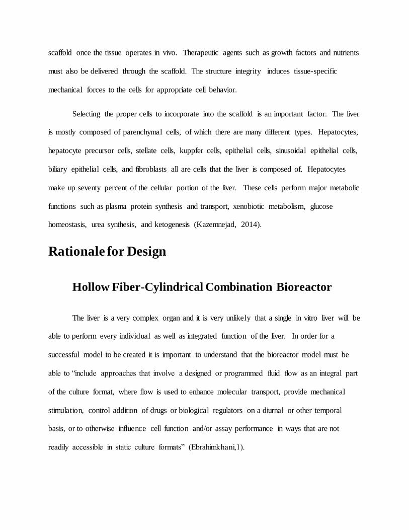
Once the scaffold has been appropriately constructed, it will be placed in a reservoir
connected to the bioreactor cartridge. Cells will move from the bioreactor cartridge into the
reservoir to attach to the scaffold. This reservoir will be attached to a motor that creates a
vibrating motion. The vibrations applied to the scaffold will cause the cells to work as a single
unit and help the liver to form together, as opposed to a collection of cells working
independently of one another. This will also promote proper vascularization of the liver tissue.
Our scaffold will be created using preexisting processes (electrostatic spinning) but out of
an unique combination of materials. This type of scaffold has not been used in practice yet. We
feel that this unique scaffold will be as strong and porous as scaffolds created mainly from
collagenous fibers (Gauvin,1). Our scaffold model will also be able to have all the neurological
adhesive support of scaffolds created with fibronectin and laminin (Kiode,1). The polyglycolic
acid will allow the scaffold to be as biocompatible and bio absorbable as possible (Boland,3).
Figure 3. Complete diagram of hollow fiber-cylindrical bioreactor combination with scaffold
In the steps after the cells are removed from the hollow fiber bioreactor, the team took

creative control to figure out the best series of steps to accomplish the goal and created a process
involving the in vitro growth of a liver that differs from anything that has been done so far in a
laboratory. Since the cells need to work as a cohesive unit on the scaffold prior to creating a
fully functioning liver, we felt that that the scaffold should be integrated into a modified
cylindrical bioreactor. A metal hook would be used to attach the liver scaffold Instead of
continuously stirring and agitating the medium after leaving the hollow fiber bioreactor, we
decided that the best way to have the cells grow as a cohesive unit and create a fully functioning
liver was to take the vibrating paddles from the cylindrical bioreactor and place them under the
scaffold. The purpose of the mechanistic change is to put some strain on the cells and force them
to start to work together in order to increase the likelihood that they will survive. The small
vibrations will force the cells to come together. It is not necessary to stir the cells after they are
removed from the hollow fiber bioreactor and pumped on to the scaffold. This is because we
need the fully differentiated cells to mesh with the scaffold so that a fully functioning,
transplantable liver can be grown in vitro rather than creating an environment that will expedite
individual cell growth. If the cells are constantly stirred it is very likely that they would link
together and start to form a functional tissue. It is rare to see one bioreactor working in junction
with one of a different type since they all have such different capabilities in terms of cell
cultivation. We felt that the best way to be able to grow a liver in a bioreactor would be to
combine all the major attributes of various bioreactors in the hopes of making the growth of an in
vitro liver a more fluid process.
Plan for Verification
Bone marrow stromal cells, as a subset of mesenchymal stem cells, prove to be a great
cell source because of their ease of isolation, manipulability, and potential for

differentiation. Mesenchymal stem cells release a spectrum of bioactive immunosuppressive
molecules providing a regenerative microenvironment for injured tissues to instigate a self-
regulated regenerative response. In addition, since all of the cells in the final product are natural
progenies of bone marrow mesenchymal stem cells (MSCs), they should be able to successfully
transdifferentiate into other kinds of cells. An in vitro differentiation strategy demonstrated that
human MSCs pre-committed to a specific mesenchymal cell lineage can convert into other cell
types in response to extracellular stimuli. In a lab we would attempt to replicate the extracellular
cues and verify this plastic property of MSCs. We would then go on to manually set the rate of
diffusion of culture medium to execute several cell types.
In a joint project conducted by Salerno and colleagues, they implemented a crossed
hollow fiber membrane bioreactor for liver tissue engineering as a tool for drug testing and
toxicology. While it had a different purpose, the bioreactor performed well in a controlled
environment that may become an inexpensive and reliable in vitro model for engineered liver
tissue constructs. The bioreactor had two kinds of hollow fiber membranes of different
molecular weight and physico-chemical properties cross assembled, using modified
polyetheretherketone(PEEK-WC) and polyethersulfone(PES) for medium inflow and
outflow. The two sets created an extracapillary network for cell adhesion and mass exchange of
culture medium. Cells were seeded in an extraluminal compartment. Primary human
hepatocytes in the PEEK-WC hollow fibers provide the oxygenated medium as the PES hollow
fibers remove cell waste products, mimicking in vivo arterial and venous blood
vessels. Hepatocytes maintained differentiated functionality at high levels up to eighteen days of
culture and established high cell adhesion on fibers surrounded by an extracellular matrix-like
structure.

Instead of following the two hollow fiber membrane system, we decided to employ a
single hollow fiber system along with a motorized base attached to the reservoir containing the
liver scaffold. The idea for the motorized foundation was derived to incorporate the mechanical
stresses generated in a cylindrical bioreactor. The perfusion system and fluid dynamics and mass
transport of nutrients in the hollow fiber bioreactor is enough to develop matured cells, however
it cannot combat problems related to static culture conditions. Without a perfusion system
installed to constantly replenish cells with fresh culture medium, there will not be uniform
distribution on the scaffold.
Depending on the function of the liver being tested, we will compare our results to those
of a healthy, functioning liver in a human or animal. For example, one of the liver’s main
functions is detoxification. When our tissue engineered liver is tested to see how well it filters
waste, if the results show that it filtered over ninety percent of the waste, we will consider the
engineered liver passed the test and can perform that function. If it filters less than ninety
percent, we will go back and analyze the cells from the hollow-fiber bioreactor and adjust the
input amounts until the desired function can be performed. The same procedure will be repeated
to test other functions of the liver.
Problems and Limits
Engineering a bioreactor that can house and develop a complex organ, such as the liver,
is obviously a difficult task as it has never been done before. Past designs are laden with
problems. Likewise our design is subject to have issues and runs into multiple flaws that, with
present technology and knowledge, will be difficult to overcome.
The most prevalent issue with our design is its overwhelming cost. The goal of any
engineering design is to make more than just one prototype. The design created must be able to
be implemented into large scale production. Essentially, creating one functioning liver will not
solve the problem the design was intended to fix. Hence, our design must be reproducible on a
large scale. Disregarding the availability of materials, this is not too difficult of a task. However,
in the real world it is not possible to disregard the availability of materials. Cost becomes a
significant issue once this is taken into account.
The cost of creating a liver has more to do with the cost of the materials than the
bioreactor itself because the materials are so complex. The bioreactor may be expensive, but this
is a one time cost, where as the materials are needed for each liver made. When a liver is made
naturally, that is through organogenesis in utero, the cost of the materials is solely the food the
mother consumes to nourish herself and the fetus she is supporting. Human biology can readily
turn basic compounds, such as carbohydrates, into more complex systems, such as cells and
eventually organs. However, human biotechnology is not nearly as advanced. With our present
understanding, stem cells are our only means to make organs. The cost of food will always be
cheaper than the cost of stem cells, because the food is what is used to make the stem cells. All in
all, it is less expensive to make a new liver through natural means than through engineering.
Cell sources are expensive not only because they are extraordinarily complex, but
because they are sufficiently rare. The main cells our design would use is bone marrow cells. A
similar extraction of biomaterials is blood donation. Major blood banks are usually on short
supply, and donating blood is much simpler than donating bone marrow cells. Hence, to assume
the supply of these cells would be sufficient if provided solely through donations is unrealistic.
The supply of these cells is not nearly available enough to expect a low cost. Coupled with the
complexity of the materials in demand, the price of the cell sources will be high.
Intercellular adhesion and/or adhesion to the hollow fibers themselves in the bioreactor is
also a potential mishap. Mesenchymal stem cells are naturally adhesive, which may be a
problem when the cells are extracted from the chamber. The experimenter would have to resort
to manually scraping cells out of the bioreactor or applying a mechanical force strong enough to
expel the cells. During this process, a good percentage of the cells may experience apoptosis.
This may also lead people to ask “How does the bioreactor know which cells are
completely matured and ready to be delivered to the scaffold?” Previous studies would have to
have been conducted and approved to produce certain cell types for experimenters to compute
the amount of starting material needed. Additionally, no other method has been published to
first include a hollow fiber bioreactor to develop cells then introduce a second bioreactor that
stores the scaffold and promotes cellular interaction. Therefore, there is no source to reference
that would explain how to discern mature from non-mature cells. Even though we can estimate
the time it normally takes cells to differentiate, they are hard enough to observe, as they are not
visible to the naked eye.
In the process of electrospinning the nanofiber mesh, we encounter the problem of not
knowing definitive rates at which scaffold material (PGA, collagen, and fibronectin) should be
pumped through the metallic needle and the strength and total current needed. Depending on
which solvent is used, we would also have to determine the ratios of scaffold solution with
different solvents. Conclusive results cannot be found because this specific combinat ion of
bioreactors has never been tested.
As for the scaffold material itself, the amalgamation of natural and synthetic fibers has
pros and cons. With collagen and fibronectin are natural materials, they are undoubtedly
biocompatible, however with PGA it is synthetic and can be manipulated. Each material has

their good qualities; however in combination there is no way to guarantee that their best features
will shine through. Since this experiment has not been performed before, we cannot state the
degradation rate of the scaffold, strength of motor vibrations, and the force driving the pulsations
through the bioreactor.
Another major issue is the lack of vascularization. Once the liver is complete, a complex
vascular system must be in place for it to function and thrive in vivo. On top of this, a biliary
system must also be in place, which is created through a similar mechanism. These systems have
both macroscopic components and microscopic components. The large arteries, veins, and bile
ducts within the liver can easily be implemented through scaffold design. However, the
microscopic components are so small that incorporating them into the scaffold would be very
difficult. Even if creating a capillary system out of the mixture of PGA, collagen, and fibronectin
was efficient, the tubules the endothelial cells would create around the scaffold would be too
small to function. Therefore, using the scaffold as a means to create a vascular and biliary system
is insufficient.
Our design attempts to mitigate the issue of microscopic vascularization through
mechanical stimulation. Previous designs have shown that mechanical stimulation, specifically
rotation, can lead to arteries forming through mass transport. While this method is a start, more
options are available to help. Our scaffold can promote angiogenesis, the formation of new
capillaries, through the inclusion of certain growth factors. Other approaches are available, but
all severely limit scaffold design. Overall, none of these methods have proven sufficient for
creating a viable vascular system. For this design to become valid, more research into
vascularization must be done.

All in all, our design takes ideas from previous examples and attempts to mesh multiple
designs into one. Without experimentation it will be impossible to tell whether our design will
work. Nonetheless, it is worthwhile to experiment with this design because it addresses problems
previous bioreactors have had. Our design may not be perfect, but ultimately it takes a step in the
right direction.
References
" Advantages of Hollow Fiber Cell Culture." Advantages of Hollow Fiber Cell Culture. Fiber Cell Systems, n.d. Web. 09 Dec. 2014. <http://www.fibercellsystems.com/advantage.htm>.
Auger, Francois A., Laure Gibot, and Dan Lacroix. "The Pivotal Role of Vascularization in
Tissue Engineering." Annual Review of Biomedical Engineering, 15(1):177. Annual Reviews,
29 Aug. 2013. Web. 1 Dec. 2014. "Bioreactors for Tissue Engineering by Varun Chalupadi." OpenWetWare RSS. N.p., n.d. Web.
10 Dec. 2014. <http://openwetware.org/wiki/Bioreactors_for_Tissue_Engineering_by_Varun_Chalupadi>.
Bruce Albert, Alexander Johnson, Julian Lewis, Martin Raff, Keith Roberts, Peter Walter,
“Molecular Biology of The Cell” fifth edition, GS Garland Science Taylor & Francis Group,
ISBN 0-8153-4105-9
Cadwell, John J.S. "IN VITRO TOXICOLOGY AND HOLLOW FIBER BIOREACTORS." Fiber Cell Systems (n.d.): n. pag. Fiber Cell Systems. Web. 9 Dec. 2014.
<http://www.fibercellsystems.com/documents/Article-In%20vitro%20Toxicology%20and%20Hollow%20Fiber%20Bioreactors.pdf>.
Caplan, Arnold I. "Adult Mesenchymal Stem Cells for Tissue Engineering versus Regenerative Medicine." Journal of Cellular Physiology 213.2 (2007): 341-47. Web.
Chaudhuri, Julian, and Mohamed Al-Rubeai. Bioreactors for Tissue Engineering: Principles,
Design and Operation. Dordrecht: Springer, 2005. Google. Web.

<http://books.google.com/books?id=6fZGf8ave3wC&pg=PA368&lpg=PA368&dq=What+happens+in+a+general+liver+bioreactor&source=bl&ots=Ce87-
9IT5y&sig=4LPTRiFa74g9J7bqjHI9Si_Qd9Y&hl=en&sa=X&ei=jQluVNv7CMy0sATtfw&ved=0CDwQ6AEwBg#v=onepage&q&f=false>.
Clemens, Latterman. "Correlation between Mass Transfer Coefficient KLa and Relevant
Operating Parameters in Cylindrical Disposable Shaken Bioreactors on a Bench-to-pilot
Scale." Journal of Biological Engineering. Journal of Biological Engineering, 28 Jan. 2013. Web. 09 Dec. 2014. <http://www.jbioleng.org/content/7/1/28>.
Ebrahimkhani, Mohommad R. "Bioreactor Technologies to Support Liver Function in Vitro."
Bioreactor Technologies to Support Liver Function in Vitro. Science Direct, 11 Feb. 2014.
Web. 03 Dec. 2014. <http://www.sciencedirect.com/science/article/pii/S0169409X14000337>.
Gauvin, Robert. "Collagen-Based Biomaterials for Tissue Engineering." MDPI. N.p., 16 Mar.
2010. Web. 09 Dec. 2014. <http://www.mdpi.com/1996-1944/3/3/1863>.
Boland, Eugene. "A STUDY OF POLY(GLYCOLIC ACID) ELECTROSPINNING." J. MACROMOL. SCI.—PURE APPL. CHEM., A38(12), 1231–1243 (2001) TAILORING
TISSUE ENGINEERING SCAFFOLDS USING ELECTROSTATIC PROCESSING TECHNIQUES: A STUDY OF POLY(GLYCOLIC ACID) ELECTROSPINNING (2001): n. pag. Virginia Commonwealth University. Web. 9 Dec. 2014.
<http://www.vcu.edu/csbc/bbsi/people/faculty/pgafinal.pdf>.
Gluck, Jessica Marie. Electrospun Nanofibrous Poly(ε-caprolactone) (PCL) Scaffolds for Liver Tissue Engineering. (Under the direction of Dr. Martin W. King.)
Hajiali, Hadi, Shapour Shahgasempour, M. Reza Naimi-Jamal, and Habibullah Peirovi. "Abstract." National Center for Biotechnology Information. U.S. National Library of
Medicine, 27 Sept. 2011. Web. 10 Dec. 2014. <http://www.ncbi.nlm.nih.gov/pmc/articles/PMC3215154/>.
Hampbell, John. "Bioreactor Design and Bioprocess Controls for Industrialized Cell Processing." Bioprocessing International. N.p., 1 June 2012. Web. 9 Dec. 2014. <http%3A%2F%2Fwww.bioprocessintl.com%2Fupstream-processing%2Fupstream-single-
use-technologies%2Fbioreactor-design-and-bioprocess-controls- for- industrialized-cell-processing-331147%2F>.
Jain, Rakesh K., Patrick Au, Josh Tam, Dan G. Duda, and Dai Fukumura. "Engineering
Vascularized Tissue." Nature Biotechnology. Nature.com, 2005. Web. 1 Dec. 2014.
Lovett, Michael, Kyongbum Lee, Aurelie Edwards, and David L. Kaplan. "Vascularization Strategies for Tissue Engineering." National Center for Biotechnology Information. N.p., 9
July 2009. Web. 1 Dec. 2014.

Kazemnejad, Somaieh. "Abstract." National Center for Biotechnology Information. U.S.
National Library of Medicine, 30 Mar. 2014. Web. 10 Dec. 2014.
Koide, A. "The Fibronectin Type III Domain as a Scaffold for Novel Binding Proteins." National Center for Biotechnology Information. U.S. National Library of Medicine, 11 Dec. 1998. Web. 09 Dec. 2014. <http://www.ncbi.nlm.nih.gov/pubmed/9837732>.
Li, Dan, and Younan Xia. Electrospinning of Nanofibers: Reinventing the Wheel?** 16.14
(2004): 1151-170. Print.
"Liver Disease Information Center." Liver Disease Information Center. American Liver
Foundation, Oct. 2014. Web. Dec. 2014.
<http://www.liverfoundation.org/abouttheliver/info/>.
"Liver Diseases: MedlinePlus." U.S National Library of Medicine. U.S. National Library of Medicine, Nov. 2014. Web. Dec. 2014. <http://www.nlm.nih.gov/medlineplus/liverdiseases.html>.
"Liver Disease." Liver Problems Causes. Mayo Clinic, July 2014. Web. Dec. 2014.
http://www.mayoclinic.org/diseases-conditions/liver-problems/basics/causes/con-20025300\ "Overview of Liver Disease." : Manifestations of Liver Disease: Merck Manual Home Edition.
Merck Manuel, Aug. 2012. Web. Dec. 2014. <http://www.merckmanuals.com/home/liver_and_gallbladder_disorders/manifestations_of_li
ver_disease/overview_of_liver_disease.html>. Rosenbaum, Andrew. "The Use of Mesenchymal Stem Cells in Tissue Engineering." NCBI. N.p.,
Jan. 2008. Web. 10 Dec. 2014. <http%3A%2F%2Fwww.ncbi.nlm.nih.gov%2Fpmc%2Farticles%2FPMC2634175%2F>.
Sakaguchi, Katsuhisa, Tatsuya Shimizu, Shigeto Horaguchi, Hidekazu Sekine, Masayuki
Yamato, Mitsuo Umezu, and Teruo Okano. "In Vitro Engineering of Vascularized Tissue
Surrogates." Scientific Reports 3 (2013): n. pag. Nature. 19 Feb. 2013. Web. 1 Dec. 2014.
Salerno, S., E. Curcio, A. Piscioneri, M. Rende, S. Morelli, F. Tasseli, A. Bader, E. Drioli, and L. Bartolo. "A Crossed Hollow Fiber Membrane Bioreactor for Liver Tissue Engineering as a Tool for Drug Testing and Toxicology." A Crossed Hollow Fiber Membrane Bioreactor for
Liver Tissue Engineering as a Tool for Drug Testing and Toxicology (n.d.): n. pag. Institute on Membrane Technology. Web.
Zabarenko, Deborah. "The Nation Has a Major Blood Shortage." ABC News. ABC News
Network, 19 Sept. 2014. Web. 10 Dec. 2014.

Dulbecco’s Modified Eagle’s Medium – DME, DHEM. Sigma-Aldrich. N.p., n.d. Web. 12 Dec.2014.
“Result Filters.” National Center for Biotechnology Information. U.S. National Library of Medicine, n.d. Web. 12 Dec. 2014
“.” HollowFiber Cell Culture. N.p., n.d. Web. 12 Dec. 2014