blood antibodies lectures/pathology/l5bloodgroup.pdf · pre‐transfusion compatibility testing abo...
TRANSCRIPT

BLOOD GROUP ANTIGENS AND ANTIBODIES Over 20 blood group systems having approximately 400 blood group antigens are currently recognised.
The ABO and Rhesus (Rh) blood group systems are of major clinical significance.
less important blood group systems are: Lewis system, P system, I system, MNS system, Kell and Duffy system, and Luthern system.

ABO SYSTEM consists of 3 major allelic genes: A, B and O, located on the long arm of chromosome 9.
There are four main typrs of blood group‐ A, B, AB, and O. Antigens of ABO system are: A (A1, A2), B and H. In addition to RBC, they are also expressed on WBC, platelets and various body secretions.
Acc to Landsteiner’s law, anti‐A or anti B antibodies are always present in plasma of individual who lack corresponding antigen on their red cells.
These antibodies are usually of IgM class.
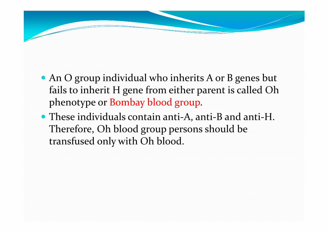
An O group individual who inherits A or B genes but fails to inherit H gene from either parent is called Oh phenotype or Bombay blood group.
These individuals contain anti‐A, anti‐B and anti‐H. Therefore, Oh blood group persons should be transfused only with Oh blood.

RHESUS SYSTEM The Rh allelic genes are C or c, D or d and E or e, located on chromosome 1.
The importance of this system lies in high immunogenecity of Rh D antigen, which can cause severe hemolytic reaction.
The presence of D in either homozygous or heterozygous state make the individual Rh positive, while Rh neg individuals are homozygous for d (d/d).
Rh antigens are expressed on RBCs only and not on any other tissue.
There are no naturally‐occurring Rh antibodies

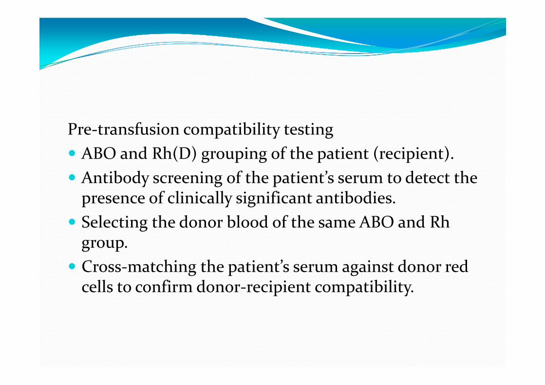
Pre‐transfusion compatibility testing ABO and Rh(D) grouping of the patient (recipient). Antibody screening of the patient’s serum to detect the presence of clinically significant antibodies.
Selecting the donor blood of the same ABO and Rhgroup.
Cross‐matching the patient’s serum against donor red cells to confirm donor‐recipient compatibility.

Complications of Blood Transfusion Immunologic transfusion reactions‐ against red blood cells (haemolytic reactions), leucocytes, platelets or immunoglobulins.
Non‐immune transfusion reactions Circulatory overload in massive transfusion transmission of an infectious agent.


Haemolytic transfusion reactions may be immediate or delayed, intravascular or extravascularABO incompatibility: Very rapid cell destruction Intravascular haemolysis naturally‐occurring antibodies, anti‐A and anti‐B, fix complement.
symptoms include restlessness, anxiety, flushing, chest or lumbar pain, tachypnoea, tachycardia and nausea, followed by shock and renal failure.

Rh incompatibility: Extravascular haemolysis anaemia due to destruction of red cells in the RE system
The clinical manifestations are relatively less severe and usually consist of malaise and fever but shock and renal failure may rarely occur

Transfusion‐related acute lung injury (TRALI) Transfusion of donor plasma containing high levels of anti‐HLA antibodies which bind to leucocytes of recipient.
leucocytes then aggregate in pulmonary microcirculation.
increased vascular permeability resulting in acute pulmonary oedema
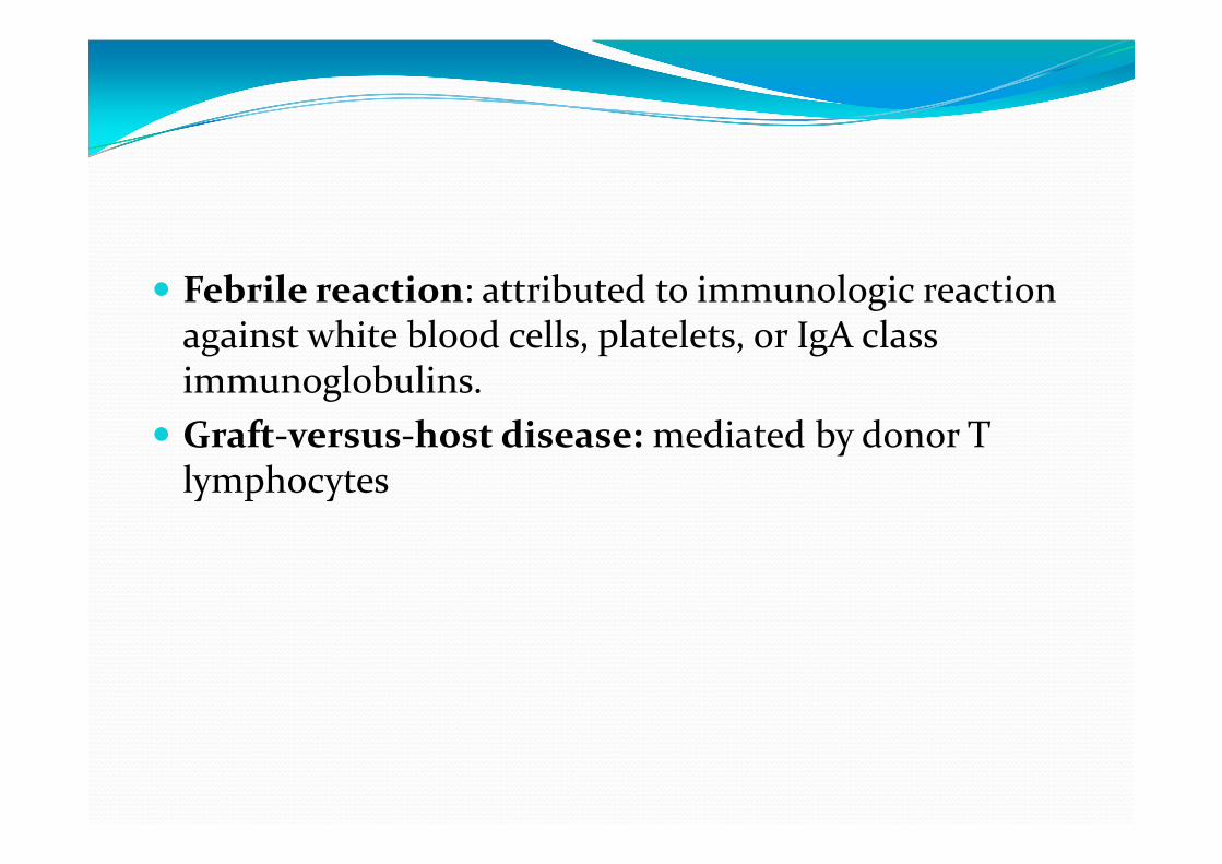
Febrile reaction: attributed to immunologic reaction against white blood cells, platelets, or IgA class immunoglobulins.
Graft‐versus‐host disease: mediated by donor T lymphocytes


Circulatory overload: result in pulmonary congestion and acute heart failure Risk factors: chronic anaemia, in infants and elderly. onset may be immediate, or may be delayed up to 24 hours.
Massive transfusion: dilutional thrombocytopenia and dilution of coagulation factors.
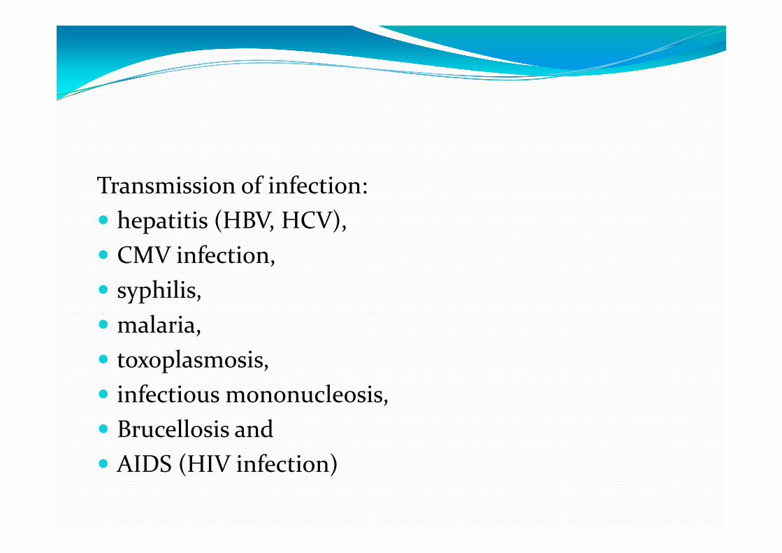
Transmission of infection: hepatitis (HBV, HCV), CMV infection, syphilis, malaria, toxoplasmosis, infectious mononucleosis, Brucellosis and AIDS (HIV infection)
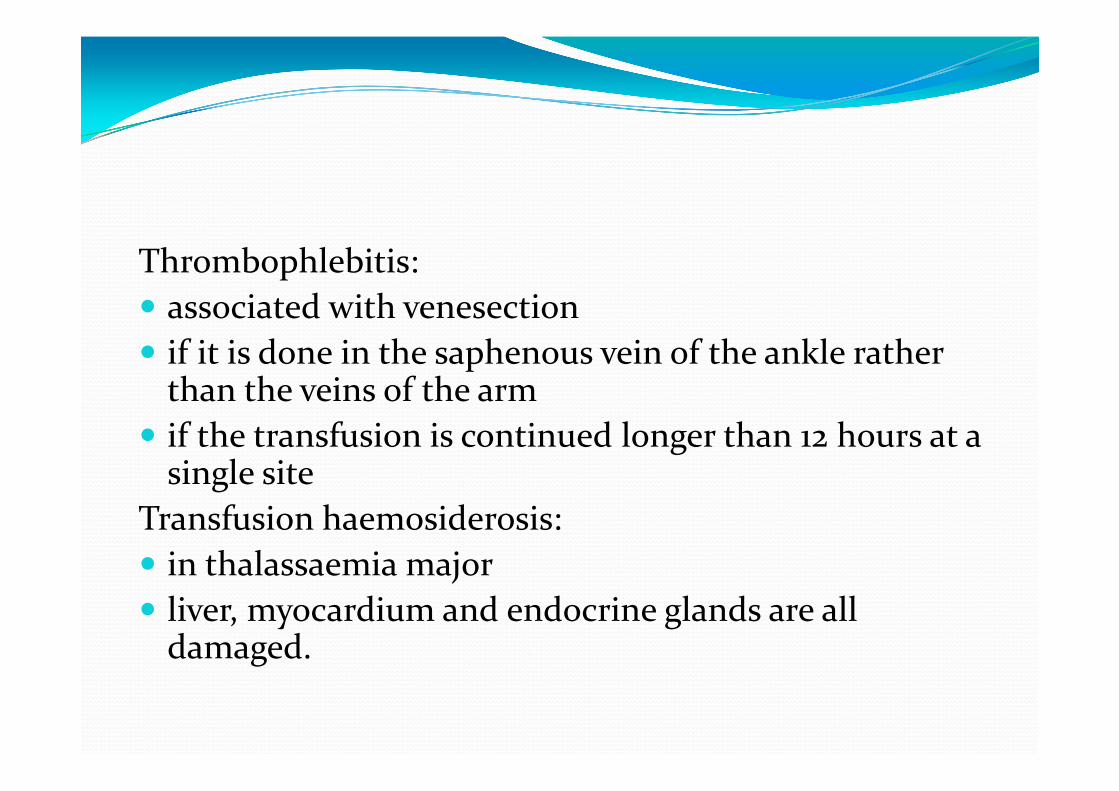
Thrombophlebitis: associated with venesection if it is done in the saphenous vein of the ankle rather than the veins of the arm
if the transfusion is continued longer than 12 hours at a single site
Transfusion haemosiderosis: in thalassaemia major liver, myocardium and endocrine glands are all damaged.

Blood components packed RBCs, platelets, fresh‐frozen plasma (FFP) and cryoprecipitate.

Collection procedure consists of initial centrifugation at low speed to separate whole blood into two parts: packed RBCs and platelet‐rich plasma (PRP).
Subsequently, PRP is centrifuged at high speed to yield two parts: random donor platelets and FFP.
Cryoprecipitates are obtained by thawing of FFP followed by centrifugation.
Apheresis is direct collection of large excess of platelets from a single donor.

Applications1. Packed RBCs: normovolaemic patients of anaemia without cardiac disease. One unit of packed RBCs‐ raise haemoglobin by 1 g/dl2. Platelets: Patient with platelet count below 10,000/μl. raise platelet count by 5,000 to 10,000/μl

3. Fresh frozen plasma: FFP contains plasma proteins and coagulation factors that include albumin, protein C and S and antithrombin.
indicated in patients of coagulation failure and TTP Each unit of FFP raises coagulation factors by about 2%4. Cryoprecipitate: plasma proteins, fibrinogen, factor VIII and vWF patients requiring fibrinogen, factor VIII and vWF Transfusion of single unit of cryoprecipitate yields about 80 IU of factor VIII

HAEMOLYTIC DISEASE OF NEWBORN passage of IgG antibodies from the maternal circulation across the placenta into the fetalcirculation.
HDN can occur from incompatibility of ABO or Rhblood group system.
ABO incompatibility is much more common but the HDN in such cases is usually mild,
Rh‐D incompatibility results in more severe form of the HDN

HDN due to Rh‐D incompatibility Rh incompatibility occurs when a Rh‐negative mother is sensitised to Rh‐positive blood
Sensitisation occurs ‐passage of Rh‐positive featlred cells across the placenta into the circulation of Rh‐negative mother
Normally, during pregnancy very few foetal red cells cross the placenta but haemorrhage during parturition causes significant sensitisation of the mother.
95% cases of Rh‐HDN are due to anti‐D, some cases are due to combination of anti‐D with other immune antibodies of the Rh system such as anti‐C and anti‐E, and rarely anti‐c alone

HDN due to ABO incompatibility Naturally‐occurring anti‐A and anti‐B antibodies which are usually of IgM class do not cross the placenta.
while immune anti‐A and anti‐B antibodies which are usually of IgG class may cross the placenta into foetal circulation and damage the foetal red cells

CLINICAL FEATURES severe form may result in intrauterine death from hydrops foetalis
Moderate disease‐ severe anaemia and jaundice due to unconjugated hyperbilirubinaemia.
When the level of unconjugated bilirubin exceeds 20 mg/dl, it may result in deposition of bile pigment in the basal ganglia of the CNS called kernicterus.
Mild disease‐ severe anaemia with or without jaundice.
LABORATORY FINDINGS Anaemia with reticulocytosis, Increased nucleated RBC and polychromasia elevated serum bilirubin positive direct Coombs’ test Mother’s blood‐ Indirect Coomb’s test‐ anti‐D antibodies.
Treatment Exchange transfusion Phototherapy‐ converts unconjucated bilirubin into soluble form, that is excreted in urine.
Infusion of bile‐ binds free bilirubin in plasma and thus decreases the risk of kernicterus.

Prevention All Rh D –ve women are given RhIg within 72 hrs of delivery of Rh +ve infant.

Rh HDN ABO HDN
Frequency Less common More common
Blood groupMotherFetus
Rh negRh positive
OA or B
Pregnancy affected Usually second Usually first
Severity Severe Mild
Blood smear Erythroblastosis Spherocytosis
DCT Strongly Positive Weakly positive or Negative
Prevention Rh immune globulin Not available