blood blood vessels - · pdf filetransports nutrients from digestive organs to cells 4. ......
TRANSCRIPT

BLOOD
&
BLOOD VESSELS
1

Cardiovascular System -- Blood
1. CV System: Blood, Heart and Blood
Vessels
2. Blood: the red body fluid that flows
through all the vessels EXCEPT the
lymph vessels
2

Blood: Physical Characteristics
1. Blood is viscous (“sticky”) – about 5 X > than
water, i.e., blood flows at about 1/5 the rate of
water flow
2. Requires physiological temperature for proper
activity – about 37C (98.6 F), more or less
3. Arterial pH of 7.35-7.45 for optimal activity
4. Between 0.85% and 0.9% NaCl
5. Constitutes about 8% of BWT
6. Males: 5-6 L; Females: 4-5 L
3

Blood Function1. Transports oxygen from lungs to all cells in the body
2. Transports carbon dioxide from cells to lungs
3. Transports nutrients from digestive organs to cells
4. Transports waste products from cells to kidneys, lungs and sweat glands
5. Transports hormones from endocrine glands to target cells
6. Regulates pH via buffers and amino acids
7. Transports enzymes to specific cells
8. Regulates body temperature due to the volume of water (heat absorber/coolant)
9. Regulates water content of cells (1 via dissolved Na+)
10. Prevents body fluid loss through clotting
11. Protects against toxins and foreign microbes via special combat unit cells
4

Blood Composition -- Plasma
• Plasma = the liquid containing dissolved substances – about 55% of blood
• Composition:
• Inorganic (1%): Na+, K+, Cl-, HCO3-,
Ca 2+ -- Na+ and Cl- are the most plentiful
• Plasma Proteins (7-9%) – contribute to the viscosity of plasma, maintains dispersion of material, amino acid reserve (very unusual, but available), provides buffers
• Water (90%) – major constituent of plasma
5

Kinds of Plasma ProteinsAlbumins Globulins Fibrinogens
Most plentiful Least plentiful
55-64% ~2%; 2-3 g/100mL of blood; largest proteins ~0.3%
~4-5 g/ 100 mL
blood
General protein functions; bind
molecules for transport
Immunoglobulins
(antibodies; Ig’s;
Ab’s)
0.15-0.3 g/100
mL blood
SOLUBLE in water Lipids, T4,
Cu, Cortisol
Fe and
cholesterol
Protect body from
chemical
challenges
Converted to
Insoluble fibrin
as blood
coagulates
Smallest of the
proteins
Produced in
liver
Produced in
liver
Produced in
plasma cells
Produced in
liver
Serves to bind
substances for
transport through
plasma: drugs
(barbiturates),
hormones
(thyroxine, T4)
IgA: Secretions
IgM: 1st to appear
IgG: Natural/
acquired Ab
(anti-HIV)
IgD: Unknown
IgE: AllergiesProduced in liver
6

Plasma Protein Concentrations• Vary little in
good health
• A/G ratio is approximately
2
• Protein concentrations decrease due to starvation, liver damage, renal disease
• The primary sign of decreased protein concentration is EDEMA
• Albumin helps to “carry” filtered plasma water back to the blood stream instead of remaining in the interstitial compartment (between the cells)
• Albumin serves to INCREASE the osmotic pressure of the blood
• In starvation, protein intake is decreased with secondary decreases in circulating amino acids’ concentration which leads to decreased plasma proteins
• Decreased plasma albumin results in plasma water staying out of the blood causing edema 7

Constituents Delivered to Blood
Stream by Body Cells
Water and Electrolytes Come from absorption across the gut
Amino acids Due to protein digestion – also absorbed
across the gut
ASIDE “OPEN GUT” – alcoholics and newborns to
about 2 weeks’ of age
Simple Sugars From carbohydrate digestion, e.g., sucrose
hydrolyzed to glucose and fructose
8

Blood Composition – Formed
Elements
• Formed elements are cells and cell-like bodies
suspended in the plasma – makes up about
45% of blood
• The process by which blood cells are produced
is called hemopoiesis or hematopoiesis
• Red blood cell synthesis is called erythropoiesis
• White blood cell synthesis is called
leuk(c)opoiesis
9

Blood Composition – Formed Elements -- 2
Clinically Relevant Formed Elements
Formed Element Concentration or Amount Notes
RBC
(Erythrocytes) ♂5.4 X 10⁶/mm3Differences due to
metabolic rate in males
and monthly blood loss
via menses in females♀4.8 X 10⁶/mm3
WBC
(Leukocytes)
Granular WBC’s Neutrophils: 60-70%
Eosinophils: 2-4%
Basophils: 0.5-1%
Agranular WBC’s Lymphocytes: 20-25%
Monocytes: 3-8%
Thrombocytes
(platelets)
250-400 X 103/mm3
10

Hematopoiesis
11

Hemopoiesis
12

Erythropoiesis
• In general, undifferentiated cells in red bone marrow are transformed into hemocytoblasts (stem cells) which develop into mature blood cells eventually
• Rubriblasts (proerythroblasts) differentiate into RBC at an ~ rate of 2 X 10⁶ produced every second
13

Reticulocyte Countaka “stipled” RBC;
gives information about the rate of erythropoiesis
< 0.5% total
RBC
(decreased
erythropoiesis)
Anemia (pernicious or aplastic); kidney
disease which effects production of
erythropoietin. (B12 deficiency because are
unable to absorb across gut due to no
secretion of intrinsic factor from stomach.)
> 1.5% total
RBC (increased
erythropoiesis)
Indicative of anemia, oxygen deficiency
(COPD), bone marrow CA with secondary
increase in erythropoiesis, hemorrhage,
hemolysis; MAY be used to check on
pernicious anemia after receiving B12
parenterally, i.e., the marrow is making up for
lost time (peaks in 4-5 days – max production
within 7 days)
14

RBC: Some Abnormalities
• Dacrocyte: A deformed RBC which is tugged to a
nipple at one end, having squeezed through a
reticuloendothelial system with increased
connective tissue; also seen in normal peripheral
blood smears as an artifact of slide preparation;
such dacrocytes are usually easily recognized as
their 'tails' all point in the same direction
• Keratocyte: An erythrocyte formed when
haemoglobin denatures—as occurs in alpha-
thalassemia or G6PD deficiency—and
precipitates—due to oxidation—into clumps that
stick to the red cell membrane
• Knizocyte: a red blood cell with two or more
concavities (triconcave erythrocyte); associated
with hemolytic anemia
• Stomatocyte: an abnormal red blood cell in which
a slit or mouthlike area replaces the normal central
circle of pallor, often caused by edema
15

Blood Groups – A, B, AB, O
Blood Types 16

Ag-Ab Response
17

HDN: Erythroblastosis Faetalis
18

HDN 2
19

Red Blood Cells: Erythrocytes
1. Biconcave discs (donut-like without the hole); increases the surface area for diffusion of gases.
2. Mature cells are very simple: LACK a nucleus, are not able to reproduce.
3. No complex metabolic activities.
4. Cell death occurs at about 120 days.
5. Mature RBC contain protein (stroma – network), some cytoplasm, lipids (including cholesterol) and a red pigment (hemoglobin; Hb or Hgb)
6. Hgb makes up about 33% of the cell volume and gives the red color to blood.
7. There are 280 X 106 molecules of Hgb/RBC
8. RBC combines with oxygen and carbon dioxide. HOW??????
20

Hemoglobin
• Tetramer – salt-linked
• Each protein contains
a heme group
• Each heme group
binds Fe 2+
• NOT Fe 3+
21
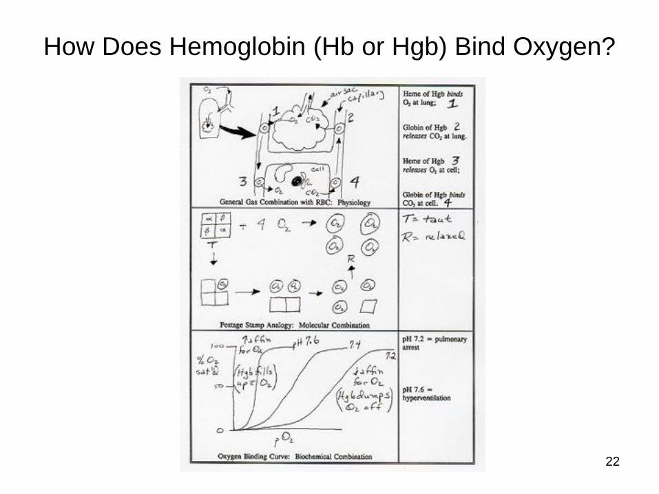
How Does Hemoglobin (Hb or Hgb) Bind Oxygen?
22

How Does Hemoglobin (Hb or Hgb) Bind Oxygen and
Act as a Buffer?
23

Oxygen Binding Curve Shift Factors – 1:
“Bohr Effect”
Left Shift
• Alkalosis
• 2,3-BPG
• Hypothermia
• Fetal Hb (> affinity for oxygen than adult Hb)
• ACD-preserved blood (acid citrate dextrose: O2
carrying capacity of RBC >2-3 days old (in bag) with Hb –PROBLEM: doesn’t release O2 to tissues for 18-24 hours after infusion)
Right Shift
• Acidosis
• 2,3-BPG
• Fever
• Anemia
• Hypoxia
24

25
Haldane Effect
• With Hb(O2)4, at some pCO2 CO2 content of blood
• With Hb(O2)4 at some pCO2 CO2 content of blood
• “Back side of Bohr effect” – greater effect than the Bohr effect on gas transport.

Regulation of Erythropoiesis
26

Erythrocyte Life Cycle
27

Leukopoiesis• In general, undifferentiated
cells in red bone marrow are transformed into hemocytoblasts (stem cells) which develop into mature blood cells eventually
• Lymphoblasts differentiate into lymphocytes
• Monoblasts differentiate into monocytes
• Myeloblasts differentiate into neutrophils, eosinophils, basophils
• WBC life span is only a couple of days due to the limit on the number of bacteria it phagocytizes.
• Graphic Source: Used under Fair Use Copyright. 28

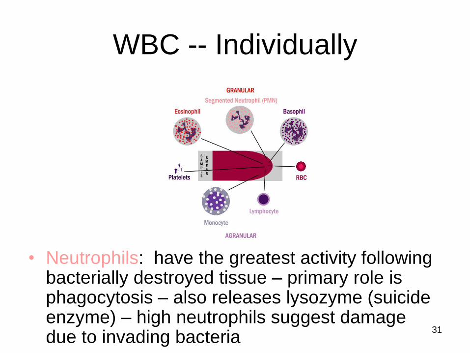
Leuk(c)ocytes
Unlike RBC, WBC HAVE nuclei – DON’T have Hgb. RBC/WBC is about 700/1.
Granular WBC Agranular WBC
From red bone marrow PLUS lymphoid tissues
Cytoplasmic granules NONE
Lobed nuclei Spherical nuclei
Neutrophils, eosinophils, basophils Lymphocytes, monocytes
FUNCTIONS/DEFINITIONS
Leukocytosis: elevated # of WBC;
“usually” >10,000 is pathological
Leukopenia: Decreased # of WBC;
“usually” < 5000 is pathological
To combat inflammation/infection Some WBC are actively phagocytic
(ingest bacteria and dispose of dead
matter).
Most WBC have the ability to crawl through capillary walls and connective
epithelial tissue = diapedesis, aka cell walking29

WBC Activities
Phagocytosis Diapedesis
30
WBC -- Individually
• Neutrophils: have the greatest activity following bacterially destroyed tissue – primary role is phagocytosis – also releases lysozyme (suicide enzyme) – high neutrophils suggest damage due to invading bacteria
31

WBC -- Individually
• Eosinophils: high eosinophils suggest allergic conditions –
believed to combat allergens which cause allergies;
elevated also in cases of porkworm infection, psoriasis,
Hodgkin’s Disease and some cancers. Decreased
numbers after period of stress (cortisol). Eosinophils
produce/release antihistamines.
Eosinophils phagocytize the AgAb complex. The AgAb complex combats infection and confers immunity; Ag’s are responsible for blood groups, allergies and organ transplant rejections.
32

WBC -- Individually
• Basophils: elevated due to allergens, Hodgkin’s Disease,
smallpox, after splenectomy, chronic hemolytic anemia and
some cancers. Elevated also during recovering lobar
pneumonia, acute rheumatic fever, anaphylactoid purpura.
Decreased in ACUTE phases of same conditions. 33

WBC -- Individually
• Monocytes: high monocytes suggest chronic infection, e.g.,
TB. Take longer than neutrophils to get to damaged site,
but arrive in greater numbers and destroy more micro-
organisms. Monocytes also phagocytic and clean up
cellular debris during an infection. 34

WBC -- Individually
• Lymphocytes: necessary for antibody production
• Antibody (Ab): special proteins which inactivate antigens
• Antigens (Ag): a substance (foreign or otherwise) that will
stimulate the production of specific Ab’s. Most Ag’s are
proteins and not synthesized by the body, e.g., pollen35
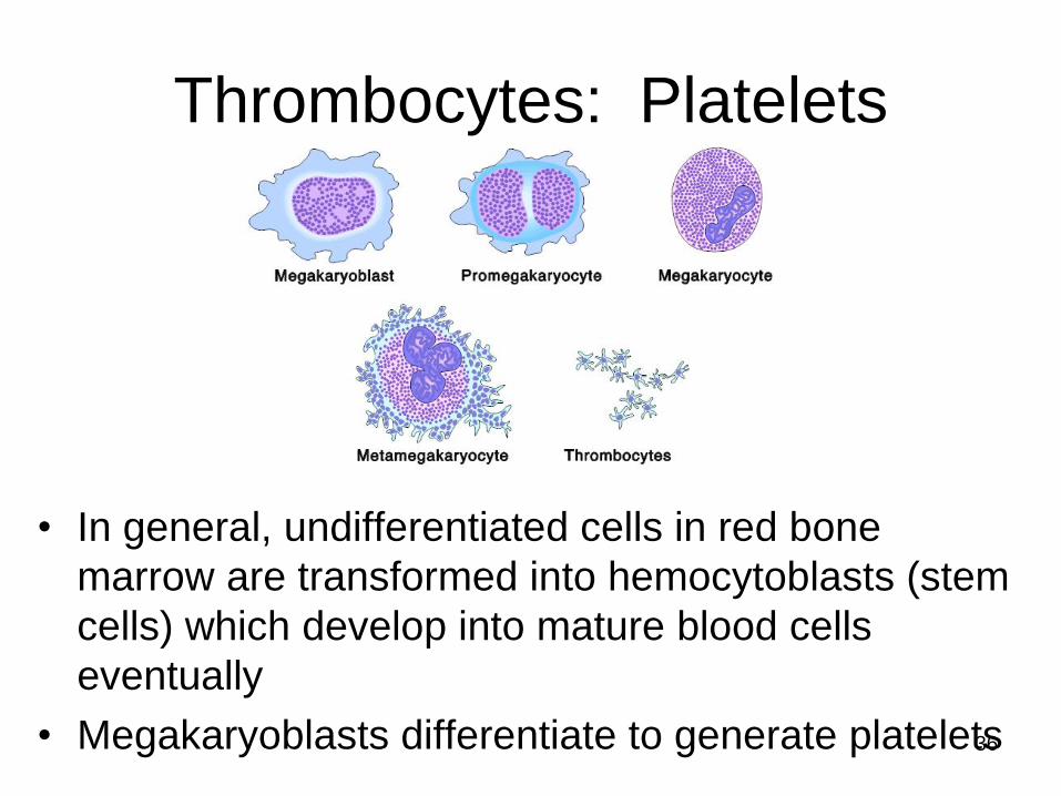
Thrombocytes: Platelets
• In general, undifferentiated cells in red bone
marrow are transformed into hemocytoblasts (stem
cells) which develop into mature blood cells
eventually
• Megakaryoblasts differentiate to generate platelets36

Thrombocytes -- Platelets
• Platelets are disc-shaped (more or less) without a
nucleus; 2-4 in diameter; they initiate a chain of
reactions that leads to blood clotting; life span is about a
week because they are 1) used up in blood clotting and
2) they are too simple to carry on much metabolism. 37

38
Blood Clotting CascadeIt's important to recognize that the liver, while of great digestive importance as a detoxification center, is also important in blood clotting. Figure, right, illustrates the effects of blood clotting after receiving a wound.
1) The skin is sliced by a knife.
2) The wound fills with blood from the damaged capillaries.
3) The capillaries then constrict to reduce the flow of blood out of the body. In the case of a small injury, this is primary hemostasis.
4) Platelets are then released. Contact between the platelets and the basement membrane causes platelet degranulation which increases the "stickiness" of the platelets that then form a platelet plug with the red blood cells (RBC) in the wound.
5) During secondary hemostasis (or following a larger wound), the next step is to form a fibrin clot.
Clotting factors come from the LIVER! Bile salts are manufactured by the liver for vitamin K absorption. If the liver is shot, expect bleeding disorders.

39
Blood Clotting Cascade
6) A hemostatic plug is formed between the RBC, fibrin and platelets.
7) Once healing begins (review A&P I) or a pathological process is in place, plasmin is released to dissolve the fibrin strands. The degradation products are removed by phagocytosis.
Clinically, "fibrin split products" are measured to determine the extent of blood clotting ability. The higher they are, the less the person may be able to clot effectively, i.e., the higher the fibrin split products, the more thrombin, fibrin polymerization and platelet aggregation are INHIBITED from forming a clot.
8) The wound has healed, more or less with or without scar formation -- review primary, secondary and tertiary wound healing in A&P I.

40
Coagulation and the Liver
•Two pathways
are used by the
body to produce
clots:
– the extrinsic
and
– intrinsic
systems.

41
Extrinsic System•The extrinsic system is generally initiated by some sort of trauma, including venipuncture. Trauma activates factor VII to factor VIIa (the "a" is for "active" factor, in this case, VII). VIIawith calcium ions and III (the factors are usually represented only by their Roman numerals), then activate X to Xa. This latter process also requires the presence of special prostaglandins called thromboxane A2 (TXA2; makes the platelets sticky, too). We'll stop here for a moment.

42
Intrinsic System
•The intrinsic
pathway is initiated
by the blood stream.
That process
activates XII to XIIa.
XIIa, in turn,
activates XI to XIa,
which activates IX to
IXa, which activates
X to Xa along with
calcium ions and
TXA2.

43
Common Pathway•Once factor Xa is formed, the clotting cascade begins the "common pathway".
•It's called the common pathway because both systems utilize the same pathway from Xa on to accomplish coagulation.
•Factor V, with Xa, and calcium ions "convert" prothrombin to thrombin.
•Thrombin causes fibrinogen to "change to" soluble fibrin. Calcium ions causes the soluble fibrin to become insoluble fibrin threads, i.e., clot-forming.
•With either wound healing or a pathological process, the thrombin and insoluble fibrin threads activate plasminogen.
•The active form of this protein is plasmin.
•Plasmin causes clot lysis.
•Plasmin not only causes clot lysis, hematologically, but also causes semen to un-clot, as well, after ejaculation (see A&P II Reproduction lecture for this process).

44
The Clotting Cascade with Anticoagulants
•Note that wherever calcium ions are required to propagate a step in the cascade that it is inhibitable with EDTA, citrate or oxalate -- lavender top tubes, light blue top tubes or gray top tubes, respectively.
•TXA2 is inhibited by aspirin through the primary enzyme of prostaglandin synthesis, cyclo-oxygenase.
• Heparin inhibits the conversion of prothrombin to thrombin.
•Clinically, the partial thromboplastin time (PTT) is used to measure the efficiency of the intrinsic system, while the protime (PT) is used to measure the efficiency of the extrinsic system.
•The PT is used, traditionally, to follow coumadin anticoagulation therapy and the PTT is used, traditionally, to follow heparin therapy for anticoagulation.
•Coumadin inhibits II, VII, IX, X, C, S, Z (latter three are clotting proteins) via Vitamin K epoxide reductase

Blood Vessels
• Blood is pumped through blood vessels by the heart.
• Heart has its own circulatory system – coronary arteries
and veins – RCA, LCA (LADCA and CXA) – Coronary
Sinus
45

“Pumping” Blood
• Atria and ventricles “pump” (contract) opposite to each other
• Relaxation = diastole; contraction = systole
• Chambers fill on diastole; chambers empty on systole
46

Arteries
1. Tunica adventia
2. Tunica media
3. Elastic lamina
4. Basement membrane
5. Endothelium
6. Lumen
7. NOT in arteries
4 & 5 = Tunica intima
1
23
4
5
6
Blood Vessel Anatomy
47

Veins
1. Tunica adventia
2. Tunica media
3. NOT in veins
4. Basement membrane
5. Endothelium
6. Lumen
7. Valve
4 & 5 = Tunica intima
1
2
4
5
67
Blood Vessel
Anatomy
48

Flow of Peripheral Blood -- Circuitry
• Blood pumped out of (AWAY from) heart into ARTERIES -- oxygenated
• Arteries branch into arterioles
• Arterioles branch into capillaries
• Nutrients into cells
• Waste out of cells
• Capillaries branch into venules
• Venules expand into veins
• Veins “pour” (“drain”) blood INTO (TOWARDS) heart – de-oxygenated
49

Blood Pressure Defined
• The pressure exerted by the blood on the wall of any vessel
• A hydrostatic pressure – key for kidneys
• Varies with age, gender, altitude, muscular development, states of mental and physical stress and fatigue
• Measured in an auscultatory manner with sphygmomanometer and stethoscope
• May be measured by palpation – only systolic, though
• May be measured electronically, too
50

Measuring Blood Pressure
• Put BP cuff over upper arm
• Put diaphragm over brachial artery
• Pump cuff to 160 mm Hg (at least – may need to go higher)
• Deflate cuff slowly, listening to sounds
• Record 1st sound pressure (in even mm Hg)
• Record 2d sound pressure (in even mm Hg)
• Remove apparatus51

52

Blood Pressure is Postural
AVP involved!
Approximate Values
Head
(mm Hg)
Heart
(mm Hg)
Feet
(mm Hg)
AVP
Levels
Position
110 120 110 Lying
Down
90 120 190 Sitting
90 120 240 Standing
53
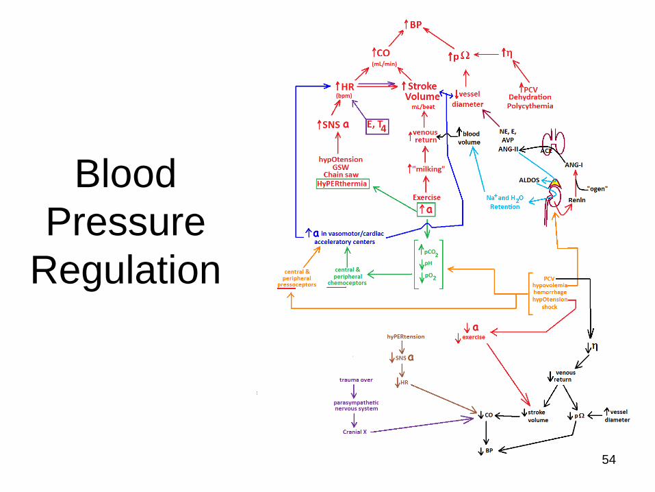
Blood
Pressure
Regulation
54

Hypertension
55
Pulse Pressure
The numeric difference between your systolic
and diastolic blood pressure is the pulse
pressure.
The most important cause of elevated pulse
pressure is stiffness of the aorta. The stiffness
may be due to high blood pressure or fatty
deposits on the walls of the arteries
(atherosclerosis). The greater the pulse
pressure, the stiffer and more damaged the
vessels are thought to be.
Mean Arterial Pressure
MAP = [(2 x diastolic)+systolic] / 3
Diastole counts twice as much as systole
because 2/3 of the cardiac cycle is spent in
diastole. An MAP of about 60 is necessary to
perfuse coronary arteries, brain, kidneys.
Usual range: 70-110; Below this range for any
appreciable time, vital organs will not get
enough Oxygen, and will become ischemic.

Major Arteries
• Aorta
– Thoracic
– Abdominal
• Common Hepatic
• Splenic
• Renal
56

1. Vertebral
2. Internal carotid
3. External carotid
4. Common carotid
5. Subclavian
6. Brachiocephalic
7. Innominate
8. Left common carotid
9. Left subclavian
10. Ascending aorta
11. Aortic arch
12. Descending aorta
1
2 3
4
5
6
7
8
9
10
11
12
57

• Note nerve, artery
and vein location
relative to rib.
• Note needle location
to MISS the three
structures
• Thoracentesis
– Hemothorax
– Pneumothorax
– Chylothorax
58

• Axillary Artery
• Brachial Artery
• Radial Artery
• Ulnar Artery
• Palmar Arch
Major
Arteries
Upper
Extremity 59
Major
Arteries
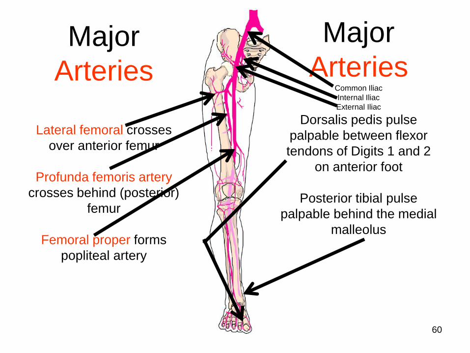
Lateral femoral crosses
over anterior femur
Profunda femoris artery
crosses behind (posterior)
femur
Femoral proper forms
popliteal artery
Major
ArteriesCommon Iliac
Internal Iliac
External Iliac
Dorsalis pedis pulse
palpable between flexor
tendons of Digits 1 and 2
on anterior foot
Posterior tibial pulse
palpable behind the medial
malleolus
60

Major Veins – NOT Inclusive
• Hepatic
portal
• Renal
• Internal
iliac
• Femoral
• Splenic
• Common iliac
• External iliac
• Greater
saphenous (used
for CABG’s)
NOTE: veins tend to follow
arteries – hence, common names
between the two kinds of vessels
61

Major Veins – NOT Inclusive
• Vertebral
vein
• External
jugular
vein
• Axillary
vein
• Internal Jugular
Vein
• Subclavian vein
• Brachiocephalic
vein
62

Major Veins – NOT Inclusive
• Cephalic
• Brachial
• Basilic
• Median
cubital
63

Major Veins – NOT Inclusive
• Lateral
femoral
• Peroneal
• Anterior tibial
• Dorsal arch
• External
iliac
• Femoral
• Great
saphenous
• Popliteal
• Posterior
tibial
64

Fetal Circulation – Differences!
1. Ductus venosus vs Ligamentum venosum
2. Umbilical arteries (2 –DE-oxygenated blood!!!) vs lateral umbilical ligaments
3. Urachus vs medial umbilical ligament –not shown here
4. Umbilical vein (1 –OXY-genated!!!) vs ligamentum teres
65

• Ductus venosus: allows nutrient-rich
blood (about 50%) to bypass liver
• Foramen ovale: allows nutrient-rich blood
to bypass lungs
• Ductus arteriosus: allows waste-rich blood
to bypass lungs and return to placenta via
umbilical arteries
Fetal Circulation – Differences!
66

Fetal Circulation – Differences!
Foramen ovale vs Fossa ovale
Ductus arteriosus vs ligamentum arteriosum
Either PG’s or Bradykinin thought to constrict D. arteriosus
after birth
Source: after http://www.natalnurses.net/images/13.jpg -- Used under Fair Use Copyright67

Tetralogy of Fallot
1. Interventricular
septal defect
2. Stenosed
pulmonic valve
3. Right ventricular
hypertrophy
4. Biventricular aorta
– dextroposed
aorta
68