blastocyst lineage formation, early embryonic asymmetries and axis patterning in the mouse
TRANSCRIPT

701
The investigation into lineage allocation and early asymmetriesin the pre- and peri-implantation mouse embryo is gainingmomentum. As we review here, new insights have been gainedinto the cellular and molecular events that lead to theestablishment of the three lineages of the blastocyst, to thedetermination of the origin and the fates of the visceralendoderm in the peri-implantation mouse embryo, and to thegeneration of cellular and molecular activities that accompanythe emergence of asymmetries in the pre-gastrulation embryo.We also discuss the continuing debate that surrounds therelative impacts of early lineage bias versus the stochasticallocation of cells with respect to the events that pattern theblastocyst and initiate its later asymmetries.
IntroductionThe progression of the mammalian embryo from fertilization togastrulation involves an ordered series of lineage specifications andaxial asymmetries (Fig. 1) that result, first, in the development of theblastocyst (see Glossary, Box 1), with its embryonic-abembryonic axis(Fig. 1; Box 2), and, later, in the formation of the embryo itself, withits anterior-posterior (AP), dorsal-ventral (DV) and left-right (LR)axes. In many invertebrates and vertebrates, asymmetries that areestablished in the egg correlate with the segregation of determinantsthat influence later lineage formation and axis development. However,in the mouse egg, the morphological asymmetries that exist, such asthe position of the second polar body (see Glossary, Box 1) and thesperm entry point, do not clearly demarcate an asymmetric domain of,for example, signaling activity or of cell fate determinants in thefertilized mouse egg. Whether there is any instructive relationshipbetween the asymmetry of the egg and the later asymmetry of theblastocyst and lineage allocation remains a controversial issue. It iswell known that the pre-implantation mammalian embryo is highlyregulative and resistant to the loss or addition of cells brought aboutby experimental manipulations. However, this does not preclude theexistence of an, as yet, uncharacterized property that could biasdevelopmental outcomes in the intact embryo.
Whether any early asymmetries in the mouse egg and/orblastocyst relate to the orientation of the definitive body axes is evenless certain. It is now clear that the AP patterning of the gastrulatingembryo is initiated prior to gastrulation by spatially localized signalsthat emanate from regionally patterned extra-embryonic tissues.Some of these asymmetries may be set up as early as the blastocyststage, linking pre-implantation patterning to post-implantationmorphogenesis.
Here, we review recent experiments that define the molecularcomponents of lineage specification in the mouse blastocyst. Wealso review the ongoing uncertainty and debate that surrounds therelative importance of early cleavage patterns at the two- to four-cellstage and of symmetric versus asymmetric divisions at the eight- to16- and 16- to 32-cell stage, and the importance of final cell positionin the late morula/early blastocyst for blastocyst lineagespecification for the positioning of the blastocyst cavity (theblastocoel) and for the establishment of the embryonic-abembryonicaxis of the blastocyst. Our critical review of the current data supportsa stochastic model of lineage specification, in which cell-cellinteractions and position effects reinforce and can override anyunderlying cell fate bias.
The asymmetries that are observed in the post-implantationdevelopment of the visceral endoderm (see Glossary, Box 1) thatlead up to gastrulation are now well defined and strongly hint at theemergence of the prospective AP body axis prior to the onset ofgastrulation. However, a definitive link between the asymmetries inthe pre-gastrula embryo and the morphological and tissueasymmetries displayed earlier in the blastocyst has still not beenestablished. Here, we also review the findings of recent experimentalstudies that help to define the events that initiate early axialpatterning in the post-implantation mouse embryo.
Lineage allocation in the blastocystThe mouse blastocyst, immediately before implantation, consists ofthree distinct cell groups: the trophectoderm (TE); the epiblast,which is derived from the earlier inner cell mass (ICM); and theprimitive endoderm. Only the epiblast gives rise to the embryo itself,whereas the other two cell types give rise to extra-embryonicstructures that support the intra-uterine development of the embryoand act as signaling sources to pattern the embryonic tissues prior togastrulation.
Although pluripotent embryonic stem cells (ES cells) can beobtained from the epiblast of the blastocyst, other progenitor cellslines that self-renew in culture can also be derived from theblastocyst, such as trophoblast stem (TS) cells, which retainproperties of the trophectoderm (see Glossary, Box 1) (Tanaka et al.,1998), and the XEN cells, which retain properties of the primitiveendoderm (Kunath et al., 2005). ES cells can be converted to TS orXEN-like cells by altering the expression of appropriatetranscription factors, providing a good assay for the identification ofkey lineage-specific factors. As we discuss below, recent progresshas been made in identifying the transcription factors that specifythe blastocyst lineages and their derived stem cells.
Lineage-specific transcription factors and trophectodermspecificationCdx2, a caudal-related homeodomain protein, is a key regulator ofthe trophectoderm lineage. The expression of Cdx2 in ES cellsinduces them to differentiate into trophoblast, and to acquire theproperties of TS cells (Niwa et al., 2005). In the embryo itself, Cdx2
Development 136, 701-713 (2009) doi:10.1242/dev.017178
Blastocyst lineage formation, early embryonic asymmetriesand axis patterning in the mouseJanet Rossant1 and Patrick P. L. Tam2
1Research Institute, The Hospital for Sick Children and Departments of MolecularGenetics, and Obstetrics and Gynecology, University of Toronto, 555 UniversityAvenue, Toronto, Ontario M5G 1X8, Canada. 2Embryology Unit, Children’s MedicalResearch Institute and Faculty of Medicine, University of Sydney, Locked Bag 23,Wentworthville, NSW 2145, Australia.
E-mails: [email protected]; [email protected]
REVIEW
DEVELO
PMENT

702
begins to be expressed around the eight-cell stage and graduallybecomes restricted and upregulated in the outside cells of the morulaahead of blastocyst formation (Dietrich and Hiiragi, 2007; Ralstonand Rossant, 2008). A loss-of-function Cdx2 mutation has no impacton the initiation of blastocyst formation (Strumpf et al., 2005), andCdx2 mutant cells are not excluded from the TE layer in chimericblastocysts (Ralston and Rossant, 2008), but in embryos carryingthis mutation, the outer epithelium of the blastocyst losesmorphological integrity and the cells do not undergo furthertrophoblast differentiation (Strumpf et al., 2005). A mutation inEomes, a T-box transcription factor, also arrests blastocystdevelopment but at a slightly later stage than is found in Cdx2mutants (Russ et al., 2000; Strumpf et al., 2005). Eomes expressionis reduced in Cdx2 mutants but Cdx2 is still expressed in Eomesmutants, placing the Eomes transcription factor downstream of Cdx2(Ralston and Rossant, 2008; Strumpf et al., 2005).
Neither mutation leads to complete failure to initiate theformation of the TE epithelium of the blastocyst, suggesting thatthere may be more players upstream of Cdx2 and Eomes. Twogroups have recently shown that another transcription factor of theTEA domain/transcription enhancer factor family, TEAD4, isrequired upstream of Cdx2 for the formation of the TE (Nishioka etal., 2008; Yagi et al., 2007). Tead4 mutants show a slightly moresevere phenotype than do Cdx2 mutants and fail to maintain Cdx2expression, placing TEAD4 currently at the top of the TE genetichierarchy. However, unlike Cdx2, Tead4 expression is not restrictedto the TE lineage during pre-implantation development, making itdifficult to reconcile this observation with the notion of TEAD4having an instructive role in TE lineage specification. In Drosophilaand mammalian cells, TEAD factors can only activate transcriptionin combination with a co-activator, Yorkie (Yki) in Drosophila orYap (Yes-associated protein) in mammals (Vassilev et al., 2001;
Zhao et al., 2008). The availability of nuclear-localized Yap couldbe the limiting factor in activating TEAD-dependent TE lineagespecification. As such, an analysis of YAP protein localizationduring cleavage could be informative. As Tead4 mutants have amore severe phenotype than do Cdx2 and Eomes mutants, TEADmay activate parallel downstream pathways that are independent ofCdx2/Eomes to specify TE fate. Clearly, the transcription factornetworks that drive TE formation are still not fully understood.
Prior to blastocyst formation, TE-specific factors such as Cdx2and Eomes become restricted to the outside cells of the morula.However, the genes known to be required for specifying thepluripotent cells of the ICM, namely Oct4 (Nichols et al., 1998),Sox2 (Avilion et al., 2003; Nichols et al., 1998) and Nanog(Chambers et al., 2003; Mitsui et al., 2003), are expressed in everycell during cleavage, and are restricted to the ICM only afterblastocyst formation (Fig. 2A-D). This restriction depends on Cdx2.In Cdx2 mutants, Oct4 and Nanog remain expressed in the TE(Ralston and Rossant, 2008). Thus blastocyst lineage specificationbegins with the activation of TE targets and repression of ICMidentity in outside cells. Later, the reciprocal repression of TE targetsby Oct4/Sox2/Nanog in the pluripotent lineages (Loh et al., 2006;Boyer et al., 2005), combined with the known autoregulatoryproperties of the Oct4 (Chew et al., 2005) and Cdx genes (Xu et al.,1999; Beland et al., 2004), ensures the maintenance of lineageidentity. In order to understand the initiation of lineage segregationat the blastocyst stage, we need to understand how factors like Cdx2become localized to the outside cells of the morula.
Polarity and position drive trophectoderm formationIt has long been proposed that the position of cells in the developingembryo somehow influences their choice to become either ICM orTE. Initially during cleavage, all blastomeres appear to be identical
REVIEW Development 136 (5)
Blastomeres (Totipotent)
(B) Two cell (E1.5) (C) Eight cell (non-compacted)
(D) Early morula (E2.5) (eight cell, compacted)
(H) Early egg cylinder (E5.5)
Exe
Epc
Epi VE
(A) One cell (E0.5)
Primitive endoderm (PE)
Trophectoderm
Inner cell mass (ICM)
Outer cells
Inner cellsVisceral endoderm (VE)
Epiblast (Epi)
Ectoplacental cone (Epc)Extra-embryonic ectoderm (Exe)
Parietal endoderm (ParE)
Polarization
Compaction
DVE ParE
Second polarbody
Zonapellucida
Pronuclei
Apical domain
Basolateraldomain
(E) Late morula (16 cell or later)
(F) Blastocyst (E3.5) (G) Late blastocyst (at implantation; E4.5)
Epi
PE
Outer cells
Innercells
Trophectoderm
Emb.Abemb.
Blastocyst axis
Fig. 1. Cell lineage formationfrom egg to egg cylinder. (A-H) Schematics of themorphological changes and celllineage specification that occur in amouse embryo, from its fertilizationat embryonic day (E) 0.5 to theearly egg cylinder stage (E5.5). Thecolored bars show the progressiveallocation of totipotent blastomeresto outer and inner cells and to thetrophectoderm and inner cell masslineages. The cell types in theembryos are color coded. Abemb.} Emb., abembryonic-embryonicaxis of the blastocyst; DVE, distalvisceral endoderm.
DEVELO
PMENT

703REVIEWDevelopment 136 (5)
in their morphology and potential, but at the eight-cell stage, theevents of compaction and polarization begin (Fig. 1; see Glossary,Box 1) (Fleming and Johnson, 1988; Johnson and McConnell,2004). Concurrent with an increase in E-cadherin-dependentintercellular adhesion (Johnson et al., 1986), cells acquire an apicaldomain that is rich in proteins, such as the atypical protein kinase C(aPKC) (Pauken and Capco, 2000), the polarity protein Par3 (Plusaet al., 2005a) and the apical membrane protein ezrin (Louvet et al.,1996). However, molecules such as Lgl (lethal giant larva homolog)and the PAR polarity protein Par1 are localized exclusively in thebasolateral regions of each blastomere (Vinot et al., 2005) (Fig. 2D).Adherens junctions and, later, tight junctions between cells (Fleminget al., 1989) separate the apical and basolateral domains of theblastomeres, resulting in the formation of a polarized epithelium. Ascells divide from the eight- to 16-cell stage and from the 16- to 32-cell stage, the outer cells retain this polarized phenotype, whereascells in the core of the cluster lose apical features and becomemorphologically apolar (Johnson and Ziomek, 1983). The outsidepolarized epithelium goes on to form the TE, while the enclosedapolar cells go on to form the ICM (Johnson and Ziomek, 1983).
Apolar cells have been proposed to arise by asymmetric celldivisions, in which some outer polarized cells divide such that onlyone daughter inherits the apical pole, whereas the remaining cell willbe apolar and take up an internal position (Johnson and Ziomek,1981). Symmetric divisions will generate two polar cells, which willstay on the outside and end up in the outer TE epithelium. A recentstudy that traced the complete cell lineages from the two-cell to the32-cell stage mouse embryo has confirmed that inside apolar cellsand outside polar cells can be generated from outside cells throughtwo rounds of polarized cell divisions, and that the final cell numberof the ICM versus the TE is determined by the proportion of insideto outside cells generated at each round of division (Bischoff et al.,2008). What is less clear at this time is whether the polarized celldivisions that occur lead to the differential inheritance of lineagedeterminants by daughters, which dictate their future fate. Thespecialized apical polar region could control the orientation of themitotic spindle, and ensure the inheritance of localized determinantsthrough asymmetric divisions, in a manner analogous to theDrosophila neuroblast lineage (Wodarz, 2005; Yu et al., 2006). Thelineage tracing experiments just described identified a division asasymmetric or symmetric based on the location of the daughter cellsafter mitosis, not based on whether the anaphase plate was orientedperpendicularly or parallel to the apical domain, or according to thedifferential inheritance of fate-determining factors.
Could there be localized TE or ICM determinants that are segregatedthrough polarized cell divisions? There is a close association betweenthe acquisition of a polar phenotype and the upregulation of Cdx2 inouter cells (Dietrich and Hiiragi, 2007; Ralston and Rossant, 2008;Suwinska et al., 2008). However, there is no evidence that Cdx2 proteinor any other TE lineage transcription factor is subcellularly localized tothe apical domain of the polarized blastomere (Dietrich and Hiiragi,2007; Ralston and Rossant, 2008). Nor is anything known about thebasal localization of known negative regulators of TE fate. A recentreport that Cdx2 mRNA might be distributed asymmetrically to thepolar regions in eight- and 16-cell blastomeres is intriguing (Jedrusiket al., 2008) and could provide a possible mechanism for the later risein Cdx2 protein levels in outside cells. However, it will be necessary tomonitor carefully the association of spindle plane orientation,inheritance of the polar region, the inheritance of Cdx2 mRNA byinside/outside daughters and, most crucially, the resultant proteindistribution, before concluding that this is the mechanism that initiateslineage specification.
It is worth remembering that Cdx2 is not at the top of the TEtranscription factor hierarchy and that its expression is not initiallylocalized to outside cells. Levels of Cdx2 and its local upregulationmay depend on post-translational events that regulate TEAD/Yapcomplex activity (Reddy and Irvine, 2008). This concept takes usback to the first hypothesis regarding ICM/TE differentiation: theinside-outside hypothesis (Tarkowski and Wroblewska, 1967). Thishypothesis is founded on the idea that inside and outside cells are indifferent micro-environments and could receive different levelsand/or types of signaling input, depending on their degree of contactwith other cells. Inside cells, by virtue of being surrounded by othercells, might perceive signaling activity differently from the outsidecells, potentially leading to the post-translational modification of oneor more key regulator(s), such as Yap. According to this hypothesis,the generation of the inside environment is the key factor to ensuringlineage segregation, rather than the segregation of determinantsthrough asymmetric cell divisions. The formation of a polarizedouter epithelium would still be important for ensuring the integrityof such an internal niche.
Despite recent advances, the exact mechanisms that link cellpolarity, cell position, the effects of the local micro-environment,signaling activity and cell fate in blastocyst formation remain to bedetermined.
Box 1. Glossary
BlastocystA vesicular mouse embryo formed 3.5 days post coitum consistingof the trophectoderm encasing an inner cell mass (ICM) and theblastocoel.
CompactionCellular changes associated with the formation of intercellularjunctions and the flattening of the blastomeres of the morula stageembryo.
EpiblastThe epithelial tissue that develops from the ICM and gives rise to theectoderm, mesoderm and definitive endoderm during gastrulation.
PolarizationAcquisition of morphological and molecular differences along theapical-basal axis of the cells.
Primitive endodermThe epithelial layer of cells that lines the blastocoelic surface of theICM.
Primitive streakThe structure that appears in the posterior region of the gastrulatingembryo where the epiblast cells undergo epithelio-mesenchymaltransition and ingressional movement to form the germ layers.
Second polar bodyThe product of second meiotic division of the oocyte, which mostlycontains haploid chromosomal material and a small amount ofcytoplasm.
TrophectodermThe outer epithelial layer of the blastocyst, consisting of a polarcomponent associated with the ICM and the mural component,which lines the blastocoel. The trophectoderm differentiates intotrophoblast during post-implantation development.
Visceral endodermThe epithelial layer of cells that envelops the extra-embryonicectoderm and the epiblast of the post-implantation embryo.
Zona pellucidaThe non-cellular covering of the oocyte, which stays with the zygotethrough development to the blastocyst.
DEVELO
PMENT

704
Primitive endoderm formation: influence of positionversus gene activityRecently, there have been new insights into how cells within the ICMof the blastocyst become segregated into the progenitors of theepiblast and primitive endoderm. Whereas cell position drives cell fatein ICM and TE formation, the converse appears to be true in epiblastversus primitive endoderm specification in the ICM: cell fate precedesand helps drives cell position. Until recently, it was thought that, atE3.5, all ICM cells were of equivalent lineage potency, with theprimitive endoderm layer then forming on the surface of the ICM (Fig.1G) by some ill-defined position-dependent mechanism. However,individual E3.5 ICM cells show the exclusive expression of eitherepiblast-specific genes (e.g. the transcription factor Nanog) orprimitive endoderm-specific genes (e.g. the transcription factorsGata4 and Gata6) in a ‘salt and pepper’ mosaic pattern prior to theappearance of the primitive endoderm layer (Chazaud et al., 2006;Gerbe et al., 2008) (Fig. 2C,D). Lineage tracing and chimera analysishas shown that the descendants of individual E3.5 ICM cells areprimarily restricted in fate to one lineage or the other (Chazaud et al.,2006). These findings have led to a new model of epiblast/primitiveendoderm formation that is based on an initial mosaic of two lineage
progenitors at E3.5, followed by their sorting and relocation to theappropriate positions in the ICM by E4.5 (Fig. 1G) (Rossant et al.,2003). Evidence in support of this hypothesis also comes from agenome-wide expression analysis that shows that individual E3.5ICM cells fall into two cohorts, one that is enriched for the expressionof epiblast genes and the other enriched for primitive endoderm-specific genes (Kurimoto et al., 2006). One of the genes identified asbeing upregulated in the primitive endoderm cohort is Pdgfra, whichencodes the platelet-derived growth factor receptor α. Plusa et al.(Plusa et al., 2008) followed the expression of Pdgfra-histone 2B(H2B)-green fluorescent protein (GFP) fusion protein to visualize theallocation of the primitive endoderm in live embryos. They found thatPdgfra-H2B was initially co-expressed in some ICM cells withepiblast factors such as Nanog. However, by E3.5, the expression ofepiblast and endoderm genes became non-overlapping among theICM cells. Videomicroscopy showed that Pdgfra-positive cells on theluminal surface of the ICM remained in place, whereas Pdgfra-positive cells that were embedded within the ICM relocated to thesuperficial position, or were eliminated by apoptosis, culminating ina sharp separation of epiblast and primitive endoderm by E4.5 (Plusaet al., 2008).
REVIEW Development 136 (5)
(A) Oct4
(C) Nanog
(B) Cdx2
(D) Gata6
Stochastic expression
Uniform expression of maternal andzygotic Oct4
Stochastic expression
Stochastic expression
Insideenvironment
Apical domain
Cdx2
Restriction to outer cells
Cdx2
Oct4
Repression in outer cells
Specific expression in ICM
Specific expression in TE
Cdx2
NanogRepression in outer cells
Grb2
Nanog
Gata6
Repression in subpopulation of ICM
Expression in subpopulation of ICM
‘Salt and pepper’ expression in ICM
‘Salt and pepper’ expression in ICM
Apical domain componentsaPKC, Par3, Par6, Ezrin, etc.
Basolateral domain componentsLgl, Par1, E-cadherin, etc.
Fig. 2. Molecular players in the formation of the first lineages in the blastocyst. Four lineage-specific transcription factors, Oct4, Cdx2,Nanog and Gata6, are important for the generation of the first three lineages in the blastocyst. The initial expression of these transcription factors isnot restricted to specific cell populations. Lineage-specific expression is gradually established in association with the maturation of cellular structures(such as apical-basolateral cell membrane domains, intercellular junctions, etc.) and of positive and negative interactions among the transcriptionfactors themselves. (A) Oct4: Oct4 protein is observed in all blastomeres throughout early cleavage stages due to maternally encoded protein. Atthe eight-cell stage, all blastomeres contain Oct4. At the blastocyst stage, Oct4 is gradually downregulated in the outer trophectoderm (TE) cells byCdx2 through direct physical interaction and transcriptional regulation. (B) Cdx2: Cdx2 protein is detected beginning at the eight- to 16-cell stage,its initial expression appears to be stochastic. By the early morula to early blastocyst stages, Cdx2 expression is ubiquitous but higher in outer,apically polarized cells. Restricted expression in outer TE cells is established by the blastocyst stage. (C) Nanog and (D) Gata6: Nanog and Gata6 aredetected from the eight-cell stage. Both proteins are expressed uniformly in all cells until the early blastocyst stage. Nanog expression isdownregulated in outer cells by Cdx2 and in a subpopulation of the ICM by Grb2-dependent signaling. By contrast, Gata6 expression is maintainedby Grb2-dependent signaling. By the late blastocyst stage, ICM cells express either Nanog or Gata6 exclusively.
DEVELO
PMENT
705REVIEWDevelopment 136 (5)
Signaling, in addition to cell position, is also involved in thecorrect specification of these lineages. In particular, active signalingthrough a Grb2 (growth receptor bound protein 2)-dependentpathway is necessary for the initiation of primitive endoderm geneexpression (Chazaud et al., 2006) (Fig. 2D). Grb2 is an adaptorprotein that links receptor tyrosine kinase activation to thedownstream Ras-MAP kinase signaling pathway in a number ofdifferent contexts. In the absence of Grb2, no primitive endodermforms and all cells of the ICM express Nanog (Fig. 2C) and areepiblast in character. It is likely that fibroblast growth factor (FGF)signaling is involved in primitive endoderm development upstreamof Grb2, based on known defects in primitive endodermdevelopment in FGF4 mutants (Feldman et al., 1995) and on theexpression patterns of FGF pathway components in the earlyembryo (Arman et al., 1998; Chai et al., 1998). However, the exacttiming and location of FGF action and its relation to early lineagemosaicism is unclear. Although recent experiments have suggestednew ways of viewing the process of epiblast/primitive endodermformation, we still lack a coherent understanding of: (1) theupstream mechanisms that lead to the initial mosaic pattern ofepiblast/primitive endoderm gene expression; (2) the exactrelationship between lineage restriction and gene expression; and (3)the pathways required for cells to segregate correctly to theirrespective positions in the ICM.
Mechanism of blastocyst axis formationThe formation of the blastocyst cell lineages essentially involvestransforming an indeterminate expression pattern of key lineageregulators into a spatially restricted and regulated pattern,concomitant with the evolving cellular properties of the blastomeres.It is not clear whether there is a need for any kind of prepattern orlineage bias in early blastomeres to achieve this end result. However,besides the divergence of cell lineages, the blastocyst has otheremergent properties. It has a clear embryonic-abembryonic axis,which is defined by the position of the ICM on one side of theblastocyst (Fig. 1F; Box 2). The second polar body, which takes upa position between the two blastomeres of the two-cell embryo (Fig.1B), remains associated with the equator of the blastocyst (see Fig.4A). These observations led to the suggestion that there might besome prepattern in early mouse development that determines theembryonic-abembryonic axis of the blastocyst (Gardner, 1997). Twostudies that used exogenous cell lineage tracers subsequentlyshowed that the progeny of one of the two-cell blastomeres have astrong tendency to contribute either to the embryonic half or to theabembryonic half of the blastocyst (Fujimori et al., 2003; Piotrowskaet al., 2001; Plusa et al., 2005b).
There have been many studies that both support and refute that arelationship exists between the cleavage pattern (and subsequentlineage) of the first two blastomeres and the axis of the blastocyst(the so-called lineage model), with a lively debate conducted in theliterature and at conferences on the relative technical merits of eachsuccessive study. Several groups, using different strains of mice anddifferent lineage-tracing techniques, have not found any significantevidence that a relationship exists between the position of theprogeny of the individual two-cell blastomeres and the embryonic-abembryonic axis (Alarcon and Marikawa, 2003; Chroscicka et al.,2004; Motosugi et al., 2005). Time-lapse movies of mouse embryosdeveloping within the zona pellucida (see Glossary, Box 1) oftenshow that they do not remain stationary but display considerablemovement during pre-blastocyst development (Kurotaki et al., 2007;Motosugi et al., 2005). Because of this, time lapse lineage tracinghas been used to observe the preferential contribution of cells to the
embryonic-abembryonic axis among a fraction of embryos(Bischoff et al., 2008). Indeed, the clearest indication that arelationship might exist between the two-cell blastomere lineagesand the embryonic-abembryonic axis came from studies in whichmouse embryos were embedded in alginate, which inhibited themovement of blastomeres within the zona (Gardner, 2001; Fujimoriet al., 2003). More recent work by the Fujimori laboratory, in whichthe two-cell blastomeres and their descendants were tracked overtime in the living unconstrained embryo by a UV-activatedfluorescent protein marker, failed to replicate any preferentialcontribution of the progeny of the two blastomeres to the embryonicversus abembryonic regions of the blastocyst (Kurotaki et al., 2007).
If no relationship exists between the lineage of the first twoblastomeres and the later blastocyst axis in the intact undisturbedembryo, how can the observation that the second polar body adoptsa consistent position from the two-cell embryo to the blastocyst beexplained? And why is the location of the blastocoel restrictedprimarily to the progeny of one of the two-cell blastomeres whenembryonic cell movement is curtailed or reduced? A mechanism,based on the mechanical constraint imposed by the zona pellucidahas been proposed to explain these apparently contradictory findings(Fig. 3) (Alarcon and Marikawa, 2003; Motosugi et al., 2005;Kurotaki et al., 2007). The zona pellucida of the egg, rather thanbeing spherical, often appears ellipsoidal (Fig. 3A), with a longerand shorter diameter (Gray et al., 2004). This shape would placephysical constraints on the embryo, resulting in the blastomeres ofthe two-cell embryo lining up along the long axis of the zona (Fig.3A). During successive stages of cleavages, as blastomeres getsmaller, the embryo as a whole is able to adjust its positionconstantly within the zona, and the packing of cells becomes lessconstrained by the shape of the zona. Subsequently, the cavity of theblastocyst begins to form, first as secretion of intracellular vacuoles,which, when externalized, coalesce to form the expandingblastocoel. As the blastocoel expands, the zona pellucida wouldagain impose a physical constraint on the embryo, and the ellipsoidalshape of the zona cavity would topologically favor the location ofthe blastocoel at one end of the long, rather than the short, axis of the
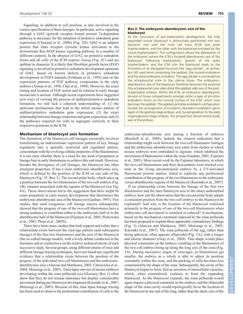
Box 2. The embryonic-abembryonic axis of theblastocystAt the conclusion of pre-implantation development, the fullyexpanded mouse blastocyst is distinctively partitioned into twodomains: one with the inner cell mass (ICM) and polartrophectoderm; and the other with the blastocoel enclosed by themural trophectoderm. This configuration allows the delineation ofthe embryonic (where the ICM is located)-abembryonic axis of theblastocyst. Following implantation, growth of the polartrophectoderm and the ICM into the blastocoel leads to theformation of an elongated structure (the ‘egg cylinder’, an archaicbut still used term) comprising the epiblast, the visceral endodermand the extra-embryonic ectoderm. The egg cylinder is connected viathe ectoplacental cone to the uterine tissue. The embryonic-abembryonic axis of the blastocyst therefore becomes the proximal(the ectoplacental cone side)-distal (the epiblast side) axis of the post-implantation embryo. Within the ICM, an embryonic-abembryonicdivision of tissue compartments emerges when a layer of primitiveendoderm forms on the luminal surface of the ICM, which nowbecomes the epiblast. The epiblast-primitive endoderm configurationheralds the arrangement of ectoderm-mesoderm-endoderm germlayers in the gastrula-stage embryo, and, by extrapolation to the earlyorganogenesis-stage embryo, the prospective dorsal-ventral bodyaxis of the embryo.
DEVELO
PMENT

706
zona (Fig. 3B). When embryos are deliberately compressed into anelongated shape from the two-cell to the blastocyst stage, theblastocoel is consistently positioned at one end of the elongatedblastocyst, regardless of the relationship to the original position ofthe first cleavage plane (Fig. 3B-D) (Motosugi et al., 2005). Acomputer simulation of blastocoel formation has also shown that aconstraining ellipsoidal capsule could help fixing the axis of theblastocyst (Honda et al., 2008).
The role of the zona in blastocoel positioning has been tested byexamining the development of zona-free embryos. In one study,removal of the zona pellucida from the morula stage onwards hadno effect on the correlation between the first cleavage plane and theorientation of the blastocoel (Gardner, 2007), whereas in another
study this association was lost (Kurotaki et al., 2007). Zona-freeembryos develop perfectly normally, which suggests that anyconstraint imposed by the zona, while imposing morphologicalconstraint on embryo development, is not relevant for thespecification of cell fates or axis formation.
Although the weight of evidence suggests that any apparentdifference in the lineage contribution of the two-cell blastomerescould be due to topological constraints, results from the Zernicka-Goetz laboratory have revealed some potential differences in thelineage potential of individual four-cell blastomeres. An analysis ofthe timing and orientation of the two- to four-cell cleavage(Piotrowska-Nitsche and Zernicka-Goetz, 2005) showed that about80% of embryos adopt a tetrahedral four-cell arrangement. This is
REVIEW Development 136 (5)
Emb.Abemb.
Emb.Abemb.
Emb.Abemb.
yzyzyz
= P2CB
yz
yz
P2CB
yz
yz
P2CB
No rotation of P2CB (yz = P2CB)
90° rotation of P2CB along y axis
90° rotation of P2CB along z axis
x
y z
xy
yz
xz
A
B C D
E
FG H
IJ
K L
Zona pellucida
Second polar body
✔ ✘ ✘
Fig. 3. The relationships among zona pellucida shape, orientation of the two-cell embryo and the embryonic-abembryonic axis of themouse blastocyst. (A) In many mouse embryos, the zona pellucida is not a sphere, but a scalene ellipsoid. It has three unequal diameters: long(marked by blue arrows and triangles), medium (orange arrows and stars) and short (green arrows and circles). The two-cell stage embryo alwaysaligns its orientation along the long axes in the zona pellucida. The broken double lines show differences in the space between the surface of theblastomere and the zona viewed at two different optical planes: left, view in the optical plane of the long and middle diameters (xy plane in E);right, view in the optical plane of the long and short diameters (yz plane in E). (B-D) With reference to the coordinates of the zona pellucidadelineated at the two-cell stage, the abembryonic-embryonic axis in the blastocyst most frequently aligns with the longest diameter of the zona (B)and rarely with the two shorter diameters (C,D). (E,F) The two-cell embryo is visualized in 3D space with the plane of two-cell boundary (P2CB)aligned with the yz plane, the plane of the middle and short diameters of the zona pellucida (each blastomere and its progeny are colored green ororange). (G,H) If the embryo does not rotate during cleavage (or rotates only along the x-axis), this alignment is maintained through (G) the eight-cell to (H) the blastocyst stage. (H) The abembryonic-embryonic axis of the blastocyst forms perpendicularly to the yz plane and the P2CB. Theprogeny of each two-cell blastomere thus predominantly occupies either the abembryonic or embryonic domain of the blastocyst. This situationoccurs in embryos in which cell movement within the zona is limited or prevented by alginate. (I-L) If the embryo rotates within the zona duringcleavage, the P2CB will no longer be aligned with the yz plane (I,K). The abembryonic-embryonic axis of the blastocyst still forms perpendicularly tothe yz plane, according to the shape of the zona pellucida, but the P2CB does not align with the abembryonic-embryonic axis (J,L). Twohypothetical examples are shown in which an embryo is rotated 90° along the y axis (I) or the z axis (K). In these situations, the progeny of eachtwo-cell blastomere shows no predictable relationship to the lineages of the blastocyst or occupancy of specific domains. In real development, theangle between P2CB and the yz plane is often oriented between I and K; thus, the position of P2CB in blastocysts varies. D
EVELO
PMENT

707REVIEWDevelopment 136 (5)
achieved by either the earlier-dividing two-cell blastomere dividingmeridionally (M, plane of cell division parallel to that of the firstcleavage), with the later division occurring equatorially (E, plane ofcell division perpendicular or oblique to that of the first cleavage),in the so-called ME pattern, which is found in 42% of embryos. Theother pattern of division may occur first equatorially thenmeridionally – the EM pattern – which is found in 39% of embryos.The later-dividing equatorial pair in the ME pattern is likely tocontribute to the abembryonic region of the blastocyst in the intactembryo, whereas the earlier-dividing equatorial pair in the EMarrangement show no such bias (Piotrowska-Nitsche and Zernicka-Goetz, 2005). Embryos generated by the re-aggregation of thedaughter cells that are located furthest away from the second polarbody after the equatorial division of one of the two-cell blastomeresshow reduced viability later in development (Piotrowska-Nitsche etal., 2005). This indicated that these so-called ‘vegetal’ blastomeresmight have an inherently deficient potential. In the ME embryo, this‘vegetal’ blastomere shows lower levels of arginine methylation ofhistone H3 (H3R26me) than do other blastomeres (Torres-Padilla etal., 2007a). The importance of this specific histone mark, which isassociated with gene activation in lineage specification, is unclear.Ectopic expression of an arginine methyltransferase CARM1, whichenhances H3R26me methylation, in a two-cell blastomere can biasthe distribution of its progeny within the blastocyst, but it is not clearhow this would affect cell lineage. Although expression of CARMImight have an impact on ICM fate (Torres-Padilla et al., 2007a), theendogenous level of histone methylation has yet to be correlatedwith lineage specification in a meaningful way. Although the vegetalblastomere of the EM embryo, like that of the ME embryo, is alsoless efficient in contributing to embryogenesis (Piotrowska-Nitscheet al., 2005), it displays H3R26me levels that are similar to the otherthree blastomeres, unlike its counterpart in the ME embryo, whichis lower than its sister blastomeres (Torres-Padilla et al., 2007a).Overall, these findings suggest that this specific histonemodification may not correlate consistently with cell fate or potency.
What of the possible importance of the inheritance of a specificregion of the egg cytoplasm in driving trophectoderm fate, asproposed from the study of the subset of embryos that haveundergone the ME pattern of cleavage up to the four-cell stage(Piotrowska-Nitsche et al., 2005)? It was proposed that the ‘vegetal’blastomere of the ME embryo is biased to acquire an abembryonicTE fate (Piotrowska-Nitsche et al., 2005), although a recent studywhich attempted to mark the same cell, found no such bias (Alarconand Marikawa, 2008). To support the case that, in this particularsubset of embryos, this vegetal blastomere has a biased lineage fate,Jedrusik et al. (Jedrusik et al., 2008) have recently reported that itsprogeny shows an elevated expression of Cdx2 at the eight-cellstage.
A priori, no pre-patterning of the egg or of the two-cell embryo isrequired to explain the formation of the cell lineages, the shape andthe axes of the mouse blastocyst. A combination of ad hoctopological constraints and cell polarity and signaling processes thatlead to the segregation of inner and outer cell populations canexplain normal development. And yet, persistent clues indicate thepossibility that asymmetries in the mammalian egg may have thepotential to bias cell fate and morphogenesis under specialconditions. Why should this be so? In many invertebrate and lowervertebrate species, it is clear that asymmetries in the distribution ofcytoplasmic determinants in the egg play major roles in establishingearly embryonic patterning. The early development of all of thesespecies depends on maternally inherited factors, with zygotictranscription occurring as a later event. In mammals, by contrast,
although maternal mRNAs and proteins are active in earlydevelopment, maternal RNA is rapidly degraded and zygotic geneactivation occurs during early cleavage and is required for blastocystdevelopment. This switch away from the dependence on maternalinheritance was presumably accompanied by a move away fromearly patterning being driven by asymmetrically distributed maternaldeterminants to being driven by zygotic transcription. Nonetheless,the systems that allow asymmetries in the egg might persist as‘evolutionary relics’ in mouse eggs, leading to the appearance ofasymmetries in some embryos. These asymmetries may biasdevelopmental pathways but can be readily overridden by processesof lineage development and blastocyst morphogenesis.
Peri-implantation asymmetry and axisspecificationIn addition to the embryonic-abembryonic axis and the ellipsoidalshape of the ICM (Fig. 1), other morphological features also revealthe asymmetry of the blastocyst. In the implanted blastocystrecovered from the mouse uterus, the ICM is often oriented in atilted position so that the ICM has an upper and a lower side (Fig.4A). Accompanying this tilted orientation of the ICM, the initialthickening of the polar trophectoderm also appears asymmetrical. Atilting of the ectoplacental cone away from the proximal-distal axis(Box 2) is seen in embryos at subsequent stages of post-implantationdevelopment (Fig. 4B-D). By the pre-primitive streak (E6.0) stage,the direction in which the cone tilts is aligned consistently with theorientation, but not with the polarity, of the prospective AP axis ofthe body, which coincides with the longer transverse diameter of thecylindrical embryo (Gardner et al., 1992) (Fig. 4D). Quiteunexpectedly, the orientation of the AP axis does not always alignwith the longer diameter of the early embryo (Mesnard et al., 2004;Perea-Gomez et al., 2004). In the younger (E.5.5-5.75) embryo, theAP axis aligns initially with the shorter diameter (Fig. 4C), whichlengthens as the embryo re-models its shape, such that the AP axislater becomes aligned with longer diameter (Fig. 4D). This re-shaping of the embryo, but not the specification of the AP axis,requires Fgf8b and Wnt3 function in the epiblast (Barrow et al.,2007; Guo and Li, 2007). It would be interesting to find out whetherthe tilting of the ICM or the ectoplacental cone has any specificorientation with respect to the short or long transverse diameter ofthe cylindrical embryo during embryogenesis.
Overall, these intriguing findings beg the question of whether thelong axis of the ICM, the angle of tilt of the ICM and the asymmetricposition of the ectoplacental cone have any developmentalrelationship with each other and with any of the three primary bodyaxes of the post-implantation embryo. The answer to this questionrequires lineages across the peri-implantation to gastrulation periodto be traced directly. In earlier studies, single ICM cells at either endof the long axis of the ICM were marked by injection and theirdescendants followed in the visceral endoderm (see Glossary, Box1) of post-implantation embryos (Weber et al., 1999). Intriguingly,clones were found to spread proximodistally in an oblique manner,suggesting that the horizontal axis of the ICM may be converted intothe proximodistal axis (Box 2) of the post-implantation embryo. Asimilar tracking study, performed by marking cells presumably atrandom positions near the surface of the ICM, showed that clonesspan the extra-embryonic and embryonic regions of the visceralendoderm, and has revealed more diverse patterns of clonaldistribution in both the proximal-distal and transverse dimensionsof the cylindrical embryo (Perea-Gomez et al., 2007). It isimperative, in view of the now available tissue- and site-specificmolecular markers of embryonic asymmetry in pre-gastrulation D
EVELO
PMENT

708
mouse embryos (Fig. 4D), to re-examine the distribution of theseICM-derived clones in the visceral endoderm to see whether thereis any consistent spatial relationship between the site of origin oftheir precursors in the ICM and their contribution to the prospectiveAP body axis. It would be particularly informative to track thedistribution of these cell clones throughout development fromblastocyst to gastrula when it becomes possible to grow the peri-implantation embryo successfully in vitro.
Visceral endoderm: tissue patterning andemerging asymmetriesThe primitive endoderm, which is formed as an epithelium initiallyon the luminal surface of the ICM (Fig. 1G), expands during peri-implantation development to form the parietal endoderm (whichlines the luminal surface of the mural trophectoderm) and thevisceral endoderm (which envelops the extra-embryonic ectodermand the epiblast) (Fig. 1H; Fig. 4B-D). The visceral endoderm is atissue of significant interest because of its crucial function inmediating the activity of transforming growth factor β (TGFβ) (bonemorphogenetic protein and Nodal) and WNT signaling pathways,which sustain the differentiation and patterning of the epiblast (Tamet al., 2006; Tam and Loebel, 2007). Furthermore, the changes in the
epithelial architecture of the endoderm, the regionalized geneexpression domains (Kemp et al., 2005; Kemp et al., 2007; Kimura-Yoshida et al., 2007; Yamamoto et al., 2004; Pfister et al., 2007) andthe pattern of morphogenetic movement of cells reflect a dynamicprocess during which structural and molecular asymmetries aretranslated into the AP patterning of the body axis (reviewed by Luet al., 2001; Zernicka-Goetz, 2002; Srinivas, 2006; Tam and Loebel,2007). Contrary to the idea that the visceral endoderm is entirelyrestricted to extra-embryonic fates, a small number of itsdescendants do contribute to the endoderm of the embryonic gut(Kwon et al., 2008).
Local visceral endoderm thickening marks emergingasymmetryChanges in epithelial morphology, revealed as a local thickening ofthe visceral endoderm, first in the distal region and then later on oneside of the pre-gastrulation embryo (Fig. 4C,D) (Kimura-Yoshida etal., 2005; Rivera-Perez et al., 2003; Yamamoto et al., 2004), aretelltale signs of the acquisition of asymmetry in the proximodistalaxis and the prospective AP body axis, respectively. By tracking theposition of the thickened population of embryonic visceralendoderm cells, in conjunction with gene expression patterns, a
REVIEW Development 136 (5)
Site of tethering of the second polar body
Tilting of the ICM
Position of Cer1- and Lefty1-expressing cellsin the primitive endoderm
Tilting of the ectoplacental cone
Prospective AP axis aligns with the longer diameter of the cylinder
Location of the AVE (broken green box)
Regionalization of gene expression in the AVE and posterior epiblast (brokenblack outline)
?Spatial correlation of asymmetric features not yet established
Verified relationship of asymmetry to prospective AP axis
? ? ?
(A) Blastocyst (B) Early egg cylinder (C) Embryo with DVE (D) Embryo with AVE
Cer1-expressing cell
Wnt3-expressing cell
Sfrp1Sfrp5Fzd5Fzd8Dkk1Cer1Lefty1Sox17Lhx1Hhex
TWnt3NodalCripto
A PA P
Plane of section
A PTransverse section of the embryo and orientation of AP axis
Tilting of the ectoplacental cone away from the proximodistal axis
Prospective AP axis aligns with the shorterdiameter of the cylinder
Lopsided distribution of Cer1- and Lefty1-expressing cells in the distal visceral endoderm (broken red box)
Localization of Wnt3 expression on one side of the visceral endoderm
Second polar body
β-catenin-expressing cell
Lefty1-expressing cell
Trophectoderm
Inner cell mass
Primitive endoderm
Visceral endoderm
Epiblast
Ectoplacentalcone
Fig. 4. The emergence of asymmetry during peri-implantation development from blastocyst to immediately before gastrulation.(A-D) Early pre-gastrulation stages of mouse development, with asymmetric features listed for each stage. The tilting of the ectoplacental cone fromthe proximal-distal axis (unbroken line) is indicated by the black broken line. Cells in the inner cells mass (ICM), the primitive endoderm and thevisceral endoderm that express β-catenin, Lefty1, Cer1 or Wnt3 are color coded. Many genes that are expressed in the anterior visceral endoderm(AVE; D broken green rectangle) are also expressed previously in the distal visceral endoderm (DVE). Four examples of genes that are expressed inthe posterior epiblast (D, broken black outline) are listed. In C (embryo with DVE) and D (embryo with AVE), a transverse section of the embryo isshown to illustrate the alignment of the prospective anterior-posterior (AP) axis first with the shorter and then with the longer diameter at therespective stage.
DEVELO
PMENT
709REVIEWDevelopment 136 (5)
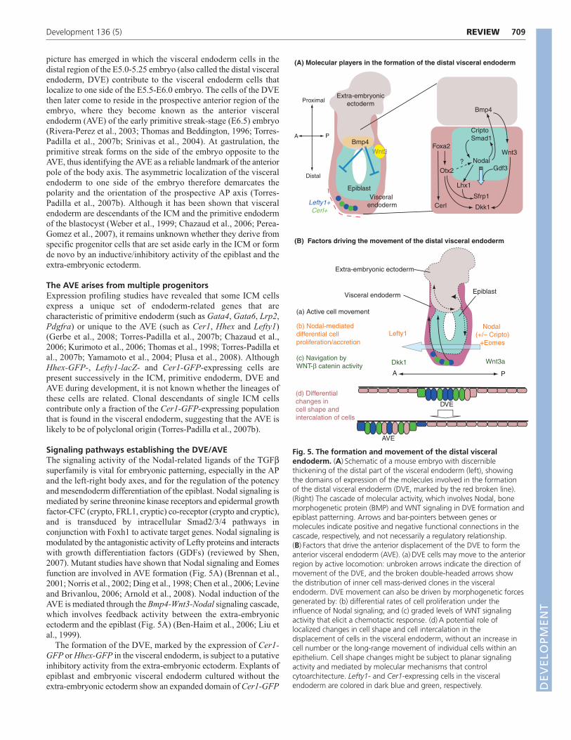
picture has emerged in which the visceral endoderm cells in thedistal region of the E5.0-5.25 embryo (also called the distal visceralendoderm, DVE) contribute to the visceral endoderm cells thatlocalize to one side of the E5.5-E6.0 embryo. The cells of the DVEthen later come to reside in the prospective anterior region of theembryo, where they become known as the anterior visceralendoderm (AVE) of the early primitive streak-stage (E6.5) embryo(Rivera-Perez et al., 2003; Thomas and Beddington, 1996; Torres-Padilla et al., 2007b; Srinivas et al., 2004). At gastrulation, theprimitive streak forms on the side of the embryo opposite to theAVE, thus identifying the AVE as a reliable landmark of the anteriorpole of the body axis. The asymmetric localization of the visceralendoderm to one side of the embryo therefore demarcates thepolarity and the orientation of the prospective AP axis (Torres-Padilla et al., 2007b). Although it has been shown that visceralendoderm are descendants of the ICM and the primitive endodermof the blastocyst (Weber et al., 1999; Chazaud et al., 2006; Perea-Gomez et al., 2007), it remains unknown whether they derive fromspecific progenitor cells that are set aside early in the ICM or formde novo by an inductive/inhibitory activity of the epiblast and theextra-embryonic ectoderm.
The AVE arises from multiple progenitorsExpression profiling studies have revealed that some ICM cellsexpress a unique set of endoderm-related genes that arecharacteristic of primitive endoderm (such as Gata4, Gata6, Lrp2,Pdgfra) or unique to the AVE (such as Cer1, Hhex and Lefty1)(Gerbe et al., 2008; Torres-Padilla et al., 2007b; Chazaud et al.,2006; Kurimoto et al., 2006; Thomas et al., 1998; Torres-Padilla etal., 2007b; Yamamoto et al., 2004; Plusa et al., 2008). AlthoughHhex-GFP-, Lefty1-lacZ- and Cer1-GFP-expressing cells arepresent successively in the ICM, primitive endoderm, DVE andAVE during development, it is not known whether the lineages ofthese cells are related. Clonal descendants of single ICM cellscontribute only a fraction of the Cer1-GFP-expressing populationthat is found in the visceral endoderm, suggesting that the AVE islikely to be of polyclonal origin (Torres-Padilla et al., 2007b).
Signaling pathways establishing the DVE/AVEThe signaling activity of the Nodal-related ligands of the TGFβsuperfamily is vital for embryonic patterning, especially in the APand the left-right body axes, and for the regulation of the potencyand mesendoderm differentiation of the epiblast. Nodal signaling ismediated by serine threonine kinase receptors and epidermal growthfactor-CFC (crypto, FRL1, cryptic) co-receptor (crypto and cryptic),and is transduced by intracellular Smad2/3/4 pathways inconjunction with Foxh1 to activate target genes. Nodal signaling ismodulated by the antagonistic activity of Lefty proteins and interactswith growth differentiation factors (GDFs) (reviewed by Shen,2007). Mutant studies have shown that Nodal signaling and Eomesfunction are involved in AVE formation (Fig. 5A) (Brennan et al.,2001; Norris et al., 2002; Ding et al., 1998; Chen et al., 2006; Levineand Brivanlou, 2006; Arnold et al., 2008). Nodal induction of theAVE is mediated through the Bmp4-Wnt3-Nodal signaling cascade,which involves feedback activity between the extra-embryonicectoderm and the epiblast (Fig. 5A) (Ben-Haim et al., 2006; Liu etal., 1999).
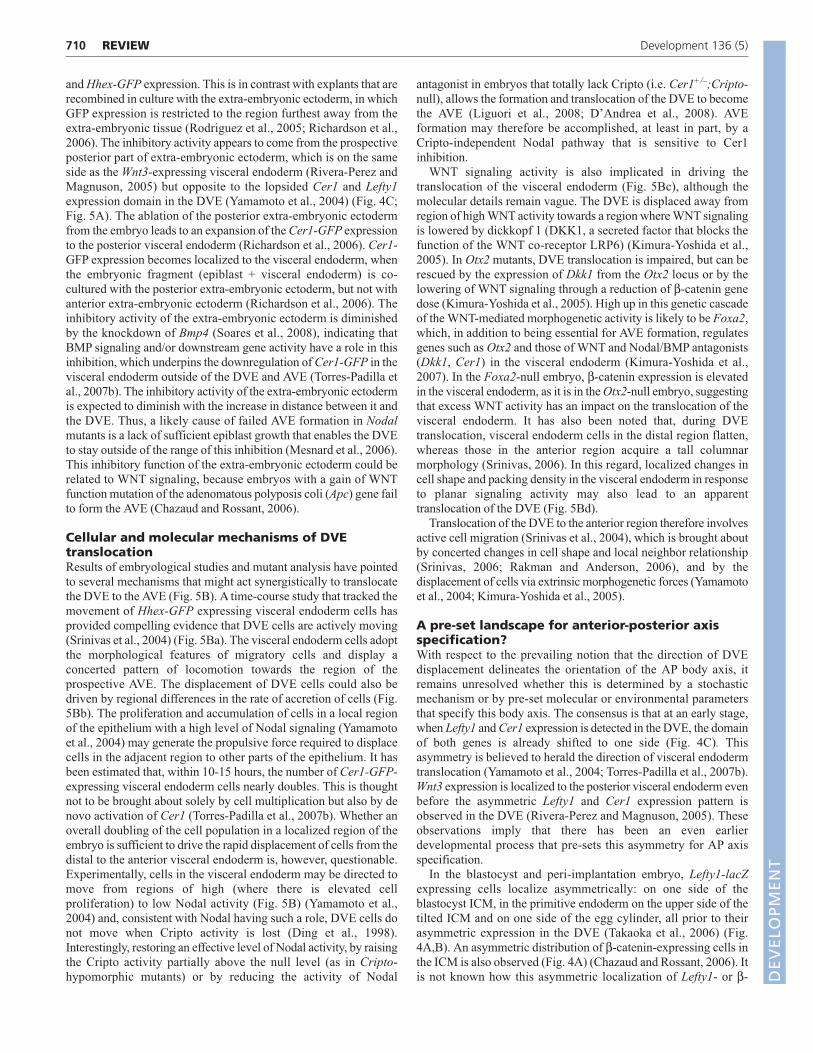
The formation of the DVE, marked by the expression of Cer1-GFP or Hhex-GFP in the visceral endoderm, is subject to a putativeinhibitory activity from the extra-embryonic ectoderm. Explants ofepiblast and embryonic visceral endoderm cultured without theextra-embryonic ectoderm show an expanded domain of Cer1-GFP
(A) Molecular players in the formation of the distal visceral endoderm
(B) Factors driving the movement of the distal visceral endoderm
Wnt3aDkk1
Nodal (+/– Cripto)
+EomesLefty1
+
Lefty1
(c) Navigation by WNT-β catenin activity
(b) Nodal-mediated differential cell proliferation/accretion
(a) Active cell movement
Foxa2
Otx2
Cerl
Bmp4
NodalGdf3
Lhx1
CriptoSmad1
Wnt3
Dkk1
Sfrp1
?
A P
Proximal
Distal
Bmp4
Lefty1+Cerl+
33WWnt3
Extra-embryonicectoderm
EpiblastVisceral
endoderm
Extra-embryonic ectoderm
Visceral endoderm Epiblast
(d) Differential changes in cell shape andintercalation of cells
A P
DVE
AVE
Fig. 5. The formation and movement of the distal visceralendoderm. (A) Schematic of a mouse embryo with discerniblethickening of the distal part of the visceral endoderm (left), showingthe domains of expression of the molecules involved in the formationof the distal visceral endoderm (DVE, marked by the red broken line).(Right) The cascade of molecular activity, which involves Nodal, bonemorphogenetic protein (BMP) and WNT signaling in DVE formation andepiblast patterning. Arrows and bar-pointers between genes ormolecules indicate positive and negative functional connections in thecascade, respectively, and not necessarily a regulatory relationship.(B) Factors that drive the anterior displacement of the DVE to form theanterior visceral endoderm (AVE). (a) DVE cells may move to the anteriorregion by active locomotion: unbroken arrows indicate the direction ofmovement of the DVE, and the broken double-headed arrows showthe distribution of inner cell mass-derived clones in the visceralendoderm. DVE movement can also be driven by morphogenetic forcesgenerated by: (b) differential rates of cell proliferation under theinfluence of Nodal signaling; and (c) graded levels of WNT signalingactivity that elicit a chemotactic response. (d) A potential role oflocalized changes in cell shape and cell intercalation in thedisplacement of cells in the visceral endoderm, without an increase incell number or the long-range movement of individual cells within anepithelium. Cell shape changes might be subject to planar signalingactivity and mediated by molecular mechanisms that controlcytoarchitecture. Lefty1- and Cer1-expressing cells in the visceralendoderm are colored in dark blue and green, respectively. D
EVELO
PMENT
710
and Hhex-GFP expression. This is in contrast with explants that arerecombined in culture with the extra-embryonic ectoderm, in whichGFP expression is restricted to the region furthest away from theextra-embryonic tissue (Rodriguez et al., 2005; Richardson et al.,2006). The inhibitory activity appears to come from the prospectiveposterior part of extra-embryonic ectoderm, which is on the sameside as the Wnt3-expressing visceral endoderm (Rivera-Perez andMagnuson, 2005) but opposite to the lopsided Cer1 and Lefty1expression domain in the DVE (Yamamoto et al., 2004) (Fig. 4C;Fig. 5A). The ablation of the posterior extra-embryonic ectodermfrom the embryo leads to an expansion of the Cer1-GFP expressionto the posterior visceral endoderm (Richardson et al., 2006). Cer1-GFP expression becomes localized to the visceral endoderm, whenthe embryonic fragment (epiblast + visceral endoderm) is co-cultured with the posterior extra-embryonic ectoderm, but not withanterior extra-embryonic ectoderm (Richardson et al., 2006). Theinhibitory activity of the extra-embryonic ectoderm is diminishedby the knockdown of Bmp4 (Soares et al., 2008), indicating thatBMP signaling and/or downstream gene activity have a role in thisinhibition, which underpins the downregulation of Cer1-GFP in thevisceral endoderm outside of the DVE and AVE (Torres-Padilla etal., 2007b). The inhibitory activity of the extra-embryonic ectodermis expected to diminish with the increase in distance between it andthe DVE. Thus, a likely cause of failed AVE formation in Nodalmutants is a lack of sufficient epiblast growth that enables the DVEto stay outside of the range of this inhibition (Mesnard et al., 2006).This inhibitory function of the extra-embryonic ectoderm could berelated to WNT signaling, because embryos with a gain of WNTfunction mutation of the adenomatous polyposis coli (Apc) gene failto form the AVE (Chazaud and Rossant, 2006).
Cellular and molecular mechanisms of DVEtranslocationResults of embryological studies and mutant analysis have pointedto several mechanisms that might act synergistically to translocatethe DVE to the AVE (Fig. 5B). A time-course study that tracked themovement of Hhex-GFP expressing visceral endoderm cells hasprovided compelling evidence that DVE cells are actively moving(Srinivas et al., 2004) (Fig. 5Ba). The visceral endoderm cells adoptthe morphological features of migratory cells and display aconcerted pattern of locomotion towards the region of theprospective AVE. The displacement of DVE cells could also bedriven by regional differences in the rate of accretion of cells (Fig.5Bb). The proliferation and accumulation of cells in a local regionof the epithelium with a high level of Nodal signaling (Yamamotoet al., 2004) may generate the propulsive force required to displacecells in the adjacent region to other parts of the epithelium. It hasbeen estimated that, within 10-15 hours, the number of Cer1-GFP-expressing visceral endoderm cells nearly doubles. This is thoughtnot to be brought about solely by cell multiplication but also by denovo activation of Cer1 (Torres-Padilla et al., 2007b). Whether anoverall doubling of the cell population in a localized region of theembryo is sufficient to drive the rapid displacement of cells from thedistal to the anterior visceral endoderm is, however, questionable.Experimentally, cells in the visceral endoderm may be directed tomove from regions of high (where there is elevated cellproliferation) to low Nodal activity (Fig. 5B) (Yamamoto et al.,2004) and, consistent with Nodal having such a role, DVE cells donot move when Cripto activity is lost (Ding et al., 1998).Interestingly, restoring an effective level of Nodal activity, by raisingthe Cripto activity partially above the null level (as in Cripto-hypomorphic mutants) or by reducing the activity of Nodal
antagonist in embryos that totally lack Cripto (i.e. Cer1+/–;Cripto-null), allows the formation and translocation of the DVE to becomethe AVE (Liguori et al., 2008; D’Andrea et al., 2008). AVEformation may therefore be accomplished, at least in part, by aCripto-independent Nodal pathway that is sensitive to Cer1inhibition.
WNT signaling activity is also implicated in driving thetranslocation of the visceral endoderm (Fig. 5Bc), although themolecular details remain vague. The DVE is displaced away fromregion of high WNT activity towards a region where WNT signalingis lowered by dickkopf 1 (DKK1, a secreted factor that blocks thefunction of the WNT co-receptor LRP6) (Kimura-Yoshida et al.,2005). In Otx2 mutants, DVE translocation is impaired, but can berescued by the expression of Dkk1 from the Otx2 locus or by thelowering of WNT signaling through a reduction of β-catenin genedose (Kimura-Yoshida et al., 2005). High up in this genetic cascadeof the WNT-mediated morphogenetic activity is likely to be Foxa2,which, in addition to being essential for AVE formation, regulatesgenes such as Otx2 and those of WNT and Nodal/BMP antagonists(Dkk1, Cer1) in the visceral endoderm (Kimura-Yoshida et al.,2007). In the Foxa2-null embryo, β-catenin expression is elevatedin the visceral endoderm, as it is in the Otx2-null embryo, suggestingthat excess WNT activity has an impact on the translocation of thevisceral endoderm. It has also been noted that, during DVEtranslocation, visceral endoderm cells in the distal region flatten,whereas those in the anterior region acquire a tall columnarmorphology (Srinivas, 2006). In this regard, localized changes incell shape and packing density in the visceral endoderm in responseto planar signaling activity may also lead to an apparenttranslocation of the DVE (Fig. 5Bd).
Translocation of the DVE to the anterior region therefore involvesactive cell migration (Srinivas et al., 2004), which is brought aboutby concerted changes in cell shape and local neighbor relationship(Srinivas, 2006; Rakman and Anderson, 2006), and by thedisplacement of cells via extrinsic morphogenetic forces (Yamamotoet al., 2004; Kimura-Yoshida et al., 2005).
A pre-set landscape for anterior-posterior axisspecification?With respect to the prevailing notion that the direction of DVEdisplacement delineates the orientation of the AP body axis, itremains unresolved whether this is determined by a stochasticmechanism or by pre-set molecular or environmental parametersthat specify this body axis. The consensus is that at an early stage,when Lefty1 and Cer1 expression is detected in the DVE, the domainof both genes is already shifted to one side (Fig. 4C). Thisasymmetry is believed to herald the direction of visceral endodermtranslocation (Yamamoto et al., 2004; Torres-Padilla et al., 2007b).Wnt3 expression is localized to the posterior visceral endoderm evenbefore the asymmetric Lefty1 and Cer1 expression pattern isobserved in the DVE (Rivera-Perez and Magnuson, 2005). Theseobservations imply that there has been an even earlierdevelopmental process that pre-sets this asymmetry for AP axisspecification.
In the blastocyst and peri-implantation embryo, Lefty1-lacZexpressing cells localize asymmetrically: on one side of theblastocyst ICM, in the primitive endoderm on the upper side of thetilted ICM and on one side of the egg cylinder, all prior to theirasymmetric expression in the DVE (Takaoka et al., 2006) (Fig.4A,B). An asymmetric distribution of β-catenin-expressing cells inthe ICM is also observed (Fig. 4A) (Chazaud and Rossant, 2006). Itis not known how this asymmetric localization of Lefty1- or β-
REVIEW Development 136 (5)
DEVELO
PMENT
711REVIEWDevelopment 136 (5)
catenin-positive cells is related to the long axis of the ICM or to theinitial tilting of the ICM prior to the formation of the primitiveendoderm. Cer1-expressing cells (as visualized by GFP reporter,mRNA and protein expression) in the primitive endoderm do notlocalize to any specific regions (Perea-Gomez et al., 2007).However, Cer1 activity, as revealed by GFP fluorescence, appearsto be uneven in the primitive endoderm and tends to be stronger inthe visceral endoderm on one side of the tilted ICM (Fig. 4A).Potentially, the regionalization of the Lefty1- and Cer1-active cellsin the visceral endoderm prior to the formation and movement of theDVE might be a manifestation of an underlying asymmetric patternthat foreshadows the orientation and polarity of the AP body axis.As the current data are primarily correlative, such inference is at bestconjectural. In blastocysts cultured in vitro over the period ofimplantation, Cer1-GFP and Lefty1-lacZ positive cells localizeunevenly in the primitive endoderm. If this pattern reflects theacquisition of embryonic asymmetry and the specification of thebody axis, such developmental capacity would have to be inherentto the embryo and not acquired by the act of implantation.
ConclusionsEmbryogenesis requires the generation of diverse cell types and theorderly assembly of these cells into an organized body plan. Duringthis process, asymmetries of anatomical and/or molecularcharacteristics emerge within cells, tissues and the whole embryo.Some of these asymmetries are relevant to cell fate, such as theradial asymmetry that differentiates outer and inner cells of themorula. Some of them drive cell rearrangements, such as themorphogenetic processes that accompany the formation of theanterior visceral endoderm by the anterior migration of the distalvisceral endoderm. Others may be incidental partners in influencingaxis formation, such as the non-spherical shape of the zonapellucida. The challenge ahead is to determine whetherdevelopmentally relevant asymmetries influence lineage allocationand to translate our knowledge of morphological asymmetries intomolecular mechanisms.
We thank Yojiro Yamanaka for useful discussions and assistance with thepreparation of figures, Amy Ralston for comments on the manuscript, andBerenika Plusa and Kat Hadjantonakis for providing materials beforepublication. We are supported by the Canadian Institute of Health Research,by the Canadian Stem Cell Network (J.R.) and by the National Health andMedical Research Council of Australia (P.P.L.T.).
ReferencesAlarcon, V. B. and Marikawa, Y. (2003). Deviation of the blastocyst axis from the
first cleavage plane does not affect the quality of mouse postimplantationdevelopment. Biol. Reprod. 69, 1208-1212.
Alarcon, V. B. and Marikawa, Y. (2008). Spatial alignment of the mouseblastocyst axis across the first cleavage plane is caused by mechanical constraintrather than developmental bias among blastomeres. Mol. Reprod. Dev. 75,1143-1153.
Arman, E., Haffner-Krausz, R., Chen, Y., Heath, J. K. and Lonai, P. (1998).Targeted disruption of fibroblast growth factor (FGF) receptor 2 suggests a rolefor FGF signaling in pregastrulation mammalian development. Proc. Natl. Acad.Sci. USA 95, 5082-5087.
Arnold, S. J., Hofmann, U. K., Bikoff, E. K. and Robertson, E. J. (2008). Pivotalroles for eomesodermin during axis formation, epithelium-to-mesenchymetransition and endoderm specification in the mouse. Development 135, 501-511.
Avilion, A. A., Nicolis, S. K., Pevny, L. H., Perez, L., Vivian, N. and Lovell-Badge, R. (2003). Multipotent cell lineages in early mouse development dependon SOX2 function. Genes Dev. 17, 126-140.
Barrow, J. R., Howell, W. D., Rule, M., Hayashi, S., Thomas, K. R., Capecchi,M. R. and McMahon, A. P. (2007). Wnt3 signaling in the epiblast is requiredfor proper orientation of the anteroposterior axis. Dev. Biol. 312, 312-320.
Beland, M., Pilon, N., Houle, M., Oh, K., Sylvestre, J. R., Prinos, P. andLohnes, D. (2004). Cdx1 autoregulation is governed by a novel Cdx1-LEF1transcription complex. Mol. Cell. Biol. 24, 5028-5038.
Ben-Haim, N., Lu, C., Guzman-Ayala, M., Pescatore, L., Mesnard, D.,Bischofberger, M., Naef, F., Robertson, E. J. and Constam, D. B. (2006). Thenodal precursor acting via activin receptors induces mesoderm by maintaining asource of its convertases and BMP4. Dev. Cell 11, 313-323.
Bischoff, M., Parfitt, D. E. and Zernicka-Goetz, M. (2008). Formation of theembryonic-abembryonic axis of the mouse blastocyst: relationships betweenorientation of early cleavage divisions and pattern of symmetric/asymmetricdivisions. Development 135, 953-962.
Boyer, L. A., Lee, T. I., Cole, M. F., Johnstone, S. E., Levine, S. S., Zucker, J. P.,Guenther, M. G., Kumar, R. M., Murray, H. L., Jenner, R. G. et al. (2005).Core transcriptional regulatory circuitry in human embryonic stem cells. Cell 122,947-956.
Brennan, J., Lu, C. C., Norris, D. P., Rodriguez, T. A., Beddington, R. S. andRobertson, E. J. (2001). Nodal signalling in the epiblast patterns the earlymouse embryo. Nature 411, 965-969.
Chai, N., Patel, Y., Jacobson, K., McMahon, J., McMahon, A. and Rappolee,D. A. (1998). FGF is an essential regulator of the fifth cell division inpreimplantation mouse embryos. Dev. Biol. 198, 105-115.
Chambers, I., Colby, D., Robertson, M., Nichols, J., Lee, S., Tweedie, S. andSmith, A. (2003). Functional expression cloning of Nanog, a pluripotencysustaining factor in embryonic stem cells. Cell 113, 643-655.
Chazaud, C. and Rossant, J. (2006). Disruption of early proximodistal patterningand AVE formation in Apc mutants. Development 133, 3379-3387.
Chazaud, C., Yamanaka, Y., Pawson, T. and Rossant, J. (2006). Early lineagesegregation between epiblast and primitive endoderm in mouse blastocyststhrough the Grb2-MAPK pathway. Dev. Cell 10, 615-624.
Chen, C., Ware, S. M., Sato, A., Houston-Hawkins, D. E., Habas, R., Matzuk,M. M., Shen, M. M. and Brown, C. W. (2006). The Vg1-related protein Gdf3acts in a Nodal signaling pathway in the pre-gastrulation mouse embryo.Development 133, 319-329.
Chew, J. L., Loh, Y. H., Zhang, W., Chen, X., Tam, W. L., Yeap, L. S., Li, P., Ang,Y. S., Lim, B., Robson, P. et al. (2005). Reciprocal transcriptional regulation ofPou5f1 and Sox2 via the Oct4/Sox2 complex in embryonic stem cells. Mol. Cell.Biol. 25, 6031-6046.
Chroscicka, A., Komorowski, S. and Maleszewski, M. (2004). Bothblastomeres of the mouse 2-cell embryo contribute to the embryonic portion ofthe blastocyst. Mol. Reprod. Dev. 68, 308-312.
D’Andrea, D., Liguori, G. L., Le Good, J. A., Lonardo, E., Andersson, O.,Constam, D. B., Persico, M. G. and Minchiotti, G. (2008). Cripto promotes A-P axis specification independently of its stimulatory effect on Nodalautoinduction. J. Cell Biol. 180, 597-605.
Dietrich, J. E. and Hiiragi, T. (2007). Stochastic patterning in the mouse pre-implantation embryo. Development 134, 4219-4231.
Ding, J., Yang, L., Yan, Y. T., Chen, A., Desai, N., Wynshaw-Boris, A. andShen, M. M. (1998). Cripto is required for correct orientation of the anterior-posterior axis in the mouse embryo. Nature 395, 702-707.
Feldman, B., Poueymirou, W., Papaioannou, V. E., DeChiara, T. M. andGoldfarb, M. (1995). Requirement of FGF-4 for postimplantation mousedevelopment. Science 267, 246-249.
Fleming, T. P. and Johnson, M. H. (1988). From egg to epithelium. Annu. Rev.Cell Biol. 4, 459-485.
Fleming, T. P., McConnell, J., Johnson, M. H. and Stevenson, B. R. (1989).Development of tight junctions de novo in the mouse early embryo: control ofassembly of the tight-junction-specific protein. J. Cell Biol. 108, 1407-1418.
Fujimori, T., Kurotaki, Y., Miyazaki, J. and Nabeshima, Y. (2003). Analysis ofcell lineage in two- and four-cell mouse embryos. Development 130, 5113-5122.
Gardner, R. L. (1997). The early blastocyst is bilaterally symmetrical and its axis ofsymmetry is aligned with the animal-vegetal axis of the zygote in the mouse.Development 124, 289-301.
Gardner, R. L. (2001). Specification of embryonic axes begins before cleavage innormal mouse development. Development 128, 839-847.
Gardner, R. L. (2007). The axis of polarity of the mouse blastocyst is specifiedbefore blastulation and independently of the zona pellucida. Hum. Reprod. 22,798-806.
Gardner, R. L., Meredith, M. R. and Altman, D. G. (1992). Is the anterior-posterior axis of the fetus specified before implantation in the mouse? J. Exp.Zool. 264, 437-443.
Gerbe, F., Cox, B., Rossant, J. and Chazaud, C. (2008). Dynamic expression ofLrp2 pathway members reveals progressive epithelial differentiation of primitiveendoderm in mouse blastocyst. Dev. Biol. 313, 594-602.
Gray, D., Plusa, B., Piotrowska, K., Na, J., Tom, B., Glover, D. M. andZernicka-Goetz, M. (2004). First cleavage of the mouse embryo responds tochange in egg shape at fertilization. Curr. Biol. 14, 397-405.
Guo, Q. and Li, J. Y. (2007). Distinct functions of the major Fgf8 spliceform,Fgf8b, before and during mouse gastrulation. Development 134, 2251-2260.
Honda, H., Motosugi, N., Nagai, T., Tanemura, M. and Hiiragi, T. (2008).Computer simulation of emerging asymmetry in the mouse blastocyst.Development 135, 1407-1414.
Jedrusik, A., Parfitt, D. E., Guo, G., Skamagki, M., Grabarek, J. B., Johnson, DEVELO
PMENT
712
M. H., Robson, P. and Zernicka-Goetz, M. (2008). Role of Cdx2 and cellpolarity in cell allocation and specification of trophectoderm and inner cell massin the mouse embryo. Genes Dev. 22, 2692-2706.
Johnson, M. H. and McConnell, J. M. (2004). Lineage allocation and cell polarityduring mouse embryogenesis. Semin. Cell Dev. Biol. 15, 583-597.
Johnson, M. H. and Ziomek, C. A. (1981). The foundation of two distinct celllineages within the mouse morula. Cell 24, 71-80.
Johnson, M. H. and Ziomek, C. A. (1983). Cell interactions influence the fate ofmouse blastomeres undergoing the transition from the 16- to the 32-cell stage.Dev. Biol. 95, 211-218.
Johnson, M. H., Maro, B. and Takeichi, M. (1986). The role of cell adhesion inthe synchronization and orientation of polarization in 8-cell mouse blastomeres.J. Embryol. Exp. Morph. 93, 239-255.
Kemp, C., Willems, E., Abdo, S., Lambiv, L. and Leyns, L. (2005). Expression ofall Wnt genes and their secreted antagonists during mouse blastocyst andpostimplantation development. Dev. Dyn. 233, 1064-1075.
Kemp, C. R., Willems, E., Wawrzak, D., Hendrickx, M., Agbor Agbor, T. andLeyns, L. (2007). Expression of Frizzled5, Frizzled7, and Frizzled10 during earlymouse development and interactions with canonical Wnt signaling. Dev. Dyn.236, 2011-2019.
Kimura-Yoshida, C., Nakano, H., Okamura, D., Nakao, K., Yonemura, S.,Belo, J. A., Aizawa, S., Matsui, Y. and Matsuo, I. (2005). Canonical Wntsignaling and its antagonist regulate anterior-posterior axis polarization byguiding cell migration in mouse visceral endoderm. Dev. Cell 9, 639-650.
Kimura-Yoshida, C., Tian, E., Nakano, H., Amazaki, S., Shimokawa, K.,Rossant, J., Aizawa, S. and Matsuo, I. (2007). Crucial roles of Foxa2 in mouseanterior-posterior axis polarization via regulation of anterior visceral endoderm-specific genes. Proc. Natl. Acad. Sci. USA 104, 5919-5924.
Kunath, T., Arnaud, D., Uy, G. D., Okamoto, I., Chureau, C., Yamanaka, Y.,Heard, E., Gardner, R. L., Avner, P. and Rossant, J. (2005). Imprinted X-inactivation in extra-embryonic endoderm cell lines from mouse blastocysts.Development 132, 1649-1661.
Kurimoto, K., Yabuta, Y., Ohinata, Y., Ono, Y., Uno, K. D., Yamada, R. G.,Ueda, H. R. and Saitou, M. (2006). An improved single-cell cDNA amplificationmethod for efficient high-density oligonucleotide microarray analysis. NucleicAcids Res. 34, e42.
Kurotaki, Y., Hatta, K., Nakao, K., Nabeshima, Y. and Fujimori, T. (2007).Blastocyst axis is specified independently of early cell lineage but aligns with theZP shape. Science 316, 719-723.
Kwon, G. S., Viotti, M. and Hadjantonakis, A. K. (2008). The endoderm of themouse embryo arises by dynamic widesapread intercalation of embryonic andextraembryonic lineages. Dev. Cell 15, 509-520.
Levine, A. J. and Brivanlou, A. H. (2006). GDF3, a BMP inhibitor, regulates cellfate in stem cells and early embryos. Development 133, 209-216.
Liguori, G. L., Borges, A. C., D’Andrea, D., Liguoro, A., Goncalves, L.,Salgueiro, A. M., Persico, M. G. and Belo, J. A. (2008). Cripto-independentNodal signaling promotes positioning of the A-P axis in the early mouse embryo.Dev. Biol. 315, 280-289.
Liu, P., Wakamiya, M., Shea, M. J., Albrecht, U., Behringer, R. R. and Bradley,A. (1999). Requirement for Wnt3 in vertebrate axis formation. Nat. Genet. 22,361-365.
Loh, Y. H., Wu, Q., Chew, J. L., Vega, V. B., Zhang, W., Chen, X., Bourque, G.,George, J., Leong, B., Liu, J. et al. (2006). The Oct4 and Nanog transcriptionnetwork regulates pluripotency in mouse embryonic stem cells. Nat. Genet. 38,431-440.
Louvet, S., Aghion, J., Santa-Maria, A., Mangeat, P. and Maro, B. (1996).Ezrin becomes restricted to outer cells following asymmetrical division in thepreimplantation mouse embryo. Dev. Biol. 177, 568-579.
Lu, C. C., Brennan, J. and Roberston, E. J. (2001). From fertilization togastrulation: axis formation in the mouse embryo. Curr. Opin. Genet. Dev. 11,384-392.
Mesnard, D., Filipe, M., Belo, J. A. and Zernicka-Goetz, M. (2004). Theanterior-posterior axis emerges respecting the morphology of the mouse embryothat changes and aligns with the uterus before gastrulation. Curr. Biol. 14, 184-196.
Mesnard, D., Guzman-Ayala, M. and Constam, D. B. (2006). Nodal specifiesembryonic visceral endoderm and sustains pluripotent cells in the epiblast beforeovert axial patterning. Development 133, 2497-2505.
Mitsui, K., Tokuzawa, Y., Itoh, H., Segawa, K., Murakami, M., Takahashi, K.,Maruyama, M., Maeda, M. and Yamanaka, S. (2003). The homeoproteinNanog is required for maintenance of pluripotency in mouse epiblast and EScells. Cell 113, 631-642.
Motosugi, N., Bauer, T., Polanski, Z., Solter, D. and Hiiragi, T. (2005). Polarityof the mouse embryo is established at blastocyst and is not prepatterned. GenesDev. 19, 1081-1092.
Nichols, J., Zevnik, B., Anastassiadis, K., Niwa, H., Klewe-Nebenius, D.,Chambers, I., Scholer, H. and Smith, A. (1998). Formation of pluripotent stemcells in the mammalian embryo depends on the POU transcription factor Oct4.Cell 95, 379-391.
Nishioka, N., Yamamoto, S., Kiyonari, H., Sato, H., Sawada, A., Ota, M.,
Nakao, K. and Sasaki, H. (2008). Tead4 is required for specification oftrophectoderm in pre-implantation mouse embryos. Mech. Dev. 125, 270-283.
Niwa, H., Toyooka, Y., Shimosato, D., Strumpf, D., Takahashi, K., Yagi, R.and Rossant, J. (2005). Interaction between Oct3/4 and Cdx2 determinestrophectoderm differentiation. Cell 123, 917-929.
Norris, D. P., Brennan, J., Bikoff, E. K. and Robertson, E. J. (2002). The Foxh1-dependent autoregulatory enhancer controls the level of Nodal signals in themouse embryo. Development 129, 3455-3468.
Pauken, C. M. and Capco, D. G. (2000). The expression and stage-specificlocalization of protein kinase C isotypes during mouse preimplantationdevelopment. Dev. Biol. 223, 411-421.
Perea-Gomez, A., Camus, A., Moreau, A., Grieve, K., Moneron, G., Dubois,A., Cibert, C. and Collignon, J. (2004). Initiation of gastrulation in the mouseembryo is preceded by an apparent shift in the orientation of the anterior-posterior axis. Curr. Biol. 14, 197-207.
Perea-Gomez, A., Meilhac, S. M., Piotrowska-Nitsche, K., Gray, D.,Collignon, J. and Zernicka-Goetz, M. (2007). Regionalization of the mousevisceral endoderm as the blastocyst transforms into the egg cylinder. BMC Dev.Biol. 7, 96.
Pfister, S., Steiner, K. A. and Tam, P. P. (2007). Gene expression pattern andprogression of embryogenesis in the immediate post-implantation period ofmouse development. Gene Expr. Patterns 7, 558-573.
Piotrowska-Nitsche, K. and Zernicka-Goetz, M. (2005). Spatial arrangement ofindividual 4-cell stage blastomeres and the order in which they are generatedcorrelate with blastocyst pattern in the mouse embryo. Mech. Dev. 122, 487-500.
Piotrowska, K., Wianny, F., Pedersen, R. A. and Zernicka-Goetz, M. (2001).Blastomeres arising from the first cleavage division have distinguishable fates innormal mouse development. Development 128, 3739-3748.
Piotrowska-Nitsche, K., Perea-Gomez, A., Haraguchi, S. and Zernicka-Goetz,M. (2005). Four-cell stage mouse blastomeres have different developmentalproperties. Development 132, 479-490.
Plusa, B., Frankenberg, S., Chalmers, A., Hadjantonakis, A. K., Moore, C. A.,Papalopulu, N., Papaioannou, V. E., Glover, D. M. and Zernicka-Goetz, M.(2005a). Downregulation of Par3 and aPKC function directs cells towards theICM in the preimplantation mouse embryo. J. Cell Sci. 118, 505-515.
Plusa, B., Hadjantonakis, A. K., Gray, D., Piotrowska-Nitsche, K., Jedrusik,A., Papaioannou, V. E., Glover, D. M. and Zernicka-Goetz, M. (2005b). Thefirst cleavage of the mouse zygote predicts the blastocyst axis. Nature 434, 391-395.
Plusa, B., Piliszek, A., Frankenberg, S., Artus, J. and Hadjantonakis, A.-K.(2008). Distinct sequential cell behaviours directing primitive endodermformation in the mouse blastocyst revealed by live imaging Development 135,3081-3091.
Rakeman, A. S. and Anderson, K. V. (2006). Axis specification andmorphogenesis in the mouse embryo require Nap1, a regulator of WAVE-mediated actin branching. Development 133, 3075-3083.
Ralston, A. and Rossant, J. (2008). Cdx2 acts downstream of cell polarization tocell-autonomously promote trophectoderm fate in the early mouse embryo. Dev.Biol. 313, 614-629.
Reddy, B. V. and Irvine, K. D. (2008). The Fat and Warts signaling pathways: newinsights into their regulation, mechanism and conservation. Development 135,2827-2838.
Richardson, L., Torres-Padilla, M. E. and Zernicka-Goetz, M. (2006).Regionalised signalling within the extraembryonic ectoderm regulates anteriorvisceral endoderm positioning in the mouse embryo. Mech. Dev. 123, 288-296.
Rivera-Perez, J. A. and Magnuson, T. (2005). Primitive streak formation in miceis preceded by localized activation of Brachyury and Wnt3. Dev. Biol. 288, 363-371.
Rivera-Perez, J. A., Mager, J. and Magnuson, T. (2003). Dynamicmorphogenetic events characterize the mouse visceral endoderm. Dev. Biol. 261,470-487.
Rodriguez, T. A., Srinivas, S., Clements, M. P., Smith, J. C. and Beddington,R. S. (2005). Induction and migration of the anterior visceral endoderm isregulated by the extra-embryonic ectoderm. Development 132, 2513-2520.
Rossant, J., Chazaud, C. and Yamanaka, Y. (2003). Lineage allocation andasymmetries in the early mouse embryo. Philos. Trans. R. Soc. Lond. B Biol. Sci.358, 1341-1348; discussion 1349.
Russ, A. P., Wattler, S., Colledge, W. H., Aparicio, S. A., Carlton, M. B.,Pearce, J. J., Barton, S. C., Surani, M. A., Ryan, K., Nehls, M. C. et al.(2000). Eomesodermin is required for mouse trophoblast development andmesoderm formation. Nature 404, 95-99.
Shen, M. M. (2007). Nodal signaling: developmental roles and regulation.Development 134, 1023-1034.
Soares, M. L., Torres-Padilla, M. E. and Zernicka-Goetz, M. (2008). Bonemorphogenetic protein 4 signaling regulates development of the anteriorvisceral endoderm in the mouse embryo. Dev. Growth Differ. 50, 615-621.
Srinivas, S. (2006). The anterior visceral endoderm-turning heads. Genesis 44,565-572.
Srinivas, S., Rodriguez, T., Clements, M., Smith, J. C. and Beddington, R. S.
REVIEW Development 136 (5)
DEVELO
PMENT
713REVIEWDevelopment 136 (5)
(2004). Active cell migration drives the unilateral movements of the anteriorvisceral endoderm. Development 131, 1157-1164.
Strumpf, D., Mao, C. A., Yamanaka, Y., Ralston, A., Chawengsaksophak, K.,Beck, F. and Rossant, J. (2005). Cdx2 is required for correct cell fatespecification and differentiation of trophectoderm in the mouse blastocyst.Development 132, 2093-2102.
Suwinska, A., Czolowska, R., Ozdzenski, W. and Tarkowski, A. K. (2008).Blastomeres of the mouse embryo lose totipotency after the fifth cleavagedivision: expression of Cdx2 and Oct4 and developmental potential of inner andouter blastomeres of 16- and 32-cell embryos. Dev. Biol. 322, 133-144.
Takaoka, K., Yamamoto, M., Shiratori, H., Meno, C., Rossant, J., Saijoh, Y.and Hamada, H. (2006). The mouse embryo autonomously acquires anterior-posterior polarity at implantation. Dev. Cell 10, 451-459.
Tam, P. P. and Loebel, D. A. (2007). Gene function in mouse embryogenesis: getset for gastrulation. Nat. Rev. Genet. 8, 368-381.
Tam, P. P., Loebel, D. A. and Tanaka, S. S. (2006). Building the mouse gastrula:signals, asymmetry and lineages. Curr. Opin. Genet. Dev. 16, 419-425.
Tanaka, S., Kunath, T., Hadjantonakis, A. K., Nagy, A. and Rossant, J. (1998).Promotion of trophoblast stem cell proliferation by FGF4. Science 282, 2072-2075.
Tarkowski, A. K. and Wroblewska, J. (1967). Development of blastomeres ofmouse eggs isolated at the 4- and 8-cell stage. J. Embryol. Exp. Morphol. 18,155-180.
Thomas, P. and Beddington, R. (1996). Anterior primitive endoderm may beresponsible for patterning the anterior neural plate in the mouse embryo. Curr.Biol. 6, 1487-1496.
Thomas, P. Q., Brown, A. and Beddington, R. S. (1998). Hex: a homeobox generevealing peri-implantation asymmetry in the mouse embryo and an earlytransient marker of endothelial cell precursors. Development 125, 85-94.
Torres-Padilla, M. E., Parfitt, D. E., Kouzarides, T. and Zernicka-Goetz, M.(2007a). Histone arginine methylation regulates pluripotency in the early mouseembryo. Nature 445, 214-218.
Torres-Padilla, M. E., Richardson, L., Kolasinska, P., Meilhac, S. M., Luetke-Eversloh, M. V. and Zernicka-Goetz, M. (2007b). The anterior visceral
endoderm of the mouse embryo is established from both preimplantationprecursor cells and by de novo gene expression after implantation. Dev. Biol.309, 97-112.
Vassilev, A., Kaneko, K. J., Shu, H., Zhao, Y. and DePamphilis, M. L. (2001).TEAD/TEF transcription factors utilize the activation domain of YAP65, a Src/Yes-associated protein localized in the cytoplasm. Genes Dev. 15, 1229-1241.
Vinot, S., Le T., Ohno, S., Pawson, T., Maro, B. and Louvet-Vallee, S. (2005).Asymmetric distribution of PAR proteins in the mouse embryo begins at the 8-cell stage during compaction. Dev. Biol. 282, 307-319.
Weber, R. J., Pedersen, R. A., Wianny, F., Evans, M. J. and Zernicka-Goetz, M.(1999). Polarity of the mouse embryo is anticipated before implantation.Development 126, 5591-5598.
Wodarz, A. (2005). Molecular control of cell polarity and asymmetric cell divisionin Drosophila neuroblasts. Curr. Opin. Cell Biol. 17, 475-481.
Xu, F., Li, H. and Jin, T. (1999). Cell type specific autoregulation of the caudal-related homeobox gene Cdx2/3. J. Biol. Chem. 247, 34310-34316.
Yagi, R., Kohn, M. J., Karavanova, I., Kaneko, K. J., Vullhorst, D.,DePamphilis, M. L. and Buonanno, A. (2007). Transcription factor TEAD4specifies the trophectoderm lineage at the beginning of mammaliandevelopment. Development 134, 3827-3836.
Yamamoto, M., Saijoh, Y., Perea-Gomez, A., Shawlot, W., Behringer, R. R.,Ang, S. L., Hamada, H. and Meno, C. (2004). Nodal antagonists regulateformation of the anteroposterior axis of the mouse embryo. Nature 428, 387-392.
Yu, F., Kuo, C. T. and Jan, Y. N. (2006). Drosophila neuroblast asymmetric celldivision: recent advances and implications for stem cell biology. Neuron 51, 13-20.
Zernicka-Goetz, M. (2002). Patterning of the embroy: the first spatial decisions inthe life of a mouse. Development 219, 815-829.
Zhao, B., Ye, X., Yu, J., Li, L., Li, W., Li, S., Yu, J., Lin, J. D., Wang, C. Y.,Chinnaiyan, A. M. et al. (2008). TEAD mediates YAP-dependent geneinduction and growth control. Genes Dev. 22, 1962-1971.
DEVELO
PMENT