bioresorbable scaffolds: a new paradigm in percutaneous
TRANSCRIPT

REVIEW Open Access
Bioresorbable scaffolds: a new paradigm inpercutaneous coronary interventionErhan Tenekecioglu1, Vasim Farooq2,8, Christos V. Bourantas3,4, Rafael Cavalcante Silva1, Yoshinobu Onuma1,Mustafa Yılmaz5 and Patrick W. Serruys1,6,7*
Abstract
Numerous advances and innovative therapies have been introduced in interventional cardiology over the recentyears, since the first introduction of balloon angioplasty, but bioresorbable scaffold is certainly one of the mostexciting and attracting one. Despite the fact that the metallic drug-eluting stents have significantly diminishedthe re-stenosis ratio, they have considerable limitations including the hypersensitivity reaction to the polymerthat can cause local inflammation, the risk of neo-atherosclerotic lesion formation which can lead to late stentfailure as well as the fact that they may preclude surgical revascularization and distort vessel physiology. Bioresorbablescaffolds overcome these limitations as they have the ability to dissolve after providing temporary scaffolding whichsafeguards vessel patency. In this article we review the recent developments in the field and provide an overview ofthe devices and the evidence that support their efficacy in the treatment of CAD. Currently 3 devices are CE markedand in clinical use. Additional 24 companies are developing these kind of coronary devices. Most frequently usedmaterial is PLLA followed by magnesium.
Keywords: Bioresorbable scaffolds, Cronary artery disease, Coronary stents
BackgroundThe need for Bioresorbable scaffoldsPlain ‘old’ balloon angioplasty (POBA) was first performedby Andreas Roland Grüntzig in 1977 and heralded the firstrevolution in the percutaneous treatment of coronary arterydisease (CAD) [1]. Despite the success in dilating andrestoring coronary flow to diseased coronary vessels, enthu-siasm to this ground-breaking technology was hamperedby issues related to acute vessel closure secondary to iatro-genic coronary dissection (occurring in approximately30–40 % of cases) and restenosis secondary to elastic re-coil, constrictive remodelling, and neointimal hyperplasia[2–5]. Bare metal stents (BMS) heralded the second revo-lution in the treatment of CAD as means to overcome thelimitations of POBA. BMS resolved the issue of acute ves-sel occlusion by sealing the dissection flaps and preventedelastic recoil and constrictive remodelling. Two landmarkstudies - BENESTENT and STRESS trials - demonstratedthe superiority of bare metal stents (BMS) over POBA
[6–8]. Nevertheless, indigenous limitations of BMS suchas the neointimal hyperplasia and consequent the in-creased risk of in-stent restenosis (ISR) precluded thewidespread adoption of this technology, particularly inmore complex CAD and diabetics [9–12].Drug eluting stents (DES) – the third revolution in inter-
ventional cardiology – were conceived as means to tacklethe iatrogenic issue of excessive neointimal hyperplasia andreduce the risk of restenosis. Land-mark studies of the firstgeneration sirolimus-eluting Bx velocity stents demon-strated the dramatic reduction in the excessive hyperplastichealing response and risk of restenosis compared to BMS[13, 14]. Subsequently the indications for DES rapidly ex-panded, with the use of DES in more complex CAD andhigher risk patient groups. Despite the promising results as-sociated with the first generation DES, safety issues arose,in particular the risk of late stent thrombosis, quoted as0.53 % per year, with a cumulative incidence of 3.3 % at4 years [15, 16]. The primary concerns with the first gener-ation DES were related 1) to the lack of biocompatibility ofthe drug eluting polymer leading to a persistent inflamma-tory response beyond the drug eluting period of the device,2) to a risk of a continued neointimal response and risk of a
* Correspondence: [email protected], Erasmus Medical Centre, Rotterdam, The Netherlands6International Centre for Circulatory Health, Imperial College, London, UKFull list of author information is available at the end of the article
© 2016 Tenekecioglu et al. Open Access This article is distributed under the terms of the Creative Commons Attribution 4.0International License (http://creativecommons.org/licenses/by/4.0/), which permits unrestricted use, distribution, andreproduction in any medium, provided you give appropriate credit to the original author(s) and the source, provide a link tothe Creative Commons license, and indicate if changes were made. The Creative Commons Public Domain Dedication waiver(http://creativecommons.org/publicdomain/zero/1.0/) applies to the data made available in this article, unless otherwise stated.
Tenekecioglu et al. BMC Cardiovascular Disorders (2016) 16:38 DOI 10.1186/s12872-016-0207-5

‘late-catch up’ phenomenon and late ISR, and 3) to a de-layed/incomplete healing, and risk of late/very late stentthrombosis. In addition, other issues were identified includ-ing, stent malapposition (early or late acquired), the risk ofearly or late stent fracture, neoatherosclerotic lesion forma-tion and late DES failure, and the permanent metallic ca-ging causing abnormal vasomotion [17]. With the latter,abnormal vasoconstriction responses to acetylcholine atthe sites distal to the DES were identified, implying the ab-normal function of the endothelial layer. Although newergeneration DES, with more biocompatible polymers, over-came many of the safety issues related to first generationDES, these concerns were not completely resolved espe-cially the longer term risk of DES failure secondary toneoatherosclerosis [18–20]. Bioresorbable scaffolds (BRS)– heralded as the fourth revolution in interventional cardi-ology – were thus designed to overcome the perceived lim-itations of DES by providing a temporary support to thevessel wall, whilst simultaneously allowing for the releaseof an anti-proliferative drug to limit the excessive response,in order to potentially allow the vessel to heal and restoreits physiological function.
Development of bioresorbable scaffoldsHistorically biodegradable materials for implants whichserve as a temporary function have been used in
therapeutic medicine in areas that include wound clos-ure – such as absorbable surgical sutures made fromglycolic and lactic acid orthopaedic devices, dentalprocedures, cardiovascular surgery, intestinal surgery,urology, nerve repair, drug delivery and oncology, andwere designed to overcome the disadvantages of per-manent metallic-based devices [21].In so far as application with BRS, this concept is still
in its infancy. Identifying the appropriate bioresorbablematerials to allow for temporary scaffolding of the vesselwall to seal dissections and prevent recoil, and allowingfor drug elution to limit the healing response has provento be a major challenge. In addition, the ideal BRSshould have as thin struts as possible to limit the healingresponse whilst providing adequate radial support for a3–6 month period to limit recoil and constrictive re-modelling, and have as low crossing profile as possibleand be flexible enough to allow delivery in more challen-ging anatomical disease.Various types of materials have been used in BRS devel-
opment (Table 1). Amongst them poly-L-lactic acid(PLLA) and magnesium appear to be the most promisingand reached clinical use. PLLA is the most commonlyused material for manufacturing BRS. The degradation ofPLLA is by hydrolysis of the ester bonds into small parti-cles that are phagocytosed by macrophages into lactic acid
Table 1 Summary of the design and structure of clinically tested bioresorbable scaffolds
Scaffold Strut material Coating material Eluted drug Strut thickness (μm) Resorption (month) Current status
Igaki-Tamai PLLA None None 170 24–36 CE mark forperipheral use
AMS-1 Mg None None 165 <4 Discontinued
DREAMS-1 Mg PLGA Paclitaxel 125 9 Clinical trials
DREAMS-2 Mg PLLA Sirolimus 150 9 Clinical trials
Absorb BVS 1.0 PLLA PDLLA Everolimus 156 18–24 Discontinued
Absorb BVS 1.1 PLLA PDLLA Everolimus 156 24–48 CE mark
Absorb BVS-New generation PLLA PDLLA Everolimus <100 NA NA
DeSolve PLLA None Myolimus 150 12–24 CE mark
DeSolve 100 PLLA PLLA Novolimus 100 24 CE mark
IDEAL biostent Polymer salicylate Salicylate Sirolimus 175 >12 Clinical trials
REVA PTD-PC None None 200 24 Discontinued
ReZolve PTD-PC None Sirolimus 115–230 4–6 Clinical trials
ReZolve2 PTD-PC None Sirolimus 100 48 Clinical trials
Fantom PTD-PC - Sirolimus 125 36 Clinical trials
Fortitude semicrystalline polylactide - None 150–200 3–6 Clinical trials
Mirage BRMS PLLA - Sirolimus 125–150 14 Clinical trials
MeRes PLLA PDLLA Sirolimus 100 24 Clinical trials
Xinsorb PLLA PDLLA Sirolimus 160 24–36 Clinical trial
ART 18AZ PDLLA None None 170 3–6 Clinical trials
Mg magnesium, PLLA poly-L-lactic acid, PDLLA poly-DL-lactic acid, BVS bioresorbable vascular scaffoldSA/AA salicylic acid/adipic acid, PTD-PC, poly-tyrosine-derived polycarbonate, CE ConformitéEuropéenne. NA not available
Tenekecioglu et al. BMC Cardiovascular Disorders (2016) 16:38 Page 2 of 11

and metabolized through the Krebs cycle into carbon di-oxide and water [22]. Magnesium is mixed with rare earthmetals to allow it to have thinner struts and control thedegradation process. In addition magnesium has beenreported to have potential antithrombotic properties em-anating from its electronegative charge during degradation[23, 24]. One of the reported challenges associated withmagnesium alloys has been the too rapid degradation ofthe material before the end of the healing process with theconsequent risk of early vessel recoil and restenosis [25].
The potential benefits of bioresorbable scaffoldsBRS allow for successful acute revascularization of coronaryartery stenosis and in preliminary studies, they have beenshown to be associated with low rates of repeat revasculari-sation and major adverse cardiac events (MACE) duringthe early follow-up period [26]. The main advantage of theBRS is that following complete bioresorption, no foreignbody remains in the vessel wall at long term follow-up,which may mitigate the increased long term risk of stentthrombosis seen with the first generation DES [22, 27, 28].In addition, a potential issue of late catch-up in restenosissecondary to a persistent low grade inflammatory responseto the polymer/device, even evident with newer generationDES [29], may be mitigated with BRS since no materialremains following bioresorption.The enhanced mechanical flexibility of the Absorb BRS
(compared to metallic DES) allows for increased conform-ability to the original vessel wall geometry, which may
have an advantageous influence on coronary blood flowand its biomechanical properties [30]. Additionally thebioresorption process allows for malapposed struts orjailed struts over the side-branch to resolve at follow-up[30]. Furthermore, the treated vessel has been shown topotentially restore its vasomotor function a year followingAbsorb BRS implantation, when the structural integrity ofthe Absorb device has been appropriately lost [31]. Con-versely, endothelial dysfunction has been shown to persistwith DES [31]. Another useful property of BRS is that itallows for a non-invasive imaging (e.g. multi-slice coron-ary computed tomography [MSCT]) without any signifi-cant imaging artefacts. Additionally, BRS potentiallyallows the surgeons to attach anastomoses to the scaf-folded segments once the bioresorption process has beencompleted.
Bioresorbable scaffolds currently in useAbsorbable magnesium stent (AMS)Magnesium (Mg) is an essential element for several en-zymes in human body and a co-factor for ATPase. Theballoon-expandable Absorbable Metal Stent (AMS-1)(Biotronik, Berlin, Germany) (Fig. 1) was the first metallicbioresorbable scaffold. The radial strength of the deviceallowed for low elastic recoil (<8 %), a high collapse pres-sure (0.8 bar), and minimal shortening after inflation(<5 %) [32]. In preclinical studies, rapid endotheliza-tion of the device and degradation into inorganic saltswas reported within 60 days [33, 34] (Fig. 2). In the
Fig. 1 Design of bioresorbable scaffolds in clinical or preclinical use
Tenekecioglu et al. BMC Cardiovascular Disorders (2016) 16:38 Page 3 of 11

prospective, multicentre, non-randomized, Clinical Per-formance and Angiographic Results of Coronary Stentingwith Absorbable Metal Stents (PROGRESS AMS) pilotstudy (63 patients with single de novo lesions, 71 AMS),no death, MI or stent thrombosis was reported at12 months follow-up, with the treated vessel attaining itsvasoreactivity within 4-months. The device was howeverassociated with an unacceptable incidence of repeat revas-cularisation (target lesion revascularization [TLR] rates23.8 % and 45 % at 4 months and 12 months respectively),which was similar to POBA [32]. Intravascular ultrasound(IVUS) imaging revealed that the late lumen loss (LLL)(1.08 mm at 4-months) was due to recoil secondary to in-adequate radial force that was ascribed to be secondary tothe too rapid degradation of the device. Afterwards the de-vice was redesigned predominantly to slow down the bior-esorption process, so as to retain its mechanical strengthfor longer in order to prevent early vessel recoil. Severaldesign iterations have emerged: AMS-2 and AMS-3. TheAMS-2 scaffold had a more refined Mg alloy which gave ita higher collapse pressure (1.5 bar compared with 0.8 barfor AMS-1), approximately 30 % thinner struts (from165 μm [AMS 1] to 125 μm), and importantly, a longerbioresorption process – with a 2–3 times slower degrad-ation process. The AMS-3 ‘DREAMS’ (Drug ElutingAbsorbable Metal Scaffold) device uses a similar platformas the AMS-2, and includes a biodegradable polymer thatallows for drug elution. The DREAMS device providesvessel scaffolding and paclitaxel drug elution for a periodof 3 months (Fig. 1). DREAMS was tested in clinical set-ting in the BIOSOLVE-I study [35]. In this prospective,multi-center, non-randomised trial, 46 patients with asingle de novo lesion with a reference diameter 3.0–3.5 mm were recruited. In total, 47 DREAMS devices weresuccessfully implanted. At 6-months the TLR rate was4.3 % and the LLL was 0.64 ± 0.50 mm. At this sametime point, improvements in the scaffolded segment
angulation were evident, from 14.9 ± 12.0° post-procedurally, to 26.1 ± 15.9° at follow-up [35]. Furthermodification of the DREAMS device alloy - made from aWE43 alloy with 6-crown 2-link design - have allowed fora slower bioresorption and dismantling process. TheDREAMS-2 device has a strut thickness of 150 μm and in-corporates tantalum based radiopaque markers at bothends to allow for more precise post-dilatation. In addition,the DREAMS-2 device was coated with a bioresorbablepolylactic acid polymer (7 μm) and a limus based anti-proliferative drug (sirolimus at a dose of 1.4 μg/mm2) –which was shown to have a more potent anti-proliferativeeffect compared to paclitaxel. DREAMS-2 has completedpreclinical evaluation and is currently being investigatedin the BIOSOLVE-II study (n = 120).
Polymeric scaffoldsThe igaki-tamai scaffoldThe Igaki-Tamai scaffold (Kyoto Medical Planning Co.,Ltd., Kyoto, Japan) was the first BRS used in humans, andis a PLLA-based, non-drug eluting and a heat treated self-expandable device [36]. For the initial expansion of thedevice, the contrast was heated up to 80 °C and appliedthrough the delivery balloon. Final expansion of the devicewas achieved at body temperature after 20–30 min follow-ing device implantation. In vivo the device took 18–24months to fully disappear. To allow for visualizationduring the follow-up, two radiopaque cylindrical goldmarkers were placed at both ends of the device. A pilotstudy examining the efficacy of this device (15 patients,19 lesions, 25 stents), demonstrated no MACE or STwithin 30 days and only 1 repeat PCI at the 6-monthfollow-up. The mean stent cross-sectional area increasedfrom 7.42 ± 1.51 mm2 at baseline to 8.18 ± 2.42 mm2 (P =0.086) at 3 months, and 8.13 ± 2.52 mm2 at 6 monthsfollow-ups (p =0.30) [36]. Notably, there was no significantneo-intimal hyperplasia on IVUS. IVUS also demonstrated
Fig. 2 Device functionality of drug-eluting absorbable metal scaffold over time. (Reprinted from European Heart Journal with permission fromOxford University Press)
Tenekecioglu et al. BMC Cardiovascular Disorders (2016) 16:38 Page 4 of 11

no significant stent recoil at day-1 but evidence of stentexpansion at 3-months following implantation.In a second study of 50 elective patients (63 lesions,
84 stents), IVUS follow up at 3-year demonstratedcomplete absence of the struts. In addition, angiographicmean diameter stenosis was 25 % compared to 38 %,29 %, and 26 % at 6, 12, and 24 months, respectively. At4-year follow-up, the overall and MACE-free and sur-vival free rates were 97.7 % and 82.0 % respectively [37].Ten-year clinical follow-up demonstrated freedom fromcardiac death, non-cardiac death, and MACE at 98 %,87 %, and 48 %, respectively [38]. Angiographic longterm follow-up demonstrated no changes in the minimallumen diameter (MLD): 1-year mean MLD 2.01 mm;10-year mean MLD 2.06 mm. Only 2 ST events werereported at 10-year follow-up. Concerns with regards tothis device arose from the use of heat to induce self-ex-pansion, which may cause arterial wall necrosis leading toan exaggerated neointimal hyperplastic response or in-creased risk of platelet adhesion and scaffold thrombosis.Another concern of this device was that it required an 8-French guiding catheter. The PERSEUS study lead to thebiodegradable peripheral Igaki-Tamai scaffolds to be usedin Europe for peripheral cases [39].
The REVA stent, a poly carbonate scaffoldThe REVA scaffold (REVA Medical, Inc., San Diego,CA, USA) is a poly (iodinated desamino tyrosyl-tyrosine ethyl ester) carbonate device composed ofiodinated-desaminotyrosinetyrosine. Following absorp-tion, water, carbon dioxide, ethanol and iodinated-desaminotyrosinetyrosine are the end products fromthe Krebs cycle and excreted from the body. The REVAscaffold has no anti-proliferative drug coating and thebioresorption time is nearly 36 months. The slide andlocking design prevented deformation and weakeningof the polymer during scaffold deployment (Fig. 1). Theradial force of the REVA scaffold has been reported tobe greater than the MULTILINK BMS [40]. In the RE-SORB study, in which 27 patients with de novo lesionswere enrolled, acute gain in lumen diameter and vesselshrinkage were satisfactory following device implantation.The mean diameter stenosis pre- and post implantationwere 70 % and 5.9 % respectively. The pre-implantationand post-implantation lumen diameters were 0.88 ±0.39 mm and 2.76 ± 0.36 mm, respectively. Despite theseresults, at 6-months follow-up LLL was 1.81 mm and TLRwas 66.7 %, predominantly secondary to vessel recoil sincethe neo-intimal hyperplasia response was shown to besimilar compared to BMS [41]. Following these findings,the scaffold has been redesigned and the second-generation ReZolve stent has stiff radiopaque polymer, aspiral ‘slide and lock’ mechanism and is coated with theantiproliferative drug sirolimus. In the RESTORE study,
with 50 patients at 12 months follow-up, acute recoil was3.8 ± 6.7 %, and LLL was 0.29 ± 0.33 mm at 12 months. At6 months there were 2 MACE events in 12 patients [42].Further improvements in the design of the scaffold haveconcluded in REVA’s current product. ReZolve2 is beingtested in the Safety and Performance Study of theReZolve2 Sirolimus-Eluting Bioresorbable Coronary Scaf-fold study (RESTORE-II) (n = 125) [43]. The company haspresented a new clinical trial program named FANTOMinvestigating Fantom bioresorbable sccaffold with thinnerstrut thickness [44].
Poly salicylic acid stent: IDEAL BRSThe IDEAL BRS (Xenogenics Corp.; Canton,Massachusetts, United States) has a backbone madeof polylactide anhydride mixed with a polymer of salicylicacid and sebacic acid. The backbone is coated with salicyl-ate that controls the release of the antiproliferative drugsirolimus (8.3 μ/mm). With salicylate and sirolimus, thescaffold has potentially both anti-inflammatory and anti-proliferative properties [45]. The IDEAL BRS was initiallytested in humans (11 patients) in 2009. In this first experi-ence, there was negligible neointimal suppression and asignificant reduction in lumen area that was associatedwith problems relating to the dose release kinetics ofsirolimus – namely that it was eluted too rapidly, with asurface area dose of only a quarter compared to Cypherdrug eluting stent [46]. The new generation IDEALBioStent device has been designed with a lower profile toaid delivery, as well as optimising the dose release kineticsof sirolimus. Preclinical studies of the IDEAL BioStentdevice are underway.
Myolimus-eluting Poly-L-Lactic acid scaffold: DESolveThe DESolve Myolimus-Eluting Bioresorbable CoronaryScaffold System has a poly L-lactic acid (PLLA) backboneand is coated with myolimus (3 mg/mm) - a sirolimusanalogue. In porcine studies, the radial strength was suffi-ciently provided over a 3 month period, and the resorptionphase was completed at up to 2-years [47]. In the multi-centre DESolve-I FIM trial, which recruited 16 patients im-planted with polylactide-based bioresorbable scaffoldcoated with bioabsorbable polymer eluting myolimus, theincidence of acute recoil was 6.4 % and the LLL was0.19 mm at 6 months. Post-procedural IVUS analysesdemonstrated a mean scaffold area 5.35 mm2 and a meanlumen area of 5.35 mm2. Six-month IVUS analyses did notsignificantly differ from the baseline IVUS with a meanscaffold area 5.61 mm2 and mean lumen area 5.10 mm2.Six-month optical coherence tomography (OCT) examin-ation at follow-up demonstrated that 98.7 % of the strutsto be covered by neointima. One-year clinical follow updemonstrated 3 MACE, 1 target vessel MI and 1 TLR; nopatient was reported to have had a scaffold thrombosis
Tenekecioglu et al. BMC Cardiovascular Disorders (2016) 16:38 Page 5 of 11

[47]. In the multi-center, prospective DESolve Nx trial, 120patients were treated with the DESolve Nx device - aPLLA-based polymer scaffold that is coated with novoli-mus (5 mg/mm), which is an active metabolite of sirolimus[48, 49]. Recruitment of patients in the trial has been com-pleted and clinical follow-up is still on-going. DESolve Nxtrial was successful in demonstrating the safety and efficacyof the DESolve scaffold, with a low 6-month LLL by QCA(0.20 ± 0.32 mm), low 6-month IVUS % volume obstruc-tion (5 %), low 6-month neointimal hyperplasia (NIH)thickness by OCT (0.10 mm), sustained neointimal sup-pression through 18 months follow-up, low 24-monthMACE rate (7.4 %), no reported late acquired incompletestrut apposition (ISA) by IVUS / OCT at 6 months andhigh percentage of strut coverage by OCT at 6 months(98.8 %) [48, 49]. The preclinical study for the next gener-ation scaffold named DEsolve 100 with reduced strutthickness (100 μm) is ongoing.
Everolimus-eluting Poly-L-lactic acid scaffold (Absorb BVS)The Abbott Vascular everolimus eluting bioresorbablevascular scaffold (ABSORB BVS) (Abbott Vascular, SantaClara, CA, USA) has a backbone of PLLA, coated withlayer of a 1:1 mixture of an amorphous matrix of poly-D,L-lactide (PDLLA) and an antiproliferative drug everoli-mus (8.2 μg/mm). The PDLLA controls the release ofeverolimus, 80 % of which is eluted at the end of the firstmonth following implantation. The first version of AbsorbBVS (Absorb BVS 1.0) had a strut thickness of 150 μm, acrossing profile of 1.4 mm, and constituted of circumfer-ential out-of-phase zigzag hoops, with the struts linkeddirectly together by thin and straight connections. Inthe first human study, ABSORB (n = 30), multimodalityintravascular imaging including IVUS, IVUS-virtualhistology (IVUS-VH), palpography and OCT were per-formed at 6-month and 2-years follow up. At 6-monthclinical follow-up, there was only one ischemic drivenmajor adverse event (non Q-wave myocardial infarc-tion); in the following 42-months there were no re-ported MACE events [22, 50]. At the 4-year clinicalfollow-up there was no ST [51]. At 5-years the overallMACE event rate was 3.4 %. At 6-months follow-upLLL was 0.44 mm. The reduction in lumen area was16.6 %, and the late recoil was 11.7 % [52]. The loss ofradial strength with bioresorption, that was considered aconsequence of scaffold shrinkage (6.94 ± 1.70 mm2 to6.29 ± 1.47 mm2 at the 6 months follow-up), prompted theredesign of the scaffold. The re-designed Absorb BVS 1.1had a strut design with in-phase hoops and straight links toprovide additional radial support, and an updated polymerto provide additional mechanical strength for the scaffold[53]. The second generation ABSORB BVS was evaluatedin the ABSORB Cohort B study. The studied populationwas divided into 2 groups; the first group (B1) had QCA,
IVUS, IVUS palpography, IVUS-VH, IVUS echogenicity,and OCT at 6 months and 2 years. The second group (B2)had the same follow-up imaging processes at 1 and at3 years. At 2 year clinical follow up overall MACE was9.0 % [54]. In Cohort B1, IVUS analyses demonstrated theminimal lumen area to decrease during the 6-monthsfollow-up (baseline: 6.60 ± 1.22 mm2 , 6-month:, P < 0.005),and to remain stable between 6-months and 2-yearsfollow-up (6-month: 6.37 ± 1.12 mm2, 24-month: 5.99 ±1.61 mm2, P = 0.26). On OCT evaluation, the scaffold areaprogressively increased (baseline: 7.47 ± 1.18 mm2, 6-months: 7.70 ± 1.34 mm2, 2-years 8.34 ± 1.83 mm2).In Cohort B2, the mean scaffold area did not significantly
change between post-implantation and 12-months in OCTand IVUS examinations. The vessel vasomotion was testedwith the application of acetylcholine or methylergonovineand the lumen measurements during these tests elicitedrestoration of the vasomotion at 12 months after scaffoldimplantation [55]. At two years, intracoronary administra-tion of nitrate was performed and a significant (p = 0.035)but modest (0.034 ± 0.09 mm) vasodilatation was demon-strated. At three years, the vasodilatation was improved(0.054 ± 0.12 mm, p = 0.005) [56]. Subsequently, prelimin-ary results from the international, multi-center ABSORBEXTEND single arm study demonstrated an incidence ofMACE of 7.3 %, ischemia driven TLR of 4.0 %, and stentthrombosis of 0.8 %, in 250 patients with 24 months ofclinical follow-up [57].ABSORB II constitutes the first randomized controlled
trial comparing the efficacy and safety of a 2nd generationbioresorbable scaffold (Absorb, Abbott Vascular, SantaClara, CA, USA) with a contemporary DES (Xience,Abbott Vascular, Santa Clara, CA, USA). The ABSORB IItrial had a 2:1 single-masked design, recruiting 501 pa-tients with stable and unstable angina symptoms to treat-ment with an everolimus eluting bioresorbable scaffold ora contemporary everolimus eluting metallic DES. The pro-cedural details of the study were shown in Table 2. Theco-primary endpoints of nitrate-induced vasomotion andchanges in minimum lumen diameter (in-stent late loss)are to be reported at 3 years. Secondary outcomes recentlyreported at 1 year demonstrated no difference in majoradverse cardiovascular events (defined as death, myocar-dial infarction or target lesion revascularization) betweenpatients treated with a bioresorbable or a contemporarymetallic DES (5 % vs. 3 %, P = 0.35). In addition, cumula-tive rates of first new or worsening angina were reportedto be lower with the bioresorbable scaffold groupcompared to contemporary metallic DES (22 % vs. 30 %,p = 0.04), whereas the performance during maximum ex-ercise and angina status by Seattle Angina Questionnairewere reported to be similar [57].In ABSORB II, pre-procedure mean lumen area in
the BVS and metallic stent groups were reported to be
Tenekecioglu et al. BMC Cardiovascular Disorders (2016) 16:38 Page 6 of 11
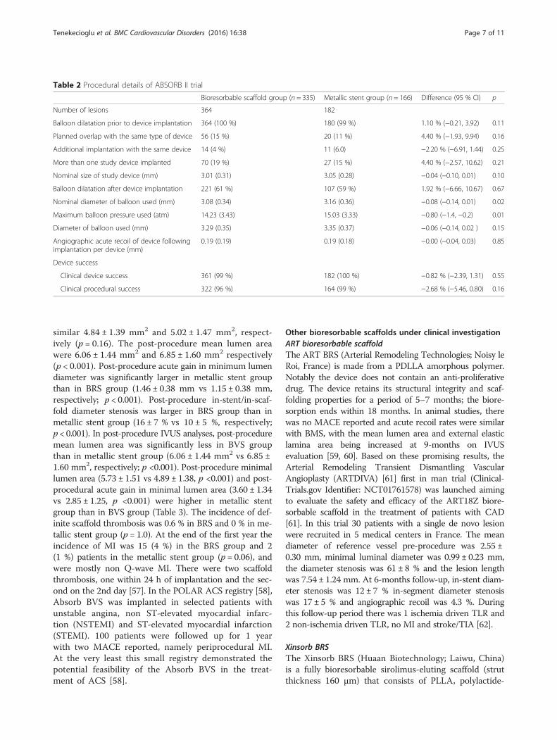
similar 4.84 ± 1.39 mm2 and 5.02 ± 1.47 mm2, respect-ively (p = 0.16). The post-procedure mean lumen areawere 6.06 ± 1.44 mm2 and 6.85 ± 1.60 mm2 respectively(p < 0.001). Post-procedure acute gain in minimum lumendiameter was significantly larger in metallic stent groupthan in BRS group (1.46 ± 0.38 mm vs 1.15 ± 0.38 mm,respectively; p < 0.001). Post-procedure in-stent/in-scaf-fold diameter stenosis was larger in BRS group than inmetallic stent group (16 ± 7 % vs 10 ± 5 %, respectively;p < 0.001). In post-procedure IVUS analyses, post-proceduremean lumen area was significantly less in BVS groupthan in metallic stent group (6.06 ± 1.44 mm2 vs 6.85 ±1.60 mm2, respectively; p <0.001). Post-procedure minimallumen area (5.73 ± 1.51 vs 4.89 ± 1.38, p <0.001) and post-procedural acute gain in minimal lumen area (3.60 ± 1.34vs 2.85 ± 1.25, p <0.001) were higher in metallic stentgroup than in BVS group (Table 3). The incidence of def-inite scaffold thrombosis was 0.6 % in BRS and 0 % in me-tallic stent group (p = 1.0). At the end of the first year theincidence of MI was 15 (4 %) in the BRS group and 2(1 %) patients in the metallic stent group (p = 0.06), andwere mostly non Q-wave MI. There were two scaffoldthrombosis, one within 24 h of implantation and the sec-ond on the 2nd day [57]. In the POLAR ACS registry [58],Absorb BVS was implanted in selected patients withunstable angina, non ST-elevated myocardial infarc-tion (NSTEMI) and ST-elevated myocardial infarction(STEMI). 100 patients were followed up for 1 yearwith two MACE reported, namely periprocedural MI.At the very least this small registry demonstrated thepotential feasibility of the Absorb BVS in the treat-ment of ACS [58].
Other bioresorbable scaffolds under clinical investigationART bioresorbable scaffoldThe ART BRS (Arterial Remodeling Technologies; Noisy leRoi, France) is made from a PDLLA amorphous polymer.Notably the device does not contain an anti-proliferativedrug. The device retains its structural integrity and scaf-folding properties for a period of 5–7 months; the biore-sorption ends within 18 months. In animal studies, therewas no MACE reported and acute recoil rates were similarwith BMS, with the mean lumen area and external elasticlamina area being increased at 9-months on IVUSevaluation [59, 60]. Based on these promising results, theArterial Remodeling Transient Dismantling VascularAngioplasty (ARTDIVA) [61] first in man trial (Clinical-Trials.gov Identifier: NCT01761578) was launched aimingto evaluate the safety and efficacy of the ART18Z biore-sorbable scaffold in the treatment of patients with CAD[61]. In this trial 30 patients with a single de novo lesionwere recruited in 5 medical centers in France. The meandiameter of reference vessel pre-procedure was 2.55 ±0.30 mm, minimal luminal diameter was 0.99 ± 0.23 mm,the diameter stenosis was 61 ± 8 % and the lesion lengthwas 7.54 ± 1.24 mm. At 6-months follow-up, in-stent diam-eter stenosis was 12 ± 7 % in-segment diameter stenosiswas 17 ± 5 % and angiographic recoil was 4.3 %. Duringthis follow-up period there was 1 ischemia driven TLR and2 non-ischemia driven TLR, no MI and stroke/TIA [62].
Xinsorb BRSThe Xinsorb BRS (Huaan Biotechnology; Laiwu, China)is a fully bioresorbable sirolimus-eluting scaffold (strutthickness 160 μm) that consists of PLLA, polylactide-
Table 2 Procedural details of ABSORB II trial
Bioresorbable scaffold group (n = 335) Metallic stent group (n = 166) Difference (95 % CI) p
Number of lesions 364 182
Balloon dilatation prior to device implantation 364 (100 %) 180 (99 %) 1.10 % (−0.21, 3.92) 0.11
Planned overlap with the same type of device 56 (15 %) 20 (11 %) 4.40 % (−1.93, 9.94) 0.16
Additional implantation with the same device 14 (4 %) 11 (6.0) −2.20 % (−6.91, 1.44) 0.25
More than one study device implanted 70 (19 %) 27 (15 %) 4.40 % (−2.57, 10.62) 0.21
Nominal size of study device (mm) 3.01 (0.31) 3.05 (0.28) −0.04 (−0.10, 0.01) 0.10
Balloon dilatation after device implantation 221 (61 %) 107 (59 %) 1.92 % (−6.66, 10.67) 0.67
Nominal diameter of balloon used (mm) 3.08 (0.34) 3.16 (0.36) −0.08 (−0.14, 0.01) 0.02
Maximum balloon pressure used (atm) 14.23 (3.43) 15.03 (3.33) −0.80 (−1.4, −0.2) 0.01
Diameter of balloon used (mm) 3.29 (0.35) 3.35 (0.37) −0.06 (−0.14, 0.02 ) 0.15
Angiographic acute recoil of device followingimplantation per device (mm)
0.19 (0.19) 0.19 (0.18) −0.00 (−0.04, 0.03) 0.85
Device success
Clinical device success 361 (99 %) 182 (100 %) −0.82 % (−2.39, 1.31) 0.55
Clinical procedural success 322 (96 %) 164 (99 %) −2.68 % (−5.46, 0.80) 0.16
Tenekecioglu et al. BMC Cardiovascular Disorders (2016) 16:38 Page 7 of 11

co-glycolide, and poly-L-lactide-co-e-caprolactone. 78 %of sirolimus is released from the Xinsorb BRS within14 days [63].In a comparison study between Xinsorb BRS and the
Excel DES (JW Medical; Shandong, China) implanted inthe coronaries of porcine models, there was no signifi-cant difference in percentage diameter stenosis (%DS) inthe Xinsorb BRS compared to the Excel DES (18.6 % vs.21.4 % at 30 days; p > 0.05 and 24.5 % vs. 27.7 % at90 days; p > 0.05, respectively) [64]. At 3-month follow-up OCT imaging demonstrated significant red significantneointimal hyperplasia in porcine models. Subsequently theLLL and %DS were noticeably reduced. At 1-month follow-up, proximal, in-scaffold, and distal LLL of scaffold were0.53 ± 0.41 mm, 0.68 ± 0.42 mm and 0.65 ± 0.24 mm, while
the %DS were 9.5 ± 7.7 %, 17.6 ± 16.8 % and 10.5 ± 7.4 % re-spectively. At 3-months, proximal, in-scaffold, and distalLLL were 0.23 ± 0.48 mm, 0.77 ± 0.48 mm and 0.11 ±0.35 mm, while %DS were 14.5 ± 9.4 %, 31.9 ± 13.6 % and5.4 ± 3.6 % respectively. At 12-months, proximal, in-scaffold, and distal LLL were −0.13 ± 0.45 mm, 0.28 ±0.41 mm and 0.18 ± 0.48 mm, while %DS were 2.4 ±2.9 %, 14.1 ± 9.1 % and 8.6 ± 8.7 % respectively. At18-month, proximal, in-scaffold, and distal LLL were0.37 ± 0.57 mm, 0.09 ± 0.31 mm and −0.01 ± 0.41 mm,while %DS were 3.9 ± 4.6 %、13.7 ± 7.3 % and 6.9 ±5.2 % respectively. Lumen area at 18-month was signifi-cantly larger than that at 3-month with a constant scaffoldarea [65]. In Xinsorb FIM trial (n = 30 patients), at 6-months follow-up, LLL was 0.18 ± 0.21 mm. In scaffolded
Table 3 Angiographic and IVUS/IVUS-VH outcomes of ABSORB II trial
Bioresorbable scaffoldgroup (n=335)
Metallic stentgroup (n=166)
Difference (95 % CI) p
Angiographic analysis
Lesion length obstruction (mm) 13.8 (6.5) 13.8 (6.6) 0.00 (−1.18, 1.18) 1.00
Total scaffolded/stented length (mm) 21.1 (8.8) 20.9 (7.4) 0.24 (−1.17, 1.65) 0.74
Reference vessel diameter
Pre-procedure diameter (mm) 2.59 (0.38) 2.63 (0.40) −0.03 (−0.10, 0.04) 0.36
Postprocedure diameter (mm) 2.64 (0.36) 2.80 (0.34) −0.16 (−0.22, −0.09) <0.001
Minimum lumen diameter
Pre-procedure diameter (mm) 1.07 (0.32) 1.05 (0.32) 0.02 (−0.03, 0.08) 0.44
Post-procedure in-stent or in-scaff old diameter (mm) 2.22 (0.33) 2.50 (0.33) −0.28 (−0.34, −0.22) <0.001
In-stent/in-scaff old acute gain (mm) 1.15±0.38 1.46±0.38 −0.30 (−0.37, −0.24) <0.001
Diameter stenosis
Pre-procedure percent diameter stenosis (%) 59±11 % 60±12 % −1.07 (−3.11, 0.97) 0.30
Post-procedure in-stent/in-scaffold diameter stenosis (%) 16±7 % 10±5 % 5.37 (4.38, 6.36) <0.001
Pre-procedural fibrotic tissue (%) 31.47±11.39 30.62±11.42 0.85 (−1.33, 3.04) 0.44
Pre-procedural fibrofatty tissue (%) 47.43±16.91 48.55±16.86 −1.12 (−4.35, 2.11) 0.50
Pre-procedural necrotic core (%) 16.20±6.86 16.15±6.90 0.05 (−1.27, 1.37) 0.94
Pre-procedural dense calcium (%) 4.90±4.73 4.68±4.10 0.22 (−0.61, 1.05) 0.60
Vessel area
Pre-procedure area (mm2) 11.51±3.40 12.34±3.42 −0.83 (−1.47, −0.19) 0.02
Post-procedure area (mm2) 13.17±3.55 14.28±3.59 −1.11 (−1.78, −0.44) 0.001
Plaque area
Pre-procedure plaque area (mm2) 6.67±2.52 7.30±2.68 0.6 (−1.12, 0.13) 0.01
Post-procedure plaque area (mm2) 7.11±2.46 7.43±2.44 −0.32 (−0.78, 0.14) 0.18
Mean lumen area
Pre-procedure mean lumen area (mm2) 4.84±1.39 5.02±1.47 −0.19 (−0.47, 0.08) 0.16
Post-procedure mean lumen area (mm2) 6.06±1.44 6.85±1.60 −0.80 (−1.09, −0.50) <0.001
Minimal lumen area
Pre-procedure minimal lumen area (mm2) 2.04±0.72 2.13±0.83 −0.10 (−0.25, 0.05) 0.20
Post-procedure minimal lumen area (mm2) 4.89±1.38 5.73±1.51 −0.84 (−1.12, −0.57) <0.001
Acute gain in minimal lumen area (mm2) 2.85±1.25 3.60±1.34 −0.75 (−0.99, −0.50) <0.001
Tenekecioglu et al. BMC Cardiovascular Disorders (2016) 16:38 Page 8 of 11
segments, the diameter stenosis was 10.0 ± 4.2 % at post-implantation and 10.6 ± 6.6 % at 6-months follow-up (p =0.70). At 6-months OCT follow-up (n = 19) the luminalarea was 6.03 ± 0.76 mm2, scaffold area was 7.74 ±0.62 mm2, in-scaffold area obstruction was 22.1 ± 6.1 %,neointimal thickness was 0.07 ± 0.04 mm with nothrombus detected. The 6-month IVUS follow-up re-vealed a mean vessel area 14.37 ± 0.90 mm2, mean neointi-mal area 3.11 ± 0.19 mm2, mean scaffold area 9.36 ±0.21 mm2 and mean luminal area 6.26 ± 0.26 mm2 [66].
Mirage bioresorbable micro-fiber scaffoldMirage Bioresorbable Micro-fiber Scaffold (Mirage BRMS,Manli Cardiology Singapore) is a PLLA-based sirolimuseluting scaffold. The device incorporates a helix coil designthat provides high flexibility with a strut thickness of125 μm in scaffolds with diameter ≤ 3 mm, and of 150 μmin scaffolds with diameter ≥ 3.5 mm. Mirage BRMS has alow crossing profile (0.044” – 0.058”), and relatively shortbioresorption time (~14 months). Results of a porcinestudy were encouraging; namely no in-scaffold restenosisat 6-month follow-up, 99 % of the struts were coveredwhile the mean NIH thickness on top of covered strutswas 0.08 ± 0.03 mm at 6-month follow-up [67]. Frequencyof covered and uncovered struts per lesion were 99.85 ±0.33 % and 0.15 ± 0.33 % respectively. The frequency ofmalapposed struts per lesion was 0.03 ± 0.08 %, andmalapposition strut-to-lumen distance was 0.28 mm (therewas only one malapposed strut at 6-month follow-up). InQCA analysis, MLD and % DS was 2.34 ± 0.49 mm and2.13 ± 0.47 mm, 17.1 ± 11.4 % and 22.8 ± 15.0 %, atpost-procedure and at 6-months, respectively. At 6-months, LLL was 0.21 ± 0.20 mm and late recoil was0.16 ± 0.12 mm. Both in-scaffold and in-segment angio-graphic binary restenosis ratios were 0 % at 6-month[67]. Patient enrolment in FIM trial was completed inSeptember 2014 and the results are expected to be pre-sented at the end of 2015.
ConclusionFor the last 20 years percutaneous coronary revasculariza-tion has evolved, with the current premise that stent im-plantation to be the standard of care in appropriatelyselected patients [68]. Considering that coronary stentingwith metallic devices may results in persistent inflamma-tion and endothelial dysfunction, an issue that has beenreduced but not eliminated with newer generation DES[69], the temporary scaffold that would safeguard vesselpatency and then it would disappear appears as the idealsolution for treating CAD [70]. These devices at the veryleast have to provide comparable performances to con-temporary DES in the short term, with the potentialpromise of enhanced longer term benefits due to freeingthe vessel wall from the metallic cage and allowing the
vessel to potentially restore its vascular function (vesselvasomotion) adaptive shear stress and would permit lateluminal enlargement, and late expansive remodelling. On-going, and future randomized trials assessing the efficacyof the multitude of bioresorbable scaffolds – currently 16different scaffolds are being developed and under investi-gation – will ultimately determine the clinical value of thisfourth revolution in interventional cardiology.
Competing interestsThe authors declare that they have no competing interests.
Authors’ contributionsET participated in its design and coordination and helped to draft themanuscript. VF, CVB,YO and MYrevised the manuscript critically for importantintellectual content. PWS has revised and given final approval of the versionto be published. All authors read and approved the final manuscript.
AcknowledgmentsWe would like to acknowledge Yapping Zhang for her intellectual input andcareful review of the literature. We would like to thank the investigators ofABSORB A, ABSORB B and ABSORB II who have contributed by theirinvestigation to the progression of the field.
DisclosuresE Tenekecioglu has research grant from TUBITAK (The Scientific andTechnological Research Council of Turkey).
Author details1ThoraxCentre, Erasmus Medical Centre, Rotterdam, The Netherlands.2Manchester Heart Centre, Manchester Royal Infirmary, Central ManchesterUniversity, Hospitals NHS Trust, Manchester, UK. 3Institute of CardiovascularSciences, University College of London, London, UK. 4Department ofCardiology, Barts Health NHS Trust, London, UK. 5Department of Cardiology,Bursa Postgraduate Education and Research Hospital, Bursa, Turkey.6International Centre for Circulatory Health, Imperial College, London, UK.7Interventional Cardiology Department, Erasmus MC, ‘s-Gravendijkwal 230,Rotterdam 3015 CE, The Netherlands. 8Institute of Cardiovascular Sciences,Manchester Academic Health Sciences Centre, University of Manchester,Manchester, UK.
Received: 22 January 2016 Accepted: 29 January 2016
References1. Gruntzig A. Transluminal dilatation of coronary-artery stenosis. Lancet. 1978;
1:263.2. Farooq V, Gogas BD, Serruys PW. Restenosis: delineating the numerous causes
of drug-eluting stent restenosis. Circ Cardiovasc Interv. 2011;4(2):195–205.3. Gruntzig AR, Senning A, Siegenthaler WE. Nonoperative dilatation of
coronary-artery stenosis: percutaneous transluminal coronary angioplasty.N Engl J Med. 1979;301:61–8.
4. Sigwart U, Urban P, Golf S, Kaufmann U, Imbert C, Fischer A, et al.Emergency stenting for acute occlusion after coronary balloon angioplasty.Circulation. 1988;78:1121–7.
5. Roubin GS, Cannon AD, Agrawal SK, Macander PJ, Dean LS, Baxley WA, et al.Intracoronary stenting for acute and threatened closure complicatingpercutaneous transluminal coronary angioplasty. Circulation. 1992;85:916–27.
6. Serruys PW, Strauss BH, Beatt KJ, Bertrand ME, Puel J, Rickards AF, et al.Angiographic follow-up after placement of a self-expanding coronary-arterystent. N Engl J Med. 1991;324:13–7.
7. Serruys PW, de Jaegere P, Kiemeneij F, Macaya C, Rutsch W, Heyndrickx G,et al. A comparison of balloon-expandable-stent implantation with balloonangioplasty in patients with coronary artery disease. Benestent Study Group.N Engl J Med. 1994;331:489–95.
8. Fischman DL, Leon MB, Baim DS, Schatz RA, Savage MP, Penn I, et al. Arandomized comparison of coronary-stent placement and balloonangioplasty in the treatment of coronary artery disease. Stent RestenosisStudy Investigators. N Engl J Med. 1994;331:496–501.
Tenekecioglu et al. BMC Cardiovascular Disorders (2016) 16:38 Page 9 of 11

9. Hoffmann R, Mintz GS, Dussaillant GR, Popma JJ, Pichard AD, Satler LF, et al.Patterns and mechanisms of in-stent restenosis. A serial intravascularultrasound study. Circulation. 1996;94:1247–54.
10. Gordon PC, Gibson CM, Cohen DJ, Carrozza JP, Kuntz RE, Baim DS.Mechanisms of restenosis and redilation within coronary stents—quantitativeangiographic assessment. J Am Coll Cardiol. 1993;21:1166–74.
11. Karas SP, Gravanis MB, Santoian EC, Robinson KA, Anderberg KA, King SB.Coronary intimal proliferation after balloon injury and stenting in swine: ananimal model of restenosis. J Am Coll Cardiol. 1992;20:467–74.
12. Dussaillant GR, Mintz GS, Pichard AD, Kent KM, Satler LF, Popma JJ, et al. Smallstent size and intimal hyperplasia contribute to restenosis: a volumetricintravascular ultrasound analysis. J Am Coll Cardiol. 1995;26:720–4.
13. Sousa JE, Costa MA, Abizaid AC, Rensing BJ, Abizaid AS, Tanajura LF, et al.Sustained suppression of neointimal proliferation by sirolimus-eluting stents:One-year angiographic and intravascularultrasound follow-up. Circulation.2001;104:2007–11.
14. Stone GW, Ellis SG, Cox DA, Hermiller J, O'Shaughnessy C, Mann JT, et al. Apolymer-based, paclitaxel-eluting stent in patients with coronary arterydisease. N Engl J Med. 2004;350:221–31.
15. Daemen J, Wenaweser P, Tsuchida K, Abrecht L, Vaina S, Morger C, et al.Early and late coronary stent thrombosis of sirolimus-eluting and paclitaxel-eluting stents in routine clinical practice: Data from a large two-institutionalcohort study. Lancet. 2007;369:667–78.
16. Wenaweser P, Daemen J, Zwahlen M, van Domburg R, Juni P, Vaina S, et al.Incidence and correlates of drug-eluting stent thrombosis in routine clinicalpractice: 4-year results from a large 2-institutional cohort study. J Am CollCardiol. 2008;52:1134–40.
17. Virmani R, Guagliumi G, Farb A, Musumeci G, Grieco N, Motta T, et al. Localizedhypersensitivity and late coronary thrombosis secondary to a sirolimus-elutingstent: should we be cautious? Circulation. 2004;109(6):701–5.
18. Serruys PW, Onuma Y, Garg S, Vranckx P, De Bruyne B, Morice MC, et al.ARTS II Investigators. 5-year clinical outcomes of the ARTS II (ArterialRevascularization Therapies Study II) of the sirolimus-eluting stent in thetreatment of patients with multivessel de novo coronary artery lesions.J Am Coll Cardiol. 2010;55:1093–101.
19. Hofma SH, van der Giessen WJ, van Dalen BM, Lemos PA, McFadden EP,Sianos G, et al. Indication of long-term endothelial dysfunction aftersirolimus-eluting stent implantation. Eur Heart J. 2006;27:166–70.
20. Togni M, Windecker S, Cocchia R, Wenaweser P, Cook S, Billinger M, et al.Sirolimus-eluting stents associated with paradoxic coronary vasoconstriction.J Am Coll Cardiol. 2005;46:231–6.
21. Shalaby SW, Burg KJL. Absorbable and Biodegradable Polymers (Advancesin Polymeric Biomaterials). Boca Raton, FL: CRC Press; 2003.
22. Ormiston JA, Serruys PW, Regar E, Dudek D, Thuesen L, Webster MW, et al.A bioabsorbable everolimus-eluting coronary stent system for patients withsingle de-novo coronary artery lesions (ABSORB): a prospective open-labeltrial. Lancet. 2008;371:899–907.
23. Heublein B, Rohde R, Kaese V, Niemeyer M, Hartung W, Haverich A.Biocorrosion of magnesium alloys: a new principle in cardiovascular implanttechnology? Heart. 2003;89:651–6.
24. Waksman R, Pakala R, Kuchulakanti PK, Baffour R, Hellinga D, Seabron R, etal. Safety and efficacy of bioabsorbable magnesium alloy stents in porcinecoronary arteries. Catheter Cardiovasc Interv. 2006;68:607–17.
25. Università degli Studi di PaviaStructural Mechanics Department. http://www.dist.unina.it/doc/seminari/corso_Auricchio/biomaterials.pdf. Accessed 04March 2015
26. Brugaletta S, Radu MD, Garcia-Garcia HM, Heo JH, Farooq V, Girasis C, et al.Circumferential evaluation of the neointima by optical coherencetomography after ABSORB bioresorbable vascular scaffold implantation: canthe scaffold cap the plaque? Atherosclerosis. 2012;221:106–12.
27. Joner M, Finn AV, Farb A, Mont EK, Kolodgie FD, Ladich E, et al. Pathologyof drug-eluting stents in humans: Delayed healing and late thrombotic risk.J Am Coll Cardiol. 2006;48:193–202.
28. Onuma Y, Serruys PW, Perkins LE, Okamura T, Gonzalo N, García-García HM,et al. Intracoronary optical coherence tomography and histology at 1month and 2, 3, and 4 years after implantation of everolimus-elutingbioresorbable vascular scaffolds in a porcine coronary artery model: anattempt to decipher the human optical coherence tomography images inthe ABSORB trial. Circulation. 2010;122:2288–300.
29. Garg S, Serruys P, Onuma Y, Dorange C, Veldhof S, Miquel-Hébert K, et al. 3-year clinical follow-up of the XIENCE V everolimus-eluting coronary stent
system in the treatment of patients with de novo coronary artery lesions:the SPIRIT II trial (Clinical Evaluation of the Xience V Everolimus ElutingCoronary Stent System in the Treatment of Patients with de novo NativeCoronary Artery Lesions). JACC Cardiovasc Interv. 2009;2(12):1190–8.
30. Verheye S, Ormiston JA, Stewart J, Webster M, Sanidas E, Costa R, et al. Anext-generation bioresorbable coronary scaffold system-from bench to firstclinical evaluation: 6- and 12-month clinical and multimodality imagingresults. J Am Coll Cardiol Intv. 2014;7:89–99.
31. Brugaletta S, Heo JH, Garcia-Garcia HM, Farooq V, van Geuns RJ, de BruyneB, et al. Endothelial-dependent vasomotion in a coronary segment treatedby ABSORB everolimus-eluting bioresorbable vascular scaffold system isrelated to plaque composition at the time of bioresorption of the polymer:indirect finding of vascular reparative therapy. Eur Heart J. 2012;33:1325–33.
32. Erbel R, Di Mario C, Bartunek J, Bonnier J, de Bruyne B, Eberli FR, et al. Temporaryscaffolding of coronary arteries with bioabsorbable magnesium stents: Aprospective, non-randomised multicentre trial. Lancet. 2007;369:1869–75.
33. Barlis P, Tanigawa J, Di Mario C. Coronary bioabsorbable magnesium stent:15-month intravascular ultrasound and optical coherence tomographyfindings. Eur Heart J. 2007;28:2319.
34. Ghimire G, Spiro J, Kharbanda R, Roughton M, Barlis P, Mason M, et al. Initialevidence for the return of coronary vasoreactivity following the absorptionof bioabsorbable magnesium alloy coronary stents. EuroIntervention.2009;4:481–4.
35. Haude M, Erbel R, Erne P, Verheye S, Degen H, Böse D, et al. Safety andperformance of the drug-eluting absorbable metal scaffold (DREAMS) inpatients with de-novo coronary lesions: 12 month results of the prospective,multicentre, first-in-man BIOSOLVE-I trial. Lancet. 2013;381(9869):836–44.
36. Tamai H, Igaki K, Kyo E, Kosuga K, Kawashima A, Matsui S, et al. Initial and6-month results of biodegradable poly-l-lactic acid coronary stents in humans.Circulation. 2000;102:399–404.
37. Tsuji T, Tamai H, Igaki K, Hsu Y-S, Kosuga K, Hata T, et al. Four-year follow-upof the biodegradable stent (Igaki-Tamai stent). Circ J. 2004;68:135.
38. Nishio S. Long-term (>10 years) clinical outcomes of first-in-manbiodegradable poly-l-lactic acid coronary stents. Eurointervenion. 2010;6:H44.
39. Biamino G, Schmidt A, Scheinert D. Treatment of SFA lesions with PLLAbiodegradable stents: results of the PERSEUS Study. J Endovasc Ther. 2005;12:5.
40. Schulze R. REVA Medical, Inc. Bioresorbable stent. In: CardiovascularRevascularization Therapies 2007.
41. Grube E. Bioabsorbable stent: the Boston Scientific and REVA technology.Presented at: EuroPCR; May 19–22, 2009; Barcelona, Spain.
42. Costa RA. REVA ReZolve clinical program update. In: TranscatheterCardiovascular Therapeutics, Miami Beach, FL, 2012.
43. ReZolve2 Clinical Investigation (RESTORE II). www.clinicaltrials.gov. Accessed20 Feb 2015
44. Abizaid A. Cardiovascular Research Technologies (CRT) 2015.45. Jabara R, Chronos N, Robinson K. Novel bioabsorbable salicylate-based polymer
as a drug-eluting stent coating. Catheter Cardiovasc Interv. 2008;72:186–94.46. Jabara R, Pendyala L, Geva S, Chen J, Chronos N, Robinson K. Novel fully
bioabsorbable salicylate-based sirolimus-eluting stent. EuroIntervention.2009;5:F58–64.
47. Verheye S. First-in-man results with a myolimus-eluting bioresorbable PLLA-based vascular scaffold.Presented in Transcatheter Cardiovasc Therapeutics.Miami Beach , FL; October 23rd 2012.USA.
48. 48:Verheye S. DESolve Nx Novolimus-Eluting PLLA-Based BRS: First Report ofthe 6-month OCT and 12-month Clinical and Imaging Result. Presented inTranscatheter Cardiovasc Therapeutics. San Fransisco, CA, USA; October 27th2013.
49. Abizaid A. Prospective, Multicenter Evaluation of the DESolve Novolimus-Eluting Bioresorbable Coronary Scaffold: Imaging Outcomes and 2-YearClinical Results. Presented in Transcatheter Cardiovasc Therapeutics.Washington, DC. USA; September 16th 2014.
50. Serruys PW, Ormiston JA, Onuma Y, Regar E, Gonzalo N, Garcia-Garcia HM,et al. A bioabsorbable everolimus-eluting coronary stent system (ABSORB):2-year outcomes and results from multiple imaging methods. Lancet. 2009;373:897–910.
51. Onuma Y, Serruys P, Ormiston J, Regar E, Webster M, Thuesen L, et al.Three-year results of clinical follow-up after a bioresorbable everolimus-eluting scaffold in patients with de novo coronary artery disease: TheABSORB trial. EuroIntervention. 2010;6:447–53.
52. Tanimoto S, Serruys PW, Thuesen L, Dudek D, de Bruyne B, Chevalier B, etal. Comparison of in vivo acute stent recoil between the bioabsorbable
Tenekecioglu et al. BMC Cardiovascular Disorders (2016) 16:38 Page 10 of 11

everolimus-eluting coronary stent and the everolimus- eluting cobaltchromium coronary stent: Insights from the ABSORB and SPIRIT Trials.Cathet Cardiovasc Interv. 2007;70:515–23.
53. Garg S, Serruys P. Biodegradable stents and nonbiodegradable stents.Minerva Cardioangiol. 2009;57:537–65.
54. Ormiston JA, Serruys PW, Onuma Y, van Geuns RJ, de Bruyne B, Dudek D, etal. First serial assessment at 6 months and 2 years of the second generationof absorb everolimus-eluting bioresorbable vascular scaffold: a multi-imaging modality study. Circ Cardiovasc Interv. 2012;5:620–32.
55. Serruys PW, Onuma Y, Dudek D, Smits PC, Koolen J, Chevalier B, et al.Evaluation of the second generation of a bioresorbable everolimus-elutingvascular scaffold for the treatment of de novo coronary artery stenosis: 12-month clinical and imaging outcomes. J Am Coll Cardiol. 2011;58:1578–88.
56. Serruys PW, Onuma Y, Garcia-Garcia HM, Muramatsu T, van Geuns RJ, de BruyneB, et al. Dynamics of vessel wall changes following the implantation of theabsorb everolimus-eluting bioresorbable vascular scaffold: a multi-imagingmodality study at 6, 12, 24 and 36 months. EuroIntervention. 2014;9(11):1271–84.
57. Serruys PW, Chevalier B, Dudek D, Cequier A, Carrie D, Iniguez A, et al. Abioresorbable everolimus-eluting scaff old versus a metallic everolimus-eluting stent for ischaemic heart disease caused by de-novo nativecoronary artery lesions (ABSORB II): an interim 1-year analysis of clinical andprocedural secondary outcomes from a randomised controlled trial. Lancet.2015;385:43–54.
58. Dudek D, Rzeszutko Ł, Zasada W, Depukat R, Siudak Z, Ochała A.Bioresorbable vascular scaffolds in patients with acute coronary syndromes :the POLAR ACS study. Pol Arch Med Wewn. 2014;124(12):669–77.
59. Lafont A, Durand E. A.R.T. concept of a bioresorbable stent without drugelution. EuroIntervention. 2009;5(Suppl F):F83–7.
60. Durand E, Lemitre M, Couty L, Sharkawi T, Brasselet C, Vert M, et al.Adjusting a polymer formulation for an optimal bioresorbable stent: a 6-month follow-up study. EuroIntervention. 2012;8:242–9.
61. Fajadet J. The ART, stent: design and early first-in-man experiences.Presented in Transcatheter Cardiovasc Therapeutics. Miami Beach , FL, USA;October 23rd 2012.
62. Lafont A. ARTDIVA. Presented in BRS 2014. Boston, MA, USA; July 6th 2014.63. Shen L, Wang Q, Wu Y, Xie J, Zhang F, Ge L, et al. Preliminary evaluation of
fully bioabsorbable PLLA sirolimus eluting stents in a porcine model. Chin JIntervent Cardiol. 2009;19:301–5.
64. Shen L, Wang Q, Wu Y, Xie J, Ge J. Short-term effects of sirolimus elutingfully bioabsorbable polymeric coronary stents in a porcine model.Transcatheter Cardiovasc Ther 2011
65. 65:Wu Y, Shen L, Yao Z, Ge L, Wang Q, Qian J, et al. Long-termAngiographic and Optical Coherence Tomography Follow-up of XINSORBScaffold in Porcine Coronary Model.Abstract presentation in TranscatheterCardiovasc Therapeutics 2014. Washington, DC. USA; September 13-17 2014.
66. Ge J. BRS Under Development III – The XINSORB BRS. Presented inTranscatheter Cardiovasc Therapeutics. Washington, DC. USA; September16th 2014.
67. Santoso T. The Mirage Bioresorbable Microfiber Scaffold (BRMS) ManliCardiology. Presented in Transcatheter Cardiovasc Therapeutics.Washington, DC. USA; September 16th 2014.
68. Windecker S, Kolh P, Alfonso F, Collet JP, Cremer J, Falk V, et al. 2014 ESC/EACTS guidelines on myocardial revascularization. EuroIntervention. 2015;10(9):1024–94.
69. Mitsutake Y, Ueno T, Ikeno F, Yokoyama S, Sasaki K, Ohtsuka M, et al.Second-generation everolimus-eluting stents demonstrate better vascularfunction, less thrombus formation, and less yellow intima than first-generation drug-eluting stents. AsiaIntervention. 2015;1:33–40.
70. Serruys PW. Innovations: resetting our thinking to solve problems.EuroIntervention. 2014;10:413.
• We accept pre-submission inquiries
• Our selector tool helps you to find the most relevant journal
• We provide round the clock customer support
• Convenient online submission
• Thorough peer review
• Inclusion in PubMed and all major indexing services
• Maximum visibility for your research
Submit your manuscript atwww.biomedcentral.com/submit
Submit your next manuscript to BioMed Central and we will help you at every step:
Tenekecioglu et al. BMC Cardiovascular Disorders (2016) 16:38 Page 11 of 11