biology and therapeutic potential of hydrogen sulfide and hydrogen sulfide-releasing chimeras
TRANSCRIPT

Biochemical Pharmacology 85 (2013) 689–703
Review
Biology and therapeutic potential of hydrogen sulfide and hydrogensulfide-releasing chimeras
Khosrow Kashfi a,*, Kenneth R. Olson b,**a Department of Physiology and Pharmacology, Sophie Davis School of Biomedical Education, City University of New York Medical School, 138th Street and Convent Avenue, New York,
NY 10031, United Statesb Department of Physiology, Indiana University School of Medicine, 1234 Notre Dame Avenue, South Bend, IN 46617, United States
Contents
1. Introduction . . . . . . . . . . . . . . . . . . . . . . . . . . . . . . . . . . . . . . . . . . . . . . . . . . . . . . . . . . . . . . . . . . . . . . . . . . . . . . . . . . . . . . . . . . . . . . . . . . . . . 690
2. H2S chemistry and biology . . . . . . . . . . . . . . . . . . . . . . . . . . . . . . . . . . . . . . . . . . . . . . . . . . . . . . . . . . . . . . . . . . . . . . . . . . . . . . . . . . . . . . . . . 690
2.1. H2S chemistry . . . . . . . . . . . . . . . . . . . . . . . . . . . . . . . . . . . . . . . . . . . . . . . . . . . . . . . . . . . . . . . . . . . . . . . . . . . . . . . . . . . . . . . . . . . . . . 690
2.2. H2S biosynthesis and metabolism . . . . . . . . . . . . . . . . . . . . . . . . . . . . . . . . . . . . . . . . . . . . . . . . . . . . . . . . . . . . . . . . . . . . . . . . . . . . . . 690
2.2.1. Other potential biosynthetic pathways . . . . . . . . . . . . . . . . . . . . . . . . . . . . . . . . . . . . . . . . . . . . . . . . . . . . . . . . . . . . . . . . . . 692
2.2.2. Metabolism (inactivation) . . . . . . . . . . . . . . . . . . . . . . . . . . . . . . . . . . . . . . . . . . . . . . . . . . . . . . . . . . . . . . . . . . . . . . . . . . . . . 692
3. H2S ‘‘donating’’ compounds. . . . . . . . . . . . . . . . . . . . . . . . . . . . . . . . . . . . . . . . . . . . . . . . . . . . . . . . . . . . . . . . . . . . . . . . . . . . . . . . . . . . . . . . . 692
3.1. Sulfide salts . . . . . . . . . . . . . . . . . . . . . . . . . . . . . . . . . . . . . . . . . . . . . . . . . . . . . . . . . . . . . . . . . . . . . . . . . . . . . . . . . . . . . . . . . . . . . . . . 692
3.2. Naturally occurring compounds . . . . . . . . . . . . . . . . . . . . . . . . . . . . . . . . . . . . . . . . . . . . . . . . . . . . . . . . . . . . . . . . . . . . . . . . . . . . . . . . 692
3.2.1. Garlic . . . . . . . . . . . . . . . . . . . . . . . . . . . . . . . . . . . . . . . . . . . . . . . . . . . . . . . . . . . . . . . . . . . . . . . . . . . . . . . . . . . . . . . . . . . . . 692
3.2.2. Sulforaphane, erucin and iberin . . . . . . . . . . . . . . . . . . . . . . . . . . . . . . . . . . . . . . . . . . . . . . . . . . . . . . . . . . . . . . . . . . . . . . . . 694
3.3. Synthetic H2S donors . . . . . . . . . . . . . . . . . . . . . . . . . . . . . . . . . . . . . . . . . . . . . . . . . . . . . . . . . . . . . . . . . . . . . . . . . . . . . . . . . . . . . . . . 694
3.3.1. GYY4137 . . . . . . . . . . . . . . . . . . . . . . . . . . . . . . . . . . . . . . . . . . . . . . . . . . . . . . . . . . . . . . . . . . . . . . . . . . . . . . . . . . . . . . . . . . 694
3.3.2. Cysteine analogs . . . . . . . . . . . . . . . . . . . . . . . . . . . . . . . . . . . . . . . . . . . . . . . . . . . . . . . . . . . . . . . . . . . . . . . . . . . . . . . . . . . . 694
3.3.3. Cysteine-activated H2S donors . . . . . . . . . . . . . . . . . . . . . . . . . . . . . . . . . . . . . . . . . . . . . . . . . . . . . . . . . . . . . . . . . . . . . . . . . 695
3.3.4. Dithiolethione and its NSAID chimeras . . . . . . . . . . . . . . . . . . . . . . . . . . . . . . . . . . . . . . . . . . . . . . . . . . . . . . . . . . . . . . . . . . 695
4. Nitric oxide and hydrogen sulfide releasing aspirin (NOSH-aspirin) . . . . . . . . . . . . . . . . . . . . . . . . . . . . . . . . . . . . . . . . . . . . . . . . . . . . . . . . . 699
4.1. Chemistry . . . . . . . . . . . . . . . . . . . . . . . . . . . . . . . . . . . . . . . . . . . . . . . . . . . . . . . . . . . . . . . . . . . . . . . . . . . . . . . . . . . . . . . . . . . . . . . . . 699
4.2. Biology. . . . . . . . . . . . . . . . . . . . . . . . . . . . . . . . . . . . . . . . . . . . . . . . . . . . . . . . . . . . . . . . . . . . . . . . . . . . . . . . . . . . . . . . . . . . . . . . . . . . 700
A R T I C L E I N F O
Article history:
Received 11 September 2012
Accepted 19 October 2012
Available online 24 October 2012
Keywords:
Hydrogen sulfide
NSAIDs
Cancer prevention
Inflammation
Cardiovascular
A B S T R A C T
Hydrogen sulfide, H2S, is a colorless gas with a strong odor that until recently was only considered to be a
toxic environmental pollutant with little or no physiological significance. However, the past few years
have demonstrated its role in many biological systems and it is becoming increasingly clear that H2S is
likely to join nitric oxide (NO) and carbon monoxide (CO) as a major player in mammalian biology. In this
review, we have provided an overview of the chemistry and biology of H2S and have summarized the
chemistry and biological activity of some natural and synthetic H2S-donating compounds. The naturally
occurring compounds discussed include, garlic, sulforaphane, erucin, and iberin. The synthetic H2S
donors reviewed include, GYY4137; cysteine analogs; S-propyl cysteine, S-allyl cysteine, S-propargyl
cysteine, and N-acetyl cysteine. Dithiolethione and its NSAID and other chimeras such as, L-DOPA,
sildenafil, aspirin, diclofenac, naproxen, ibuprofen, indomethacin, and mesalamine have also been
reviewed in detail. The newly reported NOSH-aspirin that releases both NO and H2S has also been
discussed.
� 2012 Elsevier Inc. All rights reserved.
Contents lists available at SciVerse ScienceDirect
Biochemical Pharmacology
jo u rn al h om epag e: ww w.els evier .c o m/lo cat e/b io c hem p har m
* Corresponding author. Tel.: +1 212 650 6641; fax: +1 212 650 7692.
** Corresponding author. Tel.: +1 574 631 7560; fax: +1 574 631 7821.
E-mail addresses: [email protected] (K. Kashfi), [email protected] (K.R. Olson).
0006-2952/$ – see front matter � 2012 Elsevier Inc. All rights reserved.
http://dx.doi.org/10.1016/j.bcp.2012.10.019

K. Kashfi, K.R. Olson / Biochemical Pharmacology 85 (2013) 689–703690
4.2.1. Anti-inflammatory . . . . . . . . . . . . . . . . . . . . . . . . . . . . . . . . . . . . . . . . . . . . . . . . . . . . . . . . . . . . . . . . . . . . . . . . . . . . . . . . . . . 700
4.2.2. Chemopreventive properties . . . . . . . . . . . . . . . . . . . . . . . . . . . . . . . . . . . . . . . . . . . . . . . . . . . . . . . . . . . . . . . . . . . . . . . . . . . 700
4.3. Outstanding questions . . . . . . . . . . . . . . . . . . . . . . . . . . . . . . . . . . . . . . . . . . . . . . . . . . . . . . . . . . . . . . . . . . . . . . . . . . . . . . . . . . . . . . . 700
5. Other questions remaining and future directions . . . . . . . . . . . . . . . . . . . . . . . . . . . . . . . . . . . . . . . . . . . . . . . . . . . . . . . . . . . . . . . . . . . . . . . 700
Acknowledgements . . . . . . . . . . . . . . . . . . . . . . . . . . . . . . . . . . . . . . . . . . . . . . . . . . . . . . . . . . . . . . . . . . . . . . . . . . . . . . . . . . . . . . . . . . . . . . . 700
References . . . . . . . . . . . . . . . . . . . . . . . . . . . . . . . . . . . . . . . . . . . . . . . . . . . . . . . . . . . . . . . . . . . . . . . . . . . . . . . . . . . . . . . . . . . . . . . . . . . . . . 700
1. Introduction
The initial observations by Kimura’s group [1] suggestinghydrogen sulfide (H2S) is a biologically relevant signaling moleculehave been followed by a myriad of studies demonstrating someeffect of this gas on virtually every organ system and tissue. It isonly logical that attention would soon turn to the therapeuticpotential of this signaling system and a number of H2S ‘‘releasing’’compounds have been developed and are already undergoingintensive investigation. In this review we provide an overview ofthe chemistry and biochemistry of H2S and a summary of the H2S‘‘donating’’ compounds that have been identified in nature orchemically synthesized. Emphasis is placed on areas that havereceived the most focus, namely the cardiovascular and gastroin-testinal systems, immunology and cancer biology. As would beexpected in a rapidly developing and exciting field, retrospectioncan take a back seat to prospection. Our intent is to point out theformer in order to better guide the latter.
2. H2S chemistry and biology
2.1. H2S chemistry
H2S is a malodorous gas with the smell of rotten eggs. H2S issoluble in water up to �117 mmoles/L and when dissolved acts as aweak acid with the equilibrium: H2S Ð HS� + H+Ð S2� + H+. Onlythe first reaction is physiology relevant as the pKa1 is around 6.9,which is approximately equivalent to intracellular pH in manytissues and it is not far from the pH of mammalian blood (7.4).Although there is some disagreement over the second pKa2, allestimates place it over 11 [2] and this for practical purposesrenders the divalent S2� insignificant in biological experiments.Both pKs are temperature-sensitive and this can have a significanteffect on the relative concentrations of H2S and HS� whencomparing experiments performed at room temperature (20 8C),where the pKa1 is 6.98, to those performed at body temperature(37 8C), where the pKa1 is 6.77. Failure to correct for thisdiscrepancy can account for as much as a 30% error in estimatingthe amount of dissolved H2S under physiological conditions [3].Solvation of the commonly used sulfide salts, NaHS and Na2S willalkalinize unbuffered solutions. Two protons are consumed in thehydration of Na2S and 1 mM Na2S can increase the pH ofmammalian buffers by one-half pH unit; 10 mM H2S increasespH over 2 units [3].
H2S is also relatively unstable in solution. H2S is spontaneouslyoxidized in the presence of oxygen and metal catalysts such as ferriciron and the concentration of H2S can be halved in as little as threehours [2,4]. Addition of an iron chelator such as diethylenetriami-nepentaacetic acid (DTPA) can greatly delay spontaneous oxidation,however, this can also remove ions that are vital in most biologicalprocesses. The obnoxious smell of H2S that is all to familiar to anyoneworking with sulfide salts, hints at an even greater threat to properexperimental design and that is the volatility of H2S. Biologicalexperiments are the worst-case scenario as opportunity for H2Svolatilization is exacerbated by the need to more or less continu-ously supply tissues with oxygen and remove carbon dioxide. Evenin the absence of cells, half of a dose of H2S can be lost from open
tissue culture wells in 5 min. This time is reduced in bubbled tissuebaths to around 3 min and it is even less in the Langendorff heartapparatus that is used to study cardiac function and the ability of H2Sto protect the heart from reperfusion injury [5].
The ability to measure H2S concentration in biological samplesis key to understanding its function and this appears to be one ofthe weakest links in the field of H2S biology. Two methods, themethylene blue (MB) spectrophotometric assay and S2� ionselective electrodes (ISE) are the most commonly employedmethods despite reports that both artificially inflate H2S con-centrations in tissues and blood as much as one thousand-foldwhen compared to more recent methods such as the mono-bromobimane (MBB) method [6,7], gas chromatography of headspace gas [8,9] or polarographic electrodes [10]; reviewed in Olson,[3,11]; [12]. Despite the growing evidence that H2S in blood andtissues is well under 1 mM, the MB and ISE methods are currentlyemployed, even in ongoing clinical trials, and blood values from 20to 300 mM are still being reported as ‘‘physiological.’’ It is also wellknown that H2S is rapidly oxidized by tissues under normoxicconditions (see below). This not only supports very low tissue H2Sconcentrations but suggests that H2S may be restricted to anautocrine or paracrine signal [13]. The ‘‘next generation’’ ofanalytical techniques must be able to measure intracellular H2Sconcentration and distribution. Several techniques, such as analytefilled carbon nano tubes [14] or fluorescent probes [15] show somepromise but need further refinement.
2.2. H2S biosynthesis and metabolism
Most, if not all of the biosynthesis of H2S has been attributed tothree enzymes, cystathionine b-synthase (CBS, EC 4.2.1.22),cystathionine g-lyase (CSE aka CGL, EC 4.4.1.1) and the tandemenzymes cysteine aminotransferase (CAT, EC 2.6.1.3) and 3-mercaptopyruvate sulfurtransferase (3-MST, EC 2.8.1.2). Thegenerally accepted pathways for H2S synthesis in vertebratetissues are shown in Fig. 1A. Initially CBS and CSE catalyzedreactions were thought to be the primary pathways for H2Sproduction. CBS catalyzes the b-replacement reaction of homo-cysteine with serine to form cystathionine, thereby irreversiblycommitting this reaction into the transsulfuration pathway [16].CSE then catalyzes the a,g-elimination of cystathionine to formcysteine, a-ketobutyrate and NH3. Both CBS and CSE can thengenerate H2S from cysteine via b-elimination reactions. CATtransfers the amine group from cysteine to a keto acid, typically a-ketoglutarate, forming 3-mercaptopyruvate. Subsequent desul-furation of 3-mercaptopyruvate by 3-MST forms the persulfide, 3-MST-SSH. Either thioredoxin (Trx) or dihydrolipoic acid (DHLA),both abundant in cells, then serves as the reductant, liberating H2Sfrom 3-MST [17–19].
Other pathways for H2S synthesis have also been identified.Banerjee’s group [20–22] showed that H2S can be produced byCBS-catalyzed b-replacement of two molecules of cysteine (alsoforming lanthionine) and b-replacement of one molecule ofhomocysteine and one molecule of cysteine (also formingcystathionine). CSE can catalyze b-replacement of two moleculesof cysteine (also forming lanthionine), g-elimination of homocys-teine (also forming homoserine), g-replacement of two molecules

Fig. 1. Metabolic pathways for H2S biosynthesis (A) and degradation (B). Abbreviations: DHLA; dihydrolipoic acid; CAT; cysteine aminotransferase; CBS; cystathionine b-
synthase; CSE; cystathionine g-ligase; GSH; reduced glutathione; Rde; rhodanase; SDO; sulfur dioxygenase; SO; sulfite oxidase; SQR; sulfur:quinone oxidoreductase; ST;
sulfur transferase; TR; thiosulfate reductase; Trx; thioredoxin; 3-MST; 3-mercaptopyruvate sulfur transferase.
K. Kashfi, K.R. Olson / Biochemical Pharmacology 85 (2013) 689–703 691
of homocysteine (also forming homolanthionine) and b- or g-replacement of homocysteine and cysteine (also formingcystathionine). CSE, but not CBS, activity is substantially increasedby elevated homocysteine which led Chiku et al. [20] to proposethat condensation of two homocysteine molecules and/or thecondensation of homocysteine and cysteine are importantclearance pathways in hyperhomocysteinemia and that the excessH2S produced by these reactions may contribute to the cardiovas-cular pathology associated with severe hyperhomocystenemia. Onthe other end of the spectrum, there also appears to be amechanism to prevent an excess in intracellular cysteine, whichwould otherwise potentially favor excess H2S production [16,23].Cysteine dioxygenase (CDO; EC1.13.11.20), prominent in the liverand to a lesser extent in the kidney, lung and brain, oxidizescysteine sulfur to cysteinesulfinate which is ultimately convertedto either taurine or sulfate. CDO activity is closely coupled todietary cysteine or methionine. Recent developments in thiolmetabolomics [24] provide considerable potential for furtherelucidation of these pathways.
Thiosulfate (S2O32�) is an intermediate in the mitochondrial
oxidation of H2S to sulfate (SO42�) and this is generally
considered to be a degradative pathway for sulfide excretion(see below). However, it has recently been shown that both 3-MST and rhodanase can regenerate H2S from thiosulfate in thepresence of physiological levels of DHLA, but not in the presenceof cysteine, glutathionine, NADPH or NADH (Fig. 1C; [17]).Thiosulfate reductase (TR) catalyzes the reduction of thiosulfatein the presence of glutathione and is found in mammalian liverand kidney, and to a lesser extent in brain, heart, intestine andtestis; approximately 85–90% of the TR is found in themitochondria [25]. It is intriguing to consider the possibilitythat there are physiologically relevant regenerative pathwayswithin cells that can be used for sulfide recycling andpresumably H2S signaling.
CBS and CSE are cytosolic enzymes. CBS was originallyconsidered to be the predominant enzyme for H2S production inthe brain, whereas H2S synthesis in the heart and vasculature wasattributed to CSE (reviewed in Kimura, [26]). Recent studies withimproved markers have provided a broader picture of enzymedistribution, e.g., CBS in vascular endothelium, CAT and 3-MST invascular endothelium and brain and MST, but not CAT, in vascularsmooth muscle [26,27]. CAT and 3-MST have been found in bothmitochondria and cytosol [25], although 3-MST may be morelocalized to the mitochondrial matrix [17]; see also Whitemanet al., [28] for a general summary. Few studies, however, haveexamined H2S production under the more stringent conditions ofphysiological levels of tissue enzymes and substrates. Kabil et al.[29] found that CSE predominates over CBS in both kidney and liverand at physiological levels of substrate and enzymes CSE mayaccount for 97% of H2S production by transsulfuration in the liver,where as at saturating substrate concentrations CBS is the majorsource of H2S in the kidney and brain. Recently, CBS and CSE wereidentified in human plasma where they were proposed to reducecirculating homocysteine and protect the endothelium fromoxidative stress by generating H2S [30].
CBS, CSE and CAT are pyridoxal 50-phosphate (PLP)-dependent,enzymes. S-adenosylmethionine (AdoMet), is an allosteric activa-tor of CBS that regulates sulfur flow into the transsulfurationpathway when methionine levels are low [16]. However, theability of CSE to generate H2S from homocysteine, independent ofCBS activity [20–22] suggests this is not the case and furtherstudies are called for. CBS contains a heme group and issurprisingly inhibited by CO, but not by NO binding to the heme[31]. The effects of other potential inhibitors/activators of CBS,such as redox potential and Ca-calmodlin are equivocal. Recently,Mikami et al. [32] have shown that both cytosolic and mitochon-drial CAT activity is greatest in calcium-free solutions andcompletely inhibited by 2.9 mM calcium. These results are strongly
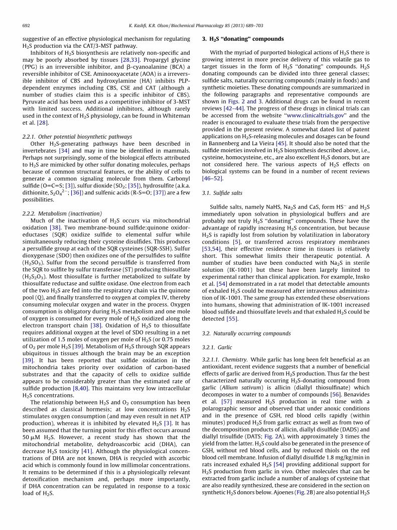
K. Kashfi, K.R. Olson / Biochemical Pharmacology 85 (2013) 689–703692
suggestive of an effective physiological mechanism for regulatingH2S production via the CAT/3-MST pathway.
Inhibitors of H2S biosynthesis are relatively non-specific andmay be poorly absorbed by tissues [28,33]. Propargyl glycine(PPG) is an irreversible inhibitor, and b-cyanoalanine (BCA) areversible inhibitor of CSE. Aminooxyacetate (AOA) is a irrevers-ible inhibitor of CBS and hydroxylamine (HA) inhibits PLP-dependent enzymes including CBS, CSE and CAT (although anumber of studies claim this is a specific inhibitor of CBS).Pyruvate acid has been used as a competitive inhibitor of 3-MSTwith limited success. Additional inhibitors, although rarelyused in the context of H2S physiology, can be found in Whitemanet al. [28].
2.2.1. Other potential biosynthetic pathways
Other H2S-generating pathways have been described ininvertebrates [34] and may in time be identified in mammals.Perhaps not surprisingly, some of the biological effects attributedto H2S are mimicked by other sulfur donating molecules, perhapsbecause of common structural features, or the ability of cells togenerate a common signaling molecule from them. Carbonylsulfide (O55C55S; [3]), sulfur dioxide (SO2; [35]), hydrosulfite (a.k.a.dithionite, S2O4
2�; [36]) and sulfenic acids (R-S55O; [37]) are a fewpossibilities.
2.2.2. Metabolism (inactivation)
Much of the inactivation of H2S occurs via mitochondrialoxidation [38]. Two membrane-bound sulfide:quinone oxidor-eductases (SQR) oxidize sulfide to elemental sulfur whilesimultaneously reducing their cysteine disulfides. This producesa persulfide group at each of the SQR cysteines (SQR-SSH). Sulfurdioxygenase (SDO) then oxidizes one of the persulfides to sulfite(H2SO3). Sulfur from the second persulfide is transferred fromthe SQR to sulfite by sulfur transferase (ST) producing thiosulfate(H2S2O3). Most thiosulfate is further metabolized to sulfate bythiosulfate reductase and sulfite oxidase. One electron from eachof the two H2S are fed into the respiratory chain via the quinonepool (Q), and finally transferred to oxygen at complex IV, therebyconsuming molecular oxygen and water in the process. Oxygenconsumption is obligatory during H2S metabolism and one moleof oxygen is consumed for every mole of H2S oxidized along theelectron transport chain [38]. Oxidation of H2S to thiosulfaterequires additional oxygen at the level of SDO resulting in a netutilization of 1.5 moles of oxygen per mole of H2S (or 0.75 molesof O2 per mole H2S [39]. Metabolism of H2S through SQR appearsubiquitous in tissues although the brain may be an exception[39]. It has been reported that sulfide oxidation in themitochondria takes priority over oxidation of carbon-basedsubstrates and that the capacity of cells to oxidize sulfideappears to be considerably greater than the estimated rate ofsulfide production [8,40]. This maintains very low intracellularH2S concentrations.
The relationship between H2S and O2 consumption has beendescribed as classical hormesis; at low concentrations H2Sstimulates oxygen consumption (and may even result in net ATPproduction), whereas it is inhibited by elevated H2S [3]. It hasbeen assumed that the turning point for this effect occurs around50 mM H2S. However, a recent study has shown that themitochondrial metabolite, dehydroascorbic acid (DHA), candecrease H2S toxicity [41]. Although the physiological concen-trations of DHA are not known, DHA is recycled with ascorbicacid which is commonly found in low millimolar concentrations.It remains to be determined if this is a physiologically relevantdetoxification mechanism and, perhaps more importantly,if DHA concentration can be regulated in response to a toxicload of H2S.
3. H2S ‘‘donating’’ compounds
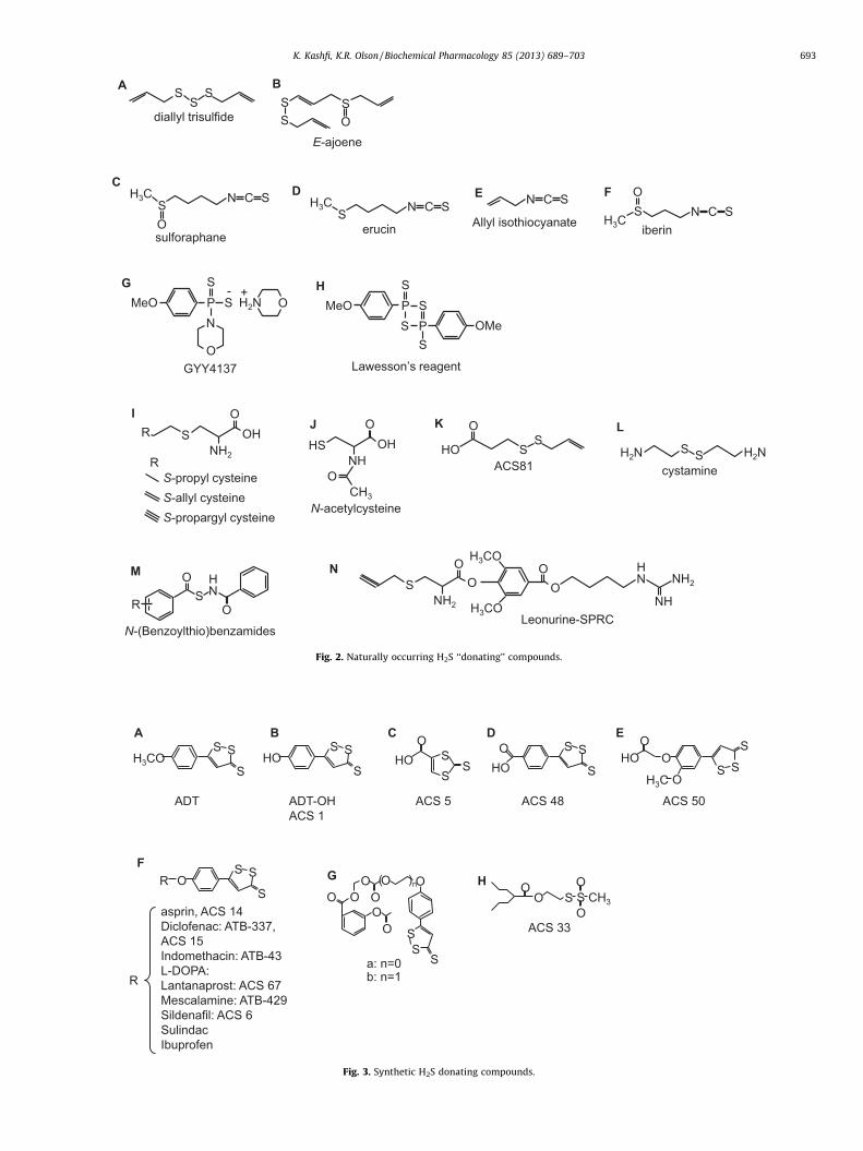
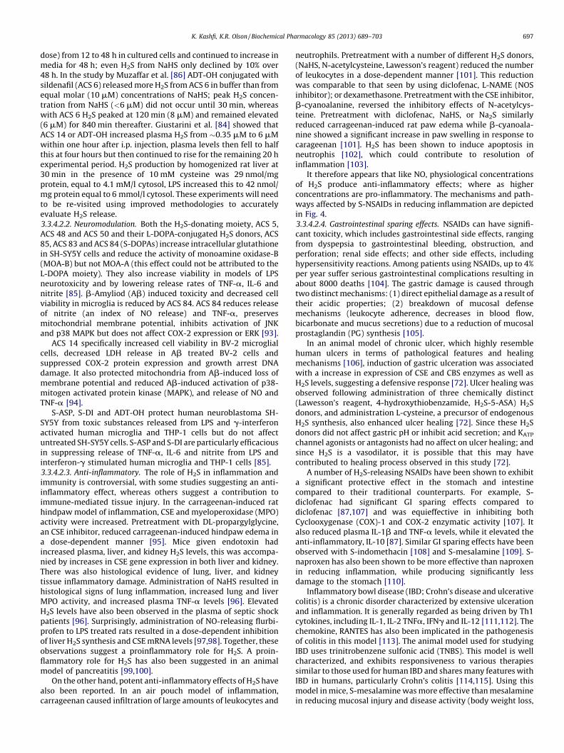
With the myriad of purported biological actions of H2S there isgrowing interest in more precise delivery of this volatile gas totarget tissues in the form of H2S ‘‘donating’’ compounds. H2Sdonating compounds can be divided into three general classes;sulfide salts, naturally occurring compounds (mainly in foods) andsynthetic moieties. These donating compounds are summarized inthe following paragraphs and representative compounds areshown in Figs. 2 and 3. Additional drugs can be found in recentreviews [42–44]. The progress of these drugs in clinical trials canbe accessed from the website ‘‘www.clinicaltrials.gov’’ and thereader is encouraged to evaluate these trials from the perspectiveprovided in the present review. A somewhat dated list of patentapplications on H2S-releasing molecules and dosages can be foundin Bannenberg and La Vieira [45]. It should also be noted that thesulfide moieties involved in H2S biosynthesis described above, i.e.,cysteine, homocysteine, etc., are also excellent H2S donors, but arenot considered here. The various aspects of H2S effects onbiological systems can be found in a number of recent reviews[46–52].
3.1. Sulfide salts
Sulfide salts, namely NaHS, Na2S and CaS, form HS� and H2Simmediately upon solvation in physiological buffers and areprobably not truly H2S ‘‘donating’’ compounds. These have theadvantage of rapidly increasing H2S concentration, but becauseH2S is rapidly lost from solution by volatilization in laboratoryconditions [5], or transferred across respiratory membranes[53,54], their effective residence time in tissues is relativelyshort. This somewhat limits their therapeutic potential. Anumber of studies have been conducted with Na2S in sterilesolution (IK-1001) but these have been largely limited toexperimental rather than clinical application. For example, Inskoet al. [54] demonstrated in a rat model that detectable amountsof exhaled H2S could be measured after intravenous administra-tion of IK-1001. The same group has extended these observationsinto humans, showing that administration of IK-1001 increasedblood sulfide and thiosulfate levels and that exhaled H2S could bedetected [55].
3.2. Naturally occurring compounds
3.2.1. Garlic
3.2.1.1. Chemistry. While garlic has long been felt beneficial as anantioxidant, recent evidence suggests that a number of beneficialeffects of garlic are derived from H2S production. Thus far the bestcharacterized naturally occurring H2S-donating compound fromgarlic (Allium sativum) is allicin (diallyl thiosulfinate) whichdecomposes in water to a number of compounds [56]. Benavideset al. [57] measured H2S production in real time with apolarographic sensor and observed that under anoxic conditionsand in the presence of GSH, red blood cells rapidly (withinminutes) produced H2S from garlic extract as well as from two ofthe decomposition products of allicin, diallyl disulfide (DADS) anddiallyl trisulfide (DATS; Fig. 2A), with approximately 3 times theyield from the latter. H2S could also be generated in the presence ofGSH, without red blood cells, and by reduced thiols on the redblood cell membrane. Infusion of diallyl disulfide 1.8 mg/kg/min inrats increased exhaled H2S [54] providing additional support forH2S production from garlic in vivo. Other molecules that can beextracted from garlic include a number of analogs of cysteine thatare also readily synthesized, these are considered in the section onsynthetic H2S donors below. Ajoenes (Fig. 2B) are also potential H2S
SSS
diallyl tris ulf ide
A
N C SS
H3C
Osulforap han e
C
MeO P
S
S
O
N
OH2N+-
GYY413 7
G
MeO P
S
S
S
S
Lawesson’s reagen t
H
P OMe
S S
S
E-ajoene
O
B
N C SS
H3C
erucin
D EN C S
Allyl isothiocy anateN C SS
H3C
O
iberin
F
S-propyl cysteine
I
NH2
O
S OHR
S-allyl cysteine
S-proparg yl cysteine
R NH
O
HS OH
CH3
O
N-acet ylcy steine
J
SS
O
HO
ACS81
K
H2NS
S H2N
cystamine
L
S NO
O
H
R
N-(Benzoylthio)benzami des
M
NH2
O
S O
HN
O
Leonuri ne-SPRCH3CO
H3CO
ONH
NH2
N
Fig. 2. Naturally occurring H2S ‘‘donating’’ compounds.
S
SSHO O
O
OH3CS
SSH3CO
OO S S CH3
O
O
ACS 33
H
S
SS
HO
O
S
SSHO
O
O
O
O
O
O
S
SS
( )nG
a: n=0b: n=1
S
SS
HO
O
B EDC
S
SSO
asprin, ACS 14
Dicl ofenac: ATB -33 7,
ACS 15
Indomethacin: ATB -43
L-DO PA:
Lantanaprost : ACS 67
Mescalami ne: ATB -42 9
Silden afil: ACS 6
Sulind ac
Ibuprofen
F
R
R
ACS 50
A
ACS 48ACS 5ADT-OH
ACS 1
ADT
Fig. 3. Synthetic H2S donating compounds.
K. Kashfi, K.R. Olson / Biochemical Pharmacology 85 (2013) 689–703 693

K. Kashfi, K.R. Olson / Biochemical Pharmacology 85 (2013) 689–703694
donors, although H2S production from these molecules has not yetbeen measured.
3.2.1.2. Biology. The beneficial effects of garlic in the prevention ofrenal, hepatic, cardiac, cerebral and pulmonary ischemia-reperfu-sion injury are well-known (reviewed in [58]), as are theirpotential as anti-cancer [59] and anti-diabetes [60] agents. Garlicextracts DATS and DADS vasodilate rat aortas via H2S production[57].
3.2.2. Sulforaphane, erucin and iberin
3.2.2.1. Chemistry. Sulforaphane (Fig. 2C), the isothiocyanatecompound from broccoli (Brassica oleracea), is rapidly absorbedby humans, reaching peak concentrations at 1 h and decliningthereafter with a half-life of 1.8 h [61]. A related isothiocyanatecompound, erucin (Fig. 2D), is found in high levels in rocket saladspecies (Eruca sativa). Allyl isothiocyanate (Fig. 2E) is derived fromwasabi, mustard and horseradish. It has not yet been determined ifany of the isothiocyanates are metabolized to produce H2S,although their purported health benefits are quite similar to thosethat are attributed to H2S or other H2S-donating drugs, suggestinga role for H2S, although this needs to be evaluated further.
3.2.2.2. Biology. Sulforaphane, Erucin and a related isothiocyanate,iberin (Fig. 2F), upregulate thioredoxin reductase 1 (EC 1.8.1.9) inhuman breast cancer MCF-7 cells. Because thioredoxin reductase isthe only enzyme that reduces thioredoxin and reduced thioredoxinis required to release H2S from 3-MST (see above) it is quitepossible that many of the health benefits of cruciferous vegetablesinvolve H2S. Sulforaphane protects vascular smooth muscle cellsand endothelial cells from oxidative and inflammatory stress andsuppresses angiogeniesis [62–64]. Sulphoraphane has neuropro-tective and anti-inflammatory actions mediated in part throughactivation of heme oxygenase-1 (HO-1) and it provides someprotection against ischemia reperfusion injury, hemorrhage andserotonin-induced toxicity [65,66].
3.3. Synthetic H2S donors
3.3.1. GYY4137
3.3.1.1. Chemistry. GYY4137 (morpholin-4-ium 4 methoxyphe-nyl(morpholino) phosphinodithioate, Cayman Chemical; Fig. 2G) isa water-soluble molecule. Formation of H2S from GYY 4137 wasfirst reported by [67]. One mmol/l of GYY4137 releases�40 mmoles of H2S in the first 10 min and then releasesapproximately 40 mmoles of H2S per hour for the ensuing80 min when dissolved in acidic (pH 3.0) phosphate buffer;relatively little (<3 mmoles) H2S appears to be released over thissame period when dissolved in buffer at pH 7.4 or 8.5. Injection of133 mmol/kg (approximately 665 mM if distributed throughoutthe extracellular compartment) either intrapertioneally (ip) orintravenously (iv) into rats, increases plasma H2S from �32 mmol/lto �80 mmol/l in 30 min and this remains elevated (50 mmol/l) for3 hours. It should be noted that plasma H2S was measured in thesestudies by DTMB (5,50-dithiobis-(2-nitrobenzoic acid; Ellman’sreagent) which reacts with thiol groups (R-SH) includingglutathione and protein thiols, therefore, the effects of GYY4137on plasma H2S per se remain to be determined. Lee et al. [68]observed a near constant 15–20 mmol/l H2S for up to 7 daysfollowing addition of GYY4137 to culture media containing humanbreast adenocarcinoma MCF-7 cells (methylene blue method). Thein vivo study of Li et al. [67] contrasts with that of Yu et al. [69] whoreported that after i.p. injection of 200 mmol/l the H2S concentra-tion in heart and liver increased, but only for 20 min.
3.3.1.2. Biology. In rats, GYY4137 relaxes aortas, mediated in partby KATP channels, vasodilates the perfused, pre-constricted kidneyand exhibits antihypertensive activity but does not affect cardiaccontractility or heart rate in vitro, furthermore, GYY4137 was notcytotoxic to cultured smooth muscle cells [67]. However, it hasbeen reported that GYY4137 interacts with endogenous NOgenerated from l-arginine to stimulate heart contraction [70].The effects of GYY4137 were slower in onset and longer lastingthan those of NaHS. Paradoxically, the same dose of GYY4137 ip(133 mmol/kg) significantly increased blood pressure in ratsrendered hypotensive by lipopolysaccharide (LPS)-induced en-dotoxic shock [71]. GYY4137 also decreased secretion of many ofthe markers of LPS-induced inflammation in cultured RAW 264.7cells including nitrate/nitrite, PGE2, TNF-a, IL-1b, IL-6, NF-kB,expression of inducible nitric oxide synthase (iNOS), andcyclooxygenase-2. One hour pre-treatment with GYY4137 priorto LPS did not provide a significant anti-inflammatory effect in rats,whereas GYY4137 administered 1 or 2 h after LPS significantlyreduced nitrite/nitrate, C-reactive protein, L-selectin and lungmyeloperoxidase activity and decreased plasma creatinine/alanineamino transferase.
Anti-cancer effects of GYY4137 have been observed in vitro andin vivo [68]. Five-day treatment with GYY4137 (400 mmol/l), butnot similar concentrations of the phosphate-substituted homolog,ZYJ1122 (morpholin-4-inum diphenylphosphinic acid), or NaHSsignificantly reduced proliferation of breast adenocarcinoma(MCF-7), acute promyelocytic leukemia (MV4-11) and myleomo-nocytic leukemia (HL-60) cells. At 800 mmol/l GYY4137 killed 90–97% of these cells and human cervical carcinoma (HeLa), colorectalcarcinoma (HCT-116), hepatocellular carcinoma (Hep-G2), osteo-sarcoma (U2OS), but did not affect survival of non-cancer humandiploid lung fibroblasts (IMR90 and WI-38). The apparent IC50 forGYY4137 in MCF-7, HL60 and MV4-11 cells was around 350 mmol/l. GYY4137 promoted apoptosis as seen by presence of cleavedPARP and cleaved caspase 9, it also triggered cell-cycle arrest in theG2/M phase in MCF-7 cancer cells without affecting non-cancerousIMR90 cells. Daily i.p. injection of GYY4137 also decreased thegrowth of HL-60 and MV4-11 tumors subcutaneously transplantedin immunodeficient mice. GYY4137 up to 2 mmol/l did not affectviability in non-tumorigenic HepG2 cells [69]. GYY4137 is aderrivative of Lawesson’s reagent (Fig. 2H), another H2S donorwhich has been used with some success as an anti-inflammatorydrug in the stomach [72].
3.3.1.3. Questions remaining. In the study by Li et al. [67] GYY4137was reported to be more potent than NaHS in relaxing rat aortas(EC50s 115.7 mmol/l vs. 274.1 mmol/l). However, when dissolved inbuffer at pH 7.4, 1 mmol/l GYY4137 produced less than 3 mmol/lH2S. Therefore, 115 mmol/l GYY4137 wouldn’t be expected toproduce more than 0.3 mmol/l of H2S. Because rat aortas did notrespond to 100 mmol/l H2S (as NaHS), it is not clear how so littleH2S from GYY4137 could be vasoactive, unless the GYY4137 wasinternalized and the H2S generated intracellularly. As describedabove, there is also some confusion surrounding the time-course ofH2S production from GYY4137. In addition, H2S formation fromGYY4137 in buffer was measured with an amperometric sensor,whereas H2S formation in plasma was not determined amper-ometrically but was measured with the methylene blue method. Itis unclear why the latter was used as it is associated withconsiderable artifact [11].
3.3.2. Cysteine analogs
3.3.2.1. Chemistry. The well-known ability of cysteine to mimicH2S effects, presumably by providing additional H2S has led to theevaluation of a number of cysteine analogs as potential substrates

K. Kashfi, K.R. Olson / Biochemical Pharmacology 85 (2013) 689–703 695
for endogenous cysteine-metabolizing enzymes. Some of the morecommon molecules are: S-propyl cysteine (SPC), S-allyl cysteine(SAC, also a derivative of garlic), S-propargyl cysteine (SPRC;Fig. 2I) and N-acetyl cysteine (NAC; Fig. 2J). Wang et al. [73]measured H2S production by homogenized rat ventricles with thedirect methylene blue method and observed that SPC, SAC andSPRC all increased H2S production by at least two-fold and the rateof H2S production increased as the terminal carbon becameprogressively less saturated. This effect was mediated by CSE. SPRCadministered i.p., dose-dependently increases H2S in LPS-treatedrats, using direct methylene blue, control H2S in hippocampus was50 mM [74].
SAC has been shown not only to serve as a substrate for H2S byCSE but also to up-regulate the enzyme in an acute myocardialinfarction rat model. Conversely in sham operated rats, SAC did notsignificantly increase plasma H2S [75]. SPRC and SAC have beenconjugated with leonurine, an alkaloid in Chinese motherwort(Fig. 2N) [50,76]. Leonurine-SPRC is somewhat more effective andit increased H2S content in hypoxic neonatal rat ventricularmyocytes. A derivative of diallyl disulfide ACS 81 (Fig. 2K), (3-(prop-2-en-1yldisulfanyl)propanoic acid) has been conjugatedwith L-DOPA and has essentially identical effects as thedithiolethiol-L-DOPA conjugates discussed below.
3.3.2.2. Biology.
3.3.2.2.1. Cardiovascular. Daily i.p. injection of cysteine analogs,SPC, SAC and SPRC for seven days prior to and two days aftercoronary artery ligation of rat hearts significantly protected thehearts from ischemia reperfusion injury and was associated withpreserved superoxide dismutase (SOD), glutathione peroxidaseactivity, and tissue GSH levels, while reducing lipid peroxidationproducts [73].
In an acute myocardial infarction rat model, pre-treatment withSAC (50 mg/kg/day for 7 days) significantly lowered mortality andreduced infarct size but did not affect blood pressure, SAC alsoincreased left ventricular CSE; inhibition of CSE with PPGeliminated the beneficial effects of SAC, increased infarct size,and significantly elevated blood pressure. Although SAC is asubstrate for H2S production, it also augments H2S by up-regulating CSE [75].
Leonurine-SPRC provides a cardioprotective effect in hypoxicneonatal rat ventricular myocytes by increasing cell viability,decreasing LDH leakage, decreasing MDA and ROS while increasingSOD, catalase activity, and have a general anti-apoptotic effectthrough an increase in bcl-2 and an decrease in bax and caspase-3[50].3.3.2.2.2. Neuromodulation. SPRC administerd i.p. attenuatedlipopolysaccharide (LPS)-induced deficits in spatial learning andmemory in rats and restored the LPS deficit in hippocampal H2Slevels. SPRC also reduced the LPS-elevation of TNF-a, TNFR1, AbPPand Ab1–40/42, increased Ikb-a degradation and NF-kb p65phosphorylation, and decreased TNF-a, TNFR1 and AbPP mRNA[74].3.3.2.2.3. Cancer. SPRC (1 mM–30 mM) produced a dose dependentinhibition of growth of cultured SGC-7901 human gastric cancercells. 1 mM SPRC produced 18% inhibition of viability andsuppressed colony forming and migration ability (�18% inhibitionwith 1 mM) significantly increased CSE protein expression and H2Sconcentration in culture media; PPG significantly decreased SPRCefficacy. SPRC effects were more pronounced than SAC. SPRCstimulated apoptosis, and induced cell cycle arrest at the G1/Sphase [77]. SPRC increased Bax and p53 but not Bcl-2 protein andmRNA expression. SAC increased p53 protein expression and BaxmRNA but did not affect other parameters. In male nude mice withSGC-7901 tumors implanted in the flank, SPRC and SAC (50 and100 mg/kg) increased plasma H2S concentration, reduced tumor
volume and increased apoptotic tumor cell number and increasedprotein expression of Bax and p53 and decreased Bcl-2. Both SPRCand SAC increased Bax and p35 mRNA expression but did not affectBcl-2 [77].3.3.2.2.4. Inflammation. In the lipopolysaccharide-induced inflam-matory response of rat embryonic ventricular myocardial cells(H9c2 cells) Pan et al. [78] observed that SPRC prevented NF-kBactivation, suppressed LPS- induced extracellular signal-regulatedkinase 1/2 (ERK1/2) phosphorylation, and intracellular ROSproduction. SPRC induced phosphorylation of Akt and reducedmRNA and protein expression of tumor necrosis factor-a (TNF-a)and these effects were abolished by PPG. SPRC (1 mM) inhibitediNOS and ICAM mRNA and increased CSE and supernatant H2Sconcentration [77].
Other: Cystamine (Fig. 2L) dose-dependently increased cellproliferation of HIV-infected CEM-6 human lymphocytes [79].Methyl disulfides (R-S-S-CH3) increase growth of murine lympho-ma L1210 cells [80].3.3.2.2.5. Questions remaining. In the study by Ma et al. [77], H2Swas measured in the supernatant above cells 24 h after addition ofSPRC or SAC and in the study by Pan et al. [78], H2S was measuredin the supernatant above cells 6.5 h after addition of SPRC. Giventhe volatility of H2S, it is unclear how much H2S was actuallypresent at these late dates.
3.3.3. Cysteine-activated H2S donors
3.3.3.1. Chemistry. Zhao et al. [81] developed a novel class of H2Sdonors based on the lability of nitrosothiol (S-N) bonds. In theirscheme a N-(benzoylthio)benzamide moiety (Fig. 2M) slowlyreleases H2S in the presence of excess cysteine. A peak of 25.4 mMH2S was observed 18 min after 40 mM of N-(Benzoylthio)benza-mide was incubated with 4 mM cysteine. The rate and quantity ofH2S released could be altered by adding side groups on thebenzoylthio ring and 11 such substitutions were examined in.Adding –CF3 to the fourth carbon decreased the peak time to13 min and increased the concentration at the peak to 35.6 mM,indicating nearly complete metabolism of the compound to H2S.An–OCH3 group in both the ortho and para-positions increased thepeak time to 50 min and decreased the concentration at the peak to23.0 mM. These reactions were most efficient when the cysteine:N-(Benzoylthio)benzamide ratio was at least 10, further increasingthis ratio did not appreciably increase yield. All of thesecompounds were soluble in phosphate buffer and in the absenceof cysteine did not generate H2S. The authors also showed that H2Scould be generated from bovine calf plasma that contained500 mM cysteine and that adding N-methylmaleimide to theplasma to block free cysteine inhibited H2S formation. Thisprovided additional evidence that cysteine is the regulator of thisH2S-generating reaction and can be used in biological systems.
3.3.3.2. Biology. To date no biological studies have been reported.3.3.3.2.1. Questions remaining. As with the study of H2S formationfrom GYY4137 (above), the formation of H2S in buffer wasmeasured amperometrically, whereas H2S generation in plasmawas measured using the methylene blue method. The actual rate ofH2S formation from cysteine and N-(Benzoylthio)benzamide inplasma needs to be verified. Furthermore, as cysteine alone cancontribute to H2S generation, additional, cysteine-only, controlstudies are necessary.
3.3.4. Dithiolethione and its NSAID chimeras
3.3.4.1. Chemistry. Anethole dithiolethione (ADT; Fig. 3A) and itsmain metabolite (ADT-OH; 5-(4-hydroxyphenyl)-3H-1,2-dithiole-3-thione; Fig. 3B) have been used extensively as a donor of H2S and

K. Kashfi, K.R. Olson / Biochemical Pharmacology 85 (2013) 689–703696
its ease of esterification with other therapeutics has led to aconsiderable variety of H2S ‘‘donating’’ drugs. ADT-OH can also bemodified prior to esterification, i.e., ACS 5, ACS 48, ASC 50 (Figs. 3C–E). A number of the ADT-OH-H2S molecules are shown in Fig. 3F.
Lee et al. [82] conjugated L-DOPA with ACS 5, ACS 48 and ACS 50to produce ACS 85, ACS 83 and ACS 84, respectively. When ACS 84was injected i.v. into rats essentially all of the dithiolethione (ACS50) was cleaved from the L-DOPA within 1 h and dopamine, L-DOPA and ACS 50 could be found in the brain. Intracellularconcentration of H2S in human monocyte (THP-1), astrocytoma(U373) and neuroblastoma (SH-SY5Y) cells increased 12 h aftertreatment with 10 mM ACS 50 to 6.5, 0.8 and 0.8 mmol/l,respectively and by 48 h post-treatment these were 6, 1.8 and1.7 mmol/l, respectively. At 48 h post-treatment, H2S concentra-tion in the extracellular fluid was between 2 and 3 mmol/l.Comparatively little (10%) H2S was released from either PBS bufferor culture media at 48 h (0.2 mmol/l). H2S production from ADT-OH, ACS 5, ACS 48, ACS 50, ACS 81 by isolated mitochondria wasrapid, over 80% within 1 h [82].
ADT-OH conjugated with aspirin (S-ASP, ACS 14) injected i.v.into rats was rapidly deacetylated then hydrolyzed at a somewhatslower rate freeing ADT-OH; plasma H2S peaked within fiveminutes of injection and fell to less than 20% of the peak value by40 min [83]. I.p. injection of ACS 14 still resulted in rapiddisappearance of the parent compound and formation of ADT-OH, however, the latter remained significantly elevated two hoursafter injection, plasma H2S peaked in about one hour after eitherACS 14 or ADT-OH injection then dipped to half the peak value inabout four hours but then slowly increased over the remaining24 h experimental period [84]. When THP-1 or U118 cells wereincubated with NaHS, ADT-OH, S-ASP, or ADT-OH conjugated withdiclofenac (S-DI) they steadily accumulated H2S for 12 h [85].
ADT-OH conjugated with sildenafil (ACS 6) released H2S slowlywith peak concentrations at 120 min, approximately 4 times asmuch H2S was released in the presence of pulmonary arteryendothelial cells compared to buffer [86].
When either ADT-OH or ADT-OH conjugated with diclofenac (S-diclofenac, S-diclo, ACS 15, ATB-337) were added to rat liverhomogenate there was a slow release of H2S (2.6% of the S-diclo) thatpersisted for at least 75 min; only one third of this amount of H2Swas released by these compounds in buffer. Inhibition ofcarboxylesterase with sodium fluoride reduced H2S productionfrom S-diclo in homogenized liver by nearly two-thirds. Whenincubated in plasma, release of H2S from S-diclo slowly increasedreaching a maximum in 90 min, declining thereafter. IP injection of47.2 mmol/kg of S-diclo increased plasma H2S from 30 to 37 mM at45 min and plasma H2S remained elevated (33 mM) after 6 h;injection of equal molar amount of NaHS increased plasma H2S from25 to 34 mM at 45 min but plasma H2S returned to control values by6 h. Plasma H2S was unaffected by IP injection of diclofenac [87].
Recently, Lazzarato et al. [88] synthesized a new class of ADT-OH esters of aspirin, ((4-(3-Thioxo-3H-1,2-dithiol-5-yl)phenoxy)-carbonyloxy)-methyl 2-(Acetyloxy)benzoate and ((2-(4-(3-Thioxo-3H-1,2-dithiol-5-yl)phenoxy)ethoxy)carbonyloxy)-methyl2-(Acetyloxy)benzoate (Fig. 3G). These compounds are reported tohave the advantage of releasing aspirin when incubated in humanserum. The compounds are relatively stable in buffered solutions(20% or less decomposition at three hours at pH 7.4) but relativelyquickly are metabolized in human serum (halftime of aspirinrelease between two and three minutes) and aspirin accounts forapproximately 1/3 of the yield.
3.3.4.2. Biology. Combining ADT-OH with a variety of compoundshas frequently shown to enhance their biological activity or,especially in the case of the nonsteroidal anti-inflammatory drugs(NSAIDs), to counter their adverse effects.
3.3.4.2.1. Cardiovascular. ACS 14 given orally (50 mg/kg) for sevendays did not affect body weight, systolic blood pressure, or heartrate in rats; ACS 14 and aspirin lowered thromboxane B2 and 6-keto-PGF1a, whereas these variables were unaffected by ADT-OHor salicyclic acid. ACS 14 but not ADT-OH lowered plasma oxidativestress marker, 8-isoprostane, and both compounds increasedplasma, cardiac and aortic tissue glutathione. ACS 14 amelioratedhemorrhagic lesions in the stomach due to oral aspirin andincreased heme oxygenase-1 (HO-1) promoter activity in NIH3T3-HO-1-luc cells [83]. I.p. injection of ACS 14 initially lowered, butthen increased, plasma levels of reduced glutathione, cysteine andthe glutathione metabolite cysteinylglycine but produced a steadyfall in plasma homocysteine, the initial fall in reduced thiols wasnot due to a higher pro-oxidant burden as oxidative stress in bloodcontinued to fall throughout the experimental period [84]. ACS 14,salicylic acid conjugated with ADT-OH (ACS 21) and ADT-OHreduced the hypertension brought about by glutathione depletionwith buthionine sulfoximine (BSO) and lowered plasma throm-boxane B2, 8-isoprostane and insulin and restored plasmaglutathione without creating gastric lesions. In addition, theyimproved endothelial function in isolated aortas and improvedoutcome in cardiac ischemia reperfusion experiments, similarbenefits were not experienced with either aspirin or salicylic acidstrongly suggesting the benefits of the H2S-releasing moiety [89].
The ‘‘pure’’ aspirin and H2S releasing drugs synthesized by [88]are reported to have good antiaggregatory effects on platelets (IC50
of 150 and 16 mM).The phosphodiesterase inhibitor, sildenafil conjugated with
ADT-OH (ACS 6) relaxes the rabbit corpus cavernosum with greatersensitivity and efficacy than NaHS, and the relaxation is due solelyto the sildenafil moiety downregulating phosphodiesterase 5.However, while sildenafil acts through a PKG-dependent mecha-nism, the H2S acts through a PKA-dependent mechanism andtogether they have a greater inhibitory effect on p47phox (andtherefore NADPH oxidase) than either sildenafil or H2S alone. ACS 6has the added benefit of antioxidant activity both in vitro and inrats in vivo [90]. Similar observations in porcine pulmonaryendothelial cells suggests that ACS 6 may also be beneficial intreating adult respiratory distress syndrome [86].
IP injection of 47.2 mmol/kg S-diclofenac, sufficient to raise ratplasma H2S from 30 to 37 mM did not affect arterial blood pressure[87]. In the isolated rabbit heart subjected to low-flow ischemia-reperfusion damage, S-diclofenac and NaHS produced a dosedependent normalization of coronary perfusion pressure, reducedleft ventricular contracture during ischemia, and at reperfusionthey improved left ventricular pressure and dp/dt while reducingcreatinine kinase and LDH and increasing GSH. These effectsappear to be mediated in part by KATP channels. Inhibition of NObiosynthesis exacerbated ischemia-reperfusion damage and thiswas offset by both S-diclofenac and NaHS. Cardiac parameters incontrol hearts were unaffected by S-diclofenac or NaHS [91].
In normal rat aortic (A-10) and immortalized (CRL-2018)smooth muscle cells S-diclofenac exhibited a concentration-dependent reduction in cell survival (�5% survival at 100 mM).Both NaHS and S-diclofenac decrease the percentage of A-10 cellsin the G1 phase and increased apoptosis. S-diclofenac increasedpro-apoptoic proteins p53, AIP1 and Bax but did not increase theanti-apoptoic protein, Bcl-2. However, S-diclofenac did not affectcells synchronized by starvation where G1 cells accumulate. Theseresults suggest that S-diclofenac may be useful for prevention ofsmooth muscle proliferation [92].3.3.4.2.1.1Questions remaining. There are a number of concernsregarding the amount, duration and stability of H2S released bythese compounds and these are most likely due to the use ofmethylene blue in the H2S assays. Lee et al. [82] reported thatintracellular H2S remained constant at 6 mM (60% of total ACS 50
K. Kashfi, K.R. Olson / Biochemical Pharmacology 85 (2013) 689–703 697
dose) from 12 to 48 h in cultured cells and continued to increase inmedia for 48 h; even H2S from NaHS only declined by 10% over48 h. In the study by Muzaffar et al. [86] ADT-OH conjugated withsildenafil (ACS 6) released more H2S from ACS 6 in buffer than fromequal molar (10 mM) concentrations of NaHS; peak H2S concen-tration from NaHS (<6 mM) did not occur until 30 min, whereaswith ACS 6 H2S peaked at 120 min (8 mM) and remained elevated(6 mM) for 840 min thereafter. Giustarini et al. [84] showed thatACS 14 or ADT-OH increased plasma H2S from �0.35 mM to 6 mMwithin one hour after i.p. injection, plasma levels then fell to halfthis at four hours but then continued to rise for the remaining 20 hexperimental period. H2S production by homogenized rat liver at30 min in the presence of 10 mM cysteine was 29 nmol/mgprotein, equal to 4.1 mM/l cytosol, LPS increased this to 42 nmol/mg protein equal to 6 mmol/l cytosol. These experiments will needto be re-visited using improved methodologies to accuratelyevaluate H2S release.3.3.4.2.2. Neuromodulation. Both the H2S-donating moiety, ACS 5,ACS 48 and ACS 50 and their L-DOPA-conjugated H2S donors, ACS85, ACS 83 and ACS 84 (S-DOPAs) increase intracellular glutathionein SH-SY5Y cells and reduce the activity of monoamine oxidase-B(MOA-B) but not MOA-A (this effect could not be attributed to theL-DOPA moiety). They also increase viability in models of LPSneurotoxicity and by lowering release rates of TNF-a, IL-6 andnitrite [85]. b-Amyliod (Ab) induced toxicity and decreased cellviability in microglia is reduced by ACS 84. ACS 84 reduces releaseof nitrite (an index of NO release) and TNF-a, preservesmitochondrial membrane potential, inhibits activation of JNKand p38 MAPK but does not affect COX-2 expression or ERK [93].
ACS 14 specifically increased cell viability in BV-2 microglialcells, decreased LDH release in Ab treated BV-2 cells andsuppressed COX-2 protein expression and growth arrest DNAdamage. It also protected mitochondria from Ab-induced loss ofmembrane potential and reduced Ab-induced activation of p38-mitogen activated protein kinase (MAPK), and release of NO andTNF-a [94].
S-ASP, S-DI and ADT-OH protect human neuroblastoma SH-SY5Y from toxic substances released from LPS and g-interferonactivated human microglia and THP-1 cells but do not affectuntreated SH-SY5Y cells. S-ASP and S-DI are particularly efficaciousin suppressing release of TNF-a, IL-6 and nitrite from LPS andinterferon-g stimulated human microglia and THP-1 cells [85].3.3.4.2.3. Anti-inflammatory. The role of H2S in inflammation andimmunity is controversial, with some studies suggesting an anti-inflammatory effect, whereas others suggest a contribution toimmune-mediated tissue injury. In the carrageenan-induced rathindpaw model of inflammation, CSE and myeloperoxidase (MPO)activity were increased. Pretreatment with DL-propargylglycine,an CSE inhibitor, reduced carrageenan-induced hindpaw edema ina dose-dependent manner [95]. Mice given endotoxin hadincreased plasma, liver, and kidney H2S levels, this was accompa-nied by increases in CSE gene expression in both liver and kidney.There was also histological evidence of lung, liver, and kidneytissue inflammatory damage. Administration of NaHS resulted inhistological signs of lung inflammation, increased lung and liverMPO activity, and increased plasma TNF-a levels [96]. ElevatedH2S levels have also been observed in the plasma of septic shockpatients [96]. Surprisingly, administration of NO-releasing flurbi-profen to LPS treated rats resulted in a dose-dependent inhibitionof liver H2S synthesis and CSE mRNA levels [97,98]. Together, theseobservations suggest a proinflammatory role for H2S. A proin-flammatory role for H2S has also been suggested in an animalmodel of pancreatitis [99,100].
On the other hand, potent anti-inflammatory effects of H2S havealso been reported. In an air pouch model of inflammation,carrageenan caused infiltration of large amounts of leukocytes and
neutrophils. Pretreatment with a number of different H2S donors,(NaHS, N-acetylcysteine, Lawesson’s reagent) reduced the numberof leukocytes in a dose-dependent manner [101]. This reductionwas comparable to that seen by using diclofenac, L-NAME (NOSinhibitor); or dexamethasone. Pretreatment with the CSE inhibitor,b-cyanoalanine, reversed the inhibitory effects of N-acetylcys-teine. Pretreatment with diclofenac, NaHS, or Na2S similarlyreduced carrageenan-induced rat paw edema while b-cyanoala-nine showed a significant increase in paw swelling in response tocarageenan [101]. H2S has been shown to induce apoptosis inneutrophis [102], which could contribute to resolution ofinflammation [103].
It therefore appears that like NO, physiological concentrationsof H2S produce anti-inflammatory effects; where as higherconcentrations are pro-inflammatory. The mechanisms and path-ways affected by S-NSAIDs in reducing inflammation are depictedin Fig. 4.3.3.4.2.4. Gastrointestinal sparing effects. NSAIDs can have signifi-cant toxicity, which includes gastrointestinal side effects, rangingfrom dyspepsia to gastrointestinal bleeding, obstruction, andperforation; renal side effects; and other side effects, includinghypersensitivity reactions. Among patients using NSAIDs, up to 4%per year suffer serious gastrointestinal complications resulting inabout 8000 deaths [104]. The gastric damage is caused throughtwo distinct mechanisms: (1) direct epithelial damage as a result oftheir acidic properties; (2) breakdown of mucosal defensemechanisms (leukocyte adherence, decreases in blood flow,bicarbonate and mucus secretions) due to a reduction of mucosalprostaglandin (PG) synthesis [105].
In an animal model of chronic ulcer, which highly resemblehuman ulcers in terms of pathological features and healingmechanisms [106], induction of gastric ulceration was associatedwith a increase in expression of CSE and CBS enzymes as well asH2S levels, suggesting a defensive response [72]. Ulcer healing wasobserved following administration of three chemically distinct(Lawesson’s reagent, 4-hydroxythiobenzamide, H2S-5-ASA) H2Sdonors, and administration L-cysteine, a precursor of endogenousH2S synthesis, also enhanced ulcer healing [72]. Since these H2Sdonors did not affect gastric pH or inhibit acid secretion; and KATP
channel agonists or antagonists had no affect on ulcer healing; andsince H2S is a vasodilator, it is possible that this may havecontributed to healing process observed in this study [72].
A number of H2S-releasing NSAIDs have been shown to exhibita significant protective effect in the stomach and intestinecompared to their traditional counterparts. For example, S-diclofenac had significant GI sparing effects compared todiclofenac [87,107] and was equieffective in inhibiting bothCyclooxygenase (COX)-1 and COX-2 enzymatic activity [107]. Italso reduced plasma IL-1b and TNF-a levels, while it elevated theanti-inflammatory, IL-10 [87]. Similar GI sparing effects have beenobserved with S-indomethacin [108] and S-mesalamine [109]. S-naproxen has also been shown to be more effective than naproxenin reducing inflammation, while producing significantly lessdamage to the stomach [110].
Inflammatory bowl disease (IBD; Crohn’s disease and ulcerativecolitis) is a chronic disorder characterized by extensive ulcerationand inflammation. It is generally regarded as being driven by Th1cytokines, including IL-1, IL-2 TNFa, IFNg and IL-12 [111,112]. Thechemokine, RANTES has also been implicated in the pathogenesisof colitis in this model [113]. The animal model used for studyingIBD uses trinitrobenzene sulfonic acid (TNBS). This model is wellcharacterized, and exhibits responsiveness to various therapiessimilar to those used for human IBD and shares many features withIBD in humans, particularly Crohn’s colitis [114,115]. Using thismodel in mice, S-mesalamine was more effective than mesalaminein reducing mucosal injury and disease activity (body weight loss,

Fig. 4. Mechanisms of action of H2S-releasing NSAIDs. When hydrolyzed, H2S-releasing NSAIDs produce the parent NSAID and the H2S-releasing moiety from which H2S is
released. The NSAID component inhibits COX-1 and COX-2 resulting in compromised mucosal defense mechanisms, which may lead to ulcers. NSAIDs can reduce renal
perfusion, which can lead to increases in blood pressure (BP) leading to cardiovascular (CV) damage. The released H2S counteracts many of the detrimental effects of NSAIDs.
These protective effects appear to be mediated through activation of KATP channels. H2S enhances the mucosal defense mechanisms; causes vasodilation thus reducing BP
leading to cardioprotective effects. Both the NSAID and H2S have anti-inflammatory effects, the former through inhibition of COX and latter through inhibition of nuclear
transcription factor kB (NF-kB).
K. Kashfi, K.R. Olson / Biochemical Pharmacology 85 (2013) 689–703698
fecal blood, diarrhea) and colonic granulocyte infiltration [109]. S-mesalamine also reduced the expression of mRNA for TNFa, IFNg,IL-1, IL-2, IL-12 and RANTES [109]. Using the same model in rats, S-mesalamine modulated expression of colonic pro-inflammatorymediators, COX-2 and IL-1b [116].3.3.4.2.5. Chemopreventive properties. NSAIDs in general andaspirin in particular represent the prototypical chemopreventiveagents against cancer. Epidemiological studies describing resultson >1 million subjects have pointed out the protective effect ofNSAIDs against colon, esophageal, gastric, breast, pancreatic andovarian cancers [117]. The epidemiological findings are inagreement with a large body of in vitro and animal data [118].However, regular NSAID use may lead to serious side effects asdescribed above. Hydrogen sulfide-releasing NSAID (HS-NSAIDsor S-NSAIDs) are a novel group of compounds that appear toovercome the shortcomings of regular NSAIDs although humansafety data on this class of compounds are not yet available.Recently, the effects of four H2S-releasing NSAIDs, (HS-aspirin,HS-sulindac, HS-ibuprofen, HS-naproxen, these are the same as S-aspirin, S-sulindac, S-ibuprofen, S-naproxen) have been describedon the growth properties of eleven different human cancer celllines of six different tissue origins [119]. These cell lines were ofadenomatous (colon, pancreatic, lung, prostate), epithelial(breast), and lymphocytic (leukemia) origin. In all of these HS-NSAIDs, the H2S-releasing moiety was ADT-OH, which wasconjugated to the parent NSAID through an ester bond [119].These HS-NSAIDs inhibited the growth of these cancer cell lineswith potencies of 28- to >3000-fold greater than that of their
traditional counterparts. HS-NSAIDs inhibited cell proliferation,induced apoptosis, and caused G0/G1 cell cycle block [119]. HS-aspirin preferentially inhibited the growth of estrogen receptor(ER)-negative breast cancer cells compared with a normalepithelial mammary cell line [120]. This effect was H2S-depen-dent as pretreatment with NaF, a carboxylesterase enzymeinhibitor, reduced the potency of HS-aspirin for cell growthinhibition by about 60-fold [120].
The transcription factor NF-kB is constitutively activated inER(-) breast cancer cell lines and primary tumors and therefore,constitutes a valid therapeutic target [121]. HS-ASA significantlyinhibited DNA binding activity of NF-kB (p65) which wasassociated with a decrease in NF-kB protein p65 levels in thenucleus. This was mediated by blockade of IkBa phosphorylationthrough inhibition of IKK activity [120]. Redox balance of the cellsmay influence the function of NF-kB by regulating its ability tobind to DNA, HS-ASA strongly inhibited thioredoxin reductase(TrxR) activity in ER(-) breast cancer cells and also increasedintracellular reactive oxygen species (ROS) [120]. In a xenograftmodel of ER(-) breast cancer, HS-ASA significantly reduced tumorvolume and tumor mass, through induction of apoptosis andinhibition of both proliferation and NF-kB [120]. Recently, usingER(-) breast cancer cells, it has been reported that dithiolethionesinhibit NF-kB transcriptional activity not through H2S release butrather via a covalent reaction with the NF-kB p50 and p65 subunits[122]. The same group has also reported that ACS-2, (4-(3-thioxo-3H-1,2-dithiol-5-yl)phenyl 2-propylpentanoate), an analog ofADT-OH which is more stable in vivo, had no effect on tumor

K. Kashfi, K.R. Olson / Biochemical Pharmacology 85 (2013) 689–703 699
volume in a murine model of ER(-) breast cancer, however, it didsignificantly reduce tumor burden in these mice [122].
The balance between phase-I carcinogen-activating and phase-II detoxifying xenobiotic metabolizing enzymes is critical todetermining an individual’s risk for cancer. HS-ASA induced theactivities of glutathione S-transferase (GST) and NAD(P) H:quinoneoxireductase (NQO1), two phase-II enzymes, in HT-29 humancolorectal cancer cells and mouse hepatoma Hepa 1c1c7 cells[123]. This induction was H2S-dependent since pretreatment of thecells with the esterase inhibitor, NaF, reversed HS-ASA-mediatedinduction of NQO1 activity to near basal levels [123]. Treatment ofboth cell lines with HS-ASA resulted in increases in NQO1, GST andUDP-glucuronyltransferase (UGT1, another phase-II enzyme)protein levels as well as increases in the phase-I enzymes CYP1A1and CYP2E1 [123]. However, in vivo studies showed that HS-ASAincreased the hepatic protein levels of phase-II enzymes, GST,NQO1, and UGT but had no effect on the protein levels of CYP1A1and CYP2E1 [123]. These finding strongly suggests that in vivo HS-ASA is a monofunctional inducer of phase-II metabolizingenzymes.
To our knowledge and according to the website, ‘‘Clinical-Trials.gov’’, none of the agents that are documented to be H2Sdonors in vivo have entered any clinical trials. However, the AntibeTherapeutics website states that ATB-429, a hydrogen sulfide-releasing derivative of mesalamine, has successfully completedanimal studies and is entering the first stage of clinical trials. This isfor inflammatory bowel disease, i.e., Crohn’s Disease and Ulcera-tive Colitis. All other H2S-releasing hybrid compounds are inpreclinical development.
4. Nitric oxide and hydrogen sulfide releasing aspirin (NOSH-aspirin)
4.1. Chemistry
Another approach in reducing the gastrointestinal side effectsof NSAIDs has been to join nitric oxide (NO) to an NSAID through anaromatic or aliphatic linker, NO-NSAIDs (reviewed in [118]). Thedevelopment of NO-NSAIDs was based on the observation that NO
Aspirin
H2S
NO
H2S
O
O
OONO2
O SS
S
NOSH-1
NOSH-aspirin
NBS-1120
O
O
OONO2
O NH2
S
NOSH-3
NBS-1121
Fig. 5. The chemical structures of NOSH compounds. NOSH-1: (4-(3-thioxo-3H-1
(nitrooxy)butyl (2-((4-(3-thioxo-3H-1,2-dithiol-5-yl)phenoxy)carbonyl)phenyl)); NOSH
(4-(nitrooxy)butyl 2-(5-((R)-1,2-dithiolan-3-yl)pentanoyloxy)benzoate).
has some of the same properties as PGs within the gastric mucosa,thus modulating some components of the mucosal defensesystems [124]. Preclinical studies have shown that NO-NSAIDsin general and NO-aspirin in particular, have chemopreventiveproperties and are much more potent than their traditionalcounterparts (reviewed in [118]). However, NO-NSAIDs posses-sing an aromatic linker can generate quinone methide inter-mediates, questioning the role of NO [125–127], or may havepotential genotoxicity [128]. NO-aspirin with an aliphatic linkerhas a very high IC50 for cell growth inhibition [129], limiting itsusefulness.
Based on the chemistry of NO and H2S, and the structuralcomponents of NO-NSAIDs and S-NSAIDs, recently it waspostulated that an NSAID possessing moieties that could releaseboth of these gasotransmeters might be more potent and effectivethan either one alone [130]. This led to the synthesis andcharacterization of a new class of anti-inflammatory and anti-cancer compounds, NOSH-aspirin, Fig. 5, [130,131]. Four NOSHcompounds have been reported, in all of these aspirin was used as ascaffold to which NO and H2S releasing moieties were coupled withone of the 1, 2 positions. Nitrate (–ONO2) was used for NO releaseand this was attached to the aspirin through an aliphatic spacer,while one of the following H2S-releasing moieties, 5-(4-hydro-xyphenyl)-3H-1, 2-dithiole-3-thione (ADT-OH), or 4-hydroxybenzothiazamide (TBZ) or lipoic acid were directly coupled toaspirin [130].
The NOSH-compounds described above were designed torelease both NO and H2S. Hence, using the methylene bluemethod, it was shown that NOSH-1 (NOSH-aspirin, NBS-1120)released H2S both in vitro [131] and in vivo [130] settings. In orderto show the actual release of H2S gas from NOSH-aspirin,homogenized mouse liver was incubated with NOSH-ASA andexamined in real time with a polarographic sensor [131]. The rateof H2S production under these conditions was significantly less inmedia than when incubated with tissue, suggesting that signifi-cantly more H2S was formed inside the cell, which may contributeto the high potency of NOSH-aspirin. Treatment of cells or animalswith NOSH-aspirin showed dose-dependent increases in NO levels[130,131].
H2S
O
OO
OONO2
O
O
SS
S
NOSH-2
NBS-1130
O
OONO2
O
OS S
NOSH-4
NBS-1131
, 2-dithiol-5-yl) phenyl 2-((4-(nitrooxy)butanoyl)oxy) benzoate); NOSH-2: (4-
-3: (4-carbamothioylphenyl 2-((4-(nitrooxy)butanoyl)oxy)benzoate); and NOSH-4:

K. Kashfi, K.R. Olson / Biochemical Pharmacology 85 (2013) 689–703700
4.2. Biology
4.2.1. Anti-inflammatory
NOSH-1 (NOSH-aspirin, NBS-1120) was shown to have anti-inflammatory properties similar to its parent compound, aspirin[130]. Using the carrageenan rat’s paw edema model, NOSH-1showed a reduction in paw inflammation, which was dose-dependent. Comparison of PGE2 content of paw exudates fromcontrol, ASA-treated, and NOSH-1-treated animals showed a clearand significant COX inhibition by aspirin and NOSH-1. EvaluationCOX expression in paw exudates showed that COX-2, whichproduces inflammatory PGE2, was barely detectable in thecontrols, was significantly induced by carrageenan, and dose-dependently inhibited by NOSH-1 [130]. Administration of ASA torats increased the proinflammatory cytokine tumor necrosisfactor-a (TNF-a) in plasma by about 20-fold; however, this risewas considerably lower in the NOSH-1 treated animals [130].
4.2.2. Chemopreventive properties
The cell growth inhibitory properties of the four NOSH-compounds were evaluated in eleven different human cancer celllines of six different histological subtypes. The cell lines were thatof colon (HT-29: COX-1 and COX-2 positive, HCT 15: COX null, andSW480: COX-1 positive, low levels of endogenous COX-2), breast(MCF7: [ER(+)], MDA MB-231 and SKBR3: [ER(�)]); T-cell leukemia(Jurkat), pancreatic (BxPC3: both COX-1 and COX-2 positive,MIAPaCa-2: COX-null), prostate (LNCaP), and lung (A549). All fourNOSH compounds were extremely effective in inhibiting thegrowth of these cell lines, however, NOSH-1 was the most potent,with IC50s for cell growth inhibition ranging from 48–280 nM at24 h [130]. In a fold comparison study of the IC50 values (ASA/NOSH 1–4), NOSH-1 was at least 100,000-fold more potent thanASA in HT-29 colon cancer cells. The increase in potency for NOSH-2, -3, and -4 in the same cell line after 24 h of treatment were>60,000-fold, >600-fold, and >16,000-fold, respectively [130]. InHT-29 colon cancer cells, the enhanced potency of NOSH-1increased to �125,000-fold and �250,000-fold after 48 h and72 h of treatment [131].
NOSH-aspirin (NBS-1120) treatment of athymic nude bearing ahuman colon cancer xenograft, caused an 85% reduction in tumorvolume after 18 days of treatment [131]. There were no overt signsof toxicity, as the mice in vehicle and NOSH-aspirin treated groupsdid not vary in their growth rate or behavior [131].
4.3. Outstanding questions
Research in the field of nitric oxide-hydrogen sulfide-releasingNSAIDs (NOSH-NSAIDs) is in its infancy. To date only a series ofcompounds based on aspirin have been reported. How otherNOSH-NSAIDs such as NOSH-naproxen, NOSH-sulindac, NOSH-ibuprofen etc. would behave has yet to be reported. The effect ofthese novel compounds on molecular targets relevant to carcino-genesis should prove very interesting indeed. NO and H2S sharemany similar actions, including modulation of leukocyte adher-ence to the vascular endothelium, both are ‘‘gasotransmitters’’, andboth bind avidly to hemoglobin. Do these similarities act topotentiate each other? Does S-nitrosylation of the transcriptionfactor NF-kB, which is central to the inflammatory process, by NOaffect its S-sulfhydration by H2S and visa versa? The answer tothese questions and many others will prove to be most interesting.
5. Other questions remaining and future directions
While there is ever increasing literature on the effects of avariety of H2S-donating compounds on physiological systemsthere have been relatively few studies of the effects of these drugs
either with, or without, the H2S-donating moiety on endogenousH2S production. This is further complicated by the fact that in mostinstances tissue H2S production or tissue H2S concentration hasbeen measured using methods of questionable accuracy. In thisregard, perhaps the most outstanding issue that must be resolvedis technical in nature; we must be able to measure and localize H2Sconcentration at the cellular and intracellular level. This will allowus to specifically correlate the therapeutic effects of manipulationof endogenous H2S metabolism on tissue function and todetermine the efficacy of exogenous H2S ‘‘donors’’ in delivery ofH2S to intracellular targets. We must also be able to identify theenigmatic artifact that appears in many assays, especially themethylene blue method and sulfide specific ion selective electro-des, that allude to perturbations in H2S/sulfide metabolism. Thatsaid, there are several studies that may suggest that some of theseanti-inflammatory drugs do themselves affect tissue H2S produc-tion. Bilska et al. [132] use the direct methylene blue method andreported that 10 mg aspirin per day (i.p.) for five days increasedH2S in brain (from 0.23 to 0.37 nmol/g wet weight; equivalent to0.33 and 0.53 mmol/l cytosol) and decreased it in the liver from1.52 to 1.15 nmol/g wet weight; equivalent to 2.17 and 1.64 mmol/l cytosol). Wilinski et al. [133] show that acetaminophen (N-acetyl-p-aminophenol) decreased H2S content in the mouse brain butincreased it in the heart liver and kidney. The authors use the directmethylene blue method and report control values of 1.5, 6.9, 3.9and 7.1 mg/g tissue forebrain heart liver and kidney, respectively,which if expressed as mmol/l cytosol (assuming 70% of the cellweight is liquid) would be approximately 61, 290, 165, and300 mmol/l. Clearly these are unrealistically high, however, theyare nevertheless suggestive of drug perturbation of intracellularsulfide. Using the same methods, this group also observed that thestatin, atorvastatin, substantially increased kidney H2S and slightlyincreased H2S in brain heart and liver [134]. Wojcicka et al. [135]also observed that atorvastatin increased H2S production inperivascular adipose tissue by producing coenzyme Q9 deficiencyand thereby inhibiting mitochondrial oxidation. Salloum et al.[136] measured H2S production in a tadalafil-treated myocardialischemia/reperfusion mouse heart model and reported that theH2S concentration went from 0.6 to 3.2 mM/mg protein. H2S wasmeasured amperometrically after homogenized tissue was incu-bated in zinc acetate (1%) for 30 min followed by TCA (10%) for60 min, all at 37 8C. These are equivalent to H2S concentrations of85.8 and 457.6 mmol/l in the cytosol, respectively and at thoselevels are lethal to cells.
In our efforts to better understand the role of H2S in health anddisease, we need to think about genetically modified animals. Thiswill allow us to delve into the mechanism(s) of cell signaling thatinvolve H2S. It would be important to understand the role of redoxmechanisms and with the NOSH compounds the relativeintracellular protein S-nitrosylation and S-sulfhydration and theirinterrelationships. In order to aim for targeted therapeuticapplications, we also need a wider range of compounds that canrelease H2S at different rates. This will go a long way in our effortsto better understand the precise cellular and intracellular sites ofaction of H2S. It appears that there will be a number of ‘‘gaseoussolutions’’ in harnessing a wide array of human maladies.
Acknowledgments
Supported in part by NIH grant R24 DA018055 (K.K.) and NSFIOS 1051627 (K.R.O.).
References
[1] Abe K, Kimura H. The possible role of hydrogen sulfide as an endogenousneuromodulator. J Neurosci 1996;16:1066–71.

K. Kashfi, K.R. Olson / Biochemical Pharmacology 85 (2013) 689–703 701
[2] Hughes MN, Centelles MN, Moore KP. Making and working with hydrogensulfide: The chemistry and generation of hydrogen sulfide in vitro and itsmeasurement in vivo: a review. Free Radic Biol Med 2009;47:1346–53.
[3] Olson KR. The therapeutic potential of hydrogen sulfide: separating hypefrom hope. Am J Physiol Regul Integr Comp Physiol 2011;301:R297–312.
[4] Chen KY, Morris JC. Oxidation of sulfide by O2: catalysis and inhibition. J SanitEng Div 1972;98:215–27.
[5] DeLeon ER, Stoy GF, Olson KR. Passive loss of hydrogen sulfide in biologicalexperiments. Anal Biochem 2012;421:203–7.
[6] Shen X, Pattillo CB, Pardue S, Bir SC, Wang R, Kevil CG. Measurement ofplasma hydrogen sulfide in vivo and in vitro. Free Radic Biol Med 2011;50:1021–31.
[7] Wintner EA, Deckwerth TL, Langston W, Bengtsson A, Leviten D, Hill P, et al. Amonobromobimane-based assay to measure the pharmacokinetic profile ofreactive sulphide species in blood. Br J Pharmacol 2010;160:941–57.
[8] Furne J, Saeed A, Levitt MD. Whole tissue hydrogen sulfide concentrations areorders of magnitude lower than presently accepted values. Am J Physiol RegulIntegr Comp Physiol 2008;295:R1479–85.
[9] Levitt MD, Abdel-Rehim MS, Furne J. Free and acid-labile hydrogen sulfideconcentrations in mouse tissues: anomalously high free hydrogen sulfide inaortic tissue. Antioxid Redox Signal 2011;15:373–8.
[10] Whitfield NL, Kreimier EL, Verdial FC, Skovgaard N, Olson KR. Reappraisal ofH2S/sulfide concentration in vertebrate blood and its potential significance inischemic preconditioning and vascular signaling. Am J Physiol Regul IntegrComp Physiol 2008;294:R1930–37.
[11] Olson KR. Is hydrogen sulfide a circulating gasotransmitter in vertebrateblood. Biochim Biophys Acta 2009;1787:856–63.
[12] Ubuka T. Assay methods and biological roles of labile sulfur in animal tissues.J Chromatogr B Analyt Technol Biomed Life Sci 2002;781:227–49.
[13] Olson KR, Whitfield NL. Hydrogen sulfide and oxygen sensing in the cardio-vascular system. Antioxid Redox Signal 2010;12:1219–34.
[14] Wu XC, Zhang WJ, Wu DQ, Sammynaiken R, Wang R, Yang Q. Using carbonnanotubes to absorb low-concentration hydrogen sulfide in fluid. IEEE TransNanobioscience 2006;5:204–9.
[15] Lippert AR, New EJ, Chang CJ. Reaction-based fluorescent probes for selec-tive imaging of hydrogen sulfide in living cells. J Am Chem Soc 2011;133:10078–80.
[16] Stipanuk MH. Sulfur amino acid metabolism: pathways for production andremoval of homocysteine and cysteine. Annu Rev Nutr 2004;24:539–77.
[17] Mikami Y, Shibuya N, Kimura Y, Nagahara N, Ogasawara Y, Kimura H.Thioredoxin and dihydrolipoic acid are required for 3-mercaptopyruvatesulfurtransferase to produce hydrogen sulfide. Biochem J 2011;439:479–85.
[18] Ishigami M, Hiraki K, Umemura K, Ogasawara Y, Ishii K, Kimura H. A source ofhydrogen sulfide and a mechanism of its release in the brain. Antioxid RedoxSignal 2009;11:205–14.
[19] Shibuya N, Tanaka M, Yoshida M, Ogasawara Y, Togawa T, Ishii K, et al. 3-Mercaptopyruvate sulfurtransferase produces hydrogen sulfide and boundsulfane sulfur in the brain. Antioxid Redox Signal 2009;11:703–14.
[20] Chiku T, Padovani D, Zhu W, Singh S, Vitvitsky V, Banerjee R. H2S biogenesisby human cystathionine gamma-lyase leads to the novel sulfur metaboliteslanthionine and homolanthionine and is responsive to the grade of hyper-homocysteinemia. J Biol Chem 2009;284:11601–12.
[21] Kabil O, Banerjee R. Redox biochemistry of hydrogen sulfide. J Biol Chem2010;285:21903–07.
[22] Singh S, Padovani D, Leslie RA, Chiku T, Banerjee R. Relative contributionsof cystathionine beta-synthase and gamma-cystathionase to H2S biogen-esis via alternative trans-sulfuration reactions. J Biol Chem 2009;284:22457–66.
[23] Stipanuk MH, Ueki I. Dealing with methionine/homocysteine sulfur: cysteinemetabolism to taurine and inorganic sulfur. J Inherit Metab Dis 2011;34:17–32.
[24] Yuan W, Edwards JL. Thiol metabolomics of endothelial cells using capillaryliquid chromatography mass spectrometry with isotope coded affinity tags. JChromatogr A 2011;1218:2561–8.
[25] Kamoun P. Endogenous production of hydrogen sulfide in mammals. AminoAcids 2004;26:243–54.
[26] Kimura H. Hydrogen sulfide: its production, release and functions. AminoAcids 2011;41:113–21.
[27] Olson KR, Whitfield NL, Bearden SE, St Leger J, Nilson E, Gao Y, et al. Hypoxicpulmonary vasodilation: a paradigm shift with a hydrogen sulfide mecha-nism. Am J Physiol Regul Integr Comp Physiol 2010;298:R51–60.
[28] Whiteman M, Le Trionnaire S, Chopra M, Fox B, Whatmore J. Emerging role ofhydrogen sulfide in health and disease: critical appraisal of biomarkers andpharmacological tools. Clin Sci (Lond) 2011;121:459–88.
[29] Kabil O, Vitvitsky V, Xie P, Banerjee R. The quantitative significance of thetranssulfuration enzymes for H2S production in murine tissues. AntioxidRedox Signal 2011;15:363–72.
[30] Bearden SE, Beard Jr RS, Pfau JC. Extracellular transsulfuration generateshydrogen sulfide from homocysteine and protects endothelium from redoxstress. Am J Physiol Heart Circ Physiol 2010;299:H1568–76.
[31] Banerjee R, Zou CG. Redox regulation and reaction mechanism of humancystathionine-beta-synthase: a PLP-dependent hemesensor protein. ArchBiochem Biophys 2005;433:144–56.
[32] Mikami Y, Shibuya N, Kimura Y, Nagahara N, Yamada M, Kimura H. Hydrogensulfide protects the retina from light-induced degeneration by the modula-tion of Ca2+ influx. J Biol Chem 2011;286:39379–86.
[33] Szabo C. Hydrogen sulphide and its therapeutic potential. Nat Rev DrugDiscov 2007;6:917–35.
[34] Julian D, Statile JL, Wohlgemuth SE, Arp AJ. Enzymatic hydrogen sulfideproduction in marine invertebrate tissues. Comp Biochem Physiol A MolIntegr Physiol 2002;133:105–15.
[35] Liang Y, Liu D, Ochs T, Tang C, Chen S, Zhang S, et al. Endogenous sulfurdioxide protects against isoproterenol-induced myocardial injury andincreases myocardial antioxidant capacity in rats. Lab Invest 2011;91:12–23.
[36] Yu MF, Gorenne I, Su X, Moreland RS, Kotlikoff MI. Sodium hydrosulfitecontractions of smooth muscle are calcium and myosin phosphorylationindependent. Am J Physiol 1998;275:L976–82.
[37] Wetzelberger K, Baba SP, Thirunavukkarasu M, Ho YS, Maulik N, Barski OA,et al. Postischemic deactivation of cardiac aldose reductase: role of glutathi-one S-transferase P and glutaredoxin in regeneration of reduced thiols fromsulfenic acids. J Biol Chem 2010;285:26135–48.
[38] Hildebrandt TM, Grieshaber MK. Three enzymatic activities catalyze theoxidation of sulfide to thiosulfate in mammalian and invertebrate mitochon-dria. FEBS J 2008;275:3352–61.
[39] Lagoutte E, Mimoun S, Andriamihaja M, Chaumontet C, Blachier F, BouillaudF. Oxidation of hydrogen sulfide remains a priority in mammalian cells andcauses reverse electron transfer in colonocytes. Biochim Biophys Acta2010;1797:1500–11.
[40] Bouillaud F, Blachier F. Mitochondria and sulfide: a very old story of poison-ing, feeding, and signaling. Antioxid Redox Signal 2011;15:379–91.
[41] Hildebrandt TM. Modulation of sulfide oxidation and toxicity in rat mito-chondria by dehydroascorbic acid. Biochim Biophys Acta 2011;1807:1206–13.
[42] Caliendo G, Cirino G, Santagada V, Wallace JL. Synthesis and biological effectsof hydrogen sulfide (H2S): development of H2S-releasing drugs as pharma-ceuticals. J Med Chem 2010;53:6275–86.
[43] Fiorucci S, Santucci L. Hydrogen sulfide-based therapies: focus on H2Sreleasing NSAIDs. Inflamm Allergy Drug Targets 2011;10:133–40.
[44] Martelli A, Testai L, Breschi MC, Blandizzi C, Virdis A, Taddei S, et al. Hydrogensulphide: novel opportunity for drug discovery. Med Res Rev 2010.
[45] Bannenberg GL, Vieira HL. Therapeutic applications of the gaseous mediatorscarbon monoxide and hydrogen sulfide. Expert Opin Ther Pat 2009;19:663–82.
[46] Gong QH, Shi XR, Hong ZY, Pan LL, Liu XH, Zhu YZ. A new hope for neurode-generation: possible role of hydrogen sulfide. J Alzheimers Dis 2011;24(Suppl2):173–82.
[47] Kimura H. Hydrogen sulfide: from brain to gut. Antioxid Redox Signal2010;12:1111–23.
[48] King AL, Lefer DJ. Cytoprotective actions of hydrogen sulfide in ischaemia-reperfusion injury. Exp Physiol 2011;96:840–6.
[49] Li L, Rose P, Moore PK. Hydrogen sulfide and cell signaling. Annu RevPharmacol Toxicol 2011;51:169–87.
[50] Liu C, Guo W, Shi X, Kaium MA, Gu X, Zhu YZ. Leonurine-cysteine analogconjugates as a new class of multifunctional anti-myocardial ischemia agent.Eur J Med Chem 2011;46:3996–4009.
[51] Tan BH, Wong PT, Bian JS. Hydrogen sulfide: a novel signaling molecule in thecentral nervous system. Neurochem Int 2010;56:3–10.
[52] Whiteman M, Winyard PG. Hydrogen sulfide and inflammation: the good,the bad, the ugly and the promising. Expert Rev Clin Pharmacol 2011;4:13–32.
[53] Derwall M, Francis RC, Kida K, Bougaki M, Crimi E, Adrie C, et al. Administra-tion of hydrogen sulfide via extracorporeal membrane lung ventilation insheep with partial cardiopulmonary bypass perfusion: a proof of conceptstudy on metabolic and vasomotor effects. Crit Care 2011;15:R51.
[54] Insko MA, Deckwerth TL, Hill P, Toombs CF, Szabo C. Detection of exhaledhydrogen sulphide gas in rats exposed to intravenous sodium sulphide. Br JPharmacol 2009;157:944–51.
[55] Toombs CF, Insko MA, Wintner EA, Deckwerth TL, Usansky H, Jamil K, et al.Detection of exhaled hydrogen sulphide gas in healthy human volunteersduring intravenous administration of sodium sulphide. Br J Clin Pharmacol2010;69:626–36.
[56] Kaschula CH, Hunter R, Hassan HT, Stellenboom N, Cotton J, Zhai XQ, et al.Anti-proliferation activity of synthetic ajoene analogues on cancer cell-lines.Anticancer Agents Med Chem 2011;11:260–6.
[57] Benavides GA, Squadrito GL, Mills RW, Patel HD, Isbell TS, Patel RP, et al.Hydrogen sulfide mediates the vasoactivity of garlic. Proc Natl Acad Sci USA2007;104:17977–82.
[58] Sener G, Sakarcan A, Yegen BC. Role of garlic in the prevention of ischemia-reperfusion injury. Mol Nutr Food Res 2007;51:1345–52.
[59] Tsubura A, Lai YC, Kuwata M, Uehara N, Yoshizawa K. Anticancer effects ofgarlic and garlic-derived compounds for breast cancer control. AnticancerAgents Med Chem 2011;11:249–53.
[60] Padiya R, Khatua TN, Bagul PK, Kuncha M, Banerjee SK. Garlic improvesinsulin sensitivity and associated metabolic syndromes in fructose fed rats.Nutr Metab (Lond) 2011;8:53.
[61] Ye L, Dinkova-Kostova AT, Wade KL, Zhang Y, Shapiro TA, Talalay P. Quanti-tative determination of dithiocarbamates in human plasma, serum, erythro-cytes and urine: pharmacokinetics of broccoli sprout isothiocyanates inhumans. Clin Chim Acta 2002;316:43–53.
[62] Jackson SJ, Singletary KW, Venema RC. Sulforaphane suppresses angiogenesisand disrupts endothelial mitotic progression and microtubule polymeriza-tion. Vascul Pharmacol 2007;46:77–84.

K. Kashfi, K.R. Olson / Biochemical Pharmacology 85 (2013) 689–703702
[63] Shan Y, Zhao R, Geng W, Lin N, Wang X, Du X, et al. Protective effect ofsulforaphane on human vascular endothelial cells against lipopolysaccha-ride-induced inflammatory damage. Cardiovasc Toxicol 2010;10:139–45.
[64] Zhu H, Jia Z, Strobl JS, Ehrich M, Misra HP, Li Y. Potent induction of totalcellular and mitochondrial antioxidants and phase 2 enzymes by cruciferoussulforaphane in rat aortic smooth muscle cells: cytoprotection against oxi-dative and electrophilic stress. Cardiovasc Toxicol 2008;8:115–25.
[65] Mukherjee S, Lekli I, Ray D, Gangopadhyay H, Raychaudhuri U, Das DK.Comparison of the protective effects of steamed and cooked broccolis onischaemia-reperfusion-induced cardiac injury. Br J Nutr 2010;103:815–23.
[66] Calabrese V, Cornelius C, Dinkova-Kostova AT, Calabrese EJ, Mattson MP.Cellular stress responses, the hormesis paradigm, and vitagenes: noveltargets for therapeutic intervention in neurodegenerative disorders. AntioxidRedox Signal 2010;13:1763–811.
[67] Li L, Whiteman M, Guan YY, Neo KL, Cheng Y, Lee SW, et al. Characterization ofa novel, water-soluble hydrogen sulfide-releasing molecule (GYY4137): newinsights into the biology of hydrogen sulfide. Circulation 2008;117:2351–60.
[68] Lee ZW, Zhou J, Chen CS, Zhao Y, Tan CH, Li L, et al. The slow-releasinghydrogen sulfide donor, GYY4137, exhibits novel anti-cancer effects in vitroand in vivo. PLoS One 2011;6:e21077.
[69] Yu F, Zhao J, Tang CS, Geng B. [Effect of synthesized GYY4137, a slowlyreleasing hydrogen sulfide donor, on cell viability and distribution of hydro-gen sulfide in mice]. Beijing Da Xue Xue Bao 2010;42:493–7.
[70] Yong QC, Cheong JL, Hua F, Deng LW, Khoo YM, Lee HS, et al. Regulation ofheart function by endogenous gaseous mediators-crosstalk between nitricoxide and hydrogen sulfide. Antioxid Redox Signal 2011;14:2081–91.
[71] Li L, Salto-Tellez M, Tan CH, Whiteman M, Moore PK. GYY4137, a novelhydrogen sulfide-releasing molecule, protects against endotoxic shock in therat. Free Radic Biol Med 2009;47:103–13.
[72] Wallace JL, Dicay M, McKnight W, Martin GR. Hydrogen sulfide enhancesulcer healing in rats. FASEB J 2007;21:4070–6.
[73] Wang Q, Wang XL, Liu HR, Rose P, Zhu YZ. Protective effects of cysteineanalogues on acute myocardial ischemia: novel modulators of endogenousH(2)S production. Antioxid Redox Signal 2010;12:1155–65.
[74] Gong QH, Wang Q, Pan LL, Liu XH, Xin H, Zhu YZ. S-propargyl-cysteine, a novelhydrogen sulfide-modulated agent, attenuates lipopolysaccharide-inducedspatial learning and memory impairment: involvement of TNF signaling andNF-kappaB pathway in rats. Brain Behav Immun 2011;25:110–9.
[75] Chuah SC, Moore PK, Zhu YZ. S-allylcysteine mediates cardioprotection in anacute myocardial infarction rat model via a hydrogen sulfide-mediatedpathway. Am J Physiol Heart Circ Physiol 2007;293:H2693–701.
[76] Liu C, Gu X, Zhu YZ. Synthesis and biological evaluation of novel leonurine-SPRC conjugate as cardioprotective agents. Bioorg Med Chem Lett 2010;20:6942–6.
[77] Ma K, Liu Y, Zhu Q, Liu CH, Duan JL, Tan BK, et al. H2S donor, S-propargyl-cysteine, increases CSE in SGC-7901 and cancer-induced mice: evidence for anovel anti-cancer effect of endogenous H2S? PLoS One 2011;6:e20525.
[78] Pan LL, Liu XH, Gong QH, Zhu YZ. S-Propargyl-cysteine (SPRC) attenuatedlipopolysaccharide-induced inflammatory response in H9c2 cells involved ina hydrogen sulfide-dependent mechanism. Amino Acids 2011;41:205–15.
[79] Toohey JI. Sulfur metabolism in AIDS: cystamine as an anti-HIV agent. AIDSRes Hum Retroviruses 2009;25:1057–60.
[80] Toohey JI. Persulfide sulfur is a growth factor for cells defective in sulfurmetabolism. Biochem Cell Biol 1986;64:758–65.
[81] Zhao Y, Wang H, Xian M. Cysteine-activated hydrogen sulfide (H2S) donors. JAm Chem Soc 2011;133:15–7.
[82] Lee M, Tazzari V, Giustarini D, Rossi R, Sparatore A, Del Soldato P, et al. Effectsof hydrogen sulfide-releasing L-DOPA derivatives on glial activation: poten-tial for treating Parkinson disease. J Biol Chem 2010;285:17318–28.
[83] Sparatore A, Perrino E, Tazzari V, Giustarini D, Rossi R, Rossoni G, et al.Pharmacological profile of a novel H(2)S-releasing aspirin. Free Radic BiolMed 2009;46:586–92.
[84] Giustarini D, Del Soldato P, Sparatore A, Rossi R. Modulation of thiolhomeostasis induced by H2S-releasing aspirin. Free Radic Biol Med 2010;48:1263–72.
[85] Lee M, Sparatore A, Del Soldato P, McGeer E, McGeer PL. Hydrogen sulfide-releasing NSAIDs attenuate neuroinflammation induced by microglial andastrocytic activation. Glia 2010;58:103–13.
[86] Muzaffar S, Jeremy JY, Sparatore A, Del Soldato P, Angelini GD, Shukla N. H2S-donating sildenafil (ACS6) inhibits superoxide formation and gp91phoxexpression in arterial endothelial cells: role of protein kinases A and G. BrJ Pharmacol 2008;155:984–94.
[87] Li L, Rossoni G, Sparatore A, Lee LC, Del Soldato P, Moore PK. Anti-inflamma-tory and gastrointestinal effects of a novel diclofenac derivative. Free RadicBiol Med 2007;42:706–19.
[88] Lazzarato L, Chegaev K, Marini E, Rolando B, Borretto E, Guglielmo S, et al.New nitric oxide or hydrogen sulfide releasing aspirins. J Med Chem 2011;54:5478–84.
[89] Rossoni G, Manfredi B, Tazzari V, Sparatore A, Trivulzio S, Del Soldato P, et al.Activity of a new hydrogen sulfide-releasing aspirin (ACS14) on pathologicalcardiovascular alterations induced by glutathione depletion in rats. Eur JPharmacol 2010;648:139–45.
[90] Shukla N, Rossoni G, Hotston M, Sparatore A, Del Soldato P, Tazzari V, et al.Effect of hydrogen sulphide-donating sildenafil (ACS6) on erectile functionand oxidative stress in rabbit isolated corpus cavernosum and in hyperten-sive rats. BJU Int 2009;103:1522–9.
[91] Rossoni G, Sparatore A, Tazzari V, Manfredi B, Del Soldato P, Berti F. Thehydrogen sulphide-releasing derivative of diclofenac protects against ischae-mia-reperfusion injury in the isolated rabbit heart. Br J Pharmacol 2008;153:100–9.
[92] Baskar R, Sparatore A, Del Soldato P, Moore PK. Effect of S-diclofenac, a novelhydrogen sulfide releasing derivative inhibit rat vascular smooth muscle cellproliferation. Eur J Pharmacol 2008;594:1–8.
[93] Liu YY, Sparatore A, Del Soldato P, Bian JS. ACS84, a novel hydrogen sulfide-releasing compound, protects against amyloid beta-induced cell cytotoxicity.Neurochem Int 2011;58:591–8.
[94] Liu YY, Sparatore A, Del Soldato P, Bian JS. H2S releasing aspirin protectsamyloid beta induced cell toxicity in BV-2 microglial cells. Neuroscience2011;193:80–8.
[95] Bhatia M, Sidhapuriwala J, Moochhala SM, Moore PK. Hydrogen sulphide is amediator of carrageenan-induced hindpaw oedema in the rat. Br J Pharmacol2005;145:141–4.
[96] Li L, Bhatia M, Zhu YZ, Zhu YC, Ramnath RD, Wang ZJ, et al. Hydrogen sulfide isa novel mediator of lipopolysaccharide-induced inflammation in the mouse.Faseb J 2005;19:1196–8.
[97] Anuar F, Whiteman M, Bhatia M, Moore PK. Flurbiprofen and its nitric oxide-releasing derivative protect against septic shock in rats. Inflamm Res 2006;55:498–503.
[98] Anuar F, Whiteman M, Siau JL, Kwong SE, Bhatia M, Moore PK. Nitric oxide-releasing flurbiprofen reduces formation of proinflammatory hydrogen sul-fide in lipopolysaccharide-treated rat. Br J Pharmacol 2006;147:966–74.
[99] Bhatia M, Wong FL, Fu D, Lau HY, Moochhala SM, Moore PK. Role of hydrogensulfide in acute pancreatitis and associated lung injury. Faseb J 2005;19:623–5.
[100] Tamizhselvi R, Moore PK, Bhatia M. Hydrogen sulfide acts as a mediator ofinflammation in acute pancreatitis: in vitro studies using isolated mousepancreatic acinar cells. J Cell Mol Med 2007;11:315–26.
[101] Zanardo RC, Brancaleone V, Distrutti E, Fiorucci S, Cirino G, Wallace JL.Hydrogen sulfide is an endogenous modulator of leukocyte-mediated in-flammation. Faseb J 2006;20:2118–20.
[102] Mariggio MA, Minunno V, Riccardi S, Santacroce R, De Rinaldis P, Fumarulo R.Sulfide enhancement of PMN apoptosis. Immunopharmacol Immunotoxicol1998;20:399–408.
[103] Gilroy DW, Lawrence T, Perretti M, Rossi AG. Inflammatory resolution: newopportunities for drug discovery. Nat Rev Drug Discov 2004;3:401–16.
[104] Bjorkman DJ. Current status of nonsteroidal anti-inflammatory drug (NSAID)use in the United States: risk factors and frequency of complications. Am JMed 1999;107:3S–8S. discussion S-10S.
[105] Wallace JL. Prostaglandins, NSAIDs, and gastric mucosal protection: whydoesn’t the stomach digest itself. Physiol Rev 2008;88:1547–65.
[106] Okabe S, Amagase K. An overview of acetic acid ulcer models–the history andstate of the art of peptic ulcer research. Biol Pharm Bull 2005;28:1321–41.
[107] Wallace JL, Caliendo G, Santagada V, Cirino G, Fiorucci S. Gastrointestinalsafety and anti-inflammatory effects of a hydrogen sulfide-releasing diclo-fenac derivative in the rat. Gastroenterology 2007;132:261–71.
[108] Wallace JL. Hydrogen sulfide-releasing anti-inflammatory drugs. TrendsPharmacol Sci 2007;28:501–5.
[109] Fiorucci S, Orlandi S, Mencarelli A, Caliendo G, Santagada V, Distrutti E, et al.Enhanced activity of a hydrogen sulphide-releasing derivative of mesala-mine (ATB-429) in a mouse model of colitis. Br J Pharmacol 2007;150:996–1002.
[110] Wallace JL, Caliendo G, Santagada V, Cirino G. Markedly reduced toxicity of ahydrogen sulphide-releasing derivative of naproxen (ATB-346). Br J Pharma-col 2010;159:1236–46.
[111] Sartor RB. Pathogenesis and immune mechanisms of chronic inflammatorybowel diseases. Am J Gastroenterol 1997;92:5S–11S.
[112] Stallmach A, Wittig B, Giese T, Pfister K, Hoffmann JC, Bulfone-Paus S, et al.Protection of trinitrobenzene sulfonic acid-induced colitis by an interleukin2-IgG2b fusion protein in mice. Gastroenterology 1999;117:866–76.
[113] Ajuebor MN, Hogaboam CM, Kunkel SL, Proudfoot AE, Wallace JL. Thechemokine RANTES is a crucial mediator of the progression from acute tochronic colitis in the rat. J Immunol 2001;166:552–8.
[114] Morris GP, Beck PL, Herridge MS, Depew WT, Szewczuk MR, Wallace JL.Hapten-induced model of chronic inflammation and ulceration in the ratcolon. Gastroenterology 1989;96:795–803.
[115] Fiorucci S, PB, Mencarelli A, Fanini C, Morelli A, Del Soldato P. NO-aspirin(NCX4016) modulates pro-inflammatory cytokines and COX isoenzymes. Anhuman study. Abstract, 2002.
[116] Distrutti E, Sediari L, Mencarelli A, Renga B, Orlandi S, Russo G, et al. 5-Amino-2-hydroxybenzoic acid 4-(5-thioxo-5H-[1,2]dithiol-3yl)-phenyl ester (ATB-429), a hydrogen sulfide-releasing derivative of mesalamine, exerts anti-nociceptive effects in a model of postinflammatory hypersensitivity. J Phar-macol Exp Ther 2006;319:447–58.
[117] Thun MJ, Henley SJ, Patrono C. Nonsteroidal anti-inflammatory drugs asanticancer agents: mechanistic, pharmacologic, and clinical issues. J NatlCancer Inst 2002;94:252–66.
[118] Kashfi K. Anti-inflammatory agents as cancer therapeutics. Adv Pharmacol2009;57:31–89.
[119] Chattopadhyay M, Kodela R, Nath N, Dastagirzada YM, Velazquez-MartinezCA, Boring D, et al. Hydrogen sulfide-releasing NSAIDs inhibit the growth ofhuman cancer cells: a general property and evidence of a tissue type-independent effect. Biochem Pharmacol 2012;83:715–22.

K. Kashfi, K.R. Olson / Biochemical Pharmacology 85 (2013) 689–703 703
[120] Chattopadhyay M, Kodela R, Nath N, Barsegian A, Boring D, Kashfi K. Hydro-gen sulfide-releasing aspirin suppresses NF-kappaB signaling in estrogenreceptor negative breast cancer cells in vitro and in vivo. Biochem Pharmacol2012;83:723–32.
[121] Biswas DK, Dai SC, Cruz A, Weiser B, Graner E, Pardee AB. The nuclear factorkappa B (NF-kappa B): a potential therapeutic target for estrogen receptornegative breast cancers. Proc Natl Acad Sci USA 2001;98:10386–91.
[122] Switzer CH, Cheng RY, Ridnour LA, Murray MC, Tazzari V, Sparatore A, et al.Dithiolethiones inhibit NF-kappaB activity via covalent modification in humanestrogen receptor-negative breast cancer. Cancer Res 2012;72:2394–404.
[123] Chattopadhyay M, Kodela R, Nath N, Street CR, Velazquez-Martinez CA,Boring D, et al. Hydrogen sulfide-releasing aspirin modulates xenobioticmetabolizing enzymes in vitro and in vivo. Biochem Pharmacol 2012;83:733–40.
[124] Wallace JL, Miller MJ. Nitric oxide in mucosal defense: a little goes a long way.Gastroenterology 2000;119:512–20.
[125] Kashfi K, Rigas B. The mechanism of action of nitric oxide-donating aspirin.Biochem Biophys Res Commun 2007;358:1096–101.
[126] Hulsman N, Medema JP, Bos C, Jongejan A, Leurs R, Smit MJ, et al. Chemicalinsights in the concept of hybrid drugs: the antitumor effect of nitric oxide-donating aspirin involves a quinone methide but not nitric oxide nor aspirin. JMed Chem 2007;50:2424–31.
[127] Dunlap T, Chandrasena RE, Wang Z, Sinha V, Wang Z, Thatcher GR. Quinoneformation as a chemoprevention strategy for hybrid drugs: balancing cyto-toxicity and cytoprotection. Chem Res Toxicol 2007;20:1903–12.
[128] BioSpace. National Cancer Institute Ends Colon Cancer Trial with NicOx SA(NCOX.F)’s NCX 4016. http://wwwbiospacecom/news_storyaspx?StoryId=60278; 2007.
[129] Kashfi K, Borgo S, Williams JL, Chen J, Gao J, Glekas A, et al. Positionalisomerism markedly affects the growth inhibition of colon cancer cells bynitric oxide-donating aspirin in vitro and in vivo. J Pharmacol Exp Ther2005;312:978–88.
[130] Kodela R, Chattopadhyay M, Kashfi K. NOSH-Aspirin. A Novel Nitric Oxide-Hydrogen Sulfide-Releasing Hybrid: A New Class of Anti-inflammatoryPharmaceuticals. ACS Med Chem Lett 2012;3:257–62.
[131] Chattopadhyay M, Kodela R, Olson KR, Kashfi K. NOSH-aspirin (NBS-1120), anovel nitric oxide- and hydrogen sulfide-releasing hybrid is a potent inhibi-tor of colon cancer cell growth in vitro and in a xenograft mouse model.Biochem Biophys Res Commun 2012;419:523–8.
[132] Bilska A, Iciek M, Kwiecien I, Kaniecki K, Paliborek M, Somogyi E, et al. Effectsof aspirin on the levels of hydrogen sulfide and sulfane sulfur in mousetissues. Pharmacol Rep 2010;62:304–10.
[133] Wilinski B, Wilinski J, Somogyi E, Goralska M, Piotrowska J. Paracetamol(acetaminophen) decreases hydrogen sulfide tissue concentration in brainbut increases it in the heart, liver and kidney in mice. Folia Biol (Krakow)2011;59:41–4.
[134] Wilinski B, Wilinski J, Somogyi E, Piotrowska J, Goralska M. Atorvastatinaffects the tissue concentration of hydrogen sulfide in mouse kidneys andother organs. Pharmacol Rep 2011;63:184–8.
[135] Wojcicka G, Jamroz-Wisniewska A, Atanasova P, Chaldakov GN, Chylinska-Kula B, Beltowski J. Differential effects of statins on endogenous H2S forma-tion in perivascular adipose tissue. Pharmacol Res 2011;63:68–76.
[136] Salloum FN, Chau VQ, Hoke NN, Abbate A, Varma A, Ockaili RA, et al.Phosphodiesterase-5 inhibitor, tadalafil, protects against myocardial ische-mia/reperfusion through protein-kinase g-dependent generation of hydro-gen sulfide. Circulation 2009;120:S31–6.