biologic sample collection and analysis … · • 21g or 23g vacutainer butterfly • vacutainer...
TRANSCRIPT

Appendix D
BIOLOGIC SAMPLE COLLECTION AND ANALYSIS PLANS:
Collection: URINE BLOOD
BUCCAL CELLS
Analysis: METALS
NON-PERSISTENT AND PERSISTENT PESTICIDES POLYCHLORINATED BIPHENYLS (PCBs)
VOLATILE ORGANIC COMPOUNDS (VOCs) INFECTIOUS AGENTS
Prepared in Support of:
CDC/NCEH Cross Sectional Assessment Study
Prepared by:
Centers for Disease Control and Prevention National Center for Environmental Health
National Center for Infectious Diseases
1

Cross-sectional Exposure Assessment of Environmental Contaminants in Churchill County, Nevada: Protocol for Collecting Urine Specimens
COLLECTION PROCEDURE Urine collection cups will be provided for each participant. Instruct each person to do the following for urine collection: - Hands should be washed with soap and water. - Do not remove the cap from cup until ready to void. - Collect at least 30 mL urine in the cup. - Do not touch the inside of the cup or cap at anytime. - Recap the specimen and deliver to investigator. - Place a label for URINE CONTAINER on cup. One aliquot is needed. Any specific analyte can be prepared at CDC from this container. - Pour 25 mL urine into a 1 oz glass bottle with the green screw cap. - Place the participant’s ID label on each container. SHIPPING LIST A collection log is provided to record samples that are collected. Please mark the appropriate spaces indicating which aliquots were collected, date collected and any problems that were encountered in collection, storage, or shipping. SHIPPING PROCEDURE 1. Pack the shipping box with the boxes of urine samples. Place each box in the
zip-lock bags before packing. Fill the shipper with dry ice, cover with the Styrofoam lid and tape down the cardboard outer flaps. Place a dry ice label on the outside of the container and write in the amount of dry ice in the shipper.
2. Ship to the following address: Charles Dodson Centers for Disease Control and Prevention 4770 Buford Highway NE
Building 17 Loading Dock Atlanta, GA 30341 3. Please call (770) 488-4305 on the day the shipment is made. Also, if any questions
arise, please call the above number.
2

Cross-sectional Exposure Assessment of Environmental Contaminants in Churchill County, Nevada: Protocol for Collecting Blood Specimens
1) Have the following items on hand and available:
• Blue Absorbent Pad • Powder free gloves • Tourniquet • Alcohol disinfectant swabs (individually wrapped) • Gauze bandages (sterile, individually wrapped) • 21g or 23g vacutainer butterfly • Vacutainer needle holder • 7 mL PURPLE top tube with Hemagard cap • 7 mL GREY top tube with Hemagard cap • 7 mL PURPLE top tube with Hemagard cap • Bandage • Sharps disposal container for used needles and butterflies
2) Select the appropriate size butterfly and attach to the Vacutainer needle holder. 3) Wipe the three tube caps with an alcohol wipe immediately before collection. 4) Tie the tourniquet onto the upper arm so that it can be quickly released with one hand. 5) Swab the venipuncture area with an alcohol pad. 6) Wipe off excess alcohol with the gauze bandages. Allow to air dry for 5 - 10 seconds. 7) Puncture the vein with the butterfly needle. 8) Insert the first 7mL purple top tube into the barrel of the vacutainer needle holder and push
until blood enters the tube. The tube will draw only 6.5 mL of blood. When full, remove tube and invert 4-6 times to mix.
9) Insert the GREY top 7mL tube into the barrel of the vacutainer needle holder and push until
blood enters the tube. When full, remove tube and invert 4-6 times to mix. 10) Insert the second 7mL purple top tube into the barrel of the vacutainer needle holder and
push until blood enters the tube. 11) Release the tourniquet when the last tube has filled half way. Allow tube to finish filling,
remove from holder, invert 4-6 times to mix, and then apply pressure with sterile gauze to venipuncture site as you remove the needle.
3
12) Carefully remove vacutainer needle or butterfly from holder and dispose of it in the sharps container.
13) Mix all of the blood tubes well by inverting 4-6 times upon removal from the holder to
ensure good distribution of the anticoagulant throughout the blood. 14) Immediately upon completion of venipuncture, while pressure is being applied to site, pick
up collected tubes and invert 6-10 more times to mix. 15) Place pressure on the venipuncture site for a few minutes with a gauze pad. Cover the
venipuncture site with a bandage. 16) Place a bar-coded ID label on the vacutainer tubes. Make sure that the label edge starts on
the edge of the tube label and that there is a “window” so that one can see the tube contents. Place the label on the tube so that the barcode looks like a “ladder” when the tube is held upright.
17) Record the sample number on the collection log indicating results of the collection and
appropriate collection comments if difficulties were encountered. 18) Place samples in the sample tube rack or box provided. Give the blood and urine specimens
to the CDC laboratory personnel. Notes: If unable to collect blood after two tries, contact the CDC laboratory personnel. If the CDC laboratory personnel are unable to collect a blood specimen after two additional tries then consult the CDC MD.
4

Cross-sectional Exposure Assessment of Environmental Contaminants in Churchill County, Nevada: Protocol for Collecting Buccal Cell Specimens
1) Read and sign the enclosed consent forms. 2) Twist it at the blue arrow to remove the cap. Take the brush out of
the tube. Be sure NOT to touch the brush end.
3) Place the brush inside the subject’s mouth, against the
inside of the cheek. Rub the brush firmly up and down along the inside of the cheek 30 times.
4) Put the brush back into the tube with the brush end first.
Cover the tube with the cap. Do not re-use this brush on another person.
5) Place the Barcode label on the tube and write the date the sa
tube label. 6) Repeat steps 2-5 with the second tube. This time, use
the brush on the other cheek. 7) Store the tubes on dry ice.
5
mple was collected on the

METALS
Laboratory Methods for the Elemental Analysis of Blood and Urine Samples Collected during the Churchill County Leukemia Investigation
Collection of Blood Samples The blood sampling was conducted by drawing from all study participants by venipuncture whole blood samples into EDTA-anticoagulated tubes. Blood specimens were refrigerated then aliquoted within two hours into cryovials immediately frozen and shipped on dry ice. All blood samples were stored frozen at or lower than -20ºC until analyzed. All blood collection supplies including butterflies, syringes, needles, evacuated tubes, transfer pipettes and cryovials were prescreened for metal contamination before use. Analysis of Blood Samples Lead and Cadmium Lead was measured by graphite furnace atomic absorption spectrometry (GFAAS). The GFAAS included simultaneous determination of lead and cadmium utilizing Zeeman background correction and resulted in a blood lead limit of detection for lead of 0.3 µg/dL and for Cadmium of 0.3 µg/L. The reported lead and cadmium results were the average of two measurements. The blood lead/cadmium instrument was calibrated by using calibrators prepared by using the National Institute of Standards (NIST) Standard Reference Material (SRM) aqueous lead. Analytical quality control was monitored by utilizing six quality control pools to assure adherence to quality control standards. Mercury Whole blood specimens were analyzed for total and inorganic mercury. Specimens were analyzed using an automated cold vapor atomic absorption spectrophotometry instrument (PerkinElmer Flow Injection Mercury System [FIMS] 400 with an AS-91 autosampler). The detection limit was 0.14 parts per billion (ppb) for total mercury and 0.4 ppb for inorganic mercury. Matrix matched calibration methods were used for both blood measurements. The total blood mercury analysis utilized a Maxidigest MX 350 in-line microwave digester connected to the FIMS-400 system. The inorganic mercury analysis utilized stannous chloride as the reducing agent and the total mercury analysis utilized sodium borohydride as the reducing agent. The blood mercury analysis requires 0.2 mL of blood for the total and an additional 0.2 mL of blood for the inorganic analysis. The reported mercury result was the average of two measurements. NIST SRM 966 was used as a bench quality control material as well as 3 levels of in-house blood pools traceable to NIST SRM 966 for daily QC. Collection of Plasma Samples The blood sampling was conducted by drawing from all participants by venipuncture whole blood samples into EDTA-anticoagulated tubes. Samples were spun down and plasma was aliquoted into cryovials within two hours then immediately frozen and shipped on dry ice. All plasma samples were stored frozen at or lower than -20ºC until analyzed. All blood collection supplies utilized for plasma collections including butterflies, syringes, needles, evacuated tubes, transfer pipettes and cryovials were prescreened for selenium contamination before use.
6
Analysis of Plasma Samples Plasma Selenium Plasma selenium concentrations were determined by using Inductively Coupled Plasma Dynamic Reaction Cell Mass Spectrometry (ICP-DRC-MS). This multi-element analytical technique is based on quadruple ICP-MS technology and includes DRC™ technology, which minimizes or eliminates many argon-based polyatomic interferences. In this method, selenium was measured at mass 80 utilizing methane as the reaction gas. Plasma samples were diluted 1+23 with 1% (v/v) double-distilled nitric acid, 0.01% Triton-X-100, 10% ethanol containing gallium for internal standardization. The detection limit for plasma selenium was 3 µg/L. The ICP-MS was calibrated using NIST traceable matrix matched calibrators. The reported selenium concentration was the average of three measurements. Quality control was monitored by utilizing three quality control pools to assure adherence to quality control standards. Collection of Urine Samples Urine samples were collected from study participants by collecting a midstream void into a sterile urine collection cup. Urine specimens were then aliquoted into cryovials, frozen and shipped on dry ice. All urine samples were stored frozen at or lower than -20º C until analyzed. All urine collection supplies including urine cups, transfer pipettes, glass bottles, and cryovials were prescreened for all metal contamination before use. The mercury urine specimen was aliquoted into a cryovial that included a preservative added for the stability of the mercury specimen. Analysis of Urine Samples Mercury Samples were analyzed for mercury using an automated cold vapor atomic absorption spectrophotometry instrument (PerkinElmer FIMS-400 with an AS-91 autosampler). The detection limit for mercury was 0.14 µg/L. A matrix matched calibration method was used. The urine mercury analysis required 0.2 mL of urine. The reported mercury result was the average of two measurements. Analytical quality control was monitored by utilizing three quality control pools to assure adherence to quality control standards. Chromium Chromium in urine was measured by GFAAS. The GFAAS determination of chromium utilizes Zeeman background correction and results in a limit of detection for urine chromium of 0.3 µg/L. The reported chromium results were the average of two measurements. The urine chromium instrument was calibrated by using calibrators prepared by using the NIST SRM aqueous SRM. Analytical quality control was monitored by utilizing four quality control pools to assure adherence to quality control standards. Nickel Nickel in urine was measured by GFAAS. The GFAAS determination of nickel utilizes Zeeman background correction and results in a limit of detection for urine nickel of 1.5 µg/L. The reported nickel results were the average of two measurements. The urine nickel instrument was
7
calibrated by using calibrators prepared by using the NIST SRM aqueous SRM. Analytical quality control was monitored by utilizing four quality control pools to assure adherence to quality control standards. Arsenic (total) Urine arsenic (total) concentrations are determined by using ICP-DRC-MS. This multielement analytical technique is based on quadruple ICP-MS technology and includes DRC™ technology, which minimizes or eliminates many argon-based polyatomic interferences. In this method, arsenic (isotope mass 75) and gallium (isotope mass 69) or tellurium (isotope mass 126) are measured in urine by ICP-DRC-MS using argon/hydrogen (90%/10%, respectively) as a reaction gas. Urine samples are diluted 1+9 with 2% (v/v) double-distilled nitric acid containing gallium or tellurium for internal standardization. The detection limit for urine arsenic is 0.3 µg/L. The ICP-MS was calibrated using NIST traceable matrix matched calibrators. The reported urine arsenic concentration was the average of three measurements. Quality control was monitored by utilizing four quality control pools to assure adherence to quality control standards. Manganese Urine Manganese concentrations were determined by using Inductively Coupled Plasma Dynamic Reaction Cell Mass Spectrometry (ICP-DRC-MS). This multielement analytical technique is based on quadruple ICP-MS technology and includes DRC™ technology, which minimizes or eliminates many argon-based polyatomic interferences. In this method, manganese was measured at mass 55 utilizing methane as the reaction gas. Plasma samples were diluted 1+23 with 1% (v/v) double-distilled nitric acid, 0.01% Triton-X-100, 10% ethanol containing rhodium for internal standardization. The detection limit for plasma selenium was 0.24 µg/L. The ICP-MS was calibrated using NIST traceable matrix matched calibrators. The reported manganese concentration was the average of three measurements. Quality control was monitored by utilizing three quality control pools to assure adherence to quality control standards. Multi-Element Multiple elements in urine were measured by ICP-MS. This ICP-MS method measured the following 12 elements in urine: Beryllium (Be), Cobalt (Co), Molybdenum (Mo), Cadmium (Cd), Antimony (Sb), Cesium (Cs), Barium (Ba), Tungsten (W), Platinum (Pt), Thallium (Tl), Lead (Pb), and Uranium (U). Urine samples were diluted 1+9 with 2% (v/v), double-distilled, concentrated nitric acid containing both iridium (Ir) and rhodium (Rh) for multi-internal standardization. The detection limits for the urine metals were: Be 0.14 µg/L, Co 0.10 µg/L, Mo 1.7 µg/L, Cd 0.09 µg/L, Sb 0.08 µg/L, Cs 0.22 µg/L, Ba 0.30 µg/L, W 0.04 µg/L, Pt 0.06 µg/L, Tl 0.03 µg/L, Pb 0.22 µg/L, and U 0.006 µg/L. The ICP-MS was calibrated using NIST traceable matrix matched calibrators. The reported urine metals concentration was the average of four measurements. Quality control was monitored by utilizing four quality control pools to assure adherence to quality control standards.
8

PESTICIDES AND POLYCHLORINATED BIPHENYLS (PCBs)
Laboratory Methods for the Analysis of Pesticides and Polychlorinated Biphenyls and/or their Metabolites in Blood and Urine Samples
Collected during the Churchill County Leukemia Investigation
Collection of Urine Samples Urine samples were collected from study participants by collecting a midstream void into a sterile urine collection cup. Urine specimens were then aliquoted into cryovials, frozen, and shipped on dry ice. All urine samples were stored frozen at -70oC until analyzed. Analysis of Urine Samples Class-Specific Metabolites of Organophosphate Pesticides Samples (2 mL) were lyophilized then the metabolites were derivatized to their respective chloropropyl phosphate esters. The esters were analyzed using gas chromatography-tandem mass spectrometry. The detection limits ranged from 0.1 to 0.6 ng/mL. A matrix-matched isotope dilution calibration method was used. Analytical quality control was monitored by utilizing two quality control pools to assure adherence to quality control standards. Chlorinated Phenols, Chemical-Specific Organophosphate Pesticide Metabolites, Carbamate Metabolites, Fungicides Samples (2 mL) were extracted with n-butyl chloride/diethyl ether then the metabolites were derivatized to their respective chloropropyl ethers. The ethers were analyzed using gas chromatography-tandem mass spectrometry. The detection limits ranged from 0.1 to 1.1 ng/mL. A matrix-matched isotope dilution calibration method was used. Analytical quality control was monitored by utilizing two quality control pools to assure adherence to quality control standards. Herbicides, Pyrethroids and Repellents Samples (2 mL) were extracted using solid phase extraction cartridges. The extracts were analyzed using high-performance liquid chromatography-atmospheric pressure chemical ionization-tandem mass spectrometry. The detection limits ranged from 0.01 to 0.5 ng/mL. A matrix-matched isotope dilution calibration method was used. Analytical quality control was monitored by utilizing two quality control pools to assure adherence to quality control standards. Collection of Plasma Samples The blood sampling was conducted by drawing from all participants by venipuncture whole blood samples into EDTA-anticoagulated tubes. Samples were spun down and plasma was aliquoted into cryovials within 2 hours then immediately frozen and shipped on dry ice. All plasma samples were stored at -70oC until analyzed. Analysis of Plasma Samples PCB/Organochlorine Pesticide Analysis Samples (1 mL) were lyophilized on hydromatrix in an accelerated solvent extraction cell. The cell was subsequently filled with florisil and inverted. The sample was extracted using
9

accelerated solvent extraction with dichloromethane:hexane and received an in-line florisil cleanup during the extraction process. The extracts were analyzed using gas chromatography-high resolution mass spectrometry. The detection limit for the PCBs and organochlorine pesticides were 5 and 20 pg/mL, respectively. An isotope dilution calibration method was used. Analytical quality control was monitored by utilizing two quality control pools to assure adherence to quality control standards.
10
VOLATILE ORGANIC COMPOUNDS (VOCs)
Laboratory Methods for the Analysis of Volatile Organic Compounds in Blood Samples Collected during the Churchill County Leukemia Investigation
Volatile organic compounds (VOCs) are measured in whole blood by solid phase microextraction/gas chromatography/isotope dilution mass spectrometry based on the method described by Cardinali, et al. (1). The analytes are in equilibrium between the whole blood matrix and the headspace above the sample. A solid-phase microextraction fiber is inserted into the headspace and the VOCs partition into the phase on the outside of the fiber shaft. This fiber is then inserted into the heated GC inlet where the VOCs rapidly desorb due to the increased temperature. Extracted VOCs are focused at the head of the GC column using a cryo-trap. Analytes are separated on a DB-VRX column and quantified using selected ion monitoring mass spectrometry (unit mass resolution). Comparison of relative response factors with known standard concentrations yields individual analyte concentrations. The method is applicable to the determination of 31 VOCs in 3 mL blood with detection limits in the low parts per trillion range. Since non-occupationally exposed individuals have blood VOC concentrations in this range, the method is applicable for determining these quantities and investigating cases of low-level exposure to VOCs.
11
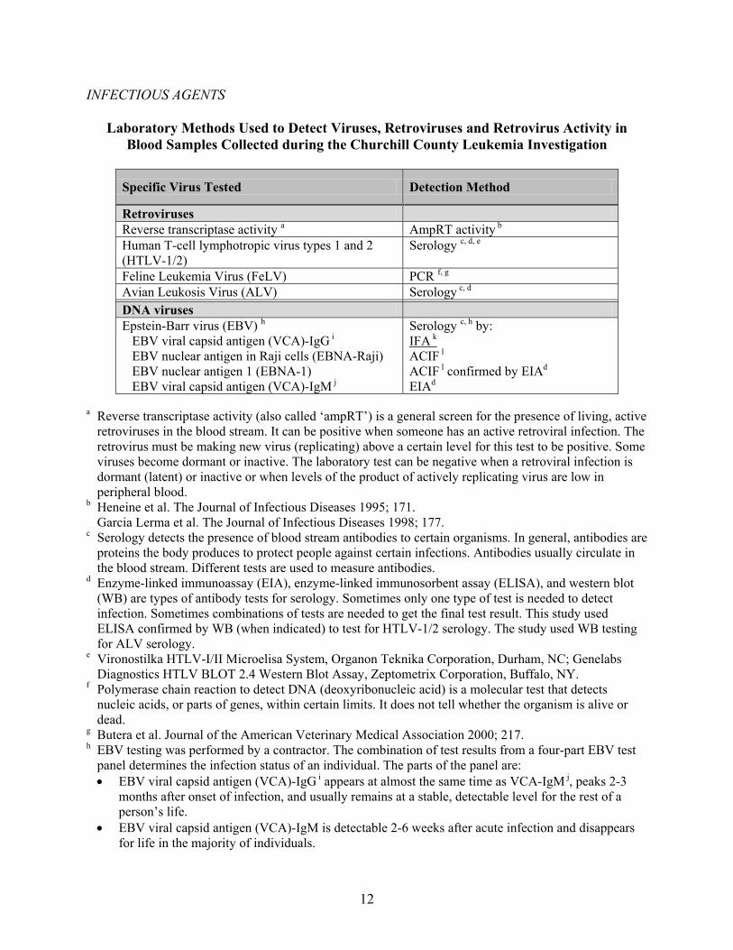
INFECTIOUS AGENTS
Laboratory Methods Used to Detect Viruses, Retroviruses and Retrovirus Activity in Blood Samples Collected during the Churchill County Leukemia Investigation
Specific Virus Tested Detection Method
Retroviruses Reverse transcriptase activity a AmpRT activity b Human T-cell lymphotropic virus types 1 and 2 (HTLV-1/2)
Serology c, d, e
Feline Leukemia Virus (FeLV) PCR f, g Avian Leukosis Virus (ALV) Serology c, d DNA viruses Epstein-Barr virus (EBV) h EBV viral capsid antigen (VCA)-IgG i EBV nuclear antigen in Raji cells (EBNA-Raji) EBV nuclear antigen 1 (EBNA-1) EBV viral capsid antigen (VCA)-IgM j
c, h by: Serology IFA k ACIF l
ACIF l confirmed by EIAd EIAd
a Reverse transcriptase activity (also called ‘ampRT’) is a general screen for the presence of living, active
retroviruses in the blood stream. It can be positive when someone has an active retroviral infection. The retrovirus must be making new virus (replicating) above a certain level for this test to be positive. Some viruses become dormant or inactive. The laboratory test can be negative when a retroviral infection is dormant (latent) or inactive or when levels of the product of actively replicating virus are low in peripheral blood.
b Heneine et al. The Journal of Infectious Diseases 1995; 171. Garcia Lerma et al. The Journal of Infectious Diseases 1998; 177. c Serology detects the presence of blood stream antibodies to certain organisms. In general, antibodies are
proteins the body produces to protect people against certain infections. Antibodies usually circulate in the blood stream. Different tests are used to measure antibodies.
d Enzyme-linked immunoassay (EIA), enzyme-linked immunosorbent assay (ELISA), and western blot (WB) are types of antibody tests for serology. Sometimes only one type of test is needed to detect infection. Sometimes combinations of tests are needed to get the final test result. This study used ELISA confirmed by WB (when indicated) to test for HTLV-1/2 serology. The study used WB testing for ALV serology.
e Vironostilka HTLV-I/II Microelisa System, Organon Teknika Corporation, Durham, NC; Genelabs Diagnostics HTLV BLOT 2.4 Western Blot Assay, Zeptometrix Corporation, Buffalo, NY.
f Polymerase chain reaction to detect DNA (deoxyribonucleic acid) is a molecular test that detects nucleic acids, or parts of genes, within certain limits. It does not tell whether the organism is alive or dead.
g Butera et al. Journal of the American Veterinary Medical Association 2000; 217. h EBV testing was performed by a contractor. The combination of test results from a four-part EBV test
panel determines the infection status of an individual. The parts of the panel are: • EBV viral capsid antigen (VCA)-IgG i appears at almost the same time as VCA-IgM j, peaks 2-3
months after onset of infection, and usually remains at a stable, detectable level for the rest of a person’s life.
• EBV viral capsid antigen (VCA)-IgM is detectable 2-6 weeks after acute infection and disappears for life in the majority of individuals.
12

13
• EBV nuclear antigen 1 (EBNA-1) is the dominant one of six proteins in the Epstein-Barr complex of nuclear antigens; antibodies to EBNA-1 first appear 10-12 weeks after acute EBV infection and so give a longer interval during which to detect acute infection than do IgM antibodies. Once the titer (level) of antibodies to EBNA-1 is ≥20 (about 3 months after onset of infection), >95% of individuals no longer have detectable VCA-IgM antibody.
• EBV nuclear antigen in Raji cells (EBNA-Raji) is the reference source of Epstein-Barr nuclear antigens.
i Immunoglobulin G (IgG) is a type of antibody in the blood stream. It usually indicates past infection and can be measured for years after an infection. IgG antibodies can also be measured during some persistent infections.
j Immunoglobulin M (IgM) is a type of antibody in the blood stream. It is usually detected during or shortly after acute infection then disappears over time.
k Indirect immunofluorescence assay (IFA) is a type of test used to detect antibodies in the blood stream or serum.
l Anticomplement immunofluorescence (ACIF) is a test used to detect specific proteins produced by the body during certain infections.