biobran-augmented maturation of human monocyte-derived
TRANSCRIPT
89
BioBran-augmented maturation of human monocyte-derived dendritic cells
D. CHOLUJOVA, J. JAKUBIKOVA, J. SEDLAK*
Laboratory of Tumor Immunology, Cancer Research Institute, Slovak Academy of Sciences, Bratislava, Slovak Republic, e-mail:[email protected]
Received September 17, 2008
BioBran, enzymatically modified arabinoxylan from rice bran was tested for its possible effects on in vitro maturation ofhuman dendritic cells (DC). Immature DC (iDC) derived from plastic-adhered, IL-4 and GM-CSF treated peripheral monocytes(Mo) were further cultured with cytokine maturation mix 1 (CMM1; TNF-α, IL-1β and IL-6) or CMM2 (LPS and IFN-γ) toinduce their maturation into mature DC (matDC1 or matDC2, respectively). Different concentrations of BioBran (10, 100, 400and 1000 μg/ml) were applied in the presence or absence of relevant CMM to assess the effects of BioBran on DC maturationprocesses. BioBran induced maturation of iDC, as these cells cultured with IL-4/GM-CSF/BioBran down-regulated CD14 andCD1a antigens on cell surface and significantly increased expression of maturation marker CD83. The increase of surfacedensity of costimulatory molecules CD80 and CD86 on iDC in the presence of BioBran was also observed. In addition,BioBran induced functional maturation of iDC, confirmed by decreased endocytic activity of iDC. Furtheremore, BioBranenhanced maturation potential of cytokine mixes, as both matDC1 and matDC2 exposed to BioBran completely lost CD14 andupregulated CD83, CD80 and CD86 antigens, in comparison to DC matured with the relevant CMM alone. BioBran alsoincreased CD123 antigen expression on all DC subsets. Interestingly, matDC2 matured in the presence of BioBran (400μg/ml)expressed higher levels of CD123 and lower levels of CD11c cell surface antigens, the phenotype represented by CD11cdim
CD123bright plasmacytoid DC population. These data demonstrate that BioBran is a potent enhancer of DC maturation andsuggest that BioBran might be a useful agent to create the environment that favours DC maturation.
Key words: Dendritic cell, maturation, BioBran, buffy coat
* Corresponding author
Dendritic cells are professional antigen-presenting cells (APC)that play the central role in initiation and regulation of immuneresponses since they control both the innate (NK cells, NKT cells,γδ Tcells) and the adaptive (T and B cells) arm of immunity [1].DC include subsets of cells with different characteristics that arederived from bone marrow progenitor cells. In humans, myeloidand lymphoid precursors can give rise to CD11cbrightCD123dim
myeloid dendritic cells (mDC) or CD11cdimCD123bright plasma-cytoid dendritic cells (pDC), respectively [2]. Peripheral tissueantigen-loaded DC migrate through the afferent lymphatics intothe draining lymph nodes where they present processed proteinand lipid antigens to T cells via both classical (major histocom-patibility complex (MHC) class I and class II) and non-classicalantigen-presenting molecules. Nonactivated iDC present anti-gens to T cells which in the absence of appropriate costimulationleads to tolerance [3, 4]. Upon encounter with pathogens or other‘danger’-associated stimuli, such as cytokines, products of dam-
aged tissues or innate lymphocytes, DC undergo a process ofactivation and maturation, wherein they loose ability to captureAg, but acquire the capacity to activate immunity. This processof maturation includes changes in morphology and motility, trans-location of MHC to the cell surface, increased expression ofcostimulatory molecules (CD80/CD86 and CD40), and produc-tion of cytokines that determine the class of immune response byselectively polarizing the development of effector T cells [5].
Due to their ability to initiate, link and regulate both innateand adaptive immunity, DC represent a potential tool for cancerimmunotherapy. Both mDC and pDC were found to infiltrateseveral types of human tumors but most of the tumor-infiltratingDC were immature DC [6]. It was also shown that several tumor-derived factors lead to inhibition of DC maturation, includingIL-10, VEGF, TGF-β, and PGE2 [7]. DC encountered by tumorantigen in the absence of pro-maturation environment remainsin immature or semi-mature state and a tolerogenic response maybe generated [4]. Therefore, promoting DC maturation was pro-posed as a therapeutic objective to counter tumor-driven toleranceand stimulate anti-tumor immunity.
NEOPLASMA 56, 2, 2009

90 D. CHOLUJOVA, J. JAKUBIKOVA, J. SEDLAK
BioBran is a food supplement which is obtained by reactingrice bran hemicellulose with multiple carbohydrate hydrolyzingenzymes from the Shiitake mushrooms. The active componentof BioBran is an arabinoxylan. Previous research has suggestedthat the product enhances natural killer (NK) cell activity in vitroand in vivo [8, 9]. The objective of this study was to investigatethe possible modulatory effects of BioBran on in vitro differen-tiation and maturation of human DC.
Materials and methods
Reagents. The following monoclonal antibodies (mAbs) wereused in this study: fluorescein isothiocyanate (FITC)-conjugatedanti-human CD11c (IgG1) mouse mAb, phycoerythrin (PE)-conjugated anti-human CD14 (IgG1) mouse mAb, PEanti-human CD45 (IgG1) mouse mAb, PE anti-human CD123(IgG1) mouse mAb, PE anti-human CD80 (IgG1) mouse mAb,PE anti-human CD83 (IgG2bκ) mouse mAb, PE anti-humanCD86 (IgG2bκ) mouse mAb, PE anti-human CD1a (IgG1κ)mouse mAb, and energy-coupled dye (ECD) anti-human HLA-DR (IgG1) mouse mAb. All mAbs and isotype-matchedcontrols were purchased from Immunotech Beckman Coulter.We also used the following cytokines and ligands: recombi-nant human granulocyte-macrophage colony-stimulating factor(rhuGM-CSF; Santa Cruz Biotechnology), recombinant humaninterleukin-4 (rhuIL-4; Santa Cruz), recombinant humaninterleukin-1β (IL-1β; Biomol), recombinant humaninterleukin-6 (IL-6; Santa Cruz), tumor necrosis factor (TNF-α; Santa Cruz), lipopolysacharide (LPS; Sigma-Aldrich), andinterferon- γ (IFN-γ; Bender MedSystem).
BioBran - denaturated hemicellulose obtained from ricebran that is treated enzymatically with an extract from Shiitakemushrooms (Lentinus edodes). The main chemical structureand an active component is an arabinoxylan with a xylose inits main chain and an arabinose polymer in its side chain.Biobran was provided by Daiwa Pharmaceuticals Co. Ltd,Tokyo, Japan. BioBran stock solution was prepared by dis-solving 1 dose of BioBran 1000 in distilled H2O at finalconcentration of 10 mg/ml.
Isolation of peripheral blood monocytes. Monocytes wereisolated from buffy coats of healthy donors (National Trans-fusion Service, Bratislava) by Pancol density gradientcentrifugation (1.077 g/ml, PAN-Biotech, Germany). Themononuclear cells from the interface were collected, washedtwice with PBS and once in AIM-V complete medium (Gibco-BRL, Paisley, UK), and then allowed to adhere in the plastic6-well plates for 2 hours at 37 °C, 5% CO2. The nonadherentcells were gently removed by washing three times with PBSand once with AIM-V medium.
Dendritic cell differentiation and maturation. Plastic-adheredmonocytes were cultured in 6-well plates for 6 days in AIM-Vcomplete medium supplemented with GM-CSF (1000 IU/ml)and IL-4 (1000 IU/ml) to generate iDC. The fresh medium withcytokines was added to cell cultures every second day. On day7 the medium was exchanged and cell were cultured in com-
plete AIM-V medium containing GM-CSF (1,000 U/mL) andIL-4 (1,000 U/mL) in the presence or absence of maturationstimuli for a further 2 days. Cytokine maturation mix 1 (CMM1;TNF-α (10 ng/ml), IL-1β (10 ng/ml) and IL-6 (10 ng/ml)), andcytokine maturation mix 2 (CMM2; LPS (250 ng/ml) and IFN-γ (1000 IU/ml)) with or without addition of BioBran (10, 100,400 and 1000 μg/ml) were used as stimuli for maturation ofDC into matDC1 and matDC2, respectively [10].
Immunophenotypic analysis of cells. Monocyte-derived DCwere collected, washed and resuspended in PBS 0.2% BSA(Applichem). Cell aliquots (50μl/well) were pipetted into 96-well V-bottomed microplate and incubated with 2 μl ofrelevant fluorochrome-conjugated mAbs for 30 min at 37 oCin the dark. At the end of incubation, samples were transferedinto cytometric test tubes and 2 μl of 7-amino-actinomycinD (7-AAD; stock solution 1 mg/ml in PBS) in 300 μl of PBSwere added to label nonviable cells. Samples were incubatedon ice for 10 min, protected from light and analysed usinga Coulter Epics Altra flow cytometer.
Endocytosis assay with FITC-dextran. The efficiency ofendocytosis was evaluated as cellular uptake of fluoresceinisothiocyanate (FITC)-conjugated dextran by monocytes, im-mature and mature DC (matDC1, matDC2) cultured in theabsence or presence of different concentrations of BioBran (100,400 and 1000 μg/ml). For the endocytosis assay, 2 x 10⁵ cellsper sample were incubated in AIM-V medium with 1 mg/ml ofFITC-dextran (Mr 40,000; Sigma) for 60 min either at 37 °C(for uptake) or 4 °C (negative control). After incubation cellswere washed three times with ice-cold PBS. Additionally, trypanblue (0.2%) was used for quenching surface-bound FITC fluo-rescence. The accumulation of FITC-dextran within the cellswas quantitated by flow cytometry. Nonviable cells and con-taminating lymphocytes were excluded according to their scatterproperties and 7-AAD staining. The values were calculated asa change in percentage of green fluorescence positivity betweencell samples incubated at 37 °C and 4 °C, respectively.
Flow cytometric analysis. Samples were analyzed using anEpics Altra (Beckman Coulter) four-colour flow cytometerequipped with an argon laser (15 mV) source operating at 488nm. The emission of fluorochromes was recorded through spe-cific band-pass fluorescence filters: FITC, 525 nm (FL1); PE,575 nm (FL2); ECD, 610 nm (FL3); 7-AAD; 675 nm (FL4).Data were analysed using WinMDI version 2.7 software (J.Trotter, Scripps Research Institute, La Jolla, CA). Forimmunophenotyping, gating was done on side scatter (SSC;ordinate) versus log-scale orange fluorescence of ECD dye(HLA-DR positivity; abscissa), to separate residual lympho-cytes. Nonviable (7-AAD-positive) cells were excluded fromanalysis. An average of 20 000 cells were collected per sample.
Results
Phenotypic characterization of Mo and Mo-derived DC atdifferent stages of in vitro maturation. The phenotypic analy-sis was performed to confirm the differentiation and
91BIOBRAN-INDUCED DC MATURATION
maturation status of DC. Human peripheral CD14+ Mo wereisolated by plastic adherence from buffy coats of healhy do-nors and cultured in the presence of GM-CSF and IL-4 for6 days to induce their differentiation into iDC. Additional 2-
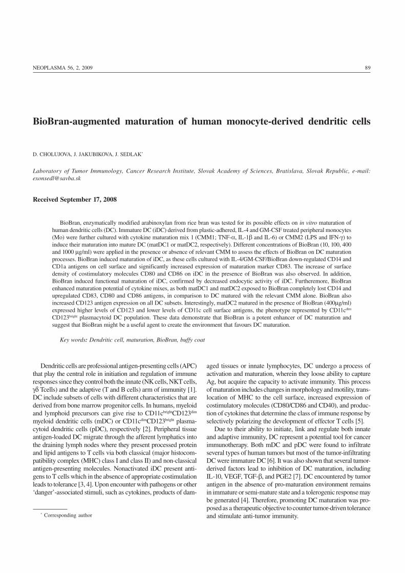
day treatment of iDC with two different maturation cytokinecocktails (CMM1, CMM2) was used to obtain 2 distinct popu-lations of mature DC, termed matDC1 and matDC2. As shownin Fig. 1, Mo prominently expressed CD14 and HLA-DR. They
Figure 1. The phenotype of Mo and DC at different stages of in vitro maturation. Human peripheral Mo were analyzed by flow cytometry and werecultured with GM-CSF and IL-4 (1000 UI/ml) for 6 days to obtain iDC. The iDC were cultured in the presence of GM-CSF and IL-4 for additional2 days either with CMM1 or CMM2 to induce their maturation into matDC1 and matDC2, respectively. At the end, iDC, matDC1 and matDC2subsets were harvested, stained with fluorochrome-conjugated mAbs against the indicated cell surface antigens, and analyzed using flow cytometry.This presented phenotype of Mo and DC is typical of more than five cell cultivations.
92 D. CHOLUJOVA, J. JAKUBIKOVA, J. SEDLAK
also showed low CD86 positivity, but almost completelylacked the expression of CD1a, CD83 and CD80. In contrastto Mo, iDC down-regulated CD14, but strongly expressedantigen-presenting molecule CD1a. They were positive for
expression of costimulatory molecules CD80 and CD86, andshowed marginal CD83 expression. Mature DC (both matDC1and matDC2), extensively increased the expression of matu-ration marker CD83 and costimulatory molecules CD80(B7-1) and CD86 (B7-2).
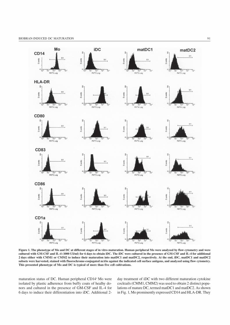
BioBran modulates expression of differentiation and matu-ration markers on DC. To study the effect of BioBran onmaturation of DC, iDC were treated with different concentra-tion (10, 100, 400 and 1000 μg/ml) of BioBran in the presenceor absence of relevant maturation mix (Fig. 2). The surfaceexpression of CD14, CD1a and CD83 antigens on iDC, matDC1and matDC2 was examined by flow cytometry on HLA-DR+gated cells. BioBran down-regulated the expression of mono-cyte marker CD14 (Fig. 2A) and antigen-presenting moleculeCD1a (Fig. 2B) on the surface of iDC, but extensively increasedthe expression of CD83 (Fig. 2C), a marker of mature DC. Simi-lar to iDC, down-regulation of CD14 and CD1a andupregulation of CD83 were observed in both mature DC popu-lations (matDC1 and matDC2) with increasing concentrationsof BioBran used.
BioBran decreases endocytic activity of iDC. BioBran wastested for its ability to affect the endocytic activity of Mo andMo-derived DC. Endocytosis was evaluated by the uptake ofFITC-conjugated dextran at 37 °C, with negative control in-cubated at 4 °C. The untreated iDC and Mo showed high levelof endocytic activity (73% and 48% of FITC-dextran-posi-tive cells, respectively), whereas both matDC1 and matDC2showed clear reduction of dextran-FITC uptake (14.9% and5.6%, respectively; Fig. 3). The significant decrease ofendocytic activity of iDC cultivated in the the presence ofBioBran was ascertained, when compared to control iDC, and
Figure 2. The effect of BioBran on the expression of differentiation/maturation antigens on DC subsets. The surface expression of CD14,CD1a and DC maturation marker CD83 was examined by flowcytometry on HLA-DR positively gated iDC, matDC1 and matDC2. Thepercentage of cell positivity is shown and is representative of threeindependent experiments.
Figure 3. Endocytic activity of Mo and DC populations. Cells wereincubated with FITC-labeled dextran (1mg/ml) for 60 min at 37 °C and4 °C (negative control). Trypan blue was used for quenching of surface-bound green fluorescence. Endocytosis was assessed by flow cytometryas the percentage of dextran positive cells. Results shown are representativefrom three similar experiments performed on cells from different donors.
93BIOBRAN-INDUCED DC MATURATION
this effect of BioBran was dose-dependent (27.7% for BioBran100 μg/ml, 17.7% for BioBran 400 μg/ml, and 14.4% of dex-tran-positive cells for BioBran 1000 μg/ml; Fig. 3). Similarresults were observed with monocytes, when 100 μg/ml ofBioBran decreased the percentage of FITC-dextran positivecells to 12.7%.
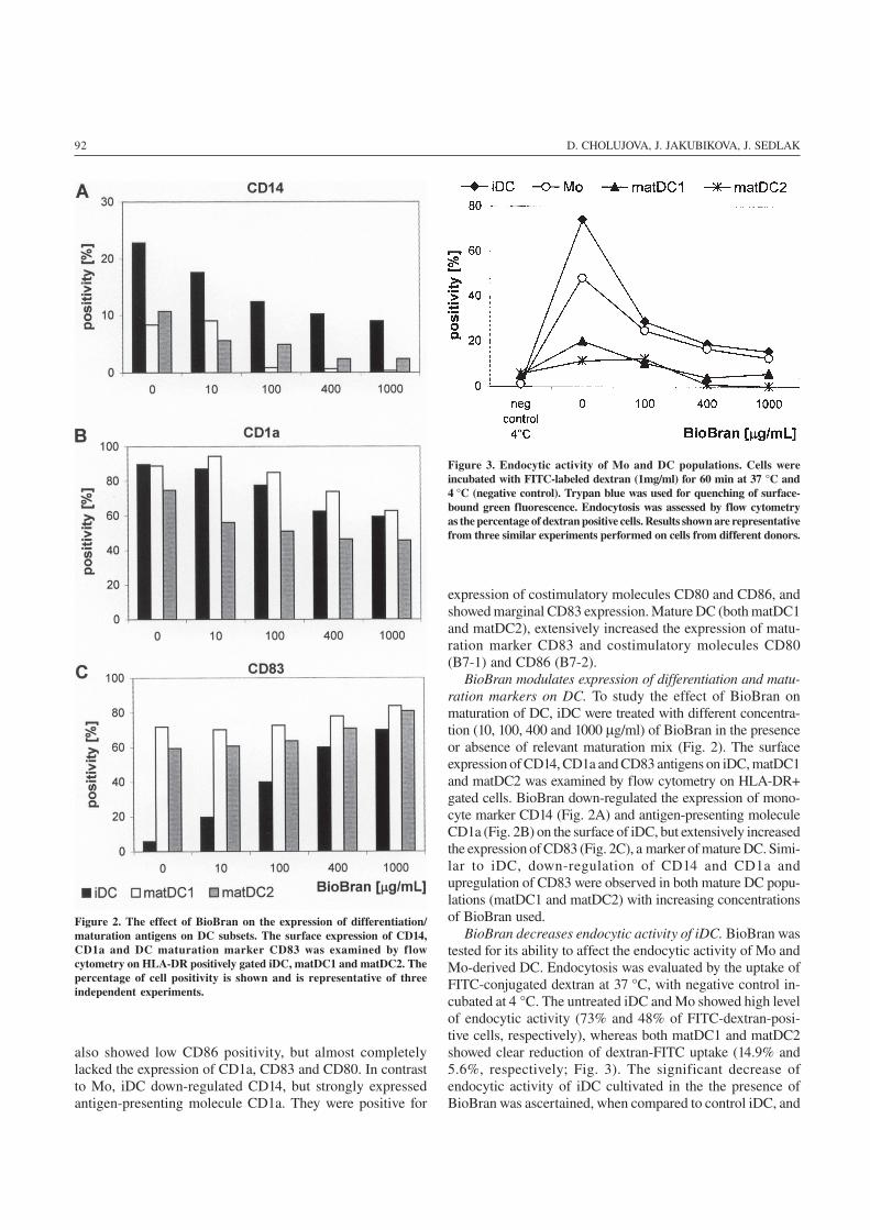
BioBran increases surface expression of costimulatorymolecules CD80 and CD86. In order to assess the effect ofBioBran on the expression of costimulatory molecules CD80and CD86, Mo-derived iDC were treated for 2 days withBioBran (10, 100, 400 μg/ml) or matured with CMM 1 orCMM2 in the presence of BioBran. The cell surface expres-sion of CD80 and CD86 on the iDC, matDC1 and matDC2were studied by flow cytometric analysis on HLA-DR posi-tively gated cells (Fig. 4). Control matDC2 matured in thepresence of LPS and IFN-γ showed the highest levels of CD80(MFI~296), compared with control matDC1 matured in the
presence of IL-1β, IL-6, and TNF-α (MFI~201) or immatureDC (MFI~156). Similarly, matDC2 expressed the highest lev-els of CD86 (MFI~829) compared with matDC1 (MFI~506)and iDC (MFI~170). BioBran increased the mean of fluores-cence intensity of costimulatory molecules CD80 and CD86in dose dependent manner on all DC subsets. In the presenceof BioBran in concentration of 400 μg/ml, the MFI of CD80increased to 368 for mDC, 319 for matDC1 and 260 for iDC.Even more BioBran (400 μg/ml) increased markedly CD86antigen (MFI~1248 for matDC2, 1027 for matDC1 and 845for iDC).
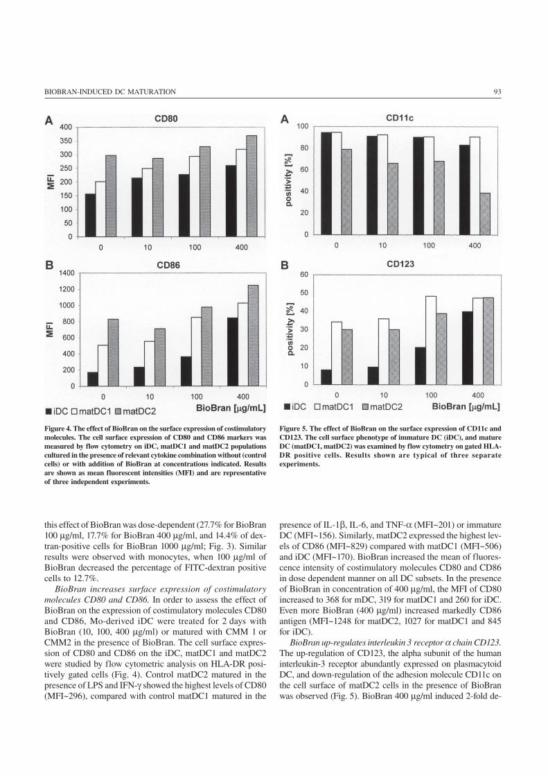
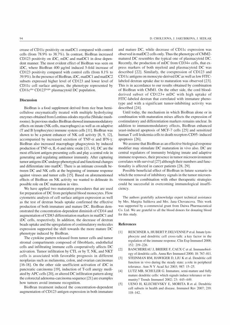
BioBran up-regulates interleukin 3 receptor α chain CD123.The up-regulation of CD123, the alpha subunit of the humaninterleukin-3 receptor abundantly expressed on plasmacytoidDC, and down-regulation of the adhesion molecule CD11c onthe cell surface of matDC2 cells in the presence of BioBranwas observed (Fig. 5). BioBran 400 μg/ml induced 2-fold de-
Figure 4. The effect of BioBran on the surface expression of costimulatorymolecules. The cell surface expression of CD80 and CD86 markers wasmeasured by flow cytometry on iDC, matDC1 and matDC2 populationscultured in the presence of relevant cytokine combination without (controlcells) or with addition of BioBran at concentrations indicated. Resultsare shown as mean fluorescent intensities (MFI) and are representativeof three independent experiments.
Figure 5. The effect of BioBran on the surface expression of CD11c andCD123. The cell surface phenotype of immature DC (iDC), and matureDC (matDC1, matDC2) was examined by flow cytometry on gated HLA-DR positive cells. Results shown are typical of three separateexperiments.
94 D. CHOLUJOVA, J. JAKUBIKOVA, J. SEDLAK
crease of CD11c positivity on matDC1 compared with controlcells (from 78.9% to 38.7%). In contrast, BioBran increasedCD123 positivity on iDC, mDC and matDC1 in dose depen-dent manner. The most evident effect of BioBran was seen oniDC, where BioBran 400 μg/ml induced 5-fold increase ofCD123 positivity compared with control cells (from 8.1% to39.9%). In the presence of BioBran, iDC, matDC1 and matDC2subsets expressed higher level of CD123 and lower level ofCD11c cell surface antigens, the phenotype represented byCD11cdim CD123bright plasmacytoid DC population.
Discussion
BioBran is a food supplement derived from rice bran hemi-cellulose enzymatically treated with multiple hydrolyzingenzymes obtained from Lentinus edodes mycelia (Shitake mush-rooms). In previous studies BioBran showed immunomodulatoryeffects on innate (NK cells, macrophages) as well as on adaptive(T and B lymphocytes) immune system cells [11]. BioBran wasshown to be a potent enhancer of NK cell activity [8, 9, 12],accompanied by increased secretion of TNF-α and IFN-γ.BioBran also increased macrophage phagocytosis by inducedproduction of TNF-α, IL-6 and nitric oxide [13, 14]. DC are themost efficient antigen-presenting cells and play a central role ingenerating and regulating antitumor immunity. After capturingtumor antigens iDC undergo phenotypical and functional changesand differentiate into matDC. There is an intimate crosstalk be-tween DC and NK cells at the beginning of immune responseagainst viruses and tumor cells [15]. Based on aforementionedeffects of BioBran on NK activity we wanted to delineate itspossible role on DC maturation in vitro.
We have applied two maturation procedures that are usedfor preparation of DC from peripheral blood monocytes. Flowcytometric analysis of cell surface antigen expression as wellas the test of dextran beads uptake confirmed the effectiveproduction of both immature and mature DC. BioBran dem-onstrated the concentration-dependent diminish of CD14 andaugmentation of CD83 differentiation markers in matDC1 andiDC cells, respectively. In addition, the decrease of dextranbeads uptake and the upregulation of costimulatory moleculesexpression supported the shift towards the more mature DCphenotype induced by BioBran.
The cytokine pattern released from tumor cells and tumorstromal compartments composed of fibroblasts, endothelialcells and infiltrating immune cells cooperatively affects DCactivation. Tumor infiltration by CTL or by T, NK, and NKTcells is associated with favorable prognosis in differentneoplasias such as melanoma, colon, and ovarian carcinomas[16-18]. On the other side unefficient activation of iDC inpancreatic carcinoma [19], induction of T-cell anergy medi-ated by APC cells [20], or altered DC infiltration pattern alongthe colorectal adenoma-carcinoma sequence [21] are exampleshow tumors avoid immune recognition.
BioBran treatment induced the concentration-dependentaugmentation of CD123 antigen expression in both immature
and mature DC, while decrease of CD11c expression wasobserved in matDC2 cells only. Thus the phenotype of CMM2-matured DC resembles the typical one of plasmacytoid DC.Recently, the production of mDC from CD34+ cells, that ex-press markers of both myeloid and plasmacytoid DC wasdescribed [22]. Similarly, the coexpression of CD123 andCD11c antigens on monocyte-derived DC as well as low FITC-labeled dextran uptake due to maturation was observed [23].This is in accordance to our results obtained by combinationof BioBran with CMM1. On the other side, the cord blood-derived subset of CD123+ mDC with high uptake ofFITC-labeled dextran that correlated with immature pheno-type and with a significant tumor-inhibiting activity wasdescribed [24].
Until today, the mechanism in which BioBran alone or incombination with maturation mixes affects the expression ofcostimulatory and differentiation markers remains unclear. Inaddition to immunomodulatory effects, BioBran enhancedyeast-induced apoptosis of MCF-7 cells [25] and sensitizedhuman T cell leukemia cells to death receptors CD95- inducedapoptosis [26].
We assume that BioBran as an effective biological responsemodifier may stimulate DC maturation in vivo also. DC arecentral regulators of immunity bridging innate and adaptiveimmune responses, their presence in tumor microenvironmentcorrelates with survival [27] although their numbers and func-tionality is affected in cancer patients [28, 29].
Possible beneficial effect of BioBran in future scenario inwhich the removal of inhibitory signals in the tumor microen-vironment in combination with other therapeutic strategiescould be successful in overcoming immunological insuffi-ciency.
The authors gratefully acknowledge expert technical assistanceby Mrs. Margita Sulikova and Mrs. Jana Chovancova. This workwas supported by a commercial grant from Daiwa PharmaceuticalCo. Ltd. We are grateful to all the blood donors for donating bloodfor this study.
References
[1] RESCHNER A, HUBERT P, DELVENNE P et al. Innate lym-phocyte and dendritic cell cross-talk: a key factor in theregulation of the immune response. Clin Exp Immunol 2008;152: 219–226.
[2] BANCHEREAU J, BRIERE F, CAUX C et al. Immunobiol-ogy of dendritic cells. Annu Rev Immunol 2000; 18: 767–811.
[3] STEINMAN RM, HAWIGER D, LIU K et al. Dendritic cellfunction in vivo during the steady state: a role in peripheraltolerance. Ann N Y Acad Sci 2003; 987: 15–25.
[4] LUTZ MB, SCHULER G. Immature, semi-mature and fullymature dendritic cells: which signals induce tolerance or im-munity? Trends Immunol 2002; 23: 445–449.
[5] UENO H, KLECHEVSKY E, MORITA R et al. Dendriticcell subsets in health and disease. Immunol Rev 2007; 219:118–142.
95BIOBRAN-INDUCED DC MATURATION
[6] PERROT I, BLANCHARD D, FREYMOND N et al. Den-dritic cells infiltrating human non-small cell lung cancerare blocked at immature stage. J Immunol 2007; 178: 2763–2769.
[7] RABINOVICH GA, GABRILOVICH D, SOTOMAYOREM. Immunosuppressive strategies that are mediated by tu-mor cells. Annu Rev Immunol 2007; 25: 267–296.
[8] GHONEUM M, JEWETT A. Production of tumor necrosisfactor-alpha and interferon-gamma from human peripheralblood lymphocytes by MGN-3, a modified arabinoxylan fromrice bran, and its synergy with interleukin-2 in vitro. CancerDetect Prev 2000; 24: 314–324.
[9] GHONEUM M, ABEDI S. Enhancement of natural killercell activity of aged mice by modified arabinoxylan ricebran (MGN-3/Biobran). J Pharm Pharmacol 2004; 56:1581–1588.
[10] HOCHREIN H, O’KEEFFE M, LUFT T et al. Interleukin(IL)-4 is a major regulatory cytokine governing bioactive IL-12 production by mouse and human dendritic cells. J ExpMed 2000; 192: 823–833.
[11] GHONEUM M. Anti-HIV activity in vitro of MGN-3, anactivated arabinoxylane from rice bran. Biochem Biophys ResCommun 1998; 243: 25–29.
[12] KIM HY, KIM JH, YANG SB et al. A polysaccharide ex-tracted from rice bran fermented with Lentinus edodesenhances natural killer cell activity and exhibits anticancereffects. J Med Food 2007; 10: 25–31.
[13] GHONEUM M, MATSUURA M. Augmentation of macroph-age phagocytosis by modified arabinoxylan rice bran(MGN-3/biobran). Int J Immunopathol Pharmacol 2004; 17:283–292.
[14] GHONEUM M, MATSUURA M, GOLLAPUDI S. Modi-fied arabinoxylan rice bran (MGN3/Biobran) enhancesintracellular killing of microbes by human phagocytic cellsin vitro. Int J Immunopathol Pharmacol 2008; 21: 87–95.
[15] MORETTA L, FERLAZZO G, BOTTINO C et al. Effectorand regulatory events during natural killer-dendritic cell in-teractions. Immunol Rev 2006; 214: 219–228.
[16] GALON J, COSTES A, SANCHEZ-CABO F et al. Type,density, and location of immune cells within human colorec-tal tumors predict clinical outcome. Science 2006; 313:1960–1964.
[17] HAANEN JB, BAARS A, GOMEZ R et al. Melanoma-spe-cific tumor-infiltrating lymphocytes but not circulatingmelanoma-specific T cells may predict survival in resectedadvanced-stage melanoma patients. Cancer Immunol Immu-nother 2006; 55: 451–458.
[18] ZHANG L, CONEJO-GARCIA JR, KATSAROS D et al.Intratumoral T cells, recurrence, and survival in epithelialovarian cancer. N Engl J Med 2003; 348: 203–213.
[19] BELLONE G, CARBONE A, SMIRNE C et al. Cooperativeinduction of a tolerogenic dendritic cell phenotype by cytok-ines secreted by pancreatic carcinoma cells. J Immunol 2006;177: 3448–3460.
[20] CUENCA A, CHENG F, WANG H et al. Extra-lymphaticsolid tumor growth is not immunologically ignored and re-sults in early induction of antigen-specific T-cell anergy:dominant role of cross-tolerance to tumor antigens. CancerRes 2003; 63: 9007–9015.
[21] YUAN A, STEIGEN SE, GOLL R et al. Dendritic cell infil-tration pattern along the colorectal adenoma-carcinomasequence. APMIS 2008; 116: 445–456.
[22] WARD KA, STEWART LA, SCHWARER AP. CD34+-de-rived CD11c+ + + BDCA-1+ + CD123+ + DC: expansion ofa phenotypically undescribed myeloid DC1 population foruse in adoptive immunotherapy. Cytotherapy 2006; 8: 130–140.
[23] HO CS, MUNSTER D, PYKE CM et al. Spontaneous gener-ation and survival of blood dendritic cells in mononuclearcell culture without exogenous cytokines. Blood 2002; 99:2897–2904.
[24] SHI J, IKEDA K, MAEDA Y et al. Identification of CD123(+)myeloid dendritic cells as an early-stage immature subset withstrong tumoristatic potential. Cancer Lett 2008; 270:19–29.
[25] GHONEUM M, GOLLAPUDI S. Modified arabinoxylan ricebran (MGN-3/Biobran) enhances yeast-induced apoptosis inhuman breast cancer cells in vitro. Anticancer Res 2005; 25:859–870.
[26] GHONEUM M, GOLLAPUDI S. Modified arabinoxylan ricebran (MGN-3/Biobran) sensitizes human T cell leukemia cellsto death receptor (CD95)-induced apoptosis. Cancer Lett2003; 201: 41–49.
[27] NAGORSEN D, VOIGT S, BERG E et al. Tumor-infiltratingmacrophages and dendritic cells in human colorectal cancer:relation to local regulatory T cells, systemic T-cell responseagainst tumor-associated antigens and survival. J Transl Med2007; 5: 62–69.
[28] MARTIN-AYUSO M, ALMEIDA J, PEREZ-ANDRES M etal. Peripheral blood dendritic cell subsets from patients withmonoclonal gammopathies show an abnormal distribution andare functionally impaired. Oncologist 2008; 13: 82–92.
[29] KOVAROVA L, BUCHLER T, POUR L et al. Dendritic cellcounts and their subsets during treatment of multiple myelo-ma. Neoplasma 2007; 54: 297–303.