bioadhesion: a review of concepts and...
TRANSCRIPT
Phil. Trans. R. Soc. A 370, 2321–2347doi:10.1098/rsta.2011.0483
Bioadhesion: a review of concepts andapplications
BY MANUEL L. B. PALACIO* AND BHARAT BHUSHAN∗
Nanoprobe Laboratory for Bio- and Nanotechnology and Biomimetics,The Ohio State University, Columbus, OH 43210, USA
Bioadhesion refers to the phenomenon where natural and synthetic materials adhereto biological surfaces. An understanding of the fundamental mechanisms that governbioadhesion is of great interest for various researchers who aim to develop newbiomaterials, therapies and technological applications such as biosensors. This reviewpaper will first describe various examples of the manifestation of bioadhesion along withthe underlying mechanisms. This will be followed by a discussion of some of the methodsfor the optimization of bioadhesion. Finally, nanoscale and macroscale characterizationtechniques for the efficacy of bioadhesion and the analysis of failure surfaces are described.
Keywords: bioadhesion; bioadhesives; cell adhesion; mucoadhesion
1. Introduction
The term ‘bioadhesion’ refers to widely diverse phenomena, which all involvethe adherence of materials (natural or synthetic) to biological surfaces. As aninterface phenomenon, bioadhesion is similar to conventional adhesion, exceptfor the special characteristics of biological organisms and surfaces. As such, thisterm covers the adhesive properties of both synthetic components and the naturalsurfaces (such as cells). Bioadhesion could also refer to the use of bioadhesives tobond two surfaces together, which is relevant in drug delivery, dental and surgicalapplications. Therefore, the wide interest in bioadhesion research is due to itsimplications for the development of new biomaterials, therapies and technologicalproducts such as biosensors [1–6].
A few examples illustrating various manifestations of bioadhesion are shownin figure 1. Pillar-directed cell growth is an area of interest for researchersinvestigating the role of substrate micro-roughness on cell behaviour in thedevelopment of new biomaterials. In figure 1a, fibroblasts attach on top of thesilicon pillar array substrate, and then extend to reach the surrounding pillars [7].The height and spacing between pillars affect cell shape and thickness, whichin turn, has implications for biological functions, such as cell growth and geneexpression. Bioadhesion can also refer to the application of adhesives for biologicalinterfaces in clinical use. Figure 1b is an example of the application of a medicaladhesive. In this case, fibrin glue was used to adhere a surgical mesh on an
*Authors for correspondence ([email protected]; [email protected]).
One contribution of 10 to a Theme Issue ‘Biosensors: surface structures and materials’.
This journal is © 2012 The Royal Society2321
on July 16, 2018http://rsta.royalsocietypublishing.org/Downloaded from

2322 M. L. B. Palacio and B. Bhushan
fatty materialon bloodvessel wall
(e)
(a)
(b)
(c)
(d)
Figure 1. Examples of bioadhesion. (a) Cell adhesion on a silicon-based micropillar array [7].(b) Example of a medical adhesive application where fibrin glue was used to attach a surgical meshafter a laparoscopic surgery procedure [8]. (c) Tooth enamel and dentin fracture and reattachmentof the tooth fragment using a dentin adhesive [9]. (d) Adhesion of a mussel on a glass slide,where multiple threads composed of the mussel adhesive protein were secreted by the animal toattach itself to the glass surface [10]. (e) Sketch depicting fat deposition on a blood vessel wall inatherosclerosis (adapted from Anonymous [11]). (Online version in colour.)
animal peritoneum after a laparoscopy [8]. Meanwhile, figure 1c shows the useof adhesives in dental applications. In this example, a dentin adhesive was usedto reattach a tooth fragment onto a fractured area [9].
Examples of naturally occurring bioadhesion are shown in figure 1d,e. Thephenomenon of mucoadhesion is illustrated in figure 1d, which shows theattachment of mussels to underwater surface, such as corals or ship surfaces.The foot of the animal extends from its shell to secrete a pad of the musseladhesive protein (MAP) on the underwater surface. It then retreats to the shellinterior, leaving a thread that secures the mussel to the pad. This process isrepeated multiple times, creating a strong bond [10]. The MAP has been used
Phil. Trans. R. Soc. A
on July 16, 2018http://rsta.royalsocietypublishing.org/Downloaded from

Bioadhesion: concepts and applications 2323
50 µm
Figure 2. Biofilm development on the inner surface of a polyurethane stent. Adapted fromTenke et al. [12].
as the model for the development of mucoadhesive materials. Lastly, a sketch isshown in figure 1e depicting the build-up of fats on the walls of animal arteries,which is part of the mechanism that leads to atherosclerosis [11].
The bioadhesion between materials can also be detrimental, and is referred toas biofouling. A biofilm is an aggregation of micro-organisms that form on a solidsubstrate, which may form during the implantation of biomaterials into the body.For humans and animals, the formation of biofilms on both living (cells) and non-living (stents and catheters, for example) surfaces has been identified as a cause ofdisease. Bacterial biofilms are known to cause diseases such as endocarditis, cysticfibrosis and other infections that are largely resistant to antibiotics. Figure 2shows an example of biofilm formation on a stent used for urological applications[12]. In other applications, dental plaque is a biofilm that can consist of morethan 500 bacterial strains [13].
This review paper is focused on the positive aspects of bioadhesion, and thus,will not discuss biofouling in detail. There are many different situations wherebioadhesion is beneficial and significant, such as cell adhesion, mucoadhesion andthe use of bioadhesives for surgical and dental applications. Examples from thesebroad applications will be cited in order to discuss mechanisms that controlthe bioadhesion process, and identify ways to optimize bioadhesion. Finally,nanoscale and macroscale experimental techniques to characterize bioadhesionwill be discussed.
2. Mechanisms that control bioadhesion
The factors that determine biological adhesion are diverse, and can be classifiedinto the effects of surface morphology, chemical interactions, physiological
Phil. Trans. R. Soc. A
on July 16, 2018http://rsta.royalsocietypublishing.org/Downloaded from

2324 M. L. B. Palacio and B. Bhushan
factors and physical–mechanical interactions. These aforementioned determinantsusually work in concert with one another in order to achieve adhesion betweenthe interfaces of interest.
(a) Surface morphology effects
Cell adhesion on synthetic biomaterial surfaces is widely studied owing to itsdirect implications for the design and clinical performance of body implants. Theadhesion of cells on biomaterials is an example that illustrates the importance ofthe micro/nanotopography of the substrate surface where the cells are cultured.During the implantation of a biomaterial into a living host, cells are not directlyattached to the biomaterial surface. Instead, the biomaterial is rapidly coatedwith a protein layer. Other proteins will displace the initial layer in a processknown as the ‘Vroman effect’. Various extracellular matrix (ECM) and serumproteins are involved, such as fibronectin, fibrinogen, albumin and vitronectin.The conformation of the adsorbed protein is partly determined by the morphologyof the biomaterial surface, which in turn, influences the cell adhesion andproliferation process [14–18].
Model synthetic surfaces have been used to investigate the effect of surfacemorphology on protein conformation and cell adhesion. These surfaces havewell-defined microscale or nanoscale topography, and are typically created usingtop-down (e.g. lithographic or dry etching methods) or bottom-up (self-assemblyprocesses or block copolymers) approaches [19–23].
Accounts vary on the optimal feature size required for effective celladhesion. Accounts of bone cell adhesion on patterned titanium indicate thatsubmicrometre (greater than 100 nm) features are better than nanometre-sized(less than 100 nm) features in facilitating cell adhesion [24]. Lehnert et al.[25] performed cell adhesion and spreading studies on patterns with distancesranging from 1 to 30 mm. They found that for patterns with small areas ofthe adhesive ECM proteins (0.1 mm2), pattern spacings larger than 5 mm showcellular adhesion, but do not support cell spreading. However, for larger dotdimensions (9 mm2), cells can effectively connect non-adhesive regions duringthe spreading process as long as the distances between features do not exceed25 mm. This variation in cell spreading as a function of the substrate geometryis shown in figure 3, where for 25 mm-spaced features, the spreading was limitedand the cells were observed to appear as either triangular, ellipsoidal or round. Inanother study, using lithographically created patterns with deposited fibronectin,it was found that cellular adhesion increases as the spacing between featuresincreases up to the optimal distance of 11 mm [26]. However, for cell adhesionon gold nanoparticles, it was found that for spacings less than 100 nm, closerpatterns (ca 60 nm) allowed for greater cell adhesion [20]. Even though theseliterature accounts are not definitive, it is apparent that dimensional thresholdsthat enhance cell adhesion are present.
(b) Chemical interactions
Various interactions between two chemically active surfaces (i.e. not inert)facilitate the bioadhesion process. Strong adhesion can occur if the two surfacesare capable of forming either covalent, ionic or metallic bonds. At the sametime, weaker forces, such as polar (dipole–dipole), hydrogen bonding or van
Phil. Trans. R. Soc. A
on July 16, 2018http://rsta.royalsocietypublishing.org/Downloaded from

Bioadhesion: concepts and applications 2325
(a) (b) (c)
( f )(e)(d )
10 µm 15 µm 20 µm
25 µm10 µm25 µm25 µm
hs <1 µm 2 µm
(g) (h) (i)
Figure 3. Fluorescence microscopy images showing the effect of substrate geometry on cell adhesionand spreading: (a) homogeneous substrate (hs), (b) 0.1 mm2 dots, approximately 1 mm apart,(c) 1 mm2 dots, 2 mm apart, (d) 9 mm2 dots, 10 mm apart, (e) 9 mm2 dots, 15 mm apart, (f ) 9 mm2
dots, 20 mm apart, (g–i) 9 mm2 dots, 25 mm apart (adapted from Lehnert et al. [25]). (Online versionin colour.)
der Waals interactions (induced dipoles) also aid in bonding the two surfaces[27–29]. A schematic of possible protein–solid surface interactions is shown infigure 4. Here, it is assumed that the surface is uniform and that there isonly one main type of interaction between the protein and the surface. Thestrength of protein adsorption depends on the net charge and polarity of theside of the protein available for adsorption and the composition of the substratesurface. The protein and substrate could either be predominantly hydrophobic,positively charged, negatively charged or neutral hydrophilic. The interactionof a neutral hydrophilic side of the protein with a surface having a similarpolarity tends to lead to weak adsorption. Ionic interactions between proteinsand substrate surfaces will lead to relatively moderate adsorption. Meanwhile,
Phil. Trans. R. Soc. A
on July 16, 2018http://rsta.royalsocietypublishing.org/Downloaded from

2326 M. L. B. Palacio and B. Bhushan
protein with + , – , neutral hydrophilic ( ),and hydrophobic ( ) faces.
neutral, hydrophilicpolymer—
weak adsorption
hydrophobicpolymer—
strong adsorption
moderateadsorption
f(I.S., pH,...)
moderateadsorption
f(I.S., pH,...)
––– +
++
––– +
++ –
–– +
++
–––
–––
+++ –––
+++
+++
Figure 4. Schematic of a protein adsorbing on surfaces with different net charge and polaritycharacteristics. The strength of adsorption depends on the net charge and polarity of theside of the protein (whether it is mainly hydrophobic, positively charged, negatively chargedor neutral hydrophilic) available for adsorption and the charge and polarity of the substratesurface [30].
the interactions between hydrophobic protein moieties and the substrate leadto strong adsorption [30]. This illustrates schematically the role of surfacechemistry in protein adsorption. However, proteins contain complex arrangementsof hydrophobic, charged and neutral hydrophilic groups, such that the resultinginteractions will be combinations of the cases presented. A discussion of proteincomposition is provided in appendix A for completeness.
Specific examples, illustrating chemical interactions in the formation ofbioadhesive bonds, are discussed below.
(i) Mussel adhesion
The case of mussel adhesion on underwater surfaces is an example of how thechemical composition of the contacting surfaces affects the adhesion mechanism.The initial interaction between the mussel and the underwater surface involvesthe removal of weak boundary layers (mostly water). If the underwater surfaceis non-polar, then the water boundary layer interacts through weak dispersiveforces. Because the MAP is larger than a water molecule, the protein experiencesan entropic gain and greater dispersive interactions with the non-polar surfacerelative to water, leading to the displacement of the water boundary layer, andadhesion of the protein. In the case of polar underwater surfaces, the waterboundary layer cannot be easily displaced. In this case, the MAP uses itshydrophilic amino acid side chains, which contain aminoalkyl, hydroxyalkyl andphenolic groups (such as 3,4-dihydroxyphenylalanine or DOPA), all of whichare capable of forming strong hydrogen bonds. Hence, the MAP is able todisplace water and the mussel is able to adhere to polar underwater surfaces as
Phil. Trans. R. Soc. A
on July 16, 2018http://rsta.royalsocietypublishing.org/Downloaded from
Bioadhesion: concepts and applications 2327
well [31,32]. The unique chemical composition and properties of the MAPs haveled to the development of synthetic analogues for potential use as mucoadhesivesfor drug-delivery systems.
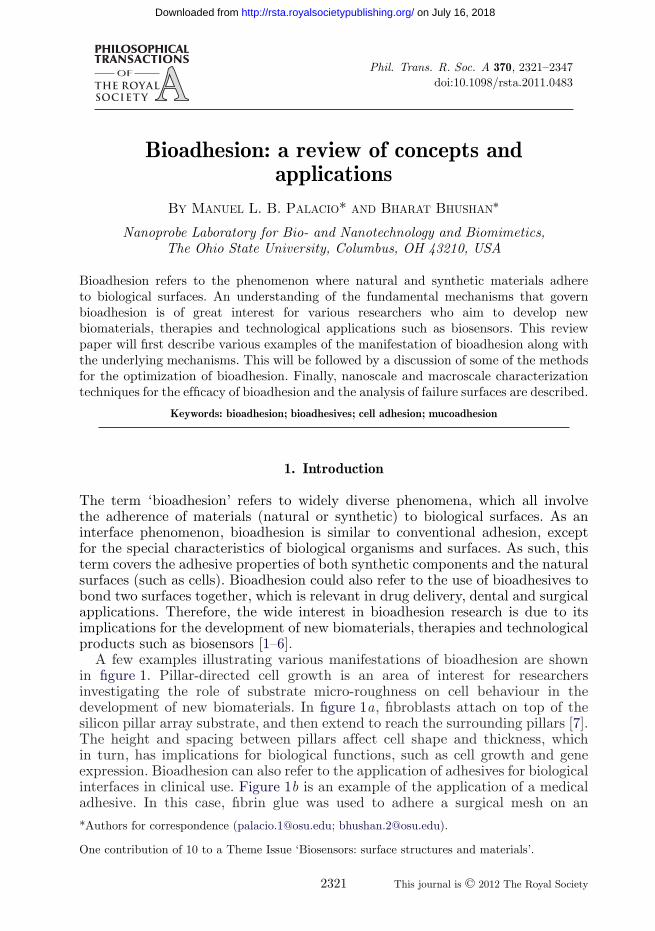
(ii) Cell adhesion to biomaterials
Cell adhesion to biomaterial surfaces is a complex phenomenon. As discussedin §2a, the micro/nanoscale surface topography has a direct impact on celladhesion and proliferation. But aside from morphology, the chemical compositionof the biomaterial surface has also been established to play a significant rolein the cell adhesion and proliferation process. For instance, cell adhesion asmediated by the integrin group of cell surface receptors has been shown todepend on the conformation of the ECM protein fibronectin, which in turn issensitive to the chemical composition of the synthetic surface. To demonstratethis concept, Keselowsky et al. [33] used various self-assembled monolayers(SAMs) with OH, COOH, NH2 and CH3 termini in order to create surfacesthat are hydrophilic and neutrally charged, hydrophilic and acidic, hydrophilicand basic, and hydrophobic, respectively. They found that the adhesion strengthof cell binding (as determined by a centrifugation assay) followed the trend:OH > COOH > NH2 > CH3. This trend was found to correlate with the integrin-binding profile on the SAMs. In another study, these researchers found that otherevents related to cell differentiation, such as osteoblast-specific gene expression,alkaline phosphatase enzyme activity and matrix mineralization, were also surfacechemistry dependent, such that OH- and NH2-terminated surfaces were moreadvantageous compared with COOH and CH3 SAMs [34].
So, how does the surface charge and hydrophilicity/hydrophobicity influencecell adhesion? The answer lies in the effect of the surface chemistry on theadsorption of the ECM proteins such as fibronectin, vitronectin, collagen andlaminin. In the context of the four model surfaces (OH-, COOH-, NH2- andCH3-terminated SAMs), it has been found that fibronectin undergoes the largestextent of denaturation on the CH3-terminated SAM, which corresponds topoor cell adhesion characteristics. The denaturation of fibronectin prevents thesurface exposure of the arginine–glycine–aspartic acid (RGD) groups on itscell-binding domain, which are known to mediate cell adhesion. In contrast,the hydrophilic, neutrally charged SAM (OH terminus) has been found toinduce the least extent of unfolding or denaturation, leading to a good celladhesion on the fibronectin/SAM surface [35]. The extent of denaturation offibronectin on the OH and CH3 surfaces is attributed to functional groupdehydration and water-restructuring effects brought about by the substratesurface [36,37].
It has also been established that the surface chemistry of the substrate playsa role in the formation of focal adhesions, which are adhesive complexes withsignalling molecules responsible for cell migration, survival and differentiation.The focal adhesion complexes (or plaques) have been found to contain integrinreceptors and cytoplasmic proteins, such as talin, vinculin and a-actinin. Anexample is shown in figure 5 of the localization of talin on fibronectin-coatedSAMs [38]. In these SAMs, the termini were also modified to possess OH,COOH, NH2 and CH3 termini. Talin formed large clusters on the OH andCOOH SAMs, fewer structures on NH2 SAMs, and the least amount on the
Phil. Trans. R. Soc. A
on July 16, 2018http://rsta.royalsocietypublishing.org/Downloaded from
2328 M. L. B. Palacio and B. Bhushan
CH3 OH
COOH NH2
Figure 5. Localization of the structural protein talin on self-assembled monolayers, illustrating howvarying the surface chemistry influences the formation of focal adhesion complexes [38].
CH3 surface. The extent of cluster formation is directly related to the amount offocal adhesions formed, which is indicative of the success of cell adhesion on thesubstrate surface.
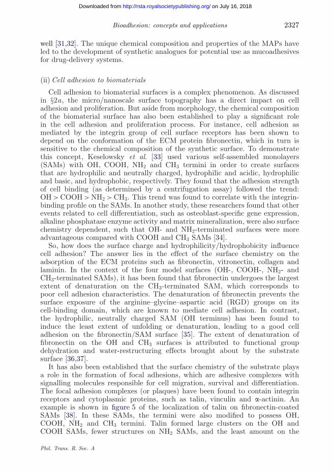
The studies on integrin binding, cell adhesion, fibronectin conformation andfocal adhesion formation are all consistent in demonstrating that modulatingthe surface chemistry of the biomaterial has a large impact on optimizing thecell adhesion process. A schematic illustrating the effect of surface chemistry onthe cell adhesion process is shown in figure 6 [39]. Figure 6a illustrates the casefor cell adhesion to a hydrophobic surface. Because the ECM protein (such asfibronectin) is denatured, specific amino acid sequences (such as the RGD groupsof fibronectin) are inaccessible for the integrin receptors. The receptors cannotcluster into focal adhesion complexes and bind to cytoplasmic proteins (suchas talin), which is required for cell adhesion. In contrast, when the biomaterialsurface is moderately hydrophilic (figure 6b), the adsorbed ECM proteins aremore flexible and not denatured, such that the receptors can form focal adhesionsand link the integrins to the actin cytoskeleton of the cells [39].
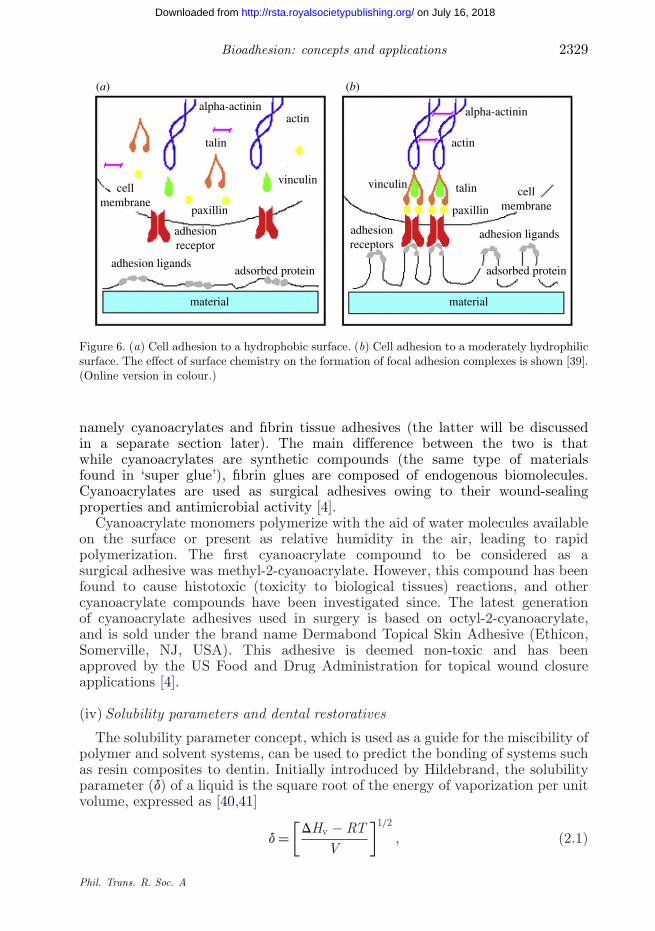
(iii) Cyanoacrylate bioadhesives
Bioadhesives for surgical applications is another area where chemicalinteractions are important. There are two main types of surgical tissue adhesives,
Phil. Trans. R. Soc. A
on July 16, 2018http://rsta.royalsocietypublishing.org/Downloaded from
Bioadhesion: concepts and applications 2329
alpha-actinin alpha-actininactin
(a) (b)
actin
vinculin vinculin
talin
talin
paxillin paxillin
adhesionreceptor
adhesionreceptors
cellmembrane
cellmembrane
adsorbed protein
material material
adhesion ligands
adhesion ligands
adsorbed protein
Figure 6. (a) Cell adhesion to a hydrophobic surface. (b) Cell adhesion to a moderately hydrophilicsurface. The effect of surface chemistry on the formation of focal adhesion complexes is shown [39].(Online version in colour.)
namely cyanoacrylates and fibrin tissue adhesives (the latter will be discussedin a separate section later). The main difference between the two is thatwhile cyanoacrylates are synthetic compounds (the same type of materialsfound in ‘super glue’), fibrin glues are composed of endogenous biomolecules.Cyanoacrylates are used as surgical adhesives owing to their wound-sealingproperties and antimicrobial activity [4].
Cyanoacrylate monomers polymerize with the aid of water molecules availableon the surface or present as relative humidity in the air, leading to rapidpolymerization. The first cyanoacrylate compound to be considered as asurgical adhesive was methyl-2-cyanoacrylate. However, this compound has beenfound to cause histotoxic (toxicity to biological tissues) reactions, and othercyanoacrylate compounds have been investigated since. The latest generationof cyanoacrylate adhesives used in surgery is based on octyl-2-cyanoacrylate,and is sold under the brand name Dermabond Topical Skin Adhesive (Ethicon,Somerville, NJ, USA). This adhesive is deemed non-toxic and has beenapproved by the US Food and Drug Administration for topical wound closureapplications [4].
(iv) Solubility parameters and dental restoratives
The solubility parameter concept, which is used as a guide for the miscibility ofpolymer and solvent systems, can be used to predict the bonding of systems suchas resin composites to dentin. Initially introduced by Hildebrand, the solubilityparameter (d) of a liquid is the square root of the energy of vaporization per unitvolume, expressed as [40,41]
d =[
DHv − RTV
]1/2
, (2.1)
Phil. Trans. R. Soc. A
on July 16, 2018http://rsta.royalsocietypublishing.org/Downloaded from
2330 M. L. B. Palacio and B. Bhushan
where DHv is the molar enthalpy of vaporization, R is the universal gas constant,T is the temperature and V is the molar volume. In this concept, liquidswith comparable solubility parameter values are predicted to be miscible witheach other. As applied to dental biomaterials, it has been found that there isa correlation between the solubility parameters of adhesive monomers and thedentin and the ability of the monomers to permeate into the substrate [42–44].
(c) Physiological factors
Biological adhesion can also take place through specific physiologically relatedmechanisms. Two examples are discussed, namely fibrin glues and the plantlectin adhesive.
(i) Fibrin tissue adhesive
The fibrin tissue adhesive is one of the two major categories of adhesivesfor surgical applications (the other type being cyanoacrylates). Fibrin tissueadhesives can be applied below the dermis as a sealant (or haemostatic clamp)for skin grafts and flaps, and for laparoscopic surgeries [4,8]. These adhesivesare typically packaged as two components, which are then mixed togetherduring surgery. The first component contains fibrinogen, plasma glutaminase (alsocalled Factor XIII) and CaCl2. The second component contains thrombin andantifibrinolytic agent. Fibrin tissue adhesives work based on the physiology ofthe blood coagulation process. Thrombin cleaves the protein fibrinogen duringclotting into smaller fibrin subunits, which then go through end-to-end and side-to-side polymerization. Factor XIII is responsible for the cross-linking of thesubunits into a stable fibrin clot in the presence of calcium [4].
(ii) Lectins
Lectins are carbohydrate-binding proteins found in both animals and plants.One of the common animal lectins is the C-type or calcium-dependent bindinglectins. One of these C-type lectins, the asialoglycoprotein lectins, is specificto liver cells and is involved in animal biological function. Meanwhile, plantlectins are abundant and can be found in common plants such as tomatoes andin the seeds of legumes. Some plant lectins are regarded as toxins when theybind to animal cells [27,45]. However, owing to their ability to bind specificallyto glycosylated cell membrane components, lectins are being investigated fortheir ability to transport macromolecules, with implications for drug deliveryto the gastrointestinal tract [46,47]. Aside from lectins, other molecules that arebeing considered for bioadhesive-based drug delivery are bacterial fimbrins andinvasins [3].
(d) Physical and mechanical effects
The physical and mechanical factors that influence bioadhesion were originallydeveloped for polymer–polymer adhesion, but are directly applicable to biologicaladhesion as well [1]. The mechanisms of wetting and interpenetration will bediscussed as follows.
Phil. Trans. R. Soc. A
on July 16, 2018http://rsta.royalsocietypublishing.org/Downloaded from

Bioadhesion: concepts and applications 2331
P
P
M M
liquid
gPM
gML
gPL
Figure 7. Schematic of the relevant interfacial energies (g) that determine the formation of abioadhesive bond, as illustrated for the adhesion between a polymer (P) and the mucous layer(M) on a biological membrane in the presence of a liquid medium (L) (adapted from Buckton [2]).
(i) Wetting phenomenon
The interfacial energy is an important determinant of successful bioadhesion. Insystems exhibiting bioadhesion, the liquid environment influences the spreading ofone material phase over another. Consider the case of a polymer surface adheringto the mucous gel layer on a biological membrane immersed in liquid medium (e.g.in drug-delivery applications). Figure 7 is a schematic illustrating the interfacialenergy components that should be considered in evaluating the thermodynamicwork of adhesion of this system. During adhesion, a unit interface between thepolymer and the liquid and between the mucus and the liquid vanishes, whereasan interface between the polymer and mucus forms. The thermodynamic workof adhesion (W adh
PM ) or the energy of adhesion per unit area between the polymerand mucus is then defined by the Dupre equation as follows [2,28]:
W adhPM = gPM − (gPL + gML), (2.2)
where g represent surface energies and the subscripts P, M and L are for polymer,mucus and liquid, respectively. A positive value for W adh
PM is regarded to be anecessary condition to achieve successful bonding between the two surfaces.
(ii) Interpenetration
While interfacial contact and chemical bonding interactions are needed for theinitial stages of bioadhesion, the interpenetration or interdiffusion between themolecules of the two contacting surfaces will maintain the adhesive bond. If apolymer is one of the contacting systems involved in the bioadhesion process,then the interpenetration process involves the mobility of the individual chainsand their entanglement in the opposing biological membrane. A related concept isthe swelling capacity of the polymer, which is the ratio of the wet to dry weights.A high swelling capacity for a given polymer implies that it has greater chainmobility, and a higher tendency towards interpenetration [2].
Phil. Trans. R. Soc. A
on July 16, 2018http://rsta.royalsocietypublishing.org/Downloaded from

2332 M. L. B. Palacio and B. Bhushan
For bioadhesion in dental prostheses, mechanical interlocking or interpenet-ration is the primary adhesive mechanism. When bonding an acrylic resinrestorative to tooth enamel, surface etching is performed to create microporeson the surface that will increase the penetration of the restorative material.This method was pioneered by Buonocore [48], who discovered that conditioninghuman enamel with 85 per cent phosphoric acid improves the adhesion ofacrylic resin to the enamel surface. This is an illustration of how a surfacechemical treatment roughens the surface and facilitates adhesion via mechanicalinterlocking effects.
The tooth dentin, on the other hand, contains tubules that radiate from thepulp. These tubular structures facilitate the penetration of resin monomers intothe dentin and their retention once the resin has been polymerized [42]. Asdiscussed earlier, chemical effects (i.e. solubility) play a role in the adhesionbetween a restorative and dentin. However, the other dimension in this adhesiveevent is the permeation of the adhesive into the collagen fibrillar network found inthe tooth dentin [5]. A list of hydrophilic monomers that have been investigatedfor this purpose is given in table 1. It is thought that the entanglement ofthe polymer network formed during the polymerization of the monomer on thecollagen network of dentin is responsible for the observed adhesion.
3. Optimization of bioadhesion
The bioadhesion between two surfaces in contact may be enhanced using variousphysical, chemical and mechanical processing techniques.
(a) Physical processes
Various methods are used to modify surfaces, such as exposure to plasma,corona discharge, ions and ultraviolet (UV)-ozone. Plasma processing is a widelyused technique for surface cleaning and modification of the surface chemicalcomposition. The plasma, which is generated by applying an electric field toa low-pressure gas in vacuum, produces reactive species such as ions and freeradicals. For surface cleaning purposes, the plasma breaks most organic bonds,such as C−H, C−C, C=C, C−O and C−N. In addition, if oxygen plasma isused, another cleaning mode is possible. The activated oxygen species (O•,O+, O−, O+
2 , O−2 , O3, among others) can combine with organic compounds to
form H2O, CO, CO2 and low molecular weight hydrocarbons, thereby removingorganic contaminants on the surface [49]. Surface modification through plasmaprocessing can render the surface more hydrophilic or hydrophobic. If the gas usedis oxygen or carbon dioxide, polar groups are introduced to the surface (suchas C−OH, C=O and COOH), enhancing surface hydrophilicity. The presenceof more polar surface groups is valuable in enhancing adhesion, e.g. betweenproteins and polymeric biomaterials used in tissue engineering applications. Theapplication of this concept is shown schematically in figure 8 [39]. Two examplesare shown to illustrate how plasma treatment leads to an activated surfacewith free radicals. After the plasma processing, molecules can be grafted ontothe activated surface, such as polyethylene glycol in figure 8a or dithiol and
Phil. Trans. R. Soc. A
on July 16, 2018http://rsta.royalsocietypublishing.org/Downloaded from

Bioadhesion: concepts and applications 2333
Table 1. Monomers used as priming agents for tooth dentin adhesives [5].
common abbreviation full name
HEMA 2-hydroxyethyl methacrylateGLUMA adduct of glutaraldehyde and HEMANPG-GMA N -phenyl glycine/glycidyl methacrylate (adduct)NTG-GMA N -(p-tolyl)glycine and glycidyl methacrylateGDMA glyceryl dimethacrylateGPDM glycerophosphoric acid dimethacrylateNMBu N -methacryloyl butyric acidNMGlu N -methacryloyl glutamic acidNAAsp N -acryloyl aspartic acidNMHyp N -methacryloyl hydroxyprolineNMGly N -methacryloyl glycineDIPENTA pentaacryloyldipentaerythritol phosphoric acidPMDM diadduct of pyromellitic anhydride with 2-hydroxyethyl methacrylateMEM 2-methacryloyloxyethyl hydrogen maleateMMEM mono-2-(methacryloyloxy)ethyl maleateMMPM mono-2-(methacryloyloxy)propyl maleateMDPM 3-methacryloxypropyl phosphateMBP 4-methacryloyloxybutyl phosphoric acidMOP 8-methacryoyloxyoctyl phosphoric acidACE acryloyloxyethyl citraconateBMEP bis[2-(methacryloyloxy)-ethyl]phosphateEGMP ethylene glycol methacrylate phosphateMDP 10-methacryloyldeacamethylene phosphoric acidPhenylP 2-methacryloyloxy phenyl phosphate4-META 4-methacryloxyethyl trimellitate anhydridePA 2-acryloyloxyethyl phosphatePM 2-methacryloyloxy ethyl phosphateSBMA 3-sulpho-2-butyl methacrylateSEMA 2-sulphoethyl methacrylateMSPMA 3-methoxy-1-sulpho-2-propyl methacrylate
Au nanoparticles in figure 8b. In these modified polymer surfaces, successfuladhesion of fibroblast cells was achieved, indicating the cytocompatibility of theplasma-processed polymer [39,50].
It should be noted that a similar effect on enhancing surface hydrophilicitycan be obtained by using UV-ozone surface treatment on polydimethylsiloxane(PDMS). Compared with the untreated polymer surface, the surface-treatedPDMS tends to be rougher and more hydrophilic. An increase in the cellattachment as well as an enhancement in cell spreading was observed with theuse of the UV-ozone-treated PDMS [51].
On the other hand, the use of fluorinated gases such as CF4 in the plasmaresults in the introduction of a low surface energy polytetrafluoroethylene-like structure, enhancing surface hydrophobicity. This approach has been usedto prevent the undesirable protein adhesion and the unwanted generation ofinflammatory cells on poly(methyl methacrylate) (PMMA) contact lenses [52].
Phil. Trans. R. Soc. A
on July 16, 2018http://rsta.royalsocietypublishing.org/Downloaded from

2334 M. L. B. Palacio and B. Bhushan
polymer
(a)
(b)
polymer
polymer
polymer
polymer
polymer
polymer
polymer
polymer
plasma
R R R R R R
R R R R R R
plasma
PEG grafting
cell culturing
dithiol grafting
Au nanoparticlesgrafting
cell culturing
Figure 8. Schematic showing the effect of plasma processing on a polymer surface, leading toits activation and capacity to graft (a) polyethylene glycol (PEG) or (b) dithiol followed by Aunanoparticles. Cells are able to grow onto the grafted surfaces [39]. (Online version in colour.)
(b) Chemical processes
Specific functional groups can be covalently attached to substrate surfacesusing chemical synthesis techniques to attach biomolecules and construct modelsystems for use in adhesion studies. Covalent bonding will lead to enhancedretention of the biomolecule relative to simply relying on its innate adsorption tothe substrate. An example is shown in figure 9, illustrating how a silica surface ischemically functionalized with the protein streptavidin [53]. In this illustration,the functionalization process consists of multiple steps, starting with the addition
Phil. Trans. R. Soc. A
on July 16, 2018http://rsta.royalsocietypublishing.org/Downloaded from

Bioadhesion: concepts and applications 2335
3-aminopropyltriethoxysilane (3-APTES)
CH2CH2OSiO2
SiO2
O
O
O HN NH
O
S
S
S
OR
OR
O
O
O
NH
NH
streptavidinhas fourbiotin-bindingpockets. Twoor one may beattached to thebiotin on thesurface, withthe remainingtwo or threeavailable tobind thebiotin analyte
HN
HN
O
O H
H
H
CH2CH2O
CH2CH2O CH2CH2CH2NH3+
CH2CH2CH2NH3+ CH2CH2CH2NH
CH2CH2CH2NH
CH2CH2CH2NH
Si
Si
O
O
O
O
O O HN NH
O
S
S
NHHN
OS
NHHN
O
O
O
O Si
O
Na+O O
O
sulpho-NHS biotin
NHHN
S
SiO2
O
ONO O
SO
O
O
O
O
O
O
O
OR
CH2CH2CH2NH3+
CH2CH2CH2NH3+
Si
Si
Si
Si
Figure 9. Schematic showing the steps in the functionalization of a silica surface with the proteinstreptavidin. Adapted from Bhushan et al. [53].
of the aminosilane 3-aminopropyltriethoxysilane (APTES) in order to introduceamine groups to the silica surface. This is then followed by reacting the surfacewith sulpho-N -hydroxysuccin-imido-biotin (abbreviated as biotin), which forms acovalent bond with the amine group of the APTES. In the final step, the proteinstreptavidin will bind to biotin. Streptavidin has a high affinity to biotin, andthis interaction is regarded as one of the strongest non-covalent bonds known.This technique can be implemented for the functionalization of the silica surfacewith other proteins as well [22,23].
(c) Mechanical interactions
Surface modification through mechanical texturing is a simple andcost-effective method to roughen biomaterials and to create anchor points forenhanced adhesion. In this method, abrasive particles (such as silicon carbideor alumina) impinge on the biomaterial, and the resulting impact increases itssurface roughness [28,54]. This method can be used on orthopaedic implantmaterials such as titanium or cobalt/chrome.
In the design of artificial cartilage scaffold, the surface modification ofa non-degradable polymer could be undertaken to enhance cell adhesion.This involves the addition of a non-woven mesh composed of polymer fibres
Phil. Trans. R. Soc. A
on July 16, 2018http://rsta.royalsocietypublishing.org/Downloaded from

2336 M. L. B. Palacio and B. Bhushan
Figure 10. Cross-section scanning electron microscopy image showing the growth andinterpenetration of a cell matrix (long arrows) on a poly(L-lactic acid) scaffold (short arrows) [55].
bonded onto a non-porous surface. The presence of the mesh on the surfacecreates a porous surface on which cells can proliferate. As the ECM develops,the fibres remain interpenetrated, and ensuring the bonding of the scaffold ontothe cartilage. An image is shown in figure 10 illustrating this concept [55]. Theshort arrows point to the fibres (made of poly(L-lactic acid)), whereas the longarrows indicate the presence of cartilage growth on the fibres.
4. Characterization techniques
In this section, nanoscale and macroscale experimental techniques for quantifyingbioadhesion are discussed.
(a) Nanoscale
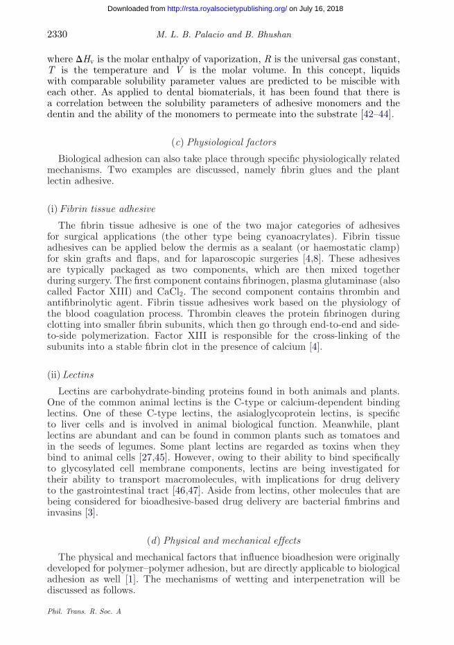
Fundamental studies of bioadhesion at the nanoscale are conducted withatomic force microscopy (AFM). Aside from its high-resolution surface imagingcapability, AFM can measure forces in the pico- to nanonewton range, allowing forthe detection of single-molecule interactions [6,56]. In AFM, force–distance curvesfor the contact between the probe and the sample surface can be obtained. Anexample is shown in figure 11. The force–distance curve consists of two segments,namely the advancing curve (corresponding to AFM piezo extension), whichshows how the probe approaches the surface, and the retracting curve, whichillustrates how the probe detaches from the surface. In the retracting force curve,a distinct snap-off point is observed, which corresponds to the force necessary toseparate the tip from the sample surface. This is the measured adhesive force.
Phil. Trans. R. Soc. A
on July 16, 2018http://rsta.royalsocietypublishing.org/Downloaded from
Bioadhesion: concepts and applications 2337
4000
3000
2000
1000
0
retracting
extending
Fadh
–10000 100 200
tip and sample separation (nm)
forc
e (p
N)
300 400 500
Figure 11. Atomic force microscopy force–distance curve between a tip functionalized with afibronectin antibody and fibronectin deposited on a polymer surface obtained in phosphate-bufferedsaline medium.
2.5
2.0
1.5
1.0
0.5
4.4pH values of phosphate-buffered saline solution
adhe
sive
for
ce (
nN)
7.4 9.10
Figure 12. Effect of pH on the adhesion between streptavidin and biotin as measured by atomicforce microscopy. Adapted from Bhushan et al. [57].
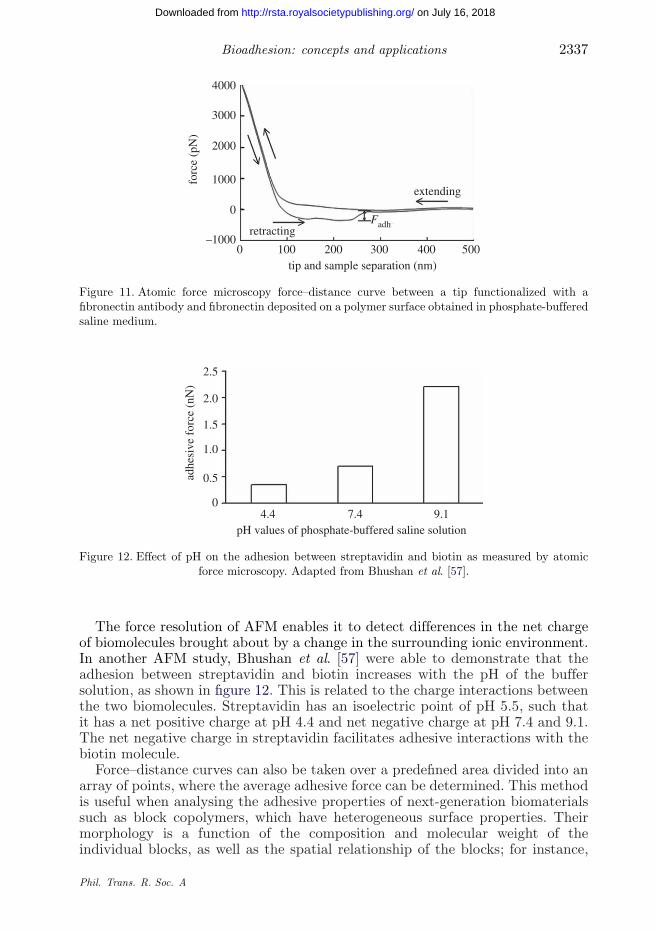
The force resolution of AFM enables it to detect differences in the net chargeof biomolecules brought about by a change in the surrounding ionic environment.In another AFM study, Bhushan et al. [57] were able to demonstrate that theadhesion between streptavidin and biotin increases with the pH of the buffersolution, as shown in figure 12. This is related to the charge interactions betweenthe two biomolecules. Streptavidin has an isoelectric point of pH 5.5, such thatit has a net positive charge at pH 4.4 and net negative charge at pH 7.4 and 9.1.The net negative charge in streptavidin facilitates adhesive interactions with thebiotin molecule.
Force–distance curves can also be taken over a predefined area divided into anarray of points, where the average adhesive force can be determined. This methodis useful when analysing the adhesive properties of next-generation biomaterialssuch as block copolymers, which have heterogeneous surface properties. Theirmorphology is a function of the composition and molecular weight of theindividual blocks, as well as the spatial relationship of the blocks; for instance,
Phil. Trans. R. Soc. A
on July 16, 2018http://rsta.royalsocietypublishing.org/Downloaded from

2338 M. L. B. Palacio and B. Bhushan
10
8
6
adhe
sive
for
ce (
nN)
4
2
0fibronectin
pH 7.4 pH 6.2 pH 7.4 pH 6.2 pH 7.4 pH 6.2
BSA collagen
PMMA-b-PAA (random 1/1)PMMA PMMA-b-PAA (1/1)
PAA-b-PMMA-b-PAA (0.5/1/0.5)
Figure 13. Atomic force microscopy (AFM) data on the average adhesive force for the interactionbetween block copolymers containing poly(methyl methacrylate) (PMMA)/poly(acrylic acid)(PAA) with the proteins fibronectin, BSA and collagen (attached to functionalized AFM tips) in pH7.4 (phosphate-buffered saline) and pH 6.2 buffer media, with data for PMMA as a reference [22].
A–B block copolymers (or diblock) will have a different morphology from A–B–Ablock copolymers (or triblock), and so on. This variation in the morphologytranslates to observable differences in the adhesive force. Figure 13 is a summaryof the average adhesive force for the interactions between three proteins, namelyfibronectin, bovine serum albumin (BSA) and collagen, on block copolymerscomposed of PMMA and poly(acrylic acid) (PAA), but with different blockarrangements (random, diblock and triblock) [22].
The data in figure 13 show the variation of the adhesive force at two ionicenvironments, pH 7.4 (phosphate-buffered saline, PBS) and pH 6.2, where itis seen that the liquid environment affects the adhesive force. For both buffermedia, the highest average adhesive force was observed in the triblock copolymer(PAA-b-PMMA-b-PAA, where ‘b’ denotes block copolymer), and the lowest forcewas obtained from the PMMA reference. The higher average adhesive force inthe triblock copolymer surface can be due to greater ordering on its surface,which would expose more PAA-rich areas than it would for the diblock orrandom copolymer.
The average adhesive forces measured at pH 6.2 are higher throughout theentire series, relative to the PBS medium (pH 7.4) data. This is attributedto higher repulsion at pH 7.4 (or conversely, increased attractive forces at thelower pH). Chemical interactions between the protein and the polymer surfaceare influenced by the surface charges of the protein and the polymer itself. Theamine groups of the proteins are protonated at both pH 7.4 and 6.2. But theseproteins carry a net negative charge when immersed in either the pH 7.4 or 6.2buffer media, because the isoelectric points of fibronectin, BSA and collagen areall lower than 6.2 [22]. There are more negative charges on the protein surfaceat pH 7.4 than at pH 6.2. Also, the acrylic acid chains are ionized (to acrylates)during the adhesive force mapping experiment as they are immersed in aqueous
Phil. Trans. R. Soc. A
on July 16, 2018http://rsta.royalsocietypublishing.org/Downloaded from

Bioadhesion: concepts and applications 2339
pH 7.4
O–
––––––– ––
–––
O–
O O
pH 6.2
protein
reduction incharge
polymer
Figure 14. Schematic illustrating the effect of pH on protein–block copolymer interactions (focusingon the effect of poly(acrylic acid), which is ionized at pH 7.4 and 6.2), showing negative chargereduction at lower pH conditions, which leads to decreased repulsion [22].
medium. The presence of negative charges on both the tip and sample surfaceleads to repulsive interactions. As illustrated in figure 14, the reduced repulsionbetween the acrylate groups in the acrylic acid and the smaller amount of surfacenegative charges in fibronectin, BSA and collagen at the lower pH increase themeasured adhesion between the two surfaces [22]. At the higher pH (7.4), theproteins carry a greater net negative charge. Because the acrylic acid in the blockcopolymer is present in its ionized form, the repulsion from the negative chargespresent on both the protein and the polymer surface is more significant at pH7.4 than at pH 6.2. This accounts for the higher adhesive force measured at pH6.2. The results demonstrate how AFM is able to measure the effect of the ionicenvironment on bioadhesion.
(b) Macroscale
Traditional adhesion characterization techniques, such as crack growth, peeland shear tests, could be applied to evaluate bioadhesion. A few examples of theuse of these methods are provided below.
For dental restorations, the Griffith energy balance model could be usedto characterize the crack growth using a bending test, where the cracksoriginate from the shrinking of the implanted dental composite material uponpolymerization [58]. The energy balance concept, as applied to restorations, dealswith the balance between the elastic energy in the tooth (which is actuallycomposed of the individual elastic energies of the tooth and the restorativematerial) and the surface energy associated with the crack. Experimentally,either the stress intensity factor (K , also referred to as the fracture toughness)or the strain energy release rate (G, also referred to as the fracture energy)is evaluated from the measurement of the crack growth. Fracture toughness isobtained from [58]
K = s(ap)1/2, (4.1)
where s is the applied stress and a is half the length of the crack. A tabulationof the fracture toughness of adhesive joints, tooth components, and restorativematerials is presented in table 2. The fracture energy can be obtained as long asthe modulus and Poisson’s ratio of the material are known.
Phil. Trans. R. Soc. A
on July 16, 2018http://rsta.royalsocietypublishing.org/Downloaded from

2340 M. L. B. Palacio and B. Bhushan
(a) (b)
5 µm
**
2 µm
Figure 15. Fractography of tooth–dental restorative interfaces using scanning electron microscopy:(a) an example of predominantly adhesive failure, except for the region marked with an asterisk,where cohesive failure occurred and (b) an example of cohesive failure (adapted from Yanget al. [59]).
Table 2. Fracture toughness of selected dental materials [58].
material fracture toughness (MPa m1/2)
adhesives bonded to dentin 0.1–1.2adhesives bonded to enamel 1.1enamel 0.65–2.5dentin 1.0–4.0dentin–enamel junction 0.8–3.4composite resins 0.7–1.9amalgams 1.4–2.4
As a qualitative tool, fractographic analysis through either scanning electronmicroscopy (SEM) or transmission electron microscopy is usually conductedon dental restoration–dentin interfaces analysed in vitro using tensile testing.Examples of fracture surface SEM images are shown in figure 15, where it isseen that either adhesive or cohesive failure can take place. The determinationof the failure mode is useful when comparing between different adhesive resinformulations [59].
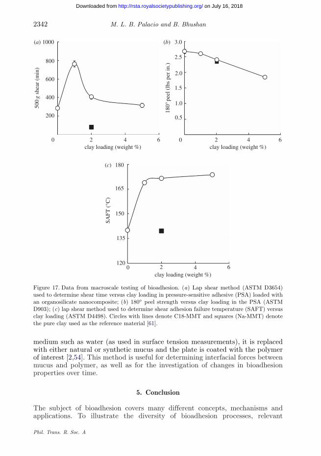
Standard American Society for Testing and Materials (ASTM) characterizationprocedures, such as the tensile test (ASTM D368), peel test (ASTM D903),shear strength (ASTM D3654) and shear adhesion failure temperature (SAFT,ASTM D4498), are conducted on interfaces where an adhesive is applied to thebiomaterial. Schematics for the peel and shear tests are shown in figure 16. Thesetests are applicable to systems such as fibrin glues used in surgical operations,PAA hydrogels and pressure-sensitive adhesives (PSAs) intended for transdermaldrug-delivery applications [8,54,60,61]. Figure 17 shows examples of macroscaleadhesion data on PSAs. The adhesive system investigated was PDMS. It wasloaded with an organo-clay based on montmorillonite (MMT) to form a compositematerial. The addition of the organo-clay was shown to improve the shear strengthand the SAFT, with a minimal reduction in the peel strength [61].
Phil. Trans. R. Soc. A
on July 16, 2018http://rsta.royalsocietypublishing.org/Downloaded from

Bioadhesion: concepts and applications 2341
adhesive tape(a) (b)
(c) (d)
(e)
adhesive tape
adhesive
tape and coatingcut to shape shown
coating
coating
coating 90° double stickadhesive tape
double stickadhesive tape
glassnormal load
backing plate
linear slide
coating
coatingsubstrate
flexiblesubstrate
substrate
flexible substrate
backingplate
substrate
Figure 16. Schematics of macroscale adhesion tests. (a, b) The scotch tape test, (c, d) examples ofthe peel test and (e) the lap shear test [54].
The flow-through method, which measures the flow rate needed to remove abioadhesive-coated sphere, is a common macroscale experiment suited for thecharacterization of mucoadhesion of potential drug-delivery systems. A widelyused biophysical assay involves the monitoring of molecular weight changethrough the variation in the sedimentation coefficient as measured by ananalytical ultracentrifuge [62]. The Wilhelmy plate experiment is anothertechnique for evaluating bioadhesion at the macroscale. Instead of having a liquid
Phil. Trans. R. Soc. A
on July 16, 2018http://rsta.royalsocietypublishing.org/Downloaded from
2342 M. L. B. Palacio and B. Bhushan
1000(a) (b)
(c)
800
600
400
200
0
3.0
2.5
2.0
1.5
1.0
0.5
0
180
165
150
135
1200 2
clay loading (weight %)
SAFT
(°C
)
180°
pee
l (lb
s pe
r in
.)
500
g sh
ear
(min
)
4 6
2clay loading (weight %)
4 6 2clay loading (weight %)
4 6
Figure 17. Data from macroscale testing of bioadhesion. (a) Lap shear method (ASTM D3654)used to determine shear time versus clay loading in pressure-sensitive adhesive (PSA) loaded withan organosilicate nanocomposite; (b) 180◦ peel strength versus clay loading in the PSA (ASTMD903); (c) lap shear method used to determine shear adhesion failure temperature (SAFT) versusclay loading (ASTM D4498). Circles with lines denote C18-MMT and squares (Na-MMT) denotethe pure clay used as the reference material [61].
medium such as water (as used in surface tension measurements), it is replacedwith either natural or synthetic mucus and the plate is coated with the polymerof interest [2,54]. This method is useful for determining interfacial forces betweenmucus and polymer, as well as for the investigation of changes in bioadhesionproperties over time.
5. Conclusion
The subject of bioadhesion covers many different concepts, mechanisms andapplications. To illustrate the diversity of bioadhesion processes, relevant
Phil. Trans. R. Soc. A
on July 16, 2018http://rsta.royalsocietypublishing.org/Downloaded from

Bioadhesion: concepts and applications 2343
O
OH
glycine (Gly, G)
valine (Val, V)
leucine (Leu, L) isoleucine (Ile, I)
lysine (Lys, K)
arginine (Arg, R)
glutamic acid (Glu, E)
histidine (His, H) phenylalanine (Phe, F)
tyrosine (Tyr, Y) tryptophan (Trp, W)
glutamine (Gin, Q) methionine (Met, M)
proline (Pro, P)
threonine (Thr, T)aspartic acid (Asp, D)
asparagine (Asn, N)
alanine (Ala, A)serine (Ser, S) cysteine (Cys, C)
H2N
O
OHH2N
Ochemical stuctures of the 20 common amino acids
OH
OH
H2N
O
OH
SH
H2N
O
OHH2N
O
OHH2N
O
OHH2N
O
OH
NH
NH2+
NH3+
H2N
H2N
O
OHH2N
OO
OHOH
NH
H2N
O
O
OH
OH
O
O
OH
O O
H2N
H2N
HN
N
H2N H2N H2N
OHH2N
NH2
OH
O O
OH
NH
OH
OH
S
H
O
OH
OH
H2N
O
OH
HO
O
H2N
O
OH
O
H2N
H2N
Figure 18. Chemical structures of the 20 amino acids found in proteins. Adapted from [63].
examples, such as cell adhesion to biomaterials, dental restorative adhesives andmucoadhesives, were discussed in this paper. Bioadhesion can be beneficial, as itcan facilitate the desired adhesion of cells and biomolecules on various natural andsynthetic substrates, which then leads to the development of novel biomaterials,
Phil. Trans. R. Soc. A
on July 16, 2018http://rsta.royalsocietypublishing.org/Downloaded from
2344 M. L. B. Palacio and B. Bhushan
DAHKSE VAHRFKDLGE ENFKALVLIA FAQYLQQCPFEDHVKLVNEV TEFAKTCVAD ESAENCDKSL HTLFGDKLCTVATLRETYGE MADCCAKQEP ERNECFLQHK DDNPNLPRLVRPEVDVMCTA FHDNEETFLK KYLYEIARRH PYFYAPELLFFAKRYKAAFT ECCQAADKAA CLLPKLDELR DEGKASSAKQRLKCASLQKF GERAFKAWAV ARLSQRFPKA EFAEVSKLVTDLTKVHTECC HGDLLECADD RADLAKYICE NQDSISSKLKECCEKPLLEK SHCIAEVEND EMPADLPSLA ADFVESKDVCKNYAEAKDVF LGMFLYEYAR RHPDYSVVLL LRLAKTYETTLEKCCAAADP HECYAKVFDE FKPLVEEPQN LlKQNCELFEQLGEYKFQNA LLVRYTKKVP QVSTPTLVEV SRNLGKVGSKCCKHPEAKRM PCAEDYLSVV LNQLCVLHEK TPVSDRVTKCCTESLVNRRP CFSALEVDET YVPKEFNAET FTFHADICTLSEKERQIKKQ TALVELVKHK PKATKEQLKA VMDDFAAFVEKCCKADDKET CFAEEGKKLV AASQAALGL
Figure 19. Amino acid sequence of the protein human serum albumin (HSA) [65].
therapies and technologies such as biosensors. Various processing techniques thatcould enhance bioadhesion are available. The characterization of bioadhesionmust be performed at the length scale appropriate to the phenomenon (either atthe nanoscale or macroscale), as it relates to the application where the adherencebetween two interfaces is desired.
Appendix A. The composition of proteins
Proteins are known as the most abundant class of macromolecules in cells. Eachprotein molecule can be envisaged as a polymer composed of amino acids, whichare molecules that contain an amine group, a carboxylic acid group and a variableside chain. The chemical structures of the 20 common amino acids found inproteins, along with their three-letter and one-letter abbreviations, are shown infigure 18 [63]. On the basis of their chemical composition, the amino acids alanine,valine, leucine, isoleucine, proline, phenylalanine, tryptophan and methionineare classified as non-polar. Glycine, serine, threonine, cysteine, asparagine andglutamine are considered as uncharged polar amino acids. Aspartic acid, glutamicacid, lysine, arginine and histidine have charged side groups [64].
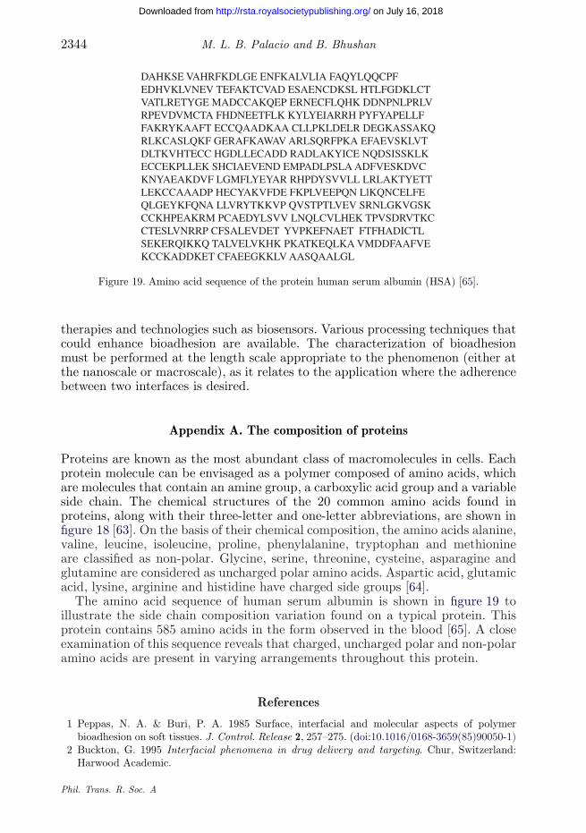
The amino acid sequence of human serum albumin is shown in figure 19 toillustrate the side chain composition variation found on a typical protein. Thisprotein contains 585 amino acids in the form observed in the blood [65]. A closeexamination of this sequence reveals that charged, uncharged polar and non-polaramino acids are present in varying arrangements throughout this protein.
References
1 Peppas, N. A. & Buri, P. A. 1985 Surface, interfacial and molecular aspects of polymerbioadhesion on soft tissues. J. Control. Release 2, 257–275. (doi:10.1016/0168-3659(85)90050-1)
2 Buckton, G. 1995 Interfacial phenomena in drug delivery and targeting. Chur, Switzerland:Harwood Academic.
Phil. Trans. R. Soc. A
on July 16, 2018http://rsta.royalsocietypublishing.org/Downloaded from
Bioadhesion: concepts and applications 2345
3 Woodley, J. 2001 Bioadhesion: new possibilities for drug administration. Clin. Pharmakokinet.40, 77–84. (doi:10.2165/00003088-200140020-00001)
4 Mobley, S. R., Hilinski, J. & Toriumi, D. M. 2002 Surgical tissue adhesives. Facial Plast. Surg.Clin. North Am. 10, 147–154. (doi:10.1016/S1064-7406(02)00014-7)
5 Vaidyanathan, T. K. & Vaidyanathan, J. 2009 Recent advances in the theory and mechanismof adhesive resin bonding to dentin: a critical review. J. Biomed. Mater. Res. B 88, 558–578.(doi:10.1002/jbm.b.31253)
6 Bhushan, B. 2010 Springer handbook of nanotechology, 3rd edn. Heidelberg, Germany: Springer.7 Su, W.-T., Liao, Y.-F., Lin, C.-Y. & Li, L.-T. 2010 Micropillar substrate influences the cellular
attachment and laminin expression. J. Biomed. Mater. Res. A 93, 1463–1469. (doi:10.1002/jbm.a.32643)
8 Eriksen, J. R., Bech, J. I., Linnemann, D. & Rosenberg, J. 2008 Laparoscopic intraperitonealmesh fixation with fibrin sealant (Tisseel�) versus titanium tacks: a randomized controlexperimental study in pigs. Hernia 12, 483–491. (doi:10.1007/s10029-008-0375-z)
9 Prashanth, K. D., Yiu, C. K.-Y. & King, N. M. 2007 A review of dentin adhesives in pediatricdentistry. Hong Kong Dent. J. 4, 80–89.
10 Burkett, J. R., Wojtas, J. L., Cloud, J. L. & Wilker, J. J. 2009 A method for measuringthe adhesion strength of marine mussels. J. Adhesion 85, 601–615. (doi:10.1080/00218460902996903)
11 Anonymous. 2011 Hardening of the arteries. A.D.A.M. Medical Encyclopedia. See http://www.ncbi.nlm.nih.gov/pubmedhealth/PMH0001224.
12 Tenke, P., Riedl, C. R., Jones, G. L., Williams, G. J., Stickler, D. & Nagy, E. 2004 Bacterialbiofilm formation on urologic devices and heparin coating as preventive strategy. Int. J.Antimicrob. Agents 23, S67–S74. (doi:10.1016/j.ijantimicag.2003.12.007)
13 Kroes, I., Lepp, P. W. & Relman, D. A. 1999 Bacterial diversity within the human subgingivalcrevice. Proc. Natl Acad. Sci. USA 96, 14 547–14 552. (doi:10.1073/pnas.96.25.14547)
14 Vroman, L. 1962 Effect of absorbed proteins on the wettability of hydrophilic and hydrophobicsolids. Nature 196, 476–477. (doi:10.1038/196476a0)
15 Turbill, P., Beugeling, T. & Poot, A. A. 1996 Proteins involved in the Vroman effect duringexposure of human blood plasma to glass and polyethylene. Biomaterials 17, 1279–1287.(doi:10.1016/0142-9612(96)88673-X)
16 Jung, S. Y., Lim, S. M., Albertorio, F., Kim, G., Gurau, M. C., Yang, R. D., Holden, M. A. &Cremer, P. S. 2003 The Vroman effect: a molecular level description of fibrinogen displacement.J. Am. Chem. Soc. 125, 12 782–12 786. (doi:10.1021/ja037263o)
17 Krishnan, A., Siedlecki, C. A. & Vogler, E. A. 2004 Mixology of protein solutions and theVroman effect. Langmuir 20, 5071–5078. (doi:10.1021/la036218r)
18 Noh, H. & Vogler, E. A. 2007 Volumetric interpretation of protein adsorption: competitionfrom mixtures and the Vroman effect. Biomaterials 28, 405–422. (doi:10.1016/j.biomaterials.2006.09.006)
19 Arnold, M., Cavalcanti-Adam, E. A., Galss, R., Blummel, J., Eck, W., Kantlehner, M.,Kessler, H. & Spatz, J. P. 2004 Activation of integrin function by nanopatterned adhesiveinterfaces. ChemPhysChem 5, 383–388. (doi:10.1002/cphc.200301014)
20 Arnold, M., Schwieder, M., Blummel, J., Cavalcanti-Adam, E. A., Lopez-Garcia, M.,Kessler, H., Geiger, B. & Spatz, J. P. 2009 Cell interactions with hierarchically structurednano-patterned adhesive surface. Soft Matter 5, 72–77. (doi:10.1039/b815634d)
21 Biggs, M. J. P., Richards, R. G., Gadegaard, N., Wilkinson, C. D. W. & Dalby, M. J. 2007Regulation of implant cell adhesion: characterization and quantification of s-phase primaryosteoblast adhesions on biomimetic nanoscale substrates. J. Orthop. Res. 25, 273–282.(doi:10.1002/jor.20319)
22 Palacio, M. L. B., Schricker, S. R. & Bhushan, B. 2011 Bioadhesion of various proteins onrandom, diblock and triblock copolymer surfaces and the effect of pH conditions. J. R. Soc.Interface 8, 630–640. (doi:10.1098/rsif.2010.0557)
23 Palacio, M. L. B., Schricker, S. R. & Bhushan, B. 2011 Protein conformation changes onblock copolymer surfaces detected by antibody-functionalized atomic force microscope tips.J. Biomed. Mater. A 100A, 18–25. (doi:10.1002/jbm.a.33219)
Phil. Trans. R. Soc. A
on July 16, 2018http://rsta.royalsocietypublishing.org/Downloaded from

2346 M. L. B. Palacio and B. Bhushan
24 Khang, D., Lu, J., Yao, C., Haberstroh, K. M. & Webster, T. J. 2008 The role of nanometerand sub-micron surface features on vascular and bone cell adhesion on titanium. Biomaterials29, 970–983. (doi:10.1016/j.biomaterials.2007.11.009)
25 Lehnert, D., Wehrle-Haller, B., David, C., Weiland, U., Ballestrem, C., Imhof, B. A. &Bastmeyer, M. 2004 Cell behaviour on micropatterned substrata: limits of extracellular matrixgeometry for spreading and adhesion. J. Cell Sci. 117, 41–52. (doi:10.1242/jcs.00836)
26 Kim, C.-H., Kim, G.-W. & Chun, H. J. 2010 Submicron-patterned fibronectin controls the biol-ogical behavior of human dermal fibroblasts. J. Nanosci. Nanotechnol. 10, 6864–6868.(doi:10.1166/jnn.2010.2989)
27 Yang, X. & Robinson, J. R. 1998 Bioadhesion in mucosal drug delivery. In Biorelated polymersand gels (ed. T. Okano), pp. 135–192. San Diego, CA: Academic Press.
28 Bhushan, B. 2002 Introduction to tribology. New York, NY: Wiley.29 Bhushan, B. 2003 Adhesion and stiction: mechanisms, measurement techniques, and methods
for reduction. J. Vac. Sci. Technol. B 21, 2262–2296. (doi:10.1116/1.1627336)30 Andrade, J. D., Hlady, V. & Wei, A. P. 1992 Adsorption of complex proteins at interfaces. Pure
Appl. Chem. 64, 1777–1781. (doi:10.1351/pac199264111777)31 Waite, J. H. 1987 Nature’s underwater adhesive specialist. Int. J. Adhesion Adhes. 7, 9–14.
(doi:10.1016/0143-7496(87)90048-0)32 Dalsin, J. L., Hu, B.-H., Lee, B. P. & Messersmith, P. B. 2003 Mussel adhesive protein
mimetic polymers for the preparation of nonfouling surfaces. J. Am. Chem. Soc. 125, 4253–4258.(doi:10.1021/ja0284963)
33 Keselowsky, B. G., Collard, D. M. & Garcia, A. J. 2003 Surface chemistry modulates fibronectinconformation and directs integrin binding and specificity to control cell adhesion. J. Biomed.Mater. Res. A 66, 247–259. (doi:10.1002/jbm.a.10537)
34 Keselowsky, B. G., Collard, D. M. & Garcia, A. J. 2005 Integrin binding specificity regulatesbiomaterial surface chemistry effects on cell differentiation. Proc. Natl Acad. Sci. USA 102,5953–5957. (doi:10.1073/pnas.0407356102)
35 Michael, K. E., Vernekar, V. N., Keselowsky, B. G., Meredith, J. C., Latour, R. A. & Garcia,A. J. 2003 Adsorption-induced conformational changes in fibronectin due to interactions withwell-defined surface chemistries. Langmuir 19, 8033–8040. (doi:10.1021/la034810a)
36 Latour Jr, R. A. & Rini, C. J. 2002 Theoretical analysis of adsorption thermodynamics forhydrophobic peptide residues on SAM surfaces of varying functionality. J. Biomed. Mater. Res.60, 564–577. (doi:10.1002/jbm.10052)
37 Basalyga, D. M. & Latour Jr, R. A. 2003 Theoretical analysis of adsorption thermodynamicsfor charged peptide residues on SAM surfaces of varying functionality. J. Biomed. Mater. Res.64, 120–130. (doi:10.1002/jbm.a.10360)
38 Keselowsky, B. G., Collard, D. M. & Garcia, A. J. 2004 Surface chemistry modulates focaladhesion composition and signaling through changes in integrin binding. Biomaterials 25, 5947–5954. (doi:10.1016/j.biomaterials.2004.01.062)
39 Bacakova, L., Filova, E., Parizek, M., Ruml, T. & Svorcik, T. 2011 Modulation of cell adhesion,proliferation and differentiation on materials designed for body implants. Biotechnol. Adv. 29,739–767. (doi:10.1016/j.biotechadv.2011.06.004)
40 Hildebrand, J. H. 1936 The solubility of non-electrolytes. New York, NY: Reinhold.41 Young, R. J. & Lovell, P. A. 1991 Introduction to polymers, 2nd edn. London, UK: Chapman
and Hall.42 Asmussen, E., Hansen, E. K. & Peutzfeldt, A. 1991 Influence of the solubility parameter
of intermediary resin on the effectiveness of the gluma bonding system. J. Dent. Res. 70,1290–1293. (doi:10.1177/00220345910700091101)
43 Asmussen, E. & Uno, S. 1993 Solubility parameters, fractional polarities, and bond strengths ofsome intermediary resins used in dentin bonding. J. Dent. Res. 72, 558–565. (doi:10.1177/00220345930720030101)
44 Chappelow, C. C., Power, M. D., Bowles, C. Q., Miller, R. G., Pinzino, C. S. & Elick, J. D.2000 Novel prining and crosslinking systems for use with isocyanomethacrylate dental adhesives.Dent. Mater. 16, 396–405. (doi:10.1016/S0109-5641(00)00034-8)
Phil. Trans. R. Soc. A
on July 16, 2018http://rsta.royalsocietypublishing.org/Downloaded from

Bioadhesion: concepts and applications 2347
45 Lord, J. M. 1985 The structure and synthesis of plant lectins. New Phytol. 101, 351–366.(doi:10.1111/j.1469-8137.1985.tb02842.x)
46 Lehr, C.-M., Bouwstra, J. A., Kok, W., Noach, A. B. J. & Junginger, H. E. 1992 Bioadhesionby means of specific binding of tomato lectin. Pharm. Res. 9, 547–553. (doi:10.1023/A:1015804816582)
47 Lehr, C.-M. 2000 Lectin-mediated drug delivery: the second generation of bioadhesives.J. Control. Release 65, 19–29. (doi:10.1016/S0168-3659(99)00228-X)
48 Buonocore, D. H. 1955 A simple method of increasing the adhesion of acryl filling materials toenamel surfaces. J. Dent. Res. 34, 849–853. (doi:10.1177/00220345550340060801)
49 Hozbor, M. A., Hansen, W. P. & McPherson, M. 1994 Plasma cleaning of metal surfaces. Precis.Cleaning 2, 46.
50 Svorcik, V. et al. 2009 Cytocompatibility of Ar plasma treated and Au nanoparticle-graftedPE. Nucl. Instrum. Methods Phys. Res. B 267, 1904–1910. (doi:10.1016/j.nimb.2009.03.099)
51 Toworfe, G. K., Composto, R. J., Adams, C. S., Shapiro, I. M. & Ducheyne, P. 2004 Fibronectinadsorption on surface-activated poly(dimethylsiloxane) and its effect on cellular function.J. Biomed. Mater. Res. A 71, 449–461. (doi:10.1002/jbm.a.30164)
52 Legeay, G. & Poncin-Epaillard, F. 2005 Surface engineering by coating of hydrophiliclayers: bioadhesion and biocontamination. In Adhesion: current research and applications (ed.W. Possat), pp. 175–188. Weinheim, Germany: Wiley-VCH.
53 Bhushan, B., Tokachichu, D. R., Keener, M. T. & Lee, S. C. 2005 Morphology andadhesion of biomolecules on silicon based surfaces. Acta Biomater. 1, 327–341. (doi:10.1016/j.actbio.2005.01.002)
54 Bhushan, B. & Gupta, B. K. 1991 Handbook of tribology: materials, coatings and surfacetreatments. New York, NY: McGraw-Hill.
55 Romito, L. & Ameer, G. A. 2006 Mechanical interlocking of engineered cartilage to anunderlying polymeric substrate: towards a biohybrid tissue equivalent. Ann. Biomed. Eng. 34,737–747. (doi:10.1007/s10439-006-9089-5)
56 Bhushan, B. 2011 Nanotribology and nanomechanics I: measurement techniques andnanomechanics. Part II nanotribology, biomimetics and industrial applications, 3rd edn.Heidelberg, Germany: Springer.
57 Bhushan, B., Tokachichu, D. R., Keener, M. T. & Lee, S. C. 2006 Nanoscale adhesion,friction and wear studies of biomolecules on silicon based surfaces. Acta Biomater. 2, 39–49.(doi:10.1016/j.actbio.2005.08.010)
58 Soderholm, K.-J. 2010 Review of the fracture toughness approach. Dent. Mater. 26, e63–e77.(doi:10.1016/j.dental.2009.11.151)
59 Yang, B., Ludwig, K., Adelung, R. & Kern, M. 2006 Micro-tensile bond strength of three lutingresins to human regional dentin. Dent. Mater. 22, 45–56. (doi:10.1016/j.dental.2005.02.009)
60 Bromberg, L., Temchenko, M., Alakhov, V. & Hatton, T. A. 2004 Bioadhesive properties andrheology of polyether-modified poly(acrylic acid) hydrogels. Int. J. Pharm. 282, 45–60.(doi:10.1016/j.ijpharm.2004.05.030)
61 Shaikh, S., Birdi, A., Qutubuddin, S., Lakatosh, E. & Baskaran, H. 2007 Controlled releasein transdermal pressure sensitive adhesives using organosilicate nanocomposites. Ann. Biomed.Eng. 35, 2130–2137. (doi:10.1007/s10439-007-9369-8)
62 Harding, S. E. 2006 Trends in mucoadhesive analysis. Trends Food Sci. Technol. 17, 255–262.(doi:10.1016/j.tifs.2005.12.007)
63 Anonymous. 2010 Amino acid. See http://en.citizendium.org/wiki/Amino_acid.64 Zubay, G. L. 1998 Biochemistry, 4th edn. Dubuque, IA: W.C. Brown.65 Anonymous. 2011 Human serum albumin. See http://en.wikipedia.org/wiki/Human_serum_
albumin.
Phil. Trans. R. Soc. A
on July 16, 2018http://rsta.royalsocietypublishing.org/Downloaded from