behavioral/systems/cognitive maternalhigh ... · behavioral/systems/cognitive...
TRANSCRIPT

Behavioral/Systems/Cognitive
Maternal High-Fat Diet and Fetal Programming: IncreasedProliferation of Hypothalamic Peptide-Producing NeuronsThat Increase Risk for Overeating and Obesity
Guo-Qing Chang, Valeriya Gaysinskaya, Olga Karatayev, and Sarah F. LeibowitzThe Rockefeller University, New York, New York 10065
Recent studies in adult and weanling rats show that dietary fat, in close association with circulating lipids, can stimulate expression ofhypothalamic peptides involved in controlling food intake and body weight. In the present study, we examined the possibility that afat-rich diet during pregnancy alters the development of these peptide systems in utero, producing neuronal changes in the offspring thatpersist postnatally in the absence of the diet and have long-term consequences. The offspring of dams on a high-fat diet (HFD) versusbalanced diet (BD), from embryonic day 6 to postnatal day 15 (P15), showed increased expression of orexigenic peptides, galanin,enkephalin, and dynorphin, in the paraventricular nucleus and orexin and melanin-concentrating hormone in the perifornical lateralhypothalamus. The increased density of these peptide-expressing neurons, evident in newborn offspring as well as P15 offspring cross-fostered at birth to dams on the BD, led us to examine events that might be occurring in utero. During gestation, the HFD stimulated theproliferation of neuroepithelial and neuronal precursor cells of the embryonic hypothalamic third ventricle. It also stimulated theproliferation and differentiation of neurons and their migration toward hypothalamic areas where ultimately a greater proportion of thenew neurons expressed the orexigenic peptides. This increase in neurogenesis, closely associated with a marked increase in lipids in theblood, may have a role in producing the long-term behavioral and physiological changes observed in offspring after weaning, includingan increase in food intake, preference for fat, hyperlipidemia, and higher body weight.
Key words: maternal high-fat diet; lipids; hypothalamus; orexigenic peptides; neurogenesis; food intake
IntroductionChildhood obesity and type II diabetes have increased markedlyin industrialized countries, more than doubling over the past 30years (Rocchini, 2002). Clinical and animal studies have attrib-uted this rise, in part, to fetal programming produced by mater-nal obesity and diabetes, which raise the offspring’s long-termrisk for these disorders, and by manipulations of maternal nutri-tion, which can affect the offspring’s dietary preferences (Mc-Millen and Robinson, 2005; Plagemann, 2006; Taylor and Pos-ton, 2007). Hypothalamic systems controlling food intake andbody weight are affected by these perinatal manipulations andmay be involved in mediating the long-term behavioral and phys-iological disturbances (McMillen and Robinson, 2005; Taylorand Poston, 2007). Studies to date have generally involved ma-ternal manipulations such as postnatal undernutrition or over-nutrition, gestational diabetes, or low-protein diets, and haverevealed changes in feeding-related peptides, particularly neu-
ropeptide Y (NPY) and galanin (GAL), in the arcuate nucleus(ARC) of weanling rats (Plagemann et al., 1999, 2000; Lopez et al.,2005; Plagemann, 2006). It is not clear whether these effects are aconsequence of changes in the offspring’s body weight andadiposity-related hormones, insulin and leptin, or whether theyreflect developmental changes in utero that persist over the longterm. The brain is clearly affected by nutrition during pregnancy,as indicated by studies measuring whole-brain NPY in embryosof diabetic dams or dams on a low-protein diet (Singh et al., 1997;Terroni et al., 2005) and also brain growth or cell generation inoffspring of malnourished dams (Debassio et al., 1994; Gressenset al., 1997; Plagemann et al., 2000).
To further elucidate mechanisms in utero that may underliefetal programming related to maternal nutrition, we focused thepresent study on a specific macronutrient, dietary fat, which canbe manipulated for brief periods without altering the body weightand hormone levels of dams and their offspring (Khan et al.,2005). In adult rats, the consumption of a high-fat diet or injec-tion of a fat emulsion stimulates the expression of orexigenicpeptides in the paraventricular nucleus (PVN) and perifornicallateral hypothalamus (PFLH), but not the ARC. These includeGAL, enkephalin (ENK), and dynorphin (DYN) in the PVN andorexin (ORX) in the PFLH, whose expression is strongly, posi-tively related to circulating levels of lipids, in particular triglycer-ides (TG) (Leibowitz and Wortley, 2004; Leibowitz et al., 2004;Chang et al., 2007), and whose injection preferentially stimulates
Received June 10, 2008; revised Oct. 3, 2008; accepted Oct. 14, 2008.This work was supported by National Institutes of Health Grant MH 43422/DA 21518. We thank Dr. Rashed Ahsan
for his help with some of the radioimmunoassays, Dr. Irene Yaroslavsky for her help with the figures, Drs. NicoleAvena, Akira Akabayashi, and Lucy Brown for their critical reading of this manuscript, and Joe Shuluk, Agnes Kim,Greg Pace, Helen Martirosova, and Jesline Alexander for technical assistance.
This article is freely available online through the J Neurosci Open Choice option.Correspondence should be addressed to Sarah F. Leibowitz, The Rockefeller University, 1230 York Avenue, New
York, NY 10065. E-mail: [email protected]:10.1523/JNEUROSCI.2642-08.2008
Copyright © 2008 Society for Neuroscience 0270-6474/08/2812107-13$15.00/0
The Journal of Neuroscience, November 12, 2008 • 28(46):12107–12119 • 12107

intake of a high-fat diet (Zhang et al., 1998; Leibowitz, 2000;Clegg et al., 2002; Yun et al., 2005). Whereas a high-fat diet duringpregnancy and lactation is known to have long-term physiologi-cal effects in adult offspring (Taylor et al., 2005; Ferezou-Viala etal., 2007), there are few studies of this dietary manipulation onthe brains of offspring at or before weaning. With one reportshowing an increase in circulating TG levels in newborn offspringfrom dams on a fat-rich diet (Guo and Jen, 1995), it is interestingthat high-fat diet exposure during pregnancy and lactation canstimulate the expression in weanling rats of PVN GAL and PFLHORX, but not NPY in the ARC (Kozak et al., 1998; Beck et al., 2006).
In the present study, we investigated the effects of in uterohigh-fat diet exposure on the development of the different fat-sensitive peptide systems in the PVN and PFLH and their patternof expression postnatally in the absence of the diet. We identifiedneuroepithelium of the hypothalamic third ventricle as a targetsite of the maternal diet and demonstrated a profound effect ofthe high-fat diet during gestation on the proliferation and differ-entiation of neurons which ultimately express these specific, fat-responsive peptides that stimulate consumption of this diet.
Materials and MethodsAnimals. Time-pregnant, Sprague Dawley rats (220 –240 g) from CharlesRiver Breeding Laboratories were delivered to the animal facility on em-bryonic day 5 (E5). The dams were individually housed in plastic cages, ina fully accredited AAALAC facility (22°C, with a 12:12 h light– dark cyclewith lights off at 2 P.M.), according to institutionally approved protocolsas specified in the NIH Guide to the Use and Care of Animals and alsowith approval of the Rockefeller University Animal Care Committee. Therats were maintained ad libitum from E6 (one experiment from E9) oneither a high-fat diet (HFD) with 50% fat or a balanced, control diet (BD)with 25% fat (see below). Standard lab chow was available for 3 addi-tional days (until E9), while the dams became fully adapted to the mixeddiet and consumed little chow. Over the course of the experiments, foodintake was measured 3 times per week, and body weight was recordedweekly. The HFD compared with the BD had no impact on the dams’daily caloric intake during pregnancy (70 –90 kcal) and lactation (100 –125 kcal) or on their body weight at parturition (320 –350 g).
The litters of the HFD dams were similar to the BD litters in terms ofsize, body length, body weight, and female/male ratio, with no sponta-neous abortions observed in either diet group. With half of the HFDlitters cross-fostered to BD dams at birth, a third group of offspringreferred to as HFD-BD was tested. Whereas the offspring of the BD andHFD dams were suckled by their own dams and maintained on theirrespective diets until they were killed at different postnatal ages, theHFD-BD offspring were removed from their HFD dam at birth andsuckled by a BD dam that had no prior exposure to the HFD. On post-natal day 2 (P2), litters studied after birth were culled to n � 8, primarilyby eliminating the females. Each experiment (except for the first one)tested only male offspring, with 1 male pup taken from each litter and thenumber of rats/group (generally n � 4 – 6) equal to the number of litters.The BD, HFD, and HFD-BD offspring were killed at different postnatalages by rapid decapitation, and whole-brain or dissected-brain tissuealong with trunk blood was collected for further analyses.
For the behavioral and physiological experiments, both male and fe-male offspring in the BD, HFD, and HFD-BD groups (n � 6 – 8/group)were examined from weaning (day 21) until 70 d of age (D70), a fewweeks after puberty. During this time, all rats were maintained on the BD,except for the period from D50 to D60, when they were given access toboth the BD and HFD. Frequent measurements (every 3– 4 d) were takenof body weight and 24 h caloric intake, as well as the rats’ preference forfat or carbohydrate (% of total caloric intake) that was calculated fromthe intake data obtained when the rats had both diets available. In addi-tion, the female rats after weaning were inspected daily to determine theday of vaginal opening. At D70, all rats were killed, and trunk blood andbrains were collected for further analyses. Unilateral body fat from 3regions (gonadal, retroperitoneal, and inguinal tissue) and the mesen-
teric fat pad were dissected and weighed, with the body fat measurereflecting the sum of the 4 individual fat pads.
Diets. The constituents of the HFD (5.15 kcal/g) and BD (4.29 kcal/g),described in detail previously (Dourmashkin et al., 2006), were as fol-lows. The BD was composed of 25% fat, consisting of 70% lard (Armour)and 30% vegetable oil (Wesson), and of 50% carbohydrate, consisting of30% dextrin (MP Biomedicals), 30% cornstarch (VWR International),and 40% sucrose (Domino). The HFD was composed of 50% fat, con-sisting of 80% lard and 20% vegetable oil, and of 25% carbohydrate,consisting of 30% dextrin, 30% cornstarch, and 40% sucrose. Both dietscontained 25% protein, composed of casein (Bioserv) with 0.3%L-cystine and DL-methionine (MP Biomedicals), and were supplementedwith 4% minerals (Briggs N Salt Mixture, MP Biomedicals) and 3%vitamins (Vitamin Diet Fortification Mixture, MP Biomedicals). Thesediets are nutritionally complete and found to have no detrimental effectson the health of the animals.
Brain dissections. Immediately after sacrifice, the postnatal brain wasplaced in a matrix with the ventral surface facing up, and three 0.5 mmcoronal sections were made, with the middle optic chiasma as the ante-rior boundary. Three hypothalamic areas, the PVN at the level of bregmaA 3.8 –3.5 mm and the PFLH and ARC at bregma A 2.9 –2.3 mm, wererapidly microdissected under a microscope using the fornix and thirdventricle as landmarks and the stereotaxic atlas of a 10-d-old rat brain forguidance (Sherwood and Timiras, 1970). The PVN was dissected as areversed isosceles triangle, 0.5 mm bilateral to the ventricle and betweenthe fornix structures. For the PFLH, the dissection was taken from thearea surrounding the fornix, within a range of 0.1 mm medial and ventralto the fornix, 0.2 mm dorsal, and 0.1 mm lateral. For the ARC, the areaadjacent to the bottom of the third ventricle was dissected parallel to theborder of the ventricle, with the width of 0.1 mm at the top graduallywidening to 0.2 mm at the bottom. These dissections were immediatelyfrozen in liquid nitrogen and stored at �80°C until processed.
Real-time quantitative PCR analysis. Real-time quantitative PCR wasused to measure mRNA levels in the PVN, PFLH, or ARC of 7 peptides inthe BD, HFD, and HFD-BD offspring at P15 (n � 4 – 6/group). Thesepeptides included GAL, ENK, and DYN expressed in the PVN and ARC,NPY and AgRP in the ARC, ORX in the PFLH, and melanin-concentrating hormone (MCH) in the PFLH, which may also show someincrease after consumption of a HFD (Kennedy et al., 2007). As previ-ously described (Chang et al., 2004), total RNA from pooled microdis-sected hypothalamic samples (n � 10) was extracted with Trizol reagentand treated with RNase-free DNase 1. The cDNA and minus RT weresynthesized using an oligo-dT primer with or without SuperScript IIreverse transcriptase. The real-time quantitative PCR was conductedwith Applied Biosystems (ABI) system. With Applied Biosystems PrimerExpress V1.5a software, primers were designed to have a melting temper-ature of 58 – 60°C and to produce an amplicon of 50 –160 bp. The last fivebases on the 3� end contained no more than 2 G and/or C bases, to reducethe possibility of nonspecific product formation. The sequences of prim-ers are shown in supplemental Table 1 A (available at www.jneurosci.orgas supplemental material).
The SYBR Green PCR core reagents kit (ABI) was used with �-actin asan endogenous control. We also tested 2 other housekeeping genes, cy-clophilin and GAPDH, and found each of these to yield consistent ex-pression patterns across tissues and experimental paradigms and showsimilar trends in the results across groups. We chose �-actin, however,because it yielded the smallest variation in the different brain areas tested.PCR was performed in MicroAmp Optic 96-well Reaction Plates (ABI)on an ABI PRISM 7900 Sequence Detection system, with the condition of2 min at 50°C, 10 min at 95°C, then 40 cycles of 15 s at 95°C and 1 min at60°C. Each study consisted of 4 independent runs of PCR in triplicate,and each run included a standard curve, nontemplate control, and neg-ative RT control. The levels of target gene expression were quantifiedrelative to the level of �-actin by standard curve method, based onthreshold with Ct value of 18 –25 for the different genes. The concentra-tions of primers were 100 –200 nM, and all reagents, unless indicated,were from Invitrogen. The specificities of RT-PCR products were con-firmed by both a single dissociation curve of the product and a singleband with a corresponding molecular weight revealed by an agarose gel
12108 • J. Neurosci., November 12, 2008 • 28(46):12107–12119 Chang et al. • Maternal High-Fat Diet and Neurogenesis
electrophoresis. In addition to the nontemplate control and a negativeRT control, the specificity of the quantitative PCR was verified with ananatomical negative control by using the corpus callosum in the samebrain. No signals above threshold of all 7 targeted genes were detected byquantitative PCR in all of the controls.
Radiolabeled in situ hybridization histochemistry. The mRNA levels ofGAL, ENK, DYN, ORX, and MCH were also measured by radiolabeled insitu hybridization histochemistry in the BD, HFD, and HFD-BD off-spring at P15 (n � 4 – 6/group). The pups were killed by rapid decapita-tion, and the brains were immediately removed and fixed in 4% parafor-maldehyde PB (0.1 M, pH 7.2) for 48 –72 h, cryoprotected in 25% sucrosefor 48 –72 h, and then frozen and stored at �80°C. The antisense andsense RNA probes were labeled with 35S-UTP (Amersham Biosciences)as described previously (Leibowitz et al., 1998; Lucas et al., 1998; Tritos etal., 1998; Wortley et al., 2003). Free-floating 30 �m coronal sections wereprocessed as follows: 10 min in 0.001% proteinase K, 5 min in 4% para-formaldehyde, and 10 min each in 0.2N HCl and acetylation solution,with 10 min wash in PB between each step. After washing, the sectionswere hybridized with 35S-labeled probe (10 3 cpm/ml) at 55°C for 18 h.After hybridization, the sections were washed in 4� SSC, and nonspe-cifically bound probe was removed by RNase (Sigma) treatment for 30min at 37°C. Then, sections were run through a series of stringencywashes with 0.1 M dithiothreitol (Sigma) in 2� SSC and 1� SSC and0.1� SSC at 55°C. Finally, sections were mounted, air dried, and exposedto Kodak BioMax MR film for 8 –18 h at �80°C, developed, and micro-scopically analyzed. The sense probe control was performed in the sametissue, and no signal was found.
Gene expression level was determined with a computer-assisted mi-crodensitometry of autoradiographic images on the MCID image analy-sis system (Image Research) as described previously (Lucas et al., 1998;Reagan et al., 2004). Microscale 14C standards (Amersham Biosciences)were exposed on the same Kodak film with the sections and digitized.Gray level/optical density calibrations were performed by using a cali-brated film strip ladder (Imaging Research) for optical density. Opticaldensity was plotted as a function of microscale calibration values. It wasdetermined that all subsequent optical density values of digitized auto-radiographic images fell within the linear range of the function. Thevalues obtained represent the average of measurements taken from10 –12 sections per animal. In each section, the optical density for thePVN, PFLH, and ARC was recorded, from which the background opticaldensity from a same size area in the thalamus was subtracted. The meanvalue of the HFD and HFD-BD groups in each experiment was reportedas percentage of the BD group.
Digoxigenin-labeled in situ hybridization histochemistry. For better vi-sualization, in situ hybridization histochemistry with a digoxigenin-labeled probe was performed, as described above, to reveal the anatom-ical distribution of changes in peptide expression in the BD and HFDoffspring (n � 4 – 6/group) at birth. AP-conjugated sheep anti-digoxigenin fragments (1:1000, Roche) and NBT/BCIP (Roche) wereused to visualize the signal, and sections were dehydrated and coverslippedfor semiquantitation, as described previously (Leibowitz et al., 2007).
5-Bromo-2-deoxyuridine immunocytochemistry. To label proliferatingcells in the hypothalamus of the embryo, the BD and HFD dams weregiven intraperitoneal (i.p.) injections of 5-bromo-2-deoxyuridine(BrdU) (20 mg/kg, Sigma) in 0.9% NaCl and 0.007N NaOH every 8 hfrom E11 to E13, E13 to E14, or E14 to E15, the period of peak cell birthin rat hypothalamus (Ifft, 1972; Altman and Bayer, 1978; Markakis andSwanson, 1997; Markakis, 2002). The offspring (n � 4 – 6/age/group)were killed at different postnatal ages (P0, P8, P15, and P21), and theirbrains were removed and processed as described above for radiolabeledin situ hybridization histochemistry. To label neuroepithelial, precursor,and migrating cells in the embryonic hypothalamus, the dams at E14were given one injection of BrdU (160 mg/kg, i.p.), and the fetuses wereremoved 3 h later. The embryonic brain was removed and processed asdescribed above.
For BrdU immunocytochemistry, 30 – 40 �m free-floating coronalsections were treated with 0.2N HCl for 60 min at 37°C, then consecu-tively processed at room temperature as follows: 30 min in 0.1 M, pH 8.5,borate buffer, 10 min in PBS, 60 min in 5% normal rabbit serum con-taining 0.5% Triton X-100 PBS, overnight in rat anti-BrdU monoclonalantibody (1:1000, Novus Biologicals), 30 min wash in PBS, 2 h incuba-tion in biotinylated rabbit anti-rat IgG (1:200, Vector Laboratories), 30min wash in PBS, and 2 h incubation in ABC (1:300, Vector Laborato-ries). After 30 min rinse in PBS, BrdU immunoreactivity was revealedwith H2O2/DAB (Sigma). After the wash in PBS, sections were mounted,dehydrated, and coverslipped for quantitative analysis (see below, Semi-quantification of digoxigenin-labeled in situ hybridization and immuno-histochemistry). Specific controls were performed with sections thatomitted the rat anti-BrdU antibody or were from non-BrdU-injectedanimals, both of which showed no BrdU-immunoreactive staining.
Immunofluorescence histochemistry. Single- and double-immunofluorescencestaining was used to label postnatal or embryonic brain tissue. The post-natal tissue was examined at P8 and P15 in most experiments, except forthose measuring double labeling of BrdU with ORX or MCH, whichexamined somewhat older, P21, offspring to obtain better peptide im-munofluorescence. For labeling of BrdU, the tissue was processed in thesame manner as described for BrdU immunocytochemistry above. Forall single labeling, after incubation in primary antibody, sections wererinsed for 30 min in PBS and then were incubated in properly conjugatedsecondary antibody for 2 h. After rinsing in PBS for 10 min, the sectionswere mounted and coverslipped with Vectashield mounting medium(Vector Laboratories). For double labeling using BrdU, the tissue wasfirst treated with HCl as described for BrdU immunocytochemistry, andthen processed following the single-labeling procedure. Information onthe antibodies and protocols for single- and double-labeling immuno-fluorescence is provided in supplemental Table 1, B and C (available at
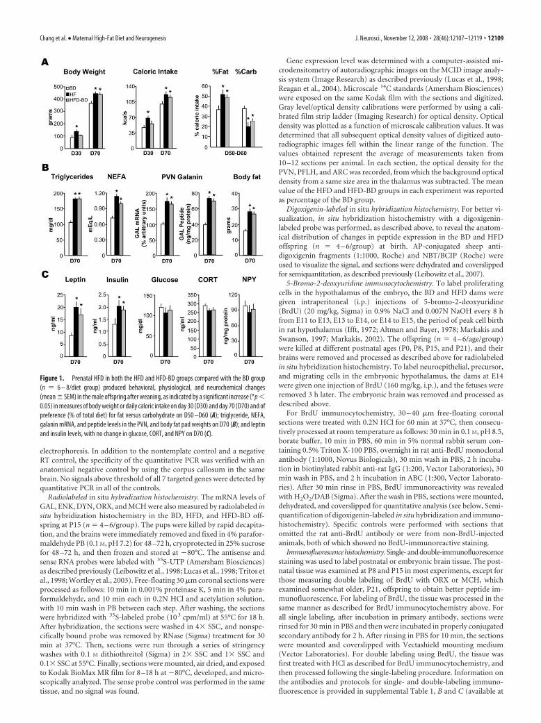
Figure 1. Prenatal HFD in both the HFD and HFD-BD groups compared with the BD group(n � 6 – 8/diet group) produced behavioral, physiological, and neurochemical changes(mean � SEM) in the male offspring after weaning, as indicated by a significant increase (*p �0.05) in measures of body weight or daily caloric intake on day 30 (D30) and day 70 (D70) and ofpreference (% of total diet) for fat versus carbohydrate on D50 –D60 (A); triglyceride, NEFA,galanin mRNA, and peptide levels in the PVN, and body fat pad weights on D70 (B); and leptinand insulin levels, with no change in glucose, CORT, and NPY on D70 (C).
Chang et al. • Maternal High-Fat Diet and Neurogenesis J. Neurosci., November 12, 2008 • 28(46):12107–12119 • 12109

www.jneurosci.org as supplemental material), and all procedures wereperformed at room temperature. Fluorescence image was captured witha Zeiss fluorescence microscope with MateVue software. Density of im-munofluorescence objects was quantified with ImagePro software as de-scribed below and reported as density (objects/�m 2). Double-labeledcells were counted and reported as percentage of total single-labeled cells.
Double labeling of digoxigenin in situ hybridization of peptide with BrdUimmunofluorescence histochemistry. Digoxigenin in situ hybridization ofpeptides, GAL, ENK, and DYN, in combination with BrdU immunoflu-orescence histochemistry was used to determine whether BrdU-labeledcells express these specific peptides. The P8 brains of BD, HFD, andHFD-BD offspring (n � 4 – 6/group) of dams with BrdU injections fromE11–13 were cut, and three sets of 30 �m free-floating alternative coronalsections were processed for digoxigenin in situ hybridization (see above).After the signal was visualized in NBT/BCIP, sections were briefly washedin 0.1 M Tris-HCl containing 0.1 M NaCl and 50 mM MgCl2, pH 9.5, andPBS, and then were treated in 0.2N HCl for 60 min at 37°C. Afterward,sections were processed for BrdU immunofluorescence and viewed on aZeiss microscope with MateVue software. Peptide-expressing neuronswith dark-blue digoxigenin-labeled signal were viewed and capturedwith DIC filter first, and then the red Texas Red/Cy3 fluorescence filterwas applied to reveal the BrdU-positive signal in the same field. Theimages were merged, and double-labeled cells were counted and expressedas percentage of total single-labeled cells.
Semiquantification of digoxigenin-labeled in situ hybridization and im-munohistochemistry. Semiquantification of cell density of GAL-, ENK-,DYN-, ORX-, and MCH-expressing neurons, BrdU-immunoreactivecells from DAB reaction, and immunofluorescence cells or fiber densityof BrdU, NeuN, Dcx, Tuj1, nestin, or vimentin were performed as de-scribed previously (Leibowitz et al., 2007). Briefly, sections were viewedon a Leitz microscope (10� objective). The images were captured with aNikon DXM 1200 digital camera (Nikon) and analyzed using ImageProPlus software (version 4.5, Media Cybernetics) on a gray-value scale from1 to 255. In each animal, 8 –10 sections at the same level were used toexamine each nucleus or area of interest, and the entire nucleus or areawas outlined and analyzed. The population density was used to deter-mine the cell or fiber density in these areas. Before measurements, athreshold for each nucleus or area was established. Using the selectedsections, this threshold was set by matching the number of objectscounted by the software in a defined area with the number of objectscounted manually in that same area. This method, which was the samefor all experiments and brain areas, yielded different threshold values(average of thresholds obtained within the same area in the 10 sections)for the different brain areas, experiments, and measurements of cellsversus fibers. This semiquantitative procedure allowed one to count thenumber of neurons or fibers in a specific area, which were then expressedas the cell density (number of cells/�m 2) or object density (number offibers/�m 2), respectively. The average cell or object density for the dif-ferent groups was then compared and statistically analyzed, with theanalyses performed by an observer unaware of the identity of the animals.
Radioimmunoassay. The PVN tissue, dissected from the BD, HFD, andHFD-BD offspring at P8 and P15 (n � 4 – 6/age/diet group), was homog-enized in 1 ml of 0.1 M acetic acid and centrifuged at 14,000 � g for 15 minat 4°C. All of the supernatant was removed, boiled for 10 min, and frozenat �80°C until use. For radioimmunoassay (RIA), GAL and NPY weremeasured as described previously (Akabayashi et al., 1994b; Leibowitz et al.,1998), and met-ENK and DYN-A were measured using commercially avail-able kits (Peninsula Laboratories).
Hormone and metabolite assays. Serum from trunk blood of BD,HFD, and HFD-BD offspring and dams was assayed using commer-cially available radioimmunoassay kits for insulin and leptin (LincoResearch) and corticosterone (MP Biomedicals). Serum TG and glu-cose levels were assayed using a Triglyceride Assay kit from Sigma orglucose kit from TECO Diagnostics, respectively. Nonesterified fattyacids (NEFAs) were assayed in serum using a kit from WAKO PureChemical Industries.
Statistical analysis. The values in the figures and tables are ex-pressed as mean � SEM. In each experiment, a direct comparisonbetween the scores for a pair of groups was made using an unpaired
Student’s t test. With multiple comparisons, data were analyzed byone-way ANOVA, with a Bonferroni post hoc test to determine signif-icant differences between the groups. The criterion for statistical sig-nificance was p � 0.05.
Figure 2. Prenatal HFD in both the HFD and HFD-BD groups compared with the BD group (n �4 – 6/age/diet group) produced changes in gene expression (mean � SD) of orexigenic peptides inthe PVN and PFLH on postnatal day 15 (P15), as measured by real-time quantitative PCR. This isindicated by a significant increase (*p � 0.05) in both prenatal HFD groups of GAL, ENK, and DYNmRNA levels in the PVN and ORX and MCH mRNA in the PFLH, along with a decrease in peptide mRNAin the ARC (A); and circulating levels of triglycerides, with no change in body weight or leptin andincreased levels only in the HFD group of NEFA, glucose, insulin, and CORT (B).
12110 • J. Neurosci., November 12, 2008 • 28(46):12107–12119 Chang et al. • Maternal High-Fat Diet and Neurogenesis

ResultsLong-term changes in adult offspring induced by prenatalHFD exposureBuilding on prior studies showing long-term, physiological ef-fects of HFD exposure extending through both pregnancy andlactation (see Introduction), we examined the effects of a shorterperiod of exposure to a HFD, 2 weeks prenatally (E6 –P0) orpostnatally (P1–P15). Also, rather than using a low-fat diet (10%fat) as a control, we compared this HFD (50% fat) to a morebalanced, moderate-fat diet (BD), which is closer in fat content(25% fat) and thus minimizes any group differences in caloricintake and body weight. This experiment had 4 sets of dams thatproduced 4 groups of offspring, as described in the Materials andMethods section. They were the (1) BD group, which was main-tained on a BD both prenatally and postnatally; (2) HFD group,which had a HFD both prenatally and postnatally; (3) HFD-BDgroup, which had the HFD only prenatally and the BD postna-tally, with the offspring cross-fostered at birth to BD dams neverpreviously exposed to a HFD; and (4) BD-HFD group, which had
the BD prenatally and HFD only duringthe postnatal period. Compared with theBD group, the two groups with prenatalHFD exposure, whether the HFD contin-ued postnatally until weaning (HFDgroup) or was removed at birth (HFD-BDgroup), exhibited similar behavioral,physiological, and neurochemical changesthat persisted after weaning (P21) in theabsence of the HFD, as illustrated for maleoffspring (Fig. 1). Although only the HFDoffspring exhibited an increase in bodyweight and daily caloric intake at 30 d ofage (D30), both the HFD and HFD-BDmale groups showed these effects after pu-berty, as indicated by the D70 measures,and exhibited an enhanced preference fordietary fat when a choice of the two dietswas provided from D50 to D60 (Fig. 1A).Further, when examined at D70, the HFDand HFD-BD offspring had increased se-rum levels of TG and NEFA, elevated GALmRNA and peptide levels in the PVN, andgreater body fat accrual (Fig. 1B). Thesechanges were accompanied by an increasein leptin and insulin, but no change in se-rum levels of glucose or CORT and in NPYlevels in the ARC (Fig. 1C). Similar effectswere observed in the female HFD andHFD-BD offspring (supplemental Table 2,available at www.jneurosci.org as supple-mental material), which showed an evengreater percentage increase ( p � 0.05)compared with males in their body fat(�140% vs �75%) and leptin levels(�240% vs �150%) and reached puberty(day of vaginal opening) at an earlier age(days 29 –32) compared with the BD off-spring (days 33–36). These changes inadult rats were not evident in the BD-HFDgroup, which was exposed for 2 weeks(P1–P15) to the HFD only during thepostnatal period (data not shown). Thesefindings underscore the importance of in
utero exposure to the HFD. They show it to be sufficient to pro-duce long-term effects, even compared with a moderate-fat BD,that persist after birth and weaning in the absence of the HFD.
Changes in preweanling offspring induced by prenatalHFD exposureThis experiment examined whether prenatal HFD exposure af-fects gene expression in preweanling offspring, similar to adults(see Introduction), of different orexigenic peptides that may con-tribute to their long-term changes in eating patterns. With damsplaced on the HFD or BD at E6, 3 groups of offspring, BD, HFD,and HFD-BD, were formed and then killed before weaning, at15 d of age (P15), just before the start of independent feeding.While having no impact on the body weight of the dams (320 –350 g) through gestational day 21, the maternal HFD had a pro-nounced, site-specific stimulatory effect on the offspring’s hypo-thalamic peptide mRNA, as measured by real-time quantitativePCR (Fig. 2A). At P15, the HFD compared with BD offspringshowed increased expression of GAL, ENK, and DYN in the PVN
Figure 3. Prenatal HFD in both the HFD and HFD-BD groups compared with the BD group (n � 4 – 6/diet group) producedchanges in the orexigenic peptides (mean � SD) in the PVN and PFLH of postnatal offspring, as measured by different techniques.This is indicated by a significant increase (*p � 0.05) in mRNA levels (% of BD group) of GAL, ENK, and DYN in the PVN and ORX andMCH in the PFLH at P15, as measured by radiolabeled in situ hybridization with 35S-labeled probes (A); photomicrographsillustrating this effect in the PVN and PFLH of the HFD-BD offspring compared with the BD (B); and a significant increase (*p �0.05) in peptide levels in the PVN, as measured by radioimmunoassay in P15 offspring, or in the PFLH, as measured by immuno-fluorescence in P21 offspring (C).
Chang et al. • Maternal High-Fat Diet and Neurogenesis J. Neurosci., November 12, 2008 • 28(46):12107–12119 • 12111

and ORX and MCH in the PFLH. This is incontrast to gene expression in the ARC,which for the 5 peptides examined was un-affected or greatly reduced. This HFD-induced increase in peptide was similarlydetected in younger pups at P8, shownwith measurements of PVN GAL andPFLH ORX (supplemental Table 3, avail-able at www.jneurosci.org as supplementalmaterial). As demonstrated in adult rats(see Introduction), these peptide changesin the HFD groups were closely associatedwith an increase in serum levels of TG inboth the P8 and P15 offspring; they weredissociated, in contrast, from the measuresof body weight and leptin, which remainedstable at both ages, and also of NEFA, glu-cose, insulin, and CORT levels, which wereelevated at P15 but not P8 (Fig. 2B; sup-plemental Table 3, available at www.jneurosci.org as supplemental material).Remarkably, the HFD-BD offspring com-pared with BD group showed a similar in-crease in peptide mRNA, once again, in thePVN and PFLH but not the ARC wheregene expression was suppressed (Fig. 2A).This provides the first evidence that a HFDrestricted to pregnancy produces changesin the brain that continue well beyondbirth and period of diet exposure. Interest-ingly, in these HFD-BD offspring, the inutero HFD also had effects on circulating TG levels that persistedin the absence of the diet (Fig. 2B; supplemental Table 3, availableat www.jneurosci.org as supplemental material). On the BD afterbirth, these lipids remained significantly elevated throughout thepostnatal period, whereas the other measures of body weight,nutrients, and hormones in the HFD-BD group were unaltered.
These findings, that prenatal HFD exposure has persistingeffects on hypothalamic peptide expression, were substantiatedin additional sets of postnatal offspring using different tech-niques. Measurements with radiolabeled in situ hybridization re-vealed significantly elevated peptide mRNA levels in the HFD andHFD-BD groups compared with BD offspring at P15 (Fig. 3A).These findings are illustrated in photomicrographs of GAL, ENK,and DYN mRNA in the PVN and of ORX and MCH mRNA in thePFLH (Fig. 3B). In accordance with these changes in gene expres-sion, examination of another set of HFD and HFD-BD offspringrevealed increased levels of the peptides in the PVN and PFLH, asmeasured by RIA or immunofluorescence (Fig. 3C). These re-sults, showing clear changes in both mRNA and peptide levels inpostnatal pups that had elevated TG similar to Figure 2B, dem-onstrate the profound vulnerability of the offspring to dietary fatduring gestation. They reveal marked changes in specific hypo-thalamic peptide systems controlling feeding and peripheral sys-tems involved in lipid metabolism, which persist long after par-turition and perhaps into adulthood and thus may be attributedto phenomena occurring in utero.
Effects of prenatal HFD exposure on the density of neuronsat birthTo visualize possible changes in peptide-expressing neurons atbirth, we used digoxigenin-labeled in situ hybridization to exam-ine the offspring of dams maintained on the HFD or BD from E6
to parturition. The BD offspring had very few detectable GAL-positive neurons in the PVN at P0; thus, the impact of the mater-nal diet on soma density was particularly notable, with the HFDoffspring at birth showing a much larger number of PVN GAL-positive neurons (Fig. 4A,B). This effect was site specific, occur-ring only in the anterior parvocellular region of this nucleus butnot in the ARC or dorsomedial nucleus (DMN). The maternalHFD also increased the density of ENK- and DYN-expressingneurons in the PVN at birth, and this effect was evident in themiddle-to-posterior region of this nucleus but, once again, not inthe ARC, DMN, or ventromedial nucleus (VMN). The peptidesexpressed in the PFLH were similarly affected by the HFD (Fig.4A,B). There was an increase in the density of ORX and MCHneurons, in both the perifornical area dorsal to the fornix (PFH)and the far lateral hypothalamus (LH), as illustrated for ORX.This increased density of neurons at P0 raises the intriguing pos-sibility that a fat-rich diet creates a particular environment inutero that affects the development of specific neurons in the PVNand PFLH that ultimately express fat-sensitive, orexigenicpeptides.
Effects of in utero HFD exposure on cell generationTo test whether the HFD stimulates the generation of new cellsduring pregnancy, dams placed on a HFD or BD at E6 were giveninjections of the cell proliferation marker, BrdU, from E11 toE13, E13 to E14, or E14 to E15. At birth (P0), the offspring fromHFD dams that received BrdU injections during these gestationalperiods exhibited a significant increase in the density of BrdU-positive (BrdU�) cells in the PVN and PFLH (Fig. 5A,B). This isin contrast to the ARC or hippocampus, where no change wasobserved (supplemental Fig. 1, available at www.jneurosci.org assupplemental material). In additional animals given BrdU injec-tions from E11 to E13, this stimulatory effect on BrdU� cell
Figure 4. Prenatal HFD compared with BD offspring (n � 4 – 6/diet group) produced changes in orexigenic peptide mRNA inthe PVN and PFLH at birth (P0), as measured using in situ hybridization with digoxigenin-labeled probes. This is indicated by asignificant increase (*p � 0.01) in mRNA levels (mean � SEM) of GAL, ENK, and DYN in the PVN and of ORX and MCH in the PFHand LH (A); and photomicrographs of GAL mRNA in the anterior PVN outlined by a dash line and ORX mRNA in the PFLH just dorsaland lateral to the fornix (F) (scale bar: 100 �m) (B).
12112 • J. Neurosci., November 12, 2008 • 28(46):12107–12119 Chang et al. • Maternal High-Fat Diet and Neurogenesis

density in the PVN and PFLH was simi-larly evident in the HFD-BD as well asHFD offspring examined at P8 or P15,whether they were initially exposed to theHFD at E6 or as late as E9 (supplementalFig. 2, available at www.jneurosci.org assupplemental material). These results un-derscore the robustness of this phenome-non. In multiple groups, they demonstratean increase in cell generation that is sitespecific, requires only a few days of in uteroHFD exposure, and persists postnatallywell beyond the period of diet exposure.
Impact of in utero HFD exposureon neurogenesisTo investigate the phenotypes of these hy-pothalamic BrdU� cells, we performedsingle- or double-labeling immunofluo-rescence histochemistry using antibodiesagainst NeuN (marker for mature neu-rons), GFAP (marker for astrocytes), andGalC (marker for oligodendrocytes). Withthe dams given diets at E6, the HFD andHFD-BD offspring compared with the BDgroup exhibited a marked increase in cola-beling of NeuN with BrdU in the PVN andPFLH at P8 (Fig. 5C). This is indicated inboth areas by the increased percentage ofdouble-labeled cells relative to the totalsingle-labeled cells immunoreactive forBrdU or for NeuN. Whereas the glialmarkers revealed relatively few labeledcells in the PVN and PFLH before P8, therewere considerably more at P15 and P25,and the number seen at these ages was unaf-fected or even slightly reduced in the HFDand HFD-BD offspring (supplemental Fig.3, available at www.jneurosci.org as supple-mental material). This demonstrates that inutero HFD exposure has its greatest impacton the generation and development of neu-rons, rather than astrocytes or oligodendro-cytes, in these hypothalamic areas.
Phenotype of new neurons generated byin utero HFD exposureThe next step, using digoxigenin in situ hy-bridization histochemistry combined withBrdU immunofluorescence or double-labeling immunofluorescence, was to de-termine whether these BrdU� neurons af-fected by the prenatal HFD can express orsynthesize the same orexigenic peptides,GAL, ENK, DYN, ORX, and MCH, knownto be stimulated by dietary fat in adults.With dams placed on a diet at E6 and givenBrdU injections from E11 to E13, the post-natal offspring in the HFD-BD as well asHFD litters exhibited a significant increasein colabeling of BrdU with the differentpeptides in the PVN and PFLH. This effect,indicated by the increased percentage of
Figure 5. Prenatal HFD stimulated cell proliferation and neurogenesis in the PVN and PFLH of postnatal offspring compared with BD(n�4 – 6/diet group). This is indicated by a significant increase (*p�0.01) in BrdU � cell density in the PVN and PFLH of the HFD versusBD offspring at P0, from dams receiving i.p. injections of BrdU from E11 to E13, E13 to E14, and E14 to E15 (mean � SEM) (A); photomi-crographsillustratingBrdU �cells inthePVN(dashline)andPFLHaroundthefornix(F)ofP0offspring(B);andasignificantincrease(*p�0.01) in double-labeled BrdU �/NeuN � cells in the PVN (scale bar: 100�m) and PFLH (scale bar: 50�m) of HFD and HFD-BD offspring atP8,presentedaspercentageofdouble-labeledneuronsrelativetototalsingle-labeledBrdU �cellsorNeuN �neurons(mean�SEM)(C).
Chang et al. • Maternal High-Fat Diet and Neurogenesis J. Neurosci., November 12, 2008 • 28(46):12107–12119 • 12113
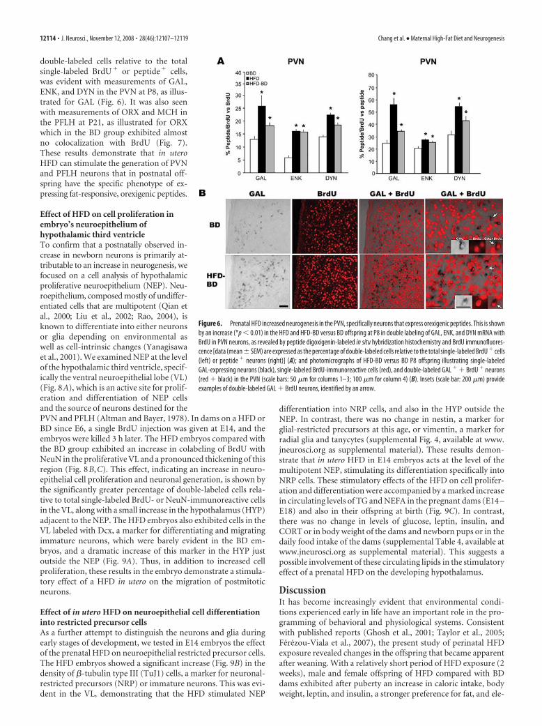
double-labeled cells relative to the totalsingle-labeled BrdU� or peptide� cells,was evident with measurements of GAL,ENK, and DYN in the PVN at P8, as illus-trated for GAL (Fig. 6). It was also seenwith measurements of ORX and MCH inthe PFLH at P21, as illustrated for ORXwhich in the BD group exhibited almostno colocalization with BrdU (Fig. 7).These results demonstrate that in uteroHFD can stimulate the generation of PVNand PFLH neurons that in postnatal off-spring have the specific phenotype of ex-pressing fat-responsive, orexigenic peptides.
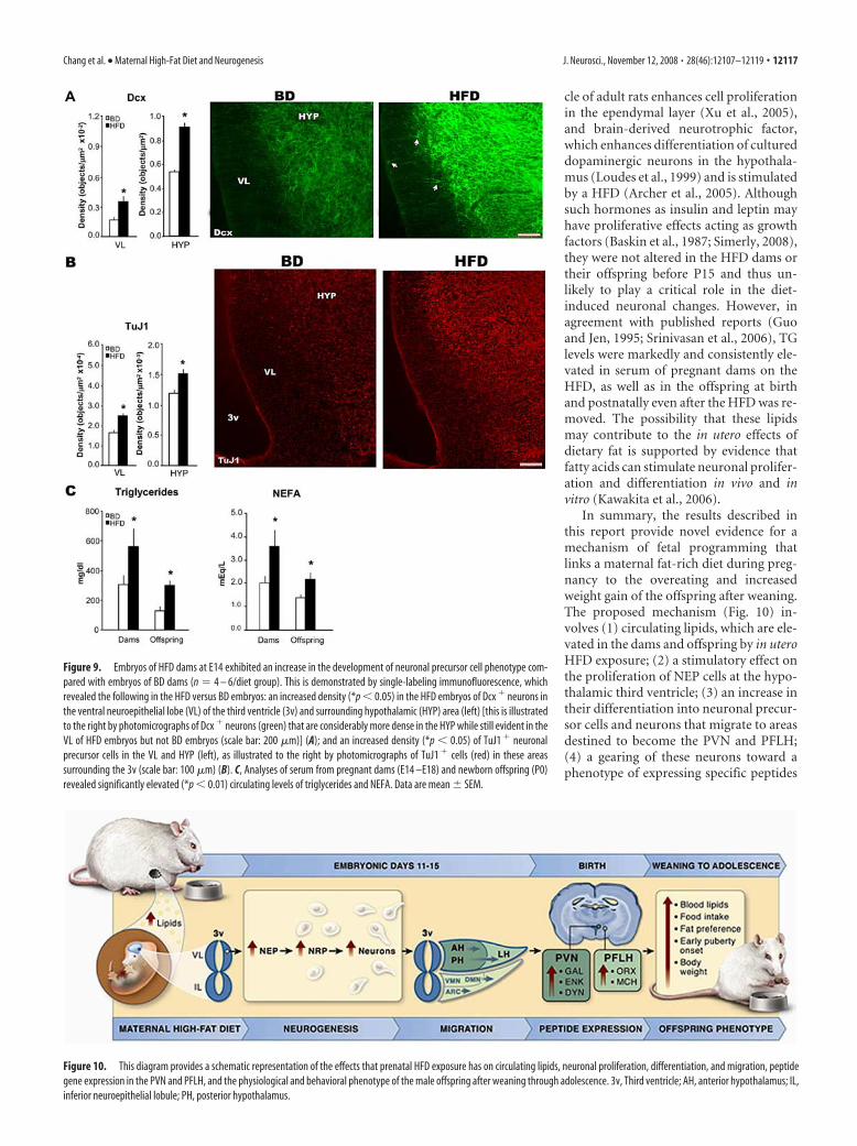
Effect of HFD on cell proliferation inembryo’s neuroepithelium ofhypothalamic third ventricleTo confirm that a postnatally observed in-crease in newborn neurons is primarily at-tributable to an increase in neurogenesis, wefocused on a cell analysis of hypothalamicproliferative neuroepithelium (NEP). Neu-roepithelium, composed mostly of undiffer-entiated cells that are multipotent (Qian etal., 2000; Liu et al., 2002; Rao, 2004), isknown to differentiate into either neuronsor glia depending on environmental aswell as cell-intrinsic changes (Yanagisawaet al., 2001). We examined NEP at the levelof the hypothalamic third ventricle, specif-ically the ventral neuroepithelial lobe (VL)(Fig. 8A), which is an active site for prolif-eration and differentiation of NEP cellsand the source of neurons destined for thePVN and PFLH (Altman and Bayer, 1978). In dams on a HFD orBD since E6, a single BrdU injection was given at E14, and theembryos were killed 3 h later. The HFD embryos compared withthe BD group exhibited an increase in colabeling of BrdU withNeuN in the proliferative VL and a pronounced thickening of thisregion (Fig. 8B,C). This effect, indicating an increase in neuro-epithelial cell proliferation and neuronal generation, is shown bythe significantly greater percentage of double-labeled cells rela-tive to total single-labeled BrdU- or NeuN-immunoreactive cellsin the VL, along with a small increase in the hypothalamus (HYP)adjacent to the NEP. The HFD embryos also exhibited cells in theVL labeled with Dcx, a marker for differentiating and migratingimmature neurons, which were barely evident in the BD em-bryos, and a dramatic increase of this marker in the HYP justoutside the NEP (Fig. 9A). Thus, in addition to increased cellproliferation, these results in the embryo demonstrate a stimula-tory effect of a HFD in utero on the migration of postmitoticneurons.
Effect of in utero HFD on neuroepithelial cell differentiationinto restricted precursor cellsAs a further attempt to distinguish the neurons and glia duringearly stages of development, we tested in E14 embryos the effectof the prenatal HFD on neuroepithelial restricted precursor cells.The HFD embryos showed a significant increase (Fig. 9B) in thedensity of �-tubulin type III (TuJ1) cells, a marker for neuronal-restricted precursors (NRP) or immature neurons. This was evi-dent in the VL, demonstrating that the HFD stimulated NEP
differentiation into NRP cells, and also in the HYP outside theNEP. In contrast, there was no change in nestin, a marker forglial-restricted precursors at this age, or vimentin, a marker forradial glia and tanycytes (supplemental Fig. 4, available at www.jneurosci.org as supplemental material). These results demon-strate that in utero HFD in E14 embryos acts at the level of themultipotent NEP, stimulating its differentiation specifically intoNRP cells. These stimulatory effects of the HFD on cell prolifer-ation and differentiation were accompanied by a marked increasein circulating levels of TG and NEFA in the pregnant dams (E14 –E18) and also in their offspring at birth (Fig. 9C). In contrast,there was no change in levels of glucose, leptin, insulin, andCORT or in body weight of the dams and newborn pups or in thedaily food intake of the dams (supplemental Table 4, available atwww.jneurosci.org as supplemental material). This suggests apossible involvement of these circulating lipids in the stimulatoryeffect of a prenatal HFD on the developing hypothalamus.
DiscussionIt has become increasingly evident that environmental condi-tions experienced early in life have an important role in the pro-gramming of behavioral and physiological systems. Consistentwith published reports (Ghosh et al., 2001; Taylor et al., 2005;Ferezou-Viala et al., 2007), the present study of perinatal HFDexposure revealed changes in the offspring that became apparentafter weaning. With a relatively short period of HFD exposure (2weeks), male and female offspring of HFD compared with BDdams exhibited after puberty an increase in caloric intake, bodyweight, leptin, and insulin, a stronger preference for fat, and ele-
Figure 6. Prenatal HFD increased neurogenesis in the PVN, specifically neurons that express orexigenic peptides. This is shownby an increase (*p � 0.01) in the HFD and HFD-BD versus BD offspring at P8 in double labeling of GAL, ENK, and DYN mRNA withBrdU in PVN neurons, as revealed by peptide digoxigenin-labeled in situ hybridization histochemistry and BrdU immunofluores-cence [data (mean�SEM) are expressed as the percentage of double-labeled cells relative to the total single-labeled BrdU � cells(left) or peptide � neurons (right)] (A); and photomicrographs of HFD-BD versus BD P8 offspring illustrating single-labeledGAL-expressing neurons (black), single-labeled BrdU-immunoreactive cells (red), and double-labeled GAL � � BrdU � neurons(red � black) in the PVN (scale bars: 50 �m for columns 1–3; 100 �m for column 4) (B). Insets (scale bar: 200 �m) provideexamples of double-labeled GAL � BrdU neurons, identified by an arrow.
12114 • J. Neurosci., November 12, 2008 • 28(46):12107–12119 Chang et al. • Maternal High-Fat Diet and Neurogenesis

vated TG levels and GAL mRNA and peptide in the PVN. Ourfinding, that these effects occurred with 2 weeks of in utero HFDexposure but not postnatal HFD, isolated the prenatal period as acritical time in programming PVN GAL neurons in associationwith enhanced preference for dietary fat and increased weightgain.
Similar to the offspring after puberty, HFD exposure duringboth the prenatal and postnatal periods stimulated expression of5 orexigenic peptides in the PVN and PFLH of preweanling off-spring. This effect is similar to that described in previous studiesof GAL and ORX in weanling rats (Kozak et al., 1998; Beck et al.,2006) and of the 5 peptides in adult rats (Akabayashi et al., 1994a;Welch et al., 1996; Leibowitz et al., 2004; Chang et al., 2007;Kennedy et al., 2007). It is very different, however, from the re-sults obtained in the ARC, where peptides are unaffected or re-duced by a HFD and low-protein diets during pregnancy andlactation, while enhanced by undernutrition and gestational dia-betes (Kozak et al., 1998; Plagemann et al., 2000; Franke et al.,2005; Beck et al., 2006; Plagemann, 2006). Examination of pups atP8, P15, and P21 showed that the change in orexigenic peptides inthe PVN and PFLH could occur in the absence of any alteration inbody weight or leptin and before the start of independent feeding,suggesting that it is transmitted via circulating factors, such asnutrients, that reach the offspring through the dam’s milk. With
each of the 5 peptides, the increase inmRNA in the PVN and PFLH of HFD off-spring was accompanied by a marked in-crease in peptide levels. This finding sup-ports the possibility that theseneurochemical changes have biologicalsignificance. Based on pharmacologicalstudies in adult rats (Zhang et al., 1998;Leibowitz, 2000; Clegg et al., 2002; Yun etal., 2005), one would expect their long-term functional consequences to be an in-crease in food intake and body weight on afat-rich diet. These effects were observedin the prenatal HFD offspring at 70 d ofage.
Most important were the findings inthe HFD-BD offspring cross-fostered atbirth to BD dams. These offspring exhib-ited the same postnatal changes in pep-tides, demonstrating that they are irrevers-ible by removal of the HFD and thus maybe occurring in utero and/or at birth. Thereare only a few reports examining prena-tally manipulated offspring at birth forphysiological or peptide effects (Guo andJen, 1995; Plagemann et al., 1998; Cerf etal., 2006). In newborn HFD offspring, weobserved a greater density of peptide-expressing neurons, raising the possibilitythat the prenatal diet exposure may be in-creasing the number of cells born duringgestation. This effect was site specific, lo-calized to the anterior parvocellular PVNfor GAL and middle-to-posterior PVN forENK and DYN and to both the perifornicaland lateral hypothalamic areas for ORXand MCH.
Investigation of the mechanism under-lying this neuronal change revealed an in-
crease in the generation of new cells and neurons in the hypothal-amus in response to in utero HFD. As with the changes in geneexpression, this phenomenon was evident at P15 in the HFD-BDas well as HFD offspring, thus not reversible by normalizing thediet, and it occurred only in the PVN and PFLH. It was not seen inthe hippocampus, where a HFD in adult rats reduces cell density(Lindqvist et al., 2006). It was also not evident in the ARC, wherethe orexigenic peptides are reduced by dietary fat or lipids inadult rats (Chang et al., 2004; Leibowitz and Wortley, 2004) asshown here in HFD offspring, and also where they are particu-larly responsive to leptin, insulin, and glucose (Leibowitz andWortley, 2004; Simerly, 2008), which were unaltered in the HFDoffspring and dams and thus unlikely to play a major role in thedevelopmental changes.
Although many cells with a glial phenotype were evident in thehypothalamic ventricular area or forebrain regions, the PVN andPFLH had relatively few even at P25, confirming reports thatmature glia in the hypothalamus develop considerably later thanneurons (Jacobson, 1991). Prenatal exposure to the HFD had noeffect on glial density in these areas. Further, in both the HFD-BDand HFD offspring, a significantly higher proportion of the newlygenerated neurons had a specific phenotype of expressing orexi-genic peptides, GAL, ENK, and DYN in the PVN and ORX andMCH in the PFLH. These results demonstrate, for the first time,
Figure 7. Prenatal HFD increased neurogenesis in the PFLH, specifically neurons that express orexigenic peptides. This is shownby an increase (*p �0.01) in the HFD and HFD-BD versus BD offspring at P21 in double labeling of ORX and MCH peptide with BrdUin PFLH neurons, as revealed by double-labeling immunofluorescence [data (mean � SEM) are expressed as the percentage ofdouble-labeled cells relative to the total single-labeled BrdU � cells (left) or peptide � neurons (right)] (A); and photomicro-graphs of HFD-BD versus BD P21 offspring illustrating single-labeled ORX-synthesizing neurons (green), single-labeled BrdU-immunoreactive cells (red), and double-labeled ORX � � BrdU � neurons (green � red) in the PFLH (scale bar: 100 �m), in thearea of the fornix (F) (B). Examples of double-labeled neurons are indicated by arrows.
Chang et al. • Maternal High-Fat Diet and Neurogenesis J. Neurosci., November 12, 2008 • 28(46):12107–12119 • 12115

that dietary fat can stimulate the genera-tion of specific new neurons, thus affectingneuronal phenotype and presumably neu-ronal differentiation. Although the mech-anisms determining hypothalamic neuro-nal phenotype are not well understood, apossible role for transcription factors inthis process is suggested by evidence thatShh can specify the identity of hypotha-lamic dopamine neurons in chick embryos(Ohyama et al., 2005) and that POU do-main transcription factors like Brn-2 arerequired for the specification of neuronallineages in developing hypothalamus (Na-kai et al., 1995). Interestingly, Shh is activeonly when bound to cholesterol (Ohyamaet al., 2005), which is known to be elevatedby prenatal HFD (Brown et al., 1990).
Further, examination of NEP at E14 re-vealed a stimulatory effect of in utero HFDexposure on the density of progenitorcells, NRP (Tuj1) rather than GRP (Nes-tin), as well as on mature new neurons(NeuN/BrdU) and migrating neurons(Dcx) at and around the third ventricle.An increase in the density of both NRPcells and neurons indicates that there wasincreased differentiation of NEP into thesecell types, making more postmitotic neu-rons available to populate different hypo-thalamic areas. It also suggests that neuro-nal proliferation rather than cell survival ismore likely to be responsible for the in-crease in new neurons seen postnatally.This is supported by the short-term natureof our experimental paradigm, which ex-amined proliferating cells 3 h after BrdUinjection before their final differentiationor migration. This paradigm allowed us toavoid cell death events that occur duringthe translocation of postmitotic cells outof the proliferative zone and into differen-tiating tissue (Barres et al., 1992). Al-though the phenomenon of cell deathneeds further investigation, it is notewor-thy that NEP lacks neurotrophin TrkC re-ceptors known to be important for cellsurvival (Hassink et al., 1999). A possiblemechanism underlying the HFD-inducedincrease in neurogenesis may involvechanges in the cell-cycle kinetics, as sug-gested for the dopamine system in thebrain (Ohtani et al., 2003).
The proliferative and differentiating ef-fects of the maternal HFD may involve theactions of growth factors, which areknown to regulate hypothalamic neuro-genesis at its different stages. These mayinclude insulin-like growth factor-1,which is regulated maternally and fetallyby nutrient availability (Gluckman et al.,1996), fibroblast growth factor whose in-jection into the hypothalamic third ventri-
Figure 8. Embryos of HFD dams at E14 exhibited an increase in neurogenesis compared with embryos of BD dams (n �4 – 6/diet group). A, The dash box in the coronal section shows the ventral neuroepithelial lobe (VL) of the hypothalamic thirdventricle (3v) and the surrounding hypothalamic area (HYP). B, C, The enhanced neurogenesis in HFD embryos is demonstrated bydouble-labeling immunofluorescence revealing an increase (*p � 0.05) in the percentage of BrdU �/NeuN � newborn neuronsrelative to the total number of single-labeled BrdU � cells (left) or NeuN � neurons (right) in the VL and HYP (mean � SEM) (B);and immunofluorescence photomicrographs of HFD versus BD embryos illustrating an increase in BrdU � cells (red, toppanel), NeuN � neurons (green, middle panel), and double-labeled BrdU � � NeuN � newborn neurons (yellow, bottompanel) in the VL, with a small effect in the HYP adjacent to the VL (scale bar: 100 �m) (C). The inset in bottom panel for HFDembryo, showing an enlargement of the area indicated by a box, illustrates examples of double-labeled neurons (yellow)detected in the VL.
12116 • J. Neurosci., November 12, 2008 • 28(46):12107–12119 Chang et al. • Maternal High-Fat Diet and Neurogenesis
cle of adult rats enhances cell proliferationin the ependymal layer (Xu et al., 2005),and brain-derived neurotrophic factor,which enhances differentiation of cultureddopaminergic neurons in the hypothala-mus (Loudes et al., 1999) and is stimulatedby a HFD (Archer et al., 2005). Althoughsuch hormones as insulin and leptin mayhave proliferative effects acting as growthfactors (Baskin et al., 1987; Simerly, 2008),they were not altered in the HFD dams ortheir offspring before P15 and thus un-likely to play a critical role in the diet-induced neuronal changes. However, inagreement with published reports (Guoand Jen, 1995; Srinivasan et al., 2006), TGlevels were markedly and consistently ele-vated in serum of pregnant dams on theHFD, as well as in the offspring at birthand postnatally even after the HFD was re-moved. The possibility that these lipidsmay contribute to the in utero effects ofdietary fat is supported by evidence thatfatty acids can stimulate neuronal prolifer-ation and differentiation in vivo and invitro (Kawakita et al., 2006).
In summary, the results described inthis report provide novel evidence for amechanism of fetal programming thatlinks a maternal fat-rich diet during preg-nancy to the overeating and increasedweight gain of the offspring after weaning.The proposed mechanism (Fig. 10) in-volves (1) circulating lipids, which are ele-vated in the dams and offspring by in uteroHFD exposure; (2) a stimulatory effect onthe proliferation of NEP cells at the hypo-thalamic third ventricle; (3) an increase intheir differentiation into neuronal precur-sor cells and neurons that migrate to areasdestined to become the PVN and PFLH;(4) a gearing of these neurons toward aphenotype of expressing specific peptides
Figure 9. Embryos of HFD dams at E14 exhibited an increase in the development of neuronal precursor cell phenotype com-pared with embryos of BD dams (n � 4 – 6/diet group). This is demonstrated by single-labeling immunofluorescence, whichrevealed the following in the HFD versus BD embryos: an increased density (*p � 0.05) in the HFD embryos of Dcx � neurons inthe ventral neuroepithelial lobe (VL) of the third ventricle (3v) and surrounding hypothalamic (HYP) area (left) [this is illustratedto the right by photomicrographs of Dcx � neurons (green) that are considerably more dense in the HYP while still evident in theVL of HFD embryos but not BD embryos (scale bar: 200 �m)] (A); and an increased density (*p � 0.05) of TuJ1 � neuronalprecursor cells in the VL and HYP (left), as illustrated to the right by photomicrographs of TuJ1 � cells (red) in these areassurrounding the 3v (scale bar: 100 �m) (B). C, Analyses of serum from pregnant dams (E14 –E18) and newborn offspring (P0)revealed significantly elevated (*p � 0.01) circulating levels of triglycerides and NEFA. Data are mean � SEM.
Figure 10. This diagram provides a schematic representation of the effects that prenatal HFD exposure has on circulating lipids, neuronal proliferation, differentiation, and migration, peptidegene expression in the PVN and PFLH, and the physiological and behavioral phenotype of the male offspring after weaning through adolescence. 3v, Third ventricle; AH, anterior hypothalamus; IL,inferior neuroepithelial lobule; PH, posterior hypothalamus.
Chang et al. • Maternal High-Fat Diet and Neurogenesis J. Neurosci., November 12, 2008 • 28(46):12107–12119 • 12117
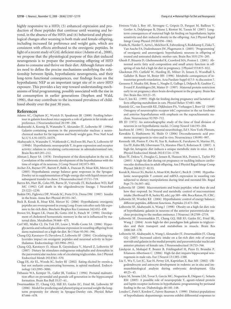
highly responsive to a HFD; (5) enhanced expression and pro-duction of these peptides that continue until weaning and be-yond, in the absence of the HFD; and (6) behavioral and physio-logical changes after weaning in both male and female offspring,including increased fat preference and weight gain, which areconsistent with effects attributed to the orexigenic peptides. Inlight of a recent study of GAL deficient mice (Adams et al., 2008),we propose that the physiological purpose of this diet-inducedneurogenesis is to prepare the postweaning offspring of HFDdams to consume and thrive on their diet. Although future stud-ies need to define the precise mechanisms underlying the rela-tionship between lipids, hypothalamic neurogenesis, and theirlong-term functional consequences, our findings focus on thehypothalamic NEP as an important target site of in utero HFDexposure. This provides a key step toward understanding mech-anisms of fetal programming, possibly associated with the rise infat consumption in earlier generations (Stephen and Wald,1990), that may contribute to the increased prevalence of child-hood obesity over the past 30 years.
ReferencesAdams AC, Clapham JC, Wynick D, Speakman JR (2008) Feeding behav-
iour in galanin knockout mice supports a role of galanin in fat intake andpreference. J Neuroendocrinol 20:199 –206.
Akabayashi A, Koenig JI, Watanabe Y, Alexander JT, Leibowitz SF (1994a)Galanin-containing neurons in the paraventricular nucleus: a neuro-chemical marker for fat ingestion and body weight gain. Proc Natl AcadSci U S A 91:10375–10379.
Akabayashi A, Watanabe Y, Wahlestedt C, McEwen BS, Paez X, Leibowitz SF(1994b) Hypothalamic neuropeptide Y, its gene expression and receptoractivity: relation to circulating corticosterone in adrenalectomized rats.Brain Res 665:201–212.
Altman J, Bayer SA (1978) Development of the diencephalon in the rat. II.Correlation of the embryonic development of the hypothalamus with thetime of origin of its neurons. J Comp Neurol 182:973–993.
Archer ZA, Rayner DV, Barrett P, Balik A, Duncan JS, Moar KM, Mercer JG(2005) Hypothalamic energy balance gene responses in the Sprague-Dawley rat to supplementation of high-energy diet with liquid ensure andsubsequent transfer to chow. J Neuroendocrinol 17:711–719.
Barres BA, Hart IK, Coles HS, Burne JF, Voyvodic JT, Richardson WD, RaffMC (1992) Cell death in the oligodendrocyte lineage. J Neurobiol23:1221–1230.
Baskin DG, Figlewicz DP, Woods SC, Porte D Jr, Dorsa DM (1987) Insulinin the brain. Annu Rev Physiol 49:335–347.
Beck B, Kozak R, Moar KM, Mercer JG (2006) Hypothalamic orexigenicpeptides are overexpressed in young Long-Evans rats after early life expo-sure to fat-rich diets. Biochem Biophys Res Commun 342:452– 458.
Brown SA, Rogers LK, Dunn JK, Gotto AM Jr, Patsch W (1990) Develop-ment of cholesterol homeostatic memory in the rat is influenced by ma-ternal diets. Metabolism 39:468 – 473.
Cerf ME, Muller CJ, Du Toit DF, Louw J, Wolfe-Coote SA (2006) Hyper-glycaemia and reduced glucokinase expression in weanling offspring fromdams maintained on a high-fat diet. Br J Nutr 95:391–396.
Chang GQ, Karatayev O, Davydova Z, Leibowitz SF (2004) Circulating trig-lycerides impact on orexigenic peptides and neuronal activity in hypo-thalamus. Endocrinology 145:3904 –3912.
Chang GQ, Karatayev O, Ahsan R, Gaysinskaya V, Marwil Z, Leibowitz SF(2007) Dietary fat stimulates endogenous enkephalin and dynorphin inthe paraventricular nucleus: role of circulating triglycerides. Am J PhysiolEndocrinol Metab 292:E561–570.
Clegg DJ, Air EL, Woods SC, Seeley RJ (2002) Eating elicited by orexin-a,but not melanin-concentrating hormone, is opioid mediated. Endocri-nology 143:2995–3000.
Debassio WA, Kemper TL, Galler JR, Tonkiss J (1994) Prenatal malnutri-tion effect on pyramidal and granule cell generation in the hippocampalformation. Brain Res Bull 35:57– 61.
Dourmashkin JT, Chang GQ, Hill JO, Gayles EC, Fried SK, Leibowitz SF(2006) Model for predicting and phenotyping at normal weight the long-term propensity for obesity in Sprague-Dawley rats. Physiol Behav87:666 – 678.
Ferezou-Viala J, Roy AF, Serougne C, Gripois D, Parquet M, Bailleux V,Gertler A, Delplanque B, Djiane J, Riottot M, Taouis M (2007) Long-term consequences of maternal high-fat feeding on hypothalamic leptinsensitivity and diet-induced obesity in the offspring. Am J Physiol RegulIntegr Comp Physiol 293:R1056 –1062.
Franke K, Harder T, Aerts L, Melchior K, Fahrenkrog S, Rodekamp E, Ziska T,Van Assche FA, Dudenhausen JW, Plagemann A (2005) ‘Programming’of orexigenic and anorexigenic hypothalamic neurons in offspring oftreated and untreated diabetic mother rats. Brain Res 1031:276 –283.
Ghosh P, Bitsanis D, Ghebremeskel K, Crawford MA, Poston L (2001) Ab-normal aortic fatty acid composition and small artery function in off-spring of rats fed a high fat diet in pregnancy. J Physiol 533:815– 822.
Gluckman PD, Cutfield W, Harding JE, Milner D, Jensen E, Woodhall S,Gallaher B, Bauer M, Breier BH (1996) Metabolic consequences of in-trauterine growth retardation. Acta Paediatr Suppl 417:3– 6; discussion 7.
Gressens P, Muaku SM, Besse L, Nsegbe E, Gallego J, Delpech B, Gaultier C,Evrard P, Ketelslegers JM, Maiter D (1997) Maternal protein restrictionearly in rat pregnancy alters brain development in the progeny. Brain ResDev Brain Res 103:21–35.
Guo F, Jen KL (1995) High-fat feeding during pregnancy and lactation af-fects offspring metabolism in rats. Physiol Behav 57:681– 686.
Hassink GC, van Esseveldt KE, Dijkhuizen PA, Verhaagen J, Boer GJ (1999)Ontogeny of neurotrophin receptor trkC expression in the rat forebrainand anterior hypothalamus with emphasis on the suprachiasmatic nu-cleus. Neuroscience 92:705–712.
Ifft JD (1972) An autoradiographic study of the time of final division ofneurons in rat hypothalamic nuclei. J Comp Neurol 144:193–204.
Jacobson M (1991) Developmental neurobiology, Ed 3. New York: Plenum.Kawakita E, Hashimoto M, Shido O (2006) Docosahexaenoic acid pro-
motes neurogenesis in vitro and in vivo. Neuroscience 139:991–997.Kennedy AR, Pissios P, Otu H, Xue B, Asakura K, Furukawa N, Marino FE,
Liu FF, Kahn BB, Libermann TA, Maratos-Flier E, Roberson R (2007) Ahigh-fat, ketogenic diet induces a unique metabolic state in mice. Am JPhysiol Endocrinol Metab 292:E1724 –1739.
Khan IY, Dekou V, Douglas G, Jensen R, Hanson MA, Poston L, Taylor PD(2005) A high-fat diet during rat pregnancy or suckling induces cardio-vascular dysfunction in adult offspring. Am J Physiol Regul Integr CompPhysiol 288:R127–R133.
Kozak R, Mercer JG, Burlet A, Moar KM, Burlet C, Beck B (1998) Hypotha-lamic neuropeptide Y content and mRNA expression in weanling ratssubjected to dietary manipulations during fetal and neonatal life. RegulPept 75–76:397– 402.
Leibowitz SF (2000) Macronutrients and brain peptides: what they do andhow they respond. In: Neural and metabolic control of macronutrientintake (Berthoud H-R, Seeley RJ, eds), pp 389 – 406. Boca Raton, FL: CRC.
Leibowitz SF, Wortley KE (2004) Hypothalamic control of energy balance:different peptides, different functions. Peptides 25:473–504.
Leibowitz SF, Akabayashi A, Wang J (1998) Obesity on a high-fat diet: roleof hypothalamic galanin in neurons of the anterior paraventricular nu-cleus projecting to the median eminence. J Neurosci 18:2709 –2719.
Leibowitz SF, Dourmashkin JT, Chang GQ, Hill JO, Gayles EC, Fried SK,Wang J (2004) Acute high-fat diet paradigms link galanin to triglycer-ides and their transport and metabolism in muscle. Brain Res1008:168 –178.
Leibowitz SF, Akabayashi A, Wang J, Alexander JT, Dourmashkin JT, ChangGQ (2007) Increased caloric intake on a fat-rich diet: role of ovariansteroids and galanin in the medial preoptic and paraventricular nuclei andanterior pituitary of female rats. J Neuroendocrinol 19:753–766.
Lindqvist A, Mohapel P, Bouter B, Frielingsdorf H, Pizzo D, Brundin P,Erlanson-Albertsson C (2006) High-fat diet impairs hippocampal neu-rogenesis in male rats. Eur J Neurol 13:1385–1388.
Liu Y, Wu Y, Lee JC, Xue H, Pevny LH, Kaprielian Z, Rao MS (2002) Oli-godendrocyte and astrocyte development in rodents: an in situ and im-munohistological analysis during embryonic development. Glia40:25– 43.
Lopez M, Seoane LM, Tovar S, García MC, Nogueiras R, Dieguez C, SenarísRM (2005) A possible role of neuropeptide Y, agouti-related proteinand leptin receptor isoforms in hypothalamic programming by perinatalfeeding in the rat. Diabetologia 48:140 –148.
Loudes C, Petit F, Kordon C, Faivre-Bauman A (1999) Distinct populationsof hypothalamic dopaminergic neurons exhibit differential responses to
12118 • J. Neurosci., November 12, 2008 • 28(46):12107–12119 Chang et al. • Maternal High-Fat Diet and Neurogenesis

brain-derived neurotrophic factor (BDNF) and neurotrophin-3 (NT3).Eur J Neurosci 11:617– 624.
Lucas F, Ackroff K, Sclafani A (1998) High-fat diet preference and overeat-ing mediated by postingestive factors in rats. Am J Physiol275:R1511–R1522.
Markakis EA (2002) Development of the neuroendocrine hypothalamus.Front Neuroendocrinol 23:257–291.
Markakis EA, Swanson LW (1997) Spatiotemporal patterns of secretomotorneuron generation in the parvicellular neuroendocrine system. Brain ResBrain Res Rev 24:255–291.
McMillen IC, Robinson JS (2005) Developmental origins of the metabolicsyndrome: prediction, plasticity, and programming. Physiol Rev85:571– 633.
Nakai S, Kawano H, Yudate T, Nishi M, Kuno J, Nagata A, Jishage K, HamadaH, Fujii H, Kawamura K, et al (1995) The POU domain transcriptionfactor Brn-2 is required for the determination of specific neuronal lin-eages in the hypothalamus of the mouse. Genes Dev 9:3109 –3121.
Ohtani N, Goto T, Waeber C, Bhide PG (2003) Dopamine modulates cellcycle in the lateral ganglionic eminence. J Neurosci 23:2840 –2850.
Ohyama K, Ellis P, Kimura S, Placzek M (2005) Directed differentiation ofneural cells to hypothalamic dopaminergic neurons. Development132:5185–5197.
Plagemann A (2006) Perinatal nutrition and hormone-dependent pro-gramming of food intake. Horm Res 65 [Suppl 3]:83– 89.
Plagemann A, Harder T, Lindner R, Melchior K, Rake A, Rittel F, Rohde W,Dorner G (1998) Alterations of hypothalamic catecholamines in thenewborn offspring of gestational diabetic mother rats. Brain Res DevBrain Res 109:201–209.
Plagemann A, Harder T, Rake A, Voits M, Fink H, Rohde W, Dorner G(1999) Perinatal elevation of hypothalamic insulin, acquired malforma-tion of hypothalamic galaninergic neurons, and syndrome X-like alter-ations in adulthood of neonatally overfed rats. Brain Res 836:146 –155.
Plagemann A, Harder T, Rake A, Melchior K, Rohde W, Dorner G (2000)Hypothalamic nuclei are malformed in weanling offspring of low proteinmalnourished rat dams. J Nutr 130:2582–2589.
Qian X, Shen Q, Goderie SK, He W, Capela A, Davis AA, Temple S (2000)Timing of CNS cell generation: a programmed sequence of neuron andglial cell production from isolated murine cortical stem cells. Neuron28:69 – 80.
Rao M (2004) Stem and precursor cells in the nervous system. J Neuro-trauma 21:415– 427.
Reagan LP, Rosell DR, Wood GE, Spedding M, Munoz C, Rothstein J, McE-wen BS (2004) Chronic restraint stress up-regulates GLT-1 mRNA andprotein expression in the rat hippocampus: reversal by tianeptine. ProcNatl Acad Sci U S A 101:2179 –2184.
Rocchini AP (2002) Childhood obesity and a diabetes epidemic. N EnglJ Med 346:854 – 855.
Sherwood NM, Timiras PS (1970) A stereotaxic atlas of the developing ratbrain. Los Angeles: University of California.
Simerly RB (2008) Hypothalamic substrates of metabolic imprinting.Physiol Behav 94:79 – 89.
Singh BS, Westfall TC, Devaskar SU (1997) Maternal diabetes-induced hy-perglycemia and acute intracerebral hyperinsulinism suppress fetal brainneuropeptide Y concentrations. Endocrinology 138:963–969.
Srinivasan M, Aalinkeel R, Song F, Mitrani P, Pandya JD, Strutt B, Hill DJ,Patel MS (2006) Maternal hyperinsulinemia predisposes rat fetuses forhyperinsulinemia, and adult-onset obesity and maternal mild food re-striction reverses this phenotype. Am J Physiol Endocrinol Metab290:E129 –E134.
Stephen AM, Wald NJ (1990) Trends in individual consumption of dietaryfat in the United States, 1920 –1984. Am J Clin Nutr 52:457– 469.
Taylor PD, Poston L (2007) Developmental programming of obesity inmammals. Exp Physiol 92:287–298.
Taylor PD, McConnell J, Khan IY, Holemans K, Lawrence KM, Asare-AnaneH, Persaud SJ, Jones PM, Petrie L, Hanson MA, Poston L (2005) Im-paired glucose homeostasis and mitochondrial abnormalities in offspringof rats fed a fat-rich diet in pregnancy. Am J Physiol Regul Integr CompPhysiol 288:R134 –139.
Terroni PL, Anthony FW, Hanson MA, Cagampang FR (2005) Expressionof agouti-related peptide, neuropeptide Y, pro-opiomelanocortin and theleptin receptor isoforms in fetal mouse brain from pregnant dams on aprotein-restricted diet. Brain Res Mol Brain Res 140:111–115.
Tritos NA, Elmquist JK, Mastaitis JW, Flier JS, Maratos-Flier E (1998) Char-acterization of expression of hypothalamic appetite-regulating peptidesin obese hyperleptinemic brown adipose tissue-deficient (uncouplingprotein-promoter-driven diphtheria toxin A) mice. Endocrinology139:4634 – 4641.
Welch CC, Kim EM, Grace MK, Billington CJ, Levine AS (1996)Palatability-induced hyperphagia increases hypothalamic Dynorphinpeptide and mRNA levels. Brain Res 721:126 –131.
Wortley KE, Chang GQ, Davydova Z, Leibowitz SF (2003) Peptides thatregulate food intake: orexin gene expression is increased during states ofhypertriglyceridemia. Am J Physiol Regul Integr Comp Physiol284:R1454 –R1465.
Xu Y, Tamamaki N, Noda T, Kimura K, Itokazu Y, Matsumoto N, Dezawa M,Ide C (2005) Neurogenesis in the ependymal layer of the adult rat 3rdventricle. Exp Neurol 192:251–264.
Yanagisawa M, Takizawa T, Ochiai W, Uemura A, Nakashima K, Taga T(2001) Fate alteration of neuroepithelial cells from neurogenesis to as-trocytogenesis by bone morphogenetic proteins. Neurosci Res41:391–396.
Yun R, Dourmashkin JT, Hill J, Gayles EC, Fried SK, Leibowitz SF (2005)PVN galanin increases fat storage and promotes obesity by causing muscleto utilize carbohydrate more than fat. Peptides 26:2265–2273.
Zhang M, Gosnell BA, Kelley AE (1998) Intake of high-fat food is selectivelyenhanced by mu opioid receptor stimulation within the nucleus accum-bens. J Pharmacol Exp Ther 285:908 –914.
Chang et al. • Maternal High-Fat Diet and Neurogenesis J. Neurosci., November 12, 2008 • 28(46):12107–12119 • 12119