bacterial infections of cns of animals
TRANSCRIPT

Microbial Infections of CNS
Ishtiaq Ahmed

Brain Abscess
• Direct extension from surroundings e.g.
calvarial fracture, infected leptomeninges
• Hematogenous: Bacteremia or septicemia
• Abscess usually occur in gray matter
• Disruption, destruction, displacement of
tissue
• Sever cases ventriculitis

• Staphylococcus, streptococcus ,
corynebacterium pale yellow to
yellow, watery to creamy exudate
• E.coli, klebsiella White to gray
• Pseudomonas: Green to bluish green
• Borders of abscess surrounded by
hyperemic zone.

Bovine Pituitary Abscess
The figure reveals a large and destructive pituitary abscess. The
inset shows the lesion extending into the hypothalamus after
fixation of the brain

Ovine Brain Abscess
Thalamus. A massive chronic abscess has created a deforming
space occupying lesion.

Ovine Brain Abscess
Cerebrum. Acute microabscesses resulting from hematogenous
bacterial infection


• Can be extension from otitis media ,
animals have facial nerve paralysis,
drooping ear
• Strangles cause brain abscess via
Hematogenous spread

Ependymitis & Choroid plexitis
• Coliform and streptococcus
• Gray white to yellow green thick to
gelatinous CSF
• Choroid plexus gray white, granular with
hyperemia and hemorrhage
• Bacteria may infect leptomeninges via
fourth ventricle.
• Exudate may block CSF flow

Feline Infectious Peritonitis - Lateral
ventricle- low magnification view of inflammatory infiltration of the choroid
plexus

Meningitis
• E.coli, streptococcus via Hematogenous
spread mostly, direct extension
• Mostly term used for leptomeninges
• While inflammation of dura mater is called
pachymeningitis
• Acute, subacute, chronic
• Suppurative, non-suppurative, eosinophillic,
granulomatous

• Pasteurella multocida & Actinomyces
pyogenes associated with pitutary fossa
abscess

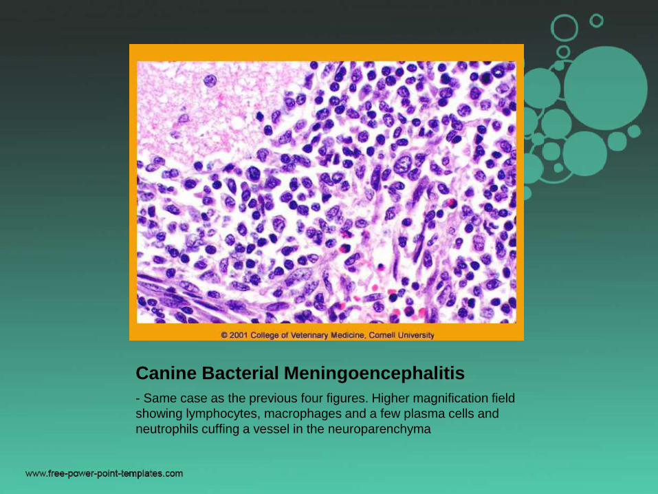
Canine Bacterial Meningoencephalitis
Higher magnification views of areas of extensive cerebral necrosis,
hemorrhage and neutrophilic infiltration

Canine Bacterial Meningoencephalitis
Same case as the previous two figures and showing higher
magnification views of acute hemorrhage, necrosis and neutrophilic
infiltration. Note the capillary thrombosis in the left panel.
Canine Bacterial Meningoencephalitis
- Same case as the previous four figures. Higher magnification field
showing lymphocytes, macrophages and a few plasma cells and
neutrophils cuffing a vessel in the neuroparenchyma

Granulomatous
Meningoencephalomyelitis -
Mixed perivascular infiltrates in the leptomeninges.

Neonatal septicemia
• E.coli, Streptococcus spp, Salmonella,
Pasteurella spp, Haemophilous
• Endotoxin & LPS, teichoic acid,
proteoglycans in vasculature TNF,
IL-1, PAF, prostaglandin, thromboxane,
leukotriens neutrophil adhesion,
injury to endothelium and blood brain
barrier
• Brain swelling, edema , increased
intracranial pressure

• Fibrinopurulent inflammation of
membranous tissue
• Infections acquired perinataly
• Onset in few days to 2 week
• Congestion, hemorrhage, focal cloudiness
or opacity of meninges
• Fibrin in ventricles

• Microscopic: Except Salmonella, Fibrin
deposition and neutrophil infiltration
around blood vessels of leptomeninges,
choroid plexus & ependymal & Sub Ep.
• Epithelium of choroid plexus and ventricles
can be disrupted by degeneration &
necrosis
• Thrombosis & vasculitis feature of E.coli

• Salmonellosis generally limited to foals
and pigs
• Inflammatory response consist of
macrophages and lymphocytes
• Thrombosis, vasculitis, necrosis
• Haemophilus parasuis cause
leptomeningitis along polyserositis and
polyarthritis.
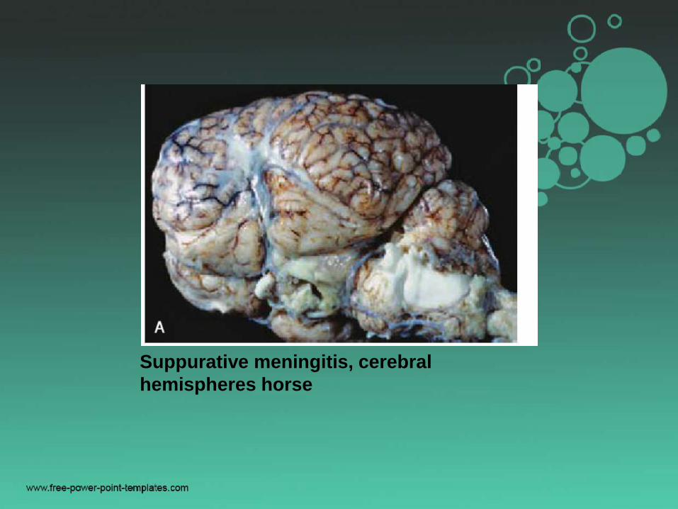
Suppurative meningitis, cerebral
hemispheres horse


Listeriosis
• Listeria monocytogenes
• Invade through the oral mucosa & into the
sensory and motor branches of trigeminal
nerve
• Other cranial nerves in the region may be
involved
• Via sensory axons using retrograde axonal
transport to trigeminal ganglion and then
brain.
• Or via motor axons directly into the mid
brain and medulla.
• Injury to neurons and axons due to
secondary bystander effect of
inflammation
• Immunological injury may also occur
• Organism also produces hemolysin
• In the brain, it can directly infect neurons,
glial cells, macrophages

• Bacteria spread cell to cell by producing
phospholipase
• Gross lesion usually absent
• Leptomeningeal opacity, foci of yellow
brown discoloration, hemorrhage, necrosis
in the terminal brainstem & cloudy csf
• Microscopically: Leptomeningitis centered
about the pons and medulla involving both
gray and white matter is characteristic

• Lesions may extend to diencephalon and
cervical spinal cord
• Early lesion: Loose clusters of microglial
cells
• Later neutrophils form microabscess
• In some foci macrophages may
predominate
• Necrosis and accumulation of gitter cells

• Gram positive bacilli can be detected
• Leptomeningitis with mononuclear cells
• 3 different disease forms
– Meningoencephalitis
– Abortion and still birth
– Septicemia
• Infection in humans do occur
• Dullness, torticollis, unilateral facial
paralysis, drooling caused by facial
paralysis

• Silage is most common source of infection

Ovine Listeriosis
Acute lesion in the medulla. Black arrows indicate a radix of cranial
nerve IX or X. There is locally extensive inflammatory infiltration
(inset) which at higher magnification is found to be predominantly
neutrophilic. Blue arrows indicate acute focal axonal swellings in the
intramedullary projection of the cranial nerve.

Ovine Listeriosis -
Caudal brainstem. Suppurative foci - numerous neutrophils and
some macrophages infiltrate the neuroparenchyma

Ovine Listeriosis -
Cervical spinal cord. Destructive inflammation, dominated in this
case by macrophages, many containing bacilli as shown by oil
immersion microscopy (arrows - right panel). Organisms are
difficult to find in many cases.

Ovine Listeriosis
Cervical spinal cord. Same case as the previous image. Inset -
bacilli within macrophages (arrows). Background - Specific
immunostaining reveals massive numbers of intracellular
organisms, often within neutrophils or macrophages. This case is
unusual in the number of organisms present.

Ovine Listeriosis
Caudal brainstem. Inset - diffuse neutrophilic infiltration of gray
matter with neuron cell bodies indicated by blue arrows. At higher
magnification bacilli are seen within a degenerating neuron (black
arrow)

Ovine Listeriosis
-Same case as the previous two figures. Higher magnification fields. Left
panel - a necrotizing pyo-granulomatous focal lesion within more diffuse
inflammatory infiltration. Right panel - degenerate neurons (arrows) have a
basophilic reticulated appearance due to dystrophic mineralization.
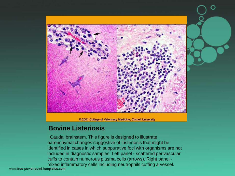
Bovine Listeriosis
Caudal brainstem. This figure is designed to illustrate
parenchymal changes suggestive of Listeriosis that might be
identified in cases in which suppurative foci with organisms are not
included in diagnostic samples. Left panel - scattered perivascular
cuffs to contain numerous plasma cells (arrows). Right panel -
mixed inflammatory cells including neutrophils cuffing a vessel.

Thrombotic
Meningoencephalitis• Histophilus somni
• Gram negative bacilli
• Septicemia
– Pneumonia
– Polyarthritis
– Myocarditis
– Abortion
– Meningoencephalitis

• More prevalent in feed lot cattle
• CNS form called TME
• Many cattle harbor in the upper digestive
tract
• Respiratory system is first site of
replication then Hematogenous spread to
CNS
• Once in circulation, emboli lodge at the
interface of white and gray matter in
microvessels

• Adherence to endothelial cells leading to
contraction and sloughing
• Subendothelial collagen is exposed
• Vasculitis, thrombosis and infarction
• Lipooligosaccharide, a bacterial toxin
protect it from defense of body
• Neutrophils, blood monocytes and alveolar
macrophages can not kill

• Irregular foci of hemorrhage & necrosis
scattered randomly
• Cerebrum, at cortical gray white interface
• Spinal cord has lesions
• Brain swelling due to edema
• Meningeal opacity
• Microscopically: Vasculitis, vascular
necrosis, thrombosis, infarction

• Clinically: Ataxic, circling, head pressing,
blindness, convulsions, comma

Fungi and Algae
• Aspergillus, Candida, Mucor
• Of the systemic fungi Coccidioides immitis,
Blastomyces dermatitides, Histoplasma
capsulatum, Cryptococcus neoformans
• Reach CNS by leukocyte trafficking and
hematogenous
• Granulomatous/pyogranulomatous
response

• Grossly, moderately well demarcated
expansile yellow brown foci
• Microscopically: neutrophils, epithelioid
macrophages, giant cells
• Organism in mononuclear cell


Canine Blastomycosis
Severely proliferative pyogranulomatous inflammation involves the
choroid plexus and ependyma of the fourth ventricle, lateral
apertures and meninges (left) and lateral and third ventricles (right).
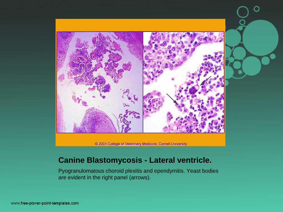
Canine Blastomycosis - Lateral ventricle.
Pyogranulomatous choroid plexitis and ependymitis. Yeast bodies
are evident in the right panel (arrows).

Canine Blastomycosis - Lateral ventricle
. Left panel - high magnification view to show intraventricular
exudate with yeast bodies within multinucleated giant cells
(arrows). Right panel - in the area indicated by the arrow are
numerous yeast bodies highlighted by silver staining.


• Cat, dog, horse
• Pathogen enters in leptomeninges and
subarachnoid space by direct extension
from the cribriform plate nasal/sinus
infection
• Hematogenous by leukocyte trafficking
from lungs infection
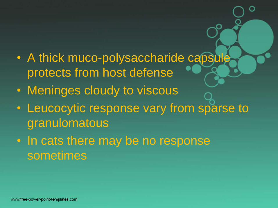
• A thick muco-polysaccharide capsule
protects from host defense
• Meninges cloudy to viscous
• Leucocytic response vary from sparse to
granulomatous
• In cats there may be no response
sometimes

• Yeast is 2-10 mm, thick non-staining
capsule
• PAS, Gomori’s methenamine silver for
organism and alcian blue for capsule
• Two virulence factors
– Mucopolysaccharide capsule
– Biochemical pathway using catecholamine's
with specific transport pathway and
phenoloxidase enzyme producing melanin like
compound
• CSF lack alternative pathway of

• Grossly: Multiple small, viscous,
gelatinous cysts in meninges and CNS
• Microscopically: Loosely organized lacy
appearance with cryptococcal organsim
• Ventriculitis, choroiditis
• Neutrophils, eosinophils, macrophages,
giant cells depending upon immune status

• Immunosuppression by Felv, FIV,
Ehrlichia canis, long term glucocorticoid
therapy increases susceptibility


Feline Cryptococcosis -
Representative high magnification field from the gelatinous lesion
seen in the previous figure. Numerous yeast bodies with unstained
capsules are embedded in mucin with a minimal inflammatory
response.

Feline Cryptococcosis
In this case there are multiple cavitating lesions in the cerebellum
containing mostly organisms and mucin with a mild macrophage
response. (Original material courtesy of Dr. W. J. Hartley)