automation in hematology until slide 23. hematology automation two general principles for cell...
TRANSCRIPT

Automation in hematologyUntil Slide 23

Hematology Automation
Two General Principles for cell counting :
Electronic impedance (resistance): called the Coulter Principle. Light scattering.
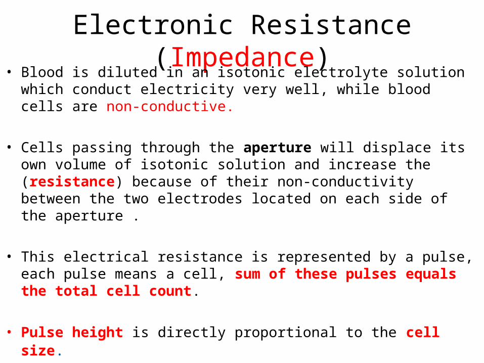
Electronic Resistance (Impedance)• Blood is diluted in an isotonic electrolyte solution which conduct
electricity very well, while blood cells are non-conductive.
• Cells passing through the aperture will displace its own volume of isotonic solution and increase the (resistance) because of their non-conductivity between the two electrodes located on each side of the aperture .
• This electrical resistance is represented by a pulse, each pulse means a cell, sum of these pulses equals the total cell count.
• Pulse height is directly proportional to the cell size.
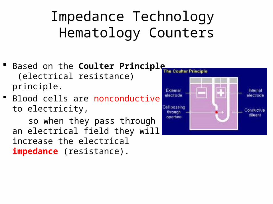
Impedance Technology Hematology Counters
Based on the Coulter Principle (electrical resistance) principle.
Blood cells are nonconductive to electricity,
so when they pass through an electrical field they will increase the electrical impedance (resistance).

Electronic Resistance (Impedance)


A sample of blood (EDTA tube) is placed in an analyzer and the cells are sorted according to size, granularity, and shape by using Electronic impedance OR Light scattering.

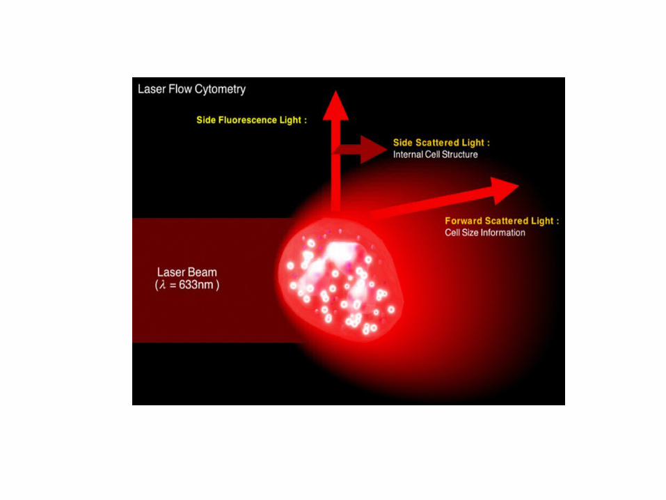
Light scattering
Cells counted as passed through focused beam of light( LASER).The amount of light scattered is proportional to the volume of the cell.
Multi angle scatter separation indicates •cell size.•Cell structure and complexity.•Nuclear lobularity
Diluted blood sample is aspirated into the counter and divided into 2 portions;
•First part will be enforced toward the RBC chamber in which red blood cells and platelets are counted and sized
•The second portion will be moved towards the WBC chamber, where it is diluted with a red blood lysing reagent .so that red blood cells will not be counted or interfere with white blood cells.
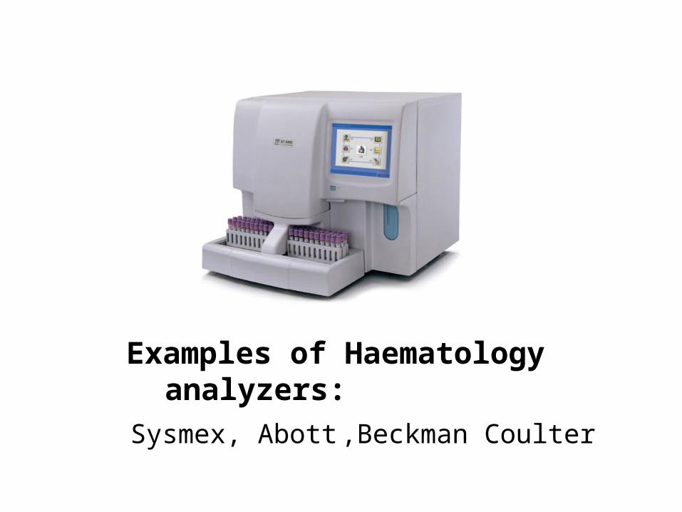
Examples of Haematology analyzers: Sysmex, Abott ,Beckman Coulter

• 3 parameters differential : Granulocytes, Lymphocytes and Monocytes.
• 5 Parameters differential :Lymphocytes, Monocytes, Neutrophils, Eosinophils, and Basophils.
• Nucleated red blood cell counts and immature granulocytes are emerging as sixth and seventh parameters

Sysmex xs 1000i
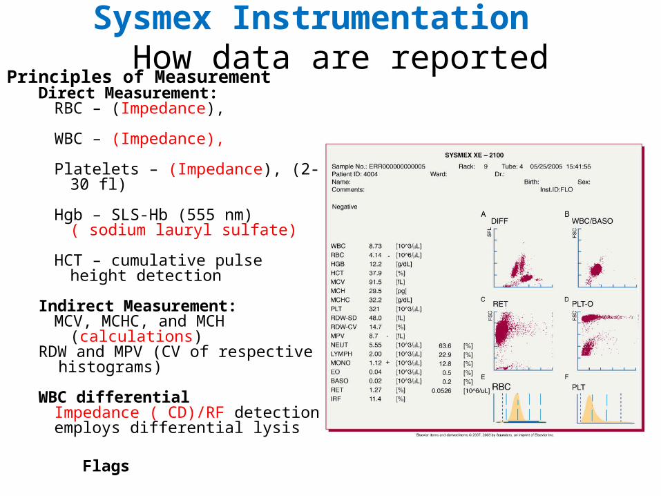
Sysmex Instrumentation How data are reported
Principles of MeasurementDirect Measurement:
RBC – (Impedance),
WBC – (Impedance),
Platelets – (Impedance), (2-30 fl)
Hgb – SLS-Hb (555 nm) ( sodium lauryl sulfate)
HCT – cumulative pulse height detection
Indirect Measurement:MCV, MCHC, and MCH (calculations)
RDW and MPV (CV of respective histograms)
WBC differential Impedance ( CD)/RF detectionemploys differential lysis
Flags

RF ( Radio frequency)and DC (Direct Current) Detection method
• Simultaneous application of DC and RF produce information on cell size and internal composition
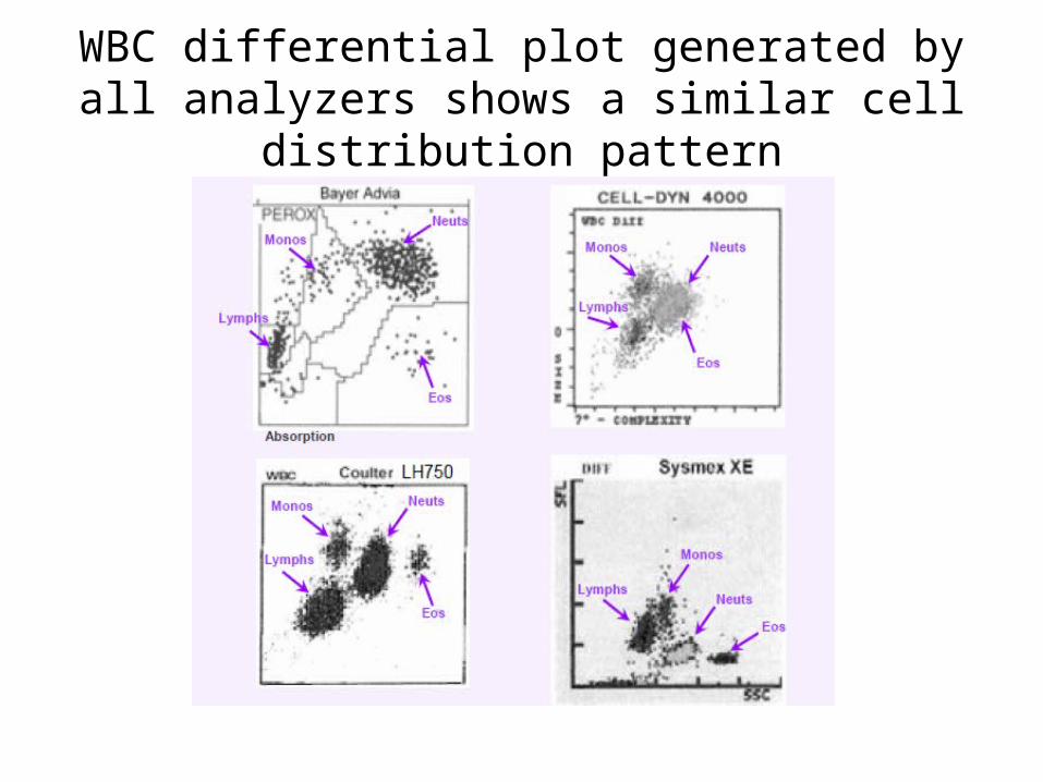
WBC differential plot generated by all analyzers shows a similar cell distribution pattern

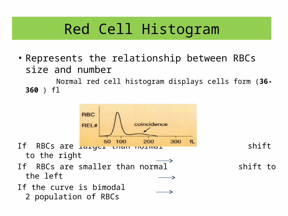
Red Cell Histogram
• Represents the relationship between RBCs size and number
Normal red cell histogram displays cells form (36- 360 ) fl
If RBCs are larger than normal shift to the rightIf RBCs are smaller than normal shift to the leftIf the curve is bimodal 2 population of RBCs
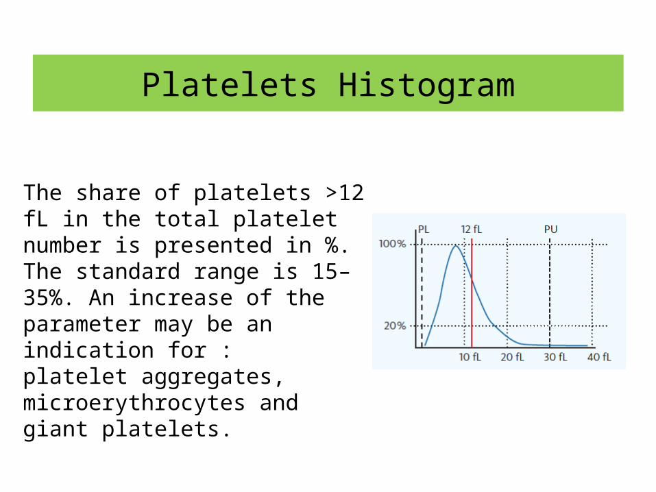
Platelets Histogram
The share of platelets >12 fL in the total platelet number is presented in %. The standard range is 15–35%. An increase of the parameter may be an indication for :platelet aggregates, microerythrocytes and giant platelets.

WBC Histogram
Sysmex XP-300™ Automated Hematology Analyzer - Shortcut.lnk


Instrument calibration
• Calibration provides the most accurate results possible.
• For best performance, calibrate all the CBC parameters.
• The WBC differential is calibrated at the factory. They do not require calibration in the laboratory.
When to CalibrateYou should calibrate your instrument:• At installation
• After the replacement of any component that involves dilution characteristics or the primary measurements (such as the apertures)
• As a routine once or twice a year

(Daily Maintenance)• Turn on the analyzer. On initial start up, the instrument will perform self-
checks• If all the self-checks is satisfactory, then the instrument is ready for
analysis.• If an error message is displayed on the analyzer screen , an alarm will
sound. The following corrective action will need to be taken before analysis can
go ahead:• Silence the alarm.• Press Help to display the error message• Press OK and the relevant corrective action will be automatically performed• Run the internal QC after start up procedure is completed.

Weekly maintenance• Check the quality control chart for evidence of drift.• Check the daily averages of (MCV, MCH and MCHC) for any drift or
sudden change outside an established 2SD.
• Clean the orifice and cell with a fine brush and flush several times with diluents. Never attempt to clear the orifice with a sharp device such as a needle or blade.
• Check seals to determine the possibility of leakage.• Check tubing.• Check stock of reagents, diluents, and disposables.• In a special logbook record the dates of all maintenance checks,
replacements of components, servicing by manufacturer's agent, recalibrations, and other necessary information.
•
Problem Solving – Troubleshooting
• An instrument problem is differentiated from a specimen-related problem by running a control.
• If the control results are acceptable, the problem is probably specimen-related. Check for:
– clots– hemolysis– lipemia
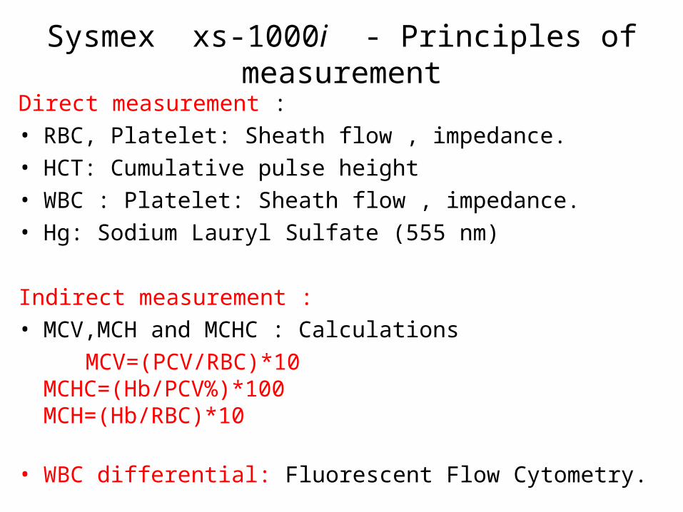
Sysmex xs-1000i - Principles of measurement
Direct measurement :• RBC, Platelet: Sheath flow , impedance. • HCT: Cumulative pulse height• WBC : Platelet: Sheath flow , impedance. • Hg: Sodium Lauryl Sulfate (555 nm)
Indirect measurement :• MCV,MCH and MCHC : Calculations MCV=(PCV/RBC)*10
MCHC=(Hb/PCV%)*100MCH=(Hb/RBC)*10
• WBC differential: Fluorescent Flow Cytometry.
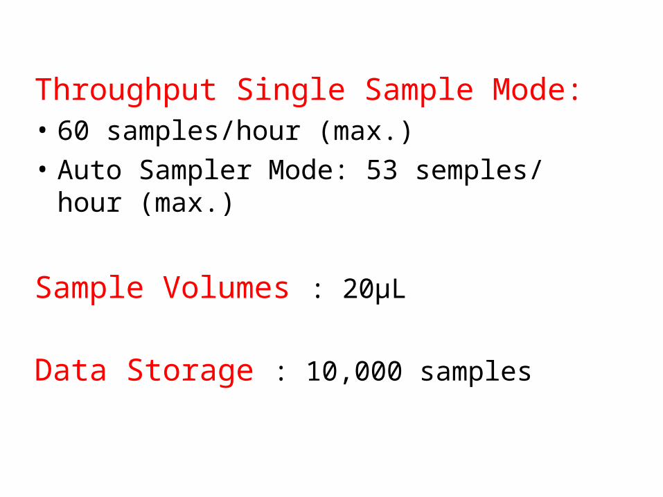
Throughput Single Sample Mode: • 60 samples/hour (max.)• Auto Sampler Mode: 53 semples/ hour (max.)
Sample Volumes : 20μL
Data Storage : 10,000 samples

Histograms
• RBC, PLT, and WBC plotted on histogram
• X-Axis– Cell size in
femtoliters (fL)• Y-Axis
– # of cells

Sysmex XP-300™ Automated Hematology Analyzer - Shortcut.lnk