author's personal copyscholar.cu.edu.eg/?q=safar/files/sulfurous-_phar_report.pdfm. safar*,...
TRANSCRIPT
This article appeared in a journal published by Elsevier. The attachedcopy is furnished to the author for internal non-commercial researchand education use, including for instruction at the authors institution
and sharing with colleagues.
Other uses, including reproduction and distribution, or selling orlicensing copies, or posting to personal, institutional or third party
websites are prohibited.
In most cases authors are permitted to post their version of thearticle (e.g. in Word or Tex form) to their personal website orinstitutional repository. Authors requiring further information
regarding Elsevier’s archiving and manuscript policies areencouraged to visit:
http://www.elsevier.com/authorsrights
Author's personal copy
Original research article
H2S donors attenuate diabetic nephropathy in rats:Modulation of oxidant status and polyol pathway
Marwa M. Safar *, Rania M. Abdelsalam
Department of Pharmacology and Toxicology, Faculty of Pharmacy, Cairo University, Cairo, Egypt
Introduction
Nephropathy is one of the serious long-term complications ofdiabetes which affects up to 40% of patients [1]. Albeit severalfactors are involved in the genesis of diabetic nephropathy,oxidative stress is regarded as a major culprit in the onset andprogression of kidney damage [2]. Chronic hyperglycemiaenhances the polyol pathway which comprises the conversion ofglucose to sorbitol by aldose reductase (AR) and the subsequentmetabolism of sorbitol to fructose by sorbitol dehydrogenase (SD).Activation of polyol pathway was formerly recognized as animportant factor in the development of diabetic nephropathy [3].
Despite its long-standing reputation as a foul smelling gas,hydrogen sulfide (H2S) is now considered the third gaseous
signaling molecule after nitric oxide and carbon monoxide [4]. In
this regard, it has emerged as a chief modulator of cardiovascular
homoeostasis, neurotransmission, as well as insulin secretion
[5,6]. Anti-apoptotic effect of H2S has also been claimed [7]. H2S
deficiency was observed in various animal models of arterial and
pulmonary hypertension, Alzheimer’s disease, gastric mucosal
injury, as well as liver cirrhosis. On the other hand, its excessive
production may contribute to the pathogenesis of inflammatory
diseases, septic shock, and cerebral stroke [8]. Hence H2S regulation
might provide a therapeutic prospect in various conditions. In the
past few years, the decline of blood H2S level in diabetic patients and
experimental animals has been demonstrated and was speculated to
contribute to vascular inflammation [9,10].Sulfurous mineral water has been long employed in thermal
medicine mainly for the treatment of dermatological and skeletal
muscle disorders in the form of mud and bath therapies [11,12]. In
addition, drinking of S containing water (hydroponic treatment) is
utilized especially in cases of gastroenteric disturbances [13]. A
promising role of S-based therapies in defending oxidative damage
Pharmacological Reports 67 (2015) 17–23
A R T I C L E I N F O
Article history:
Received 20 December 2013
Received in revised form 26 July 2014
Accepted 1 August 2014
Available online 20 August 2014
Keywords:
Diabetic nephropathy
Sulfurous mineral water
H2S
Insulin like growth factor-I (IGF-I)
Polyol pathway
A B S T R A C T
Background: Sulfurous mineral water and its main active ingredient sodium hydrosulfide (NaHS) are
major sources of H2S. The present study aimed to explore their protective effect on one of the serious
long-term complications of diabetes; diabetic nephropathy.
Methods: Sulfurous mineral water (as drinking water), NaHS (14 mmol/kg/day; ip), and gliclazide
(10 mg/kg; po) were administered daily for 6 weeks to streptozotocin (STZ)-diabetic rats.
Results: STZ-induced diabetes was associated with body weight reduction, hyperglycemia, overproduc-
tion of glycated hemoglobin, as well as decline in serum insulin, C-peptide, and insulin like growth
factor-I. Besides, diabetes impaired kidney functions and imposed oxidative and nitrosative stress as
manifested by elevated contents of renal thiobarbituric acid reactive substances and nitric oxide, parallel
to reduced glutathione content. These deleterious effects were antagonized by sulfurous water and to a
better extent by NaHS. Activities of myeloperoxidase and sorbitol dehydrogenase were not altered by
STZ or any of the treatments. However, STZ-induced diabetes was accompanied by an increment of
aldose reductase which was only mitigated by gliclazide and NaHS. Histopathological examination of
kidney sections corroborated the biochemical findings.
Conclusion: This study suggests a novel therapeutic approach for diabetic nephropathy using H2S
donors.
� 2014 Institute of Pharmacology, Polish Academy of Sciences. Published by Elsevier Urban & Partner Sp.
z o.o. All rights reserved.
Abbreviations: AR, aldose reductase; HbA1C %, glycated hemoglobin; IGF-I, insulin
like growth factor-I; MPO, myeloperoxidase; GSH, reduced glutathione; NaHS,
sodium hydrosulfide; SD, sorbitol dehydrogenase; STZ, streptozotocin; TBARs,
thiobarbituric acid reactive substance content; NOx, total nitrate/nitrite content.
* Corresponding author.
E-mail addresses: [email protected], [email protected]
(M.M. Safar).
Contents lists available at ScienceDirect
Pharmacological Reports
jou r nal h o mep ag e: w ww .e lsev ier . co m / loc ate /p h arep
http://dx.doi.org/10.1016/j.pharep.2014.08.001
1734-1140/� 2014 Institute of Pharmacology, Polish Academy of Sciences. Published by Elsevier Urban & Partner Sp. z o.o. All rights reserved.

Author's personal copy
associated with aging and age-related degenerative diseases hasbeen reported [14]. Nevertheless, until now, the response of theantioxidant defense system to drinking therapies involving H2S-rich water has been poorly scrutinized. Thus, the present studyaimed to investigate the effects of sulfurous mineral water and itsmain active ingredient NaHS on diabetic nephropathy in rats ascompared to the widely used and well tolerated sulfonyl urea;gliclazide. Modulation of renal oxidant status and polyol pathwayhas been targeted.
Materials and methods
Animals
Sixty-six male Wistar rats weighing 200–220 g were used inthis study. The study was conducted in accordance with the ethicalprocedures and policies approved by Animal Care and UseCommittee of Faculty of Pharmacy Cairo University, Cairo, Egypt.Animals had free access to standard rat chow and waterthroughout the study.
Drugs and chemicals
Streptozotocin was purchased from Sigma Chemicals Company,St. Louis, MO, USA. Sulfurous mineral water was obtained from theThermal Center of Helwan (Helwan Kabritage, Helwan Province;south of Cairo, Egypt), which has a sulfuric degree of 8.4 mg/L, asreported in Table 1. Gliclazide (Servier, France) was also used inthis study. All other chemicals used were of the highest purity andanalytical grade.
Experimental design
Diabetes was induced by a single intraperitoneal injection ofSTZ (50 mg/kg), freshly prepared in 0.1 M citrate buffer, pH 4.5[15]. A normal control group (n = 10) was injected with theappropriate volume of the citrate buffer. The STZ-treated rats wereallowed to drink 5% glucose solution during the first 24 h ofdiabetes induction to overcome the drug-induced hypoglycemia[16]. Four days later, blood samples were collected from rats’ tailsand hyperglycemia was confirmed by a blood glucose levelreaching 300 mg/dl. Glucose was measured using an analyzer(Roche Diagnostic Accu-Check test strips, Germany).
Diabetic rats were randomly divided into four groups (n = 10).The first one served as a control untreated diabetic group. Thesecond group was supplied daily with sulfurous mineral waterinstead of their drinking water [17]. Rats of the third group wereintraperitoneally injected with NaHS at a dose of 14 mmol/kg/day[18], while those of the fourth group were treated with gliclazide(10 mg/kg/day, po) [19]. Since NaHS is considered a toxicsubstance, we injected NaHS (14 mmol/kg/day) alone to controlnon-diabetic rats (n = 8) as the treatment control. [20] reported anLD50 value of 15 mg/kg (approximately 192 mmol/kg) for NaHS in
rats, which is far beyond the used dosage. Another control group(n = 8) was supplied with sulfurous mineral water to exclude anytoxic effect of H2S rich water consumption.
After 6 weeks and immediately before scarification, the bodyweight of each rat was determined. Blood samples werewithdrawn from the retro-orbital plexus and divided on twospecimens; one was processed for serum preparation used forassaying the levels of glucose, insulin, IGF-I, C-peptide, urea, andcreatinine. The other was collected in heparinized tubes and usedfor the estimation of glycated hemoglobin (HbA1C %). Immediatelyafter blood sampling, animals were euthanized by decapitation,and their kidneys were excised, washed with ice-cold physiologicalsaline, blotted dry, and weighed. The left kidneys were homoge-nized in ice-cold saline (10%, w/v) and used for assessment ofthiobarbituric acid reactive substance content (TBARs), reducedglutathione (GSH), total nitrate/nitrite content (NOx), as well asmyeloperoxidase (MPO), SDH, and AR activities.
Biochemical investigations in blood and serum
Serum level of glucose and the percentage of HbA1C wereassayed using commercially available kits provided by Stanbio, SanAntonio, TX, USA. Serum insulin level was measured by immunor-adiometric assay using kits provided by Immunotech, France.Serum C-peptide was measured using solid phase, two sitechemiluminescent immunometric assay using kits supplied byDPC, USA on Immulite analyzer. IGF-I was assessed using an ELISAkit provided by DRG Instruments GmbH, Germany. Serum levels ofurea and creatinine were estimated using commercial test reagentkits provided by Biodiagnostic, Giza, Egypt.
Biochemical investigations in kidney homogenate
Renal content of TBARS was determined according to themethod described by [21] using malondialdehyde (MDA) as astandard. GSH content was measured using Ellman’s reagent asdescribed by [22]. Total tissue nitrite and nitrate content wasestimated according to the method described by [23] based on theGriess reaction and employing vanadium trichloride as reducingagent. The kinetic method described by [24] was employed todetermine MPO activity. Renal activities of AR and SDH weredetermined according to the methods of [25,26], respectively. Theprotein content was measured by the method of [27].
Histopathological studies
Immediately following sacrifice, sections of the left kidneyswere fixed in 10% formalin/saline, embedded in paraffin wax andcut at 5 mm thickness. Sections were processed, stained withhematoxylin and eosin dye (H&E), and examined under lightmicroscope.
Statistical analysis
Data were expressed as means � SEM. Comparisons betweenmeans were carried out using one-way ANOVA followed by Tukey–Kramer multiple comparisons test using Instat software, version 3(GraphPad Software, Inc., San Diego, USA); probability level of lessthan 0.05 was accepted as statistically significant.
Results
No difference was detected between the normal rats and thosewhich received sulfurous mineral water or NaHS (at the useddosage) alone in any of the tested parameters, indicating that theyare safe and had no toxic effects.
Table 1Physico-chemical characteristics of the sulfurous mineral water of Helwan
Kabritage.
Parameter Unit Parameter Unit
pH 6.69 K+ 2 mg/L
Conductivity 6590 mS/Cm HCO3� 180 mg/L
Fixed residue (at 180 8C) 6480 mg/L F� 1.5 mg/L
Sulfuric degree 8.4 mg/L Cl� 1560 mg/L
CO2 80 mg/L NO2� <0.01 mg/L
Ca2+ 403 mg/L SO42� 884 mg/L
Mg2+ 49 mg/L NO3� 0.04 mg/L
Na+ 1300 mg/L Iron 0.006 mg/L
M.M. Safar, R.M. Abdelsalam / Pharmacological Reports 67 (2015) 17–2318
Author's personal copy
General features (Table 2)
Diabetic rats were severely hyperglycemic and exhibitedoverproduction of glycated hemoglobin accompanied by reducedserum levels of insulin (80.5%), C-peptide (70%), and IGF-I (54%) ascompared to normal animals. These were associated with a markeddecline in body weight (33%) and increment of relative kidneyweight (77%). Sulfurous mineral water, NaHS, as well as thereference antidiabetic drug, gliclazide, antagonized hyperglycemiaby 47%, 59%, and 57%, respectively. In addition, the treated groupsrevealed reduced levels of HbA1C by 50%, 54%, and 33%,respectively, as compared to the diabetic group. Similarly, relativekidney weight was lowered by sulfurous mineral water (28%),NaHS (35%), and gliclazide (36%) without normalizing the bodyweight. Albeit none of the treatments restored the C-peptide level,NaHS leveled up serum insulin and IGF-I by nearly 4 folds and2 folds, respectively, as compared to the diabetic rats. Bothsulfurous mineral water and gliclazide tended to increase IGF-Ilevel but did not reach statistical significance.
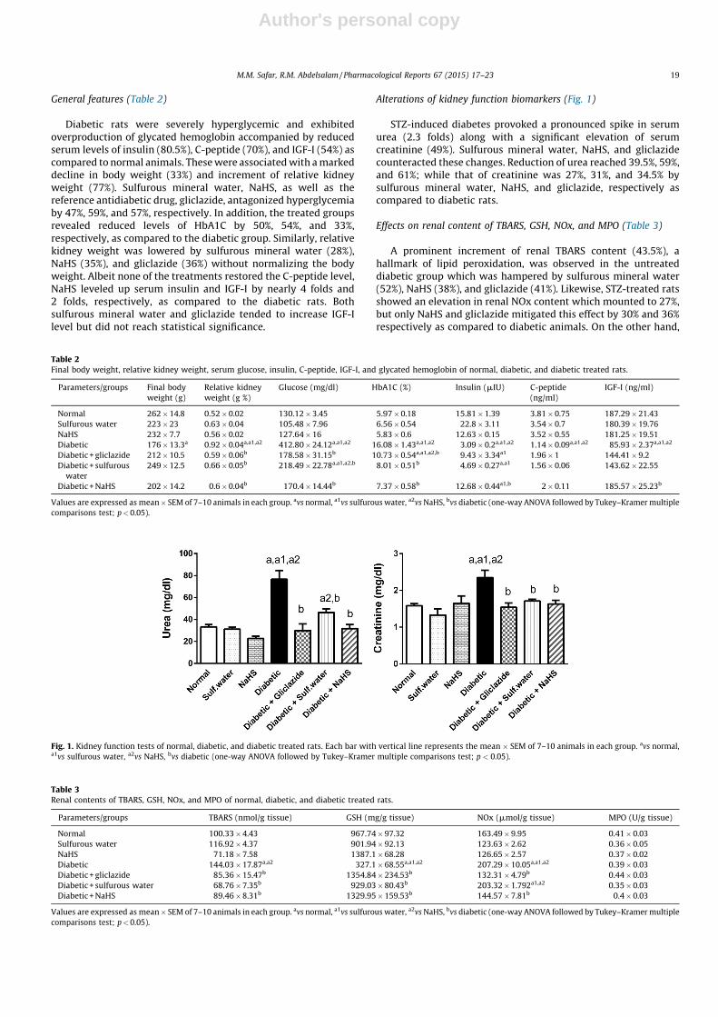
Alterations of kidney function biomarkers (Fig. 1)
STZ-induced diabetes provoked a pronounced spike in serumurea (2.3 folds) along with a significant elevation of serumcreatinine (49%). Sulfurous mineral water, NaHS, and gliclazidecounteracted these changes. Reduction of urea reached 39.5%, 59%,and 61%; while that of creatinine was 27%, 31%, and 34.5% bysulfurous mineral water, NaHS, and gliclazide, respectively ascompared to diabetic rats.
Effects on renal content of TBARS, GSH, NOx, and MPO (Table 3)
A prominent increment of renal TBARS content (43.5%), ahallmark of lipid peroxidation, was observed in the untreateddiabetic group which was hampered by sulfurous mineral water(52%), NaHS (38%), and gliclazide (41%). Likewise, STZ-treated ratsshowed an elevation in renal NOx content which mounted to 27%,but only NaHS and gliclazide mitigated this effect by 30% and 36%respectively as compared to diabetic animals. On the other hand,
Table 2Final body weight, relative kidney weight, serum glucose, insulin, C-peptide, IGF-I, and glycated hemoglobin of normal, diabetic, and diabetic treated rats.
Parameters/groups Final body
weight (g)
Relative kidney
weight (g %)
Glucose (mg/dl) HbA1C (%) Insulin (mIU) C-peptide
(ng/ml)
IGF-I (ng/ml)
Normal 262 � 14.8 0.52 � 0.02 130.12 � 3.45 5.97 � 0.18 15.81 � 1.39 3.81 � 0.75 187.29 � 21.43
Sulfurous water 223 � 23 0.63 � 0.04 105.48 � 7.96 6.56 � 0.54 22.8 � 3.11 3.54 � 0.7 180.39 � 19.76
NaHS 232 � 7.7 0.56 � 0.02 127.64 � 16 5.83 � 0.6 12.63 � 0.15 3.52 � 0.55 181.25 � 19.51
Diabetic 176 � 13.3a 0.92 � 0.04a,a1,a2 412.80 � 24.12a,a1,a2 16.08 � 1.43a,a1,a2 3.09 � 0.2a,a1,a2 1.14 � 0.09a,a1,a2 85.93 � 2.37a,a1,a2
Diabetic + gliclazide 212 � 10.5 0.59 � 0.06b 178.58 � 31.15b 10.73 � 0.54a,a1,a2,b 9.43 � 3.34a1 1.96 � 1 144.41 � 9.2
Diabetic + sulfurous
water
249 � 12.5 0.66 � 0.05b 218.49 � 22.78a,a1,a2,b 8.01 � 0.51b 4.69 � 0.27a,a1 1.56 � 0.06 143.62 � 22.55
Diabetic + NaHS 202 � 14.2 0.6 � 0.04b 170.4 � 14.44b 7.37 � 0.58b 12.68 � 0.44a1,b 2 � 0.11 185.57 � 25.23b
Values are expressed as mean � SEM of 7–10 animals in each group. avs normal, a1vs sulfurous water, a2vs NaHS, bvs diabetic (one-way ANOVA followed by Tukey–Kramer multiple
comparisons test; p < 0.05).
Table 3Renal contents of TBARS, GSH, NOx, and MPO of normal, diabetic, and diabetic treated rats.
Parameters/groups TBARS (nmol/g tissue) GSH (mg/g tissue) NOx (mmol/g tissue) MPO (U/g tissue)
Normal 100.33 � 4.43 967.74 � 97.32 163.49 � 9.95 0.41 � 0.03
Sulfurous water 116.92 � 4.37 901.94 � 92.13 123.63 � 2.62 0.36 � 0.05
NaHS 71.18 � 7.58 1387.1 � 68.28 126.65 � 2.57 0.37 � 0.02
Diabetic 144.03 � 17.87a,a2 327.1 � 68.55a,a1,a2 207.29 � 10.05a,a1,a2 0.39 � 0.03
Diabetic + gliclazide 85.36 � 15.47b 1354.84 � 234.53b 132.31 � 4.79b 0.44 � 0.03
Diabetic + sulfurous water 68.76 � 7.35b 929.03 � 80.43b 203.32 � 1.792a1,a2 0.35 � 0.03
Diabetic + NaHS 89.46 � 8.31b 1329.95 � 159.53b 144.57 � 7.81b 0.4 � 0.03
Values are expressed as mean � SEM of 7–10 animals in each group. avs normal, a1vs sulfurous water, a2vs NaHS, bvs diabetic (one-way ANOVA followed by Tukey–Kramer multiple
comparisons test; p < 0.05).
Fig. 1. Kidney function tests of normal, diabetic, and diabetic treated rats. Each bar with vertical line represents the mean � SEM of 7–10 animals in each group. avs normal,a1vs sulfurous water, a2vs NaHS, bvs diabetic (one-way ANOVA followed by Tukey–Kramer multiple comparisons test; p < 0.05).
M.M. Safar, R.M. Abdelsalam / Pharmacological Reports 67 (2015) 17–23 19

Author's personal copy
renal GSH was depleted in diabetic rats by 66%. All tested drugsreinstated normal GSH content. No significant change wasdetected in MPO content.
Effects on polyol pathway (Fig. 2)
STZ-induced diabetes instigated a significant increase in renalAR activity (66%) without affecting the SDH activity. NaHS andgliclazide reduced the elevated AR activity by 46% and 50%,respectively. No improvement was depicted in the sulfurousmineral water treated group.
Histopathological examination of kidney
Fig. 3 illustrates photomicrographs of kidneys from normal,diabetic, and treated rats. A normal architecture of the glomeruliand tubules in the cortex and the tubules at the corticomedullaryjunction was manifested in the normal group (a and b), whilediabetic rats demonstrated focal inflammatory cells infiltration inbetween the glomeruli, tubules, and blood vessels at the cortex (c).There were swelling and vacuolization in the lining endothelium ofthe glomerular tufts as well as swelling and degeneration in thelining epithelium of the tubules (d). The tubular lumen of themedullary portion had homogenous eosinophilic casts as well asred blood cells (e). Congestion and dilatation were detected in theblood vessels of both the cortex and the medulla (f and g). No majorhistopathological alteration was observed in treated rats whichportrayed normal histological structures of the glomeruli andtubules at the cortex (h–j).
Discussion
The current investigation depicts antidiabetic effects ofsulfurous mineral water and to a better extent its main activeconstituent NaHS which are quite comparable to gliclazide. Thisgoes in line with the findings of [28]. A recent study demonstratedthat NaHS possesses an insulin sensitizing effect both in vitro andin vivo [29]. Decline in plasma H2S level and its vascular productionhave been previously reported in diabetic animals and humans[10]. Consequently, the exogenous administration H2S donors inthis study might have provided compensation. Sulfur is alsonecessary for the formation of insulin and IGF-I [30]. Several linesof evidence accentuate the antidiabetic effects of sulfur containingcompounds via increasing insulin secretion and insulin sensitivity[31,32].
STZ-induced diabetes was associated with marked kidneyfunction impairment along with renal lipid peroxidation, excessivenitric oxide production, and depletion of GSH; effects which clearly
indicate enhanced oxidative stress. Similar findings have beenpreviously documented [33,34]. Glucose auto-oxidation, non-enzymatic glycation, and the overproduction of reactive oxygenspecies (ROS) by mitochondria are potential sources of hypergly-cemia-induced oxidative stress [3,35]. Increased HbA1C levelobserved herein represents a key point in diabetes associatednephropathy. Oxidant injury of proteins and lipids along with theaccumulation of AGEs instigate dysfunction of apical transportersin renal proximal tubule cells [36], proliferation of various renalcell types [37], microvascular complications [38], and apoptosis[39]. C-peptide, a peptide hormone that possibly acts through a Gprotein-coupled membrane receptor, was shown to protect againstapoptosis in kidney proximal tubular cells [40]. In this context, thereduced serum level of C-peptide in diabetic rats has contributed tothe kidney malfunction and histopathological changes. None of thetested drugs antagonized this effect.
In the present study, sulfurous mineral water and NaHScounteracted the elevation of renal TBARS and replenished GSHcontents; the latter was also able to normalize the NOx content.Improvement in renal redox balance was reflected on kidneyfunction tests, its hypertrophy and histopathological features.Several lines of evidence emphasize these findings where sulfurousmineral water drinking therapy improved body redox status inhealthy volunteers [17] and in cardiac tissue of diabetic rats[28]. Similarly, [41] reported the decrease in NOx production inosteoarthritic mice subjected to mud therapy. The protectiveeffects of sulfur water against the free-radical damage of lipids andproteins may be possibly related to the increase in total –SH levels,which include both protein (principally albumin) and non-protein(cysteine and GSH) thiol groups [17].
H2S may react with reactive oxygen and/or nitrogen specieslimiting their toxic effects and might be involved in the reductionof thiols, thus being directly implicated in redox reactions as anantioxidant [8]. In vitro, the H2S donor NaHS directly scavengedhydrogen peroxide and superoxide anions [18] while in vivo, itsadministration alleviated ischemia/reperfusion injury-inducedrenal dysfunction [42] and mitigated ureteral obstruction-induced kidney fibrosis [43]. The endogenously produced H2Sapparently ameliorates oxidative injury by increasing theintracellular concentrations of GSH via increasing the activityof g-glutamylcysteine synthetase and upregulating cystinetransport [5]. Besides its observed effect as an antioxidant dueto its H2S content’s, sulfurous mineral water may combat the stateof hypomagnesaemia often encountered in diabetes [44] due to itsenrichment with Mg2+. The sulfur content might also play a role;sulfur compounds have been found to be beneficial in counter-acting oxidative stress and ameliorating diabetic nephropathy[45,46].
Fig. 2. Sorbitol dehydrogenase and aldose reductase activities of normal, diabetic, and treated rats. Each bar with vertical line represents the mean � SEM of 7–10 animals in
each group. avs normal, a1vs sulfurous water, a2vs NaHS, bvs diabetic (one-way ANOVA followed by Tukey–Kramer multiple comparisons test; p < 0.05).
M.M. Safar, R.M. Abdelsalam / Pharmacological Reports 67 (2015) 17–2320

Author's personal copy
Elevation of renal AR along with a deranged oxidant status iscoherent with previous studies [33]. Activation of NADPH-dependent aldose reductase (AR) diminishes the quantity ofNADPH available for the reduction of oxidized glutathione (GSSG)by glutathione reductase [47]. This suggests that the reduced renalGSH content observed herein is due to its reduced synthesis ratherthan its increased degradation. Taken in consideration that GSHrepresents the key non-enzymatic tissue antioxidant, its depletionindicates a reduced capacity of kidney antioxidant defensesystem making it more vulnerable to the hyperglycemic ROS- and
RNS-induced damage. This is confirmed by the finding thatabnormal polyol pathway in diabetic rats resulted in glomerulo-pathy and renal hypofunction [3]. Notably, aldose reductaseinhibitors exhibited protective effect on diabetic nephropathypossibly via counteracting oxidative and nitrosative stress indiabetic kidney [48] and/or reducing renal vascular endothelialgrowth factor VEGF overexpression [49]. Normalization of ARactivity by NaHS is a key finding in this study.
Renal SD was not altered in this study demonstrating that ARactivity, the first step in the polyol pathway, makes a greater
Fig. 3. Photomicrographs of sections from the kidneys of normal, diabetic, diabetic treated rats Kidneys from (a) normal rat treated with 1% Tween 80 showing normal
histological structure of the glomeruli (g) and tubules (t) at the cortex; (b) normal rat treated with 1% Tween 80 showing normal histological structure of the tubules (t) at the
corticomedullary portion; (c) diabetic rat showing focal inflammatory cells infiltration (m) in between the glomeruli (g) and blood vessels (v); (d) diabetic rat showing
vacuolization and swelling in the lining endothelium of the glomerular tuft (g) with swelling and degeneration in the lining epithelium of the tubules (d); (e) diabetic rat
showing homogenous eosinophilic casts (c) and red blood cells (r) in the lumen of the tubules at the medullary portion; (f) diabetic rat showing severe congestion in cortical
blood vessels (v); (g) diabetic rat showing severe congestion in medullary blood vessels (v); (h–j) rats treated with gliclazide, sulfurous water, NaHS, respectively showing
normal histological structure of the glomeruli (g) and tubules (t) at the cortex (H&E �40 except f and g �80).
M.M. Safar, R.M. Abdelsalam / Pharmacological Reports 67 (2015) 17–23 21

Author's personal copy
contribution to the etiology of diabetic nephropathy than does thesecond step involving SD. Interestingly, it was claimed that geneexpression of the two enzymes are not regulated via a commonmechanism and are tissue-specific; where STZ-diabetes increasedSD gene expression in testis but not in kidney or brain [50].
In conclusion, this study suggests that H2S donors possess anti-diabetic effects and ameliorate associated nephropathy possiblyvia modulation of renal redox status and polyol pathway. Hence,may promise a novel and applicable approach in preventing and/ortreating diabetic complications.
Conflicts of interest statement
The authors declare that there are no conflicts of interest thatcould prejudice the impartiality of this scientific work.
Funding
This study received no funds from any sources. It was carried onthe author’s own expenses from their salaries they receive from thefaculty.
Acknowledgments
The authors gratefully acknowledge Dr. Tarek Samir, WaterPollution Control Department, National Research Center, Cairo,Egypt, for the analysis of sulfurous mineral water. The authors alsothank Prof. Dr. Adel Bakeir, Histology Department, Faculty ofVeterinary Medicine, Cairo University, for performing the histo-pathological examinations of this study.
References
[1] Ritz E, Orth SR. Nephropathy in patients with type 2 diabetes mellitus. N Engl JMed 1999;341(15):1127–33.
[2] Hakim FA, Pflueger A. Role of oxidative stress in diabetic kidney disease. MedSci Monit 2010;16(2):RA37–48.
[3] Itagaki I, Shimizu K, Kamanaka Y, Ebata K, Kikkawa R, Haneda M, et al. Theeffect of an aldose reductase inhibitor (epalrestat) on diabetic nephropathy inrats. Diabetes Res Clin Pract 1994;25(3):147–54.
[4] Wang R. Hydrogen sulfide: the third gasotransmitter in biology and medicine.Antioxid Redox Signal 2010;12(9):1061–4.
[5] Kimura H. Hydrogen sulfide: its production, release and functions. AminoAcids 2011;41(1):113–21.
[6] Szabo C. Hydrogen sulphide and its therapeutic potential. Nat Rev Drug Discov2007;6(11):917–35.
[7] Rinaldi L, Gobbi G, Pambianco M, Micheloni C, Mirandola P, Vitale M. Hydrogensulfide prevents apoptosis of human PMN via inhibition of p38 and caspase 3.Lab Invest 2006;86(4):391–7.
[8] Lowicka E, Beltowski J. Hydrogen sulfide (H2S) – the third gas of interest forpharmacologists. Pharmacol Rep 2007;59(1):4–24.
[9] Brancaleone V, Roviezzo F, Vellecco V, De Gruttola L, Bucci M, Cirino G.Biosynthesis of H2S is impaired in non-obese diabetic (nod) mice. Br J Phar-macol 2008;155(5):673–80.
[10] Jain SK, Bull R, Rains JL, Bass PF, Levine SN, Reddy S, et al. Low levels of hydrogensulfide in the blood of diabetes patients and streptozotocin-treated rats causesvascular inflammation? Antioxid Redox Signal 2010;12(11):1333–7.
[11] Gupta AK, Nicol K. The use of sulfur in dermatology. J Drugs Dermatol2004;3(4):427–31.
[12] Sukenik S, Flusser D, Abu-Shakra M. The role of spa therapy in variousrheumatic diseases. Rheum Dis Clin North Am 1999;25(4):883–97.
[13] Costantino M, Giuberti G, Caraglia M, Lombardi A, Misso G, Abbruzzese A, et al.Possible antioxidant role of spa therapy with chlorine–sulphur–bicarbonatemineral water. Amino Acids 2009;36(2):161–5.
[14] Casetta I, Govoni V, Granieri E. Oxidative stress, antioxidants and neurode-generative diseases. Curr Pharm Des 2005;11(16):2033–52.
[15] Saito M, Kinoshita Y, Satoh I, Shinbori C, Kono T, Hanada T, et al. N-hexacosanolameliorates streptozotocin-induced diabetic rat nephropathy. Eur J Pharmacol2006;544(1–3):132–7.
[16] Matthaei S, Horuk R, Olefsky JM. Blood–brain glucose transfer in diabetesmellitus. Decreased number of glucose transporters at blood–brain barrier.Diabetes 1986;35(10):1181–4.
[17] Benedetti S, Benvenuti F, Nappi G, Fortunati NA, Marino L, Aureli T, et al.Antioxidative effects of sulfurous mineral water: protection against lipid andprotein oxidation. Eur J Clin Nutr 2009;63(1):106–12.
[18] Geng B, Chang L, Pan C, Qi Y, Zhao J, Pang Y, et al. Endogenous hydrogen sulfideregulation of myocardial injury induced by isoproterenol. Biochem BiophysRes Commun 2004;318(3):756–63.
[19] Attia HN, Al-Rasheed NM, Maklad YA, Ahmed AA, Kenawy SA. Protectiveeffects of combined therapy of gliclazide with curcumin in experimentaldiabetic neuropathy in rats. Behav Pharmacol 2012;23(2):153–61.
[20] Warenycia MW, Smith KA, Blashko CS, Kombian SB, Reiffenstein RJ. Mono-amine oxidase inhibition as a sequel of hydrogen sulfide intoxication:increases in brain catecholamine and 5-hydroxytryptamine levels. Arch Tox-icol 1989;63(2):131–6.
[21] Mihara M, Uchiyama M. Determination of malonaldehyde precursor in tissuesby thiobarbituric acid test. Anal Biochem 1978;86(1):271–8.
[22] Beutler E, Duron O, Kelly BM. Improved method for the determination of bloodglutathione. J Lab Clin Med 1963;61:882–8.
[23] Miranda KM, Espey MG, Wink DA. A rapid, simple spectrophotometricmethod for simultaneous detection of nitrate and nitrite. Nitric Oxide2001;5(1):62–71.
[24] Bradley PP, Priebat DA, Christensen RD, Rothstein G. Measurement of cutane-ous inflammation: estimation of neutrophil content with an enzyme marker. JInvest Dermatol 1982;78(3):206–9.
[25] Hoffman PL, Wermuth B, von Wartburg JP. Human brain aldehyde reductases:relationship to succinic semialdehyde reductase and aldose reductase. JNeurochem 1980;35(2):354–66.
[26] Chauncey B, Leite MV, Goldstein L. Renal sorbitol accumulation and associatedenzyme activities in diabetes. Enzyme 1988;39(4):231–4.
[27] Lowry OH, Rosebrough NJ, Farr AL, Randall RJ. Protein measurement with thefolin phenol reagent. J Biol Chem 1951;193(1):265–75.
[28] El-Seweidy MM, Sadik NA, Shaker OG. Role of sulfurous mineral water andsodium hydrosulfide as potent inhibitors of fibrosis in the heart of diabeticrats. Arch Biochem Biophys 2011;506(1):48–57.
[29] Xue R, Hao DD, Sun JP, Li WW, Zhao MM, Li XH, et al. Hydrogen sulfidetreatment promotes glucose uptake by increasing insulin receptor sensitivityand ameliorates kidney lesions in type 2 diabetes. Antioxid Redox Signal2013;19(1):5–23.
[30] Rinderknecht E, Humbel RE. The amino acid sequence of human insulin-likegrowth factor I and its structural homology with proinsulin. J Biol Chem1978;253(8):2769–76.
[31] Kook S, Kim GH, Choi K. The antidiabetic effect of onion and garlic inexperimental diabetic rats: meta-analysis. J Med Food 2009;12(3):552–60.
[32] Klimek M, Wang S, Ogunkanmi A. Safety and efficacy of red yeast rice(Monascus purpureus) as an alternative therapy for hyperlipidemia. P T2009;34(6):313–27.
[33] Morsy MD, Hassan WN, Zalat SI. Improvement of renal oxidative stressmarkers after ozone administration in diabetic nephropathy in rats. DiabetolMetab Syndr 2010;2(1):29.
[34] Xu Y, Osborne BW, Stanton RC. Diabetes causes inhibition of glucose-6-phosphate dehydrogenase via activation of PKA, which contributes tooxidative stress in rat kidney cortex. Am J Physiol Renal Physiol 2005;289(5):F1040–47.
[35] Forbes JM, Coughlan MT, Cooper ME. Oxidative stress as a major culprit inkidney disease in diabetes. Diabetes 2008;57(6):1446–54.
[36] Hamilton KL, Butt AG. The molecular basis of renal tubular transport disorders.Comp Biochem Physiol A: Mol Integr Physiol 2000;126(3):305–21.
[37] Forbes JM, Cooper ME, Oldfield MD, Thomas MC. Role of advanced glycationend products in diabetic nephropathy. J Am Soc Nephrol 2003;14(8 Suppl.3):S254–8.
[38] Karachalias N, Babaei-Jadidi R, Ahmed N, Thornalley PJ. Accumulation offructosyl-lysine and advanced glycation end products in the kidney, retinaand peripheral nerve of streptozotocin-induced diabetic rats. Biochem SocTrans 2003;31(Pt 6):1423–5.
[39] Ha H, Yu MR, Choi YJ, Kitamura M, Lee HB. Role of high glucose-inducednuclear factor-kappab activation in monocyte chemoattractant protein-1expression by mesangial cells. J Am Soc Nephrol 2002;13(4):894–902.
[40] Al-Rasheed NM, Willars GB, Brunskill NJ. C-peptide signals via Galpha i toprotect against TNF-alpha-mediated apoptosis of opossum kidney proximaltubular cells. J Am Soc Nephrol 2006;17(4):986–95.
[41] Caraglia M, Beninati S, Giuberti G, D’Alessandro AM, Lentini A, Abbruzzese A,et al. Alternative therapy of earth elements increases the chondroprotectiveeffects of chondroitin sulfate in mice. Exp Mol Med 2005;37(5):476–81.
[42] Tripatara P, Patel NS, Brancaleone V, Renshaw D, Rocha J, Sepodes B, et al.Characterisation of cystathionine gamma-lyase/hydrogen sulphide pathwayin ischaemia/reperfusion injury of the mouse kidney: an in vivo study. Eur JPharmacol 2009;606(1–3):205–9.
[43] Jung KJ, Jang HS, Kim JI, Han SJ, Park JW, Park KM. Involvement of hydrogensulfide and homocysteine transsulfuration pathway in the progression ofkidney fibrosis after ureteral obstruction. Biochim Biophys Acta 2013;1832(12):1989–97.
[44] Barbagallo M, Dominguez LJ, Galioto A, Ferlisi A, Cani C, Malfa L, et al. Role ofmagnesium in insulin action, diabetes and cardio-metabolic syndrome X. MolAspects Med 2003;24(1–3):39–52.
[45] Liu CT, Wong PL, Lii CK, Hse H, Sheen LY. Antidiabetic effect of garlic oil but notdiallyl disulfide in rats with streptozotocin-induced diabetes. Food ChemToxicol 2006;44(8):1377–84.
[46] Nandhini TA, Anuradha CV. Inhibition of lipid peroxidation, protein glycationand elevation of membrane ion pump activity by taurine in RBC exposed tohigh glucose. Clin Chim Acta 2003;336(1–2):129–35.
M.M. Safar, R.M. Abdelsalam / Pharmacological Reports 67 (2015) 17–2322

Author's personal copy
[47] Dominguez C, Ruiz E, Gussinye M, Carrascosa A. Oxidative stress at onset andin early stages of type 1 diabetes in children and adolescents. Diabetes Care1998;21(10):1736–42.
[48] Drel VR, Pacher P, Stevens MJ, Obrosova IG. Aldose reductase inhibitioncounteracts nitrosative stress and poly(ADP-ribose) polymerase activationin diabetic rat kidney and high-glucose-exposed human mesangial cells. FreeRadic Biol Med 2006;40(8):1454–65.
[49] Sung JK, Koh JH, Lee MY, Kim BH, Nam SM, Kim JH, et al. Aldose reductaseinhibitor ameliorates renal vascular endothelial growth factor expres-sion in streptozotocin-induced diabetic rats. Yonsei Med J 2010;51(3):385–91.
[50] Kicic E, Palmer TN. Is sorbitol dehydrogenase gene expression affected bystreptozotocin-diabetes in the rat? Biochim Biophys Acta 1994;1226(2):213–8.
M.M. Safar, R.M. Abdelsalam / Pharmacological Reports 67 (2015) 17–23 23