authors: lauren berstler eric woodard
TRANSCRIPT

For research use only. Not for use in diagnostic procedures.
AlphaLISA Technology and JANUS Workstations
A P P L I C A T I O N N O T E
Authors:
Lauren Berstler
Eric Woodard
PerkinElmer, Inc. Hopkinton, MA
Introduction
Monoclonal antibody therapeutics are a class of highly effective drugs that can treat a variety of conditions from lung cancer to systemic autoimmune disorders. With over 90 approved monoclonal antibody drugs in the United States, this class of drugs represents one of the fastest growing
research and development areas.1 Monoclonal antibodies (mAb) have the advantage of superior specificity; via inhibiting targets and pathways that may have evaded small molecule drugs. The potency and efficacy of biologics stem from their immunogenic properties with fewer adverse side effects, compared to small molecule treatments. More recently, emerging antibody engineering has introduced fragments and fusion antibodies to append specific modalities in a highly optimized fashion.2,3
A number of screening methodologies can be applied for monoclonal antibody discovery. Phage display is a technique where a combinatorial library of antibodies is encoded into bacteriophage. The genes fuse with the phage coat protein and are expressed on the phage surface. The phage is introduced to the screening antigen and any high-affinity binders are captured and characterized to determine the cognate antibody sequence. Phage display can also be used in antibody maturation to improve complementary-determining regions and affinity by various types of mutagenesis and recombination strategies. To ensure diversity in the antibodies displayed, a large library must be produced and screened which can be cumbersome and expensive. One of the most notable developed drugs produced from a phage display screen is adalimumab (Humira®), an anti-TNFα antibody used for the treatment of rheumatoid arthritis and other autoimmune disorders.4
A Systems Approach Leading to an Integrated and Automated Workflow Solution for Screening of Monoclonal Antibody Hybridomas

2
More routinely, a hybridoma strategy is employed to produce mAbs. This strategy involves fusion of two cell types, typically a murine immune cell (e.g., B lymphocytes) with a highly proliferative mouse or human cell line (Figure 1). A mouse is challenged or mutated to develop resistance to a desired antigen. Isolated mouse immune cells are fused with a myeloma cell line to aid in growth and replication. The fused cell line is selected using specialized media that forces nucleotide synthesis through the HGPRT gene, found from the B cell lineage. Properly fused hybridoma cells will be able to survive and replicate in hypoxanthine-aminopterin-thymidine (HAT) media. The resulting cell line is screened by the production of antibodies against the chosen antigen.
Techniques such as phage display and hybridoma screening are labor-intensive, time consuming and produce variable scientific results. To overcome these bottlenecks, we have developed an
Immunization of mice with a specific antigen to produce a
desired antibody
Isolation of antibody secreting cells
Fusion and generation of hybridoma cells
Selection in HAT media and immunoassay screening
Figure 1. Hybridoma screening workflow.
integrated workflow solution to alleviate these issues and enable an efficient hybridoma selection process. This application note outlines an optimized and streamlined workflow (Figure 2) using automated liquid handling followed by assay reagents, detection and GxP compliant analysis software as a solution from PerkinElmer.
An antigen-binding assay using the AlphaLISA® bead-based luminescent homogeneous ELISA-like technology was dispensed using the JANUS® automated liquid dispensing workstation. All assays were read on the EnVision® multimode plate reader to support high throughput screening demands. Lastly, data analysis was carried out using the MyAssays® desktop or web-based platform with user friendly preloaded plate layouts and analysis methods. The collection of platforms described above offers the flexibility to adapt any monoclonal antibody discovery platform, such as hybridoma or phage display screens, to an efficient, automated workflow.
AlphaLISA Toolbox Beads
JANUS Workstation
EnVision® MyAssays Desktop
FULLY AUTOMATED HYBRIDOMA SCREENING WORKFLOW
Figure 2. PerkinElmer products to support mAb screening research.
Immunoassay Liquid Handling Multimode Detector Data Processing

3
Materials and Methods
AlphaLISA Screening AssaysDetection of specific mAbs produced from the hybridoma cells must be performed to select which cells should be investigated further. Replacing traditional, wash-based ELISAs with a homogeneous assay can provide a number of advantages. PerkinElmer’s AlphaLISA reagents are homogeneous immunoassays, suitable for detection of hybridoma mAbs (Figure 3). Alpha technology utilizes a donor bead and a europium-based acceptor bead to generate signal in a sandwich assay. When these beads come into proximity, through binding of the hybridoma mAb, excitation with 680 nm light produces signal with an emission at 615 nm. With this assay, cells that secrete high levels of the desired hybridoma mAb will elicit an AlphaLISA signal over background indicating the cells should be investigated further.
The homogeneous assay format affords less hands-on steps for the scientist. In the assays described below, the hybridoma supernatant and detection reagents can be directly added to the plate in one or two addition steps. Eliminating the numerous wash and aspiration steps from an immunoassay workflow significantly reduces the total assay time and yields more reproducible results. AlphaLISA assays only require a short incubation time to ensure the detection reagents come to equilibrium and bind to the hybridoma mAb.
Outlined below are steps to develop a primary screening assay using off the shelf AlphaLISA reagents for an anti-CTLA-4 hybridoma mouse cell line. To probe for a hybridoma mAb that binds antigen, a biotinylated CTLA-4 protein will be used and captured with a streptavidin coated bead. Anti-mouse beads will be used to capture the Fc-portion of the hybridoma mAb. Reagent conditions and bead orientations will be tested to find the optimal configuration and concentration for each assay component. To adapt for an automation workflow, we have outlined various addition and incubation steps which were tested for assay optimization.
Reagents • Anti-CTLA-4, clone A3.6B10, Hybridoma Cell Line
(ATCC® HB-12318™)
• DMEM (ATCC® 30-2002)
• Fetal bovine serum (ThermoFisher, #26140079)
• CTLA-4 protein (R&D Systems, #325-CT)
• Anti-CTLA-4 antibody, clone A3.6B10 (BioLegend, #525401)
• Anti-CTLA-4 antibody, clone L3D10 (BioLegend, #349901)
• Anti-B7-2/CD86 antibody (R&D Systems, #MAB141)
• Anti-B7-1/CD80 antibody (R&D Systems, #MAB140)
• Anti-mouse IgG AlphaLISA Acceptor beads (PerkinElmer, #AL105)
• Streptavidin Alpha Donor beads (PerkinElmer, #6760002)
• AlphaLISA CTLA-4/CD80 Binding Assay (PerkinElmer, #AL3046)
• AlphaLISA CTLA-4/CD86 Binding Assay (PerkinElmer, #AL3047)
Figure 3. Assay protocol for the antigen-binding hybridoma screening assay using AlphaLISA toolbox beads (A). The assay schematic using anti-mouse IgG Acceptor AlphaLISA bead and Streptavidin Donor bead to bind a hybridoma mAb (B).
Figure 4. The EnVision multimode plate reader.
A
B
Dynamic Sensitivity for High Throughput Multimode DetectionAll AlphaLISA assay data were acquired with the EnVision multimode plate reader, a state-of-the-art high throughput detector (Figure 4). This plate reader delivers superior sensitivity across a number of assay technologies. The laser source provides the proper excitation for Alpha detection as well as precise excitation for time-resolved fluorescent measurements. It is a filter-based reader that can be outfitted with a monochromator for absorbance and fluorescence wavelength scanning. The versatility of optics supports luminescence, fluorescence, TRF, fluorescence polarization, and absorbance measurement types in 96 through 1536 well plate formats.

4
The EnVision platform comes preloaded with settings for all microtiter plates offered by PerkinElmer. A number of plate types, formats, and colors are available for particular assays. PerkinElmer offers technology-optimized plates, such as the AlphaPlate™-384 microplate which is uniquely engineered gray to maximize the Alpha technology signal without contributing to additional well-to-well crosstalk. The CulturPlate™-96 and CulturPlate-384 plates are sterile and tissue culture treated, and suitable for cell culture. Customized barcoding and specialized plate coatings are available to facilitate automation workflows and specialized cell culture needs.
MyAssays for Data AnalysisThe MyAssays software is a solution for computing and analyzing data from any multimode detector. This easy-to-use software can be desktop or web-based and data can be seamlessly transferred from any plate reader and analyzed to generate a report. Custom scripting can allow the user to design analyses and reports to include tables of assay parameters or metrics as well as plots of the data. For hybridoma screening, MyAssays software can perform hit list generation from a threshold preset to determine well locations of positive hits.This hit list can then be used for cherry picking positive well locations using the JANUS workstation. MyAssays software can also fit curve data to generate best fit and IC50/EC50 values from AlphaLISA biomarker or binding kits. This software solution provides flexible data analysis options that can be effortlessly integrated from the plate reader to give results in faster time.
Results
Developing a Robust Immunoassay for the Screening of HybridomasTwo pairs of Alpha toolbox beads were tested to ensure binding of the selected antigen and hybridoma mAb produce signal. One assay contained anti-mouse IgG AlphaLISA Acceptor beads with streptavidin Donor beads, while the other assay contained anti-mouse IgG Donor beads with streptavidin AlphaLISA Acceptor beads (Figure 5). Both pairs of beads were tested at 20 µg/mL using a positive control supernatant from A3.6B10 cells. The streptavidin Acceptor/anti-mouse Donor configuration produced less signal and spanned a range less than three logs. The anti-mouse IgG Acceptor/streptavidin Donor configuration produced the greatest signal with a dynamic range over three logs, making it the superior assay configuration. It is important to note additional assay optimization, as detailed in the steps below, can further enhance the assay's dynamic range.
To optimize the antigen concentration in the assay, the biotinylated CTLA-4 protein was titrated with the streptavidin Donor and anti-mouse IgG Acceptor beads. Three dilutions of positive control supernatant were tested over a range of CTLA-4 protein, which was biotinylated accordingly to standard procedures (Figure 6).5 The maximum assay signal was reached between 3-10 µM biotinylated CTLA-4. Higher antigen concentrations (30, 100 µM) produced lower signal, as these concentrations have saturated the assay. Each AlphaLISA bead has a theoretical bead capacity; the maximal amount of reagent that can bind to the bead. Exceeding these concentrations hook the assay or block possible binding
100
1000
10000
100000
1000000
10000000
5 50 500 5000 50000
Alph
aLIS
A Si
gnal
Dilution (1/X)
SA AcceptorSA DonorSA Acceptor BackgroundSA Donor Background
Figure 5. Hybridoma supernatant, expressing anti-CTLA-4 mAb, was serially diluted in media (DMEM supplemented with 10 % FBS) and assayed with anti-mouse IgG Acceptor beads and streptavidin (SA) Donor beads (A, red) or anti-mouse IgG Donor beads and streptavidin (SA) Acceptor beads (B, blue).
A
B
C
interactions to the beads which sharply decreases the assay signal. If experimental hybridoma supernatants fall over the hook point in the assay, data will appear as false negatives. To ensure all hits are correctly identified, it is paramount to determine the concentration of assay components to give the greatest dynamic range.
Figure 6. Hybridoma supernatant assayed with various concentrations of biotinylated CTLA-4 protein with 20 µg/mL anti-mouse IgG Acceptor and streptavidin Donor beads.

5
After selecting the optimal bead types and antigen concentrations, the assay components were tested under different incubation paradigms. While the assay was read under equilibrium conditions, dispensing the reagents in a different order may allow for a greater signal or change the dynamic range of the assay. The assay components were delivered sequentially to facilitate antibody-antigen binding or simultaneously with a single incubation (Figure 7). Though the signal from each assay appears similar, nuanced changes can be seen when the signal to background is plotted. The simultaneous assay gave higher signal to background, though both assays span the same range. To pursue an automation-friendly workflow, the simultaneous assay was selected to undergo further refinement.
The final optimization step in developing the primary screening assay is to test different concentrations of Alpha beads. While 20 µg/mL is a typical concentration of beads to use in an AlphaLISA assay, changing the bead concentration can alter the signal to background. Optimizing this parameter can ensure that the assay window can identify both high and low expressing hybridoma cells. The assay was performed with equal ratio Acceptor and Donor beads (20 µg/mL) and in a 1:4 and 4:1 ratio (10 µg/mL - 40 µg/mL). Changing the amount of streptavidin Donor beads in the assay can improve assay performance when using media containing biotin. In the anti-CTLA-4 hybridoma screening assay, using the 10 µg/mL Acceptor beads, 40 µg/mL Donor beads gave lower maximum signal than the assay with 20 µg/mL Acceptor and Donor beads (Figure 8). However, when the signal-to-background was calculated, the 10 µg/mL Acceptor beads, 40 µg/mL Donor beads gave the greatest S/B.
Figure 7. Anti-CTLA-4 supernatant assayed with 10 nM biotinylated CTLA-4 protein and 20 µg/mL anti-mouse IgG Acceptor and streptavidin Donor beads. Components were added individually with a short incubation after each addition (A, red) or simultaneously with a two-hour incubation (B, blue).
Color Description Max Signal Hook PointBackground
SignalDynamic Range
S/B
Blue Simultaneously bead + antigen addition (1 incubation) 1010548 1 to 3 4871 4.3 log 207
Red Sequential antigen + bead additions (3 incubations) 547093 1 to 3 5361 4.3 log 102
A B

6
Figure 8. A3.6B10 (anti-CTLA-4) supernatant tested with three concentrations of anti-mouse IgG Acceptor beads and streptavidin Donor beads (A). The signal to background was calculated for each assay to ascertain the assay with the largest window (B).
1000
10000
100000
1000000
1 100 10000 1000000
Alph
aLIS
A Si
gnal
Dilution (1/X)
20 µg/mL Acceptor, 20 µg/mLDonor20 µg/mL Acceptor, 20 µg/mLDonor Background40 µg/mL Acceptor, 10 µg/mLDonor40 µg/mL Acceptor, 10 µg/mLDonor Background10 µg/mL Acceptor, 40 µg/mLDonor10 µg/mL Acceptor, 40 µg/mLDonor Background
A B
Automating the Hybridoma AlphaLISA Screening AssayTo reduce the hands-on time involved in hybridoma screening, Alpha assays can be automated. Pre-programmed liquid dispensing and robotic plate movements require less dedicated time from a highly trained scientist. Further, automation allows for easy miniaturization of assays which can reduce costly reagents and yield more data in a single run. PerkinElmer has automated liquid handling solutions for low throughput workflows to high throughput screening platforms (Figure 9). The automation workstations can be customized to meet specific requirements for space, throughput, and utility. These innovative and flexible workstations can be modified to adapt any liquid transfer or pipetting steps into an automated, walk away workflow that can run unassisted.
The JANUS workstation is an efficient automation system with programmed liquid handling steps for automating a standard AlphaLISA assay. The workstation deck comes equipped with a
variety of additional features to meet the requirements of running an immunoassay and can be customized to enclose the deck to meet light sensitivity requirements. The workstation can accommodate a variety of sample plates and generate cell lysates or extract cell supernatants depending on the specific assay requirement. The liquid transfer steps and plate movements allow the user to design a workflow that meets their research needs.
To automate larger screening efforts, the explorer™ G3 platforms are equipped to handle 20-100 plates per 12 hours. These dedicated platforms are designed to house and perform the entire screen and can be integrated with various instruments and accessories. The highlighted explorer G3 systems are built on a two platform table that can house cell incubators, plate washers, and a multimode detector such as the VICTOR® NIVO™ or EnVision. These platforms contain multiple robotic arms to move plates from stackers to each area of the workstation. These systems can be customized to integrate specific laboratory
explorer G3 Workstation
Figure 9. Automation solutions for low to high throughput screening.
JANUS Workstation
• Increased reproducibility, reliability • Customizable assay workflows
• Reduced hands-on time • Direct feeding to multimode detector
BENEFITS:
Color Bead Concentration Max Signal Background Signal Dynamic Range S/B
Blue 20 µg/mL Acceptor 20 µg/mL Donor 960526 6001 4.3 log 225
Red 40 µg/mL Acceptor 10 µg/mL Donor 709094 9588 4.3 log 82.4
Green 10 µg/mL Acceptor 40 µg/mL Donor 496476 1997 4.3 log 313
7
instruments and devices not commonly used in automated workflows. PerkinElmer’s plate works™ software can seamlessly track plate movements to design an efficient workflow with concurrent activities.
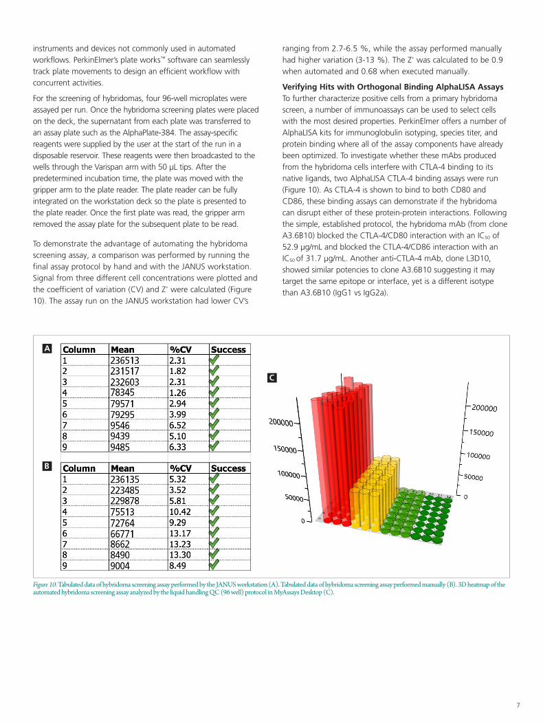
For the screening of hybridomas, four 96-well microplates were assayed per run. Once the hybridoma screening plates were placed on the deck, the supernatant from each plate was transferred to an assay plate such as the AlphaPlate-384. The assay-specific reagents were supplied by the user at the start of the run in a disposable reservoir. These reagents were then broadcasted to the wells through the Varispan arm with 50 µL tips. After the predetermined incubation time, the plate was moved with the gripper arm to the plate reader. The plate reader can be fully integrated on the workstation deck so the plate is presented to the plate reader. Once the first plate was read, the gripper arm removed the assay plate for the subsequent plate to be read.
To demonstrate the advantage of automating the hybridoma screening assay, a comparison was performed by running the final assay protocol by hand and with the JANUS workstation. Signal from three different cell concentrations were plotted and the coefficient of variation (CV) and Z' were calculated (Figure 10). The assay run on the JANUS workstation had lower CV’s
ranging from 2.7-6.5 %, while the assay performed manually had higher variation (3-13 %). The Z' was calculated to be 0.9 when automated and 0.68 when executed manually.
Verifying Hits with Orthogonal Binding AlphaLISA AssaysTo further characterize positive cells from a primary hybridoma screen, a number of immunoassays can be used to select cells with the most desired properties. PerkinElmer offers a number of AlphaLISA kits for immunoglobulin isotyping, species titer, and protein binding where all of the assay components have already been optimized. To investigate whether these mAbs produced from the hybridoma cells interfere with CTLA-4 binding to its native ligands, two AlphaLISA CTLA-4 binding assays were run (Figure 10). As CTLA-4 is shown to bind to both CD80 and CD86, these binding assays can demonstrate if the hybridoma can disrupt either of these protein-protein interactions. Following the simple, established protocol, the hybridoma mAb (from clone A3.6B10) blocked the CTLA-4/CD80 interaction with an IC50 of 52.9 µg/mL and blocked the CTLA-4/CD86 interaction with an IC50 of 31.7 µg/mL. Another anti-CTLA-4 mAb, clone L3D10, showed similar potencies to clone A3.6B10 suggesting it may target the same epitope or interface, yet is a different isotype than A3.6B10 (IgG1 vs IgG2a).
Figure 10. Tabulated data of hybridoma screening assay performed by the JANUS workstation (A). Tabulated data of hybridoma screening assay performed manually (B). 3D heatmap of the automated hybridoma screening assay analyzed by the liquid handling QC (96 well) protocol in MyAssays Desktop (C).
A
B
C

For a complete listing of our global offices, visit www.perkinelmer.com/ContactUs
Copyright ©2020, PerkinElmer, Inc. All rights reserved. PerkinElmer® is a registered trademark of PerkinElmer, Inc. All other trademarks are the property of their respective owners. 143953 PKI
PerkinElmer, Inc. 940 Winter Street Waltham, MA 02451 USA P: (800) 762-4000 or (+1) 203-925-4602www.perkinelmer.com
Figure 11. AlphaLISA CTLA-4/CD80 and CTLA-4/CD86 assay workflow (A) and CTLA-4/CD80 assay data (B) CTLA-4/CD86 assay data (C). The profiled hybridoma (anti-CTLA-4, clone A3.6B10) disrupts both protein-protein interactions, demonstrating its function to impact biological processes.
A
B C
References
1. Reichert, Janice M. The Antibody Society. 2020. <https://www.antibodysociety.org/resources/approved-antibodies/>.
2. R uei-Min Lu, Yu-Chyi Hwang, I-Ju Liu, Chi-Chiu Lee, Han-Zen
Tsai, Hsin-Jung Li and Han-Chung Wu. "Development of therapeutic antibodies for the treatment of diseases." Journal of Biomedical Science 27.1 (2020).
3. Vicki Sifniotis, Esteban Cruz, Barbaros Eroglu and Veysel Kayser. "Current Advancements in Addressing Key Challenges of Therapeutic Antibody Design, Manufacture, and Formulation." Antibodies 8.36 (2019).
4. Chia Chiu Lim, Yee Siew Choong, Theam Soon Lim. "Cognizance of Molecular Methods for the Generation of Mutagenic Phage Display Antibody Libraries for Affinity Maturation." International Journal of Molecular Sciences 20 (2019): 1861.
5. "ELISA to Alpha Immunoassay Conversion Guide," PerkinElmer, [Online]. Available: https://www.perkinelmer.com/lab-solutions/resources/docs/GDE_ELISA-to-AlphaLISA.pdf.
Conclusion
The synergy of automated liquid dispensing and AlphaLISA screening assays provide a workflow solution for therapeutic monoclonal antibody discovery. By using a flexible, homogenous assay format, an Alpha assay was developed with a wide dynamic range (4 logs) that ensure cells expressing mAbs can be specifically identified as hits in the screen. To carry out the optimized hybridoma screening assay, the JANUS workstation was utilized for automated liquid dispensing to improve reproducibility and minimize hands-on time. Microplates containing precious sample and assay reagents were then read on the EnVision multimode detector. The raw signal output was imported into the MyAssays software which calculated and plotted the datasets. The integrated and automated workflow developed above with PerkinElmer’s end-to-end solutions represent an advancement to streamline hybridoma screening, a key step within the upstream discovery of the biologics process.