attractive and repulsive factors act through multi...
TRANSCRIPT

COMMENTARY
Attractive and repulsive factors act through multi-subunit receptorcomplexes to regulate nerve fiber growthNina K. Thiede-Stan* and Martin E. Schwab
ABSTRACTIn the nervous system, attractive and repulsive factors guide neuronalgrowth, pathfinding and target innervationduringdevelopment, learningand regeneration after injury. Repulsive and growth-inhibitory factors,such as some ephrins, semaphorins, netrins and myelin-associatedgrowth inhibitors, restrict nerve fiber growth, whereas neurotrophins,and other ephrins, semaphorins and netrins attract fibers and promoteneurite growth. Several of these guidance molecules also play crucialroles invasculogenesis, and regulate cellmigrationand tissue formationin different organs. Precise and highly specific signal transduction inspace and time is required in all these cases, which primarily dependson the presence and function of specific receptors. Interestingly, manyof these ligands act through multi-subunit receptor complexes. In thisCommentary,we review thecurrent knowledgeof howcomplexesof thereceptors for attractive and repulsive neurite growth regulatory factorsare reorganized in a spatial and temporal manner, and reveal theimplications that such dynamics have on the signaling events thatcoordinate neurite fiber growth.
KEY WORDS: Ephrin, Functional microdomain, Myelin-associatedinhibitors, Neurotrophins, Receptor complex
IntroductionThe outgrowth of axons and pathfinding to their targets is accuratelyguided by environmental cues in the developing central andperipheral nervous system. The modulation of the neuronal growthcones by growth-promoting or -restricting neurotrophic and axonalguidance factors is not only relevant for axonal patterning duringdevelopment, but also for plastic circuit rearrangements and fiberregeneration following injury. Interestingly, many of these ligandsare also present at synapses in the adult nervous system, wherethey influence synaptic plasticity and learning. Axon guidancemolecules, such as ephrins, netrins, as well as neurotrophins andmyelin-associated growth inhibitory molecules, signal throughdynamically assembled multi-subunit receptor complexes, whichcomprise several, often structurally unrelated binding partners(Fig. 1). Furthermore, semaphorin (SEMA)-family members, actingas repulsive or growth-promoting cues, signal through plexins(PLXNs) and a diversity of co-receptors. SEMA3 proteinsadditionally require neuropilins (NRP1 or NRP2) for repulsiveneuronal signaling. Semaphorin-mediated axon pathfinding is ofparticular importance during development of the early spinal cordand the retinothalamic tract (Kuwajima et al., 2012; Pasterkamp,2012; Yoshida, 2012). In this Commentary, we will first introducethe main interaction partners of neurotrophins, ephrins, netrins andmyelin-associated inhibitors that are relevant in this context, as well
as their effects on nerve fiber growth. We will then review andcompare the events during formation of the early receptor complex,such as the recruitment and assembly of receptors and co-receptors,before discussing studies of ligand-induced membrane mobility andcomplex internalization, and emphasizing their implications indownstream signaling events.
Neurotrophicandaxonalguidancefactors,andtheir receptorsNeurotrophinsThe family of neurotrophins comprises nerve growth factor (NGF),brain-derived neurotrophic factor (BDNF), neurotrophin-3 (NT3, alsoknown as NTF3) and neurotrophin-4 (NT4, also known as NTF4 andneurotrophin-5) (Chao, 2003; Chao et al., 2006). All neurotrophin-family members bind to high-affinity tropomyosin receptor kinases(Trks); however, they bind with different preferences – NGF bindspreferentially to TrkA, BDNF and NT4 to TrkB, and NT3 to TrkC(TrkA, TrkB and TrkC are also known as NTRK1, NTRK2 andNTRK3, respectively) (Barbacid, 1995; Ip et al., 1992). The lowaffinity tumor necrosis factor (TNF)-receptor-family member p75(TNFRSF1B) interacts with all neurotrophins, both in the presenceand absence of Trks as co-receptors (Hempstead et al., 1991).Neurotrophin signaling through Trks and p75 promotes neuriteoutgrowth, differentiation and cell survival (Huang and Reichardt,2001), whereas the precursor peptide pro-NGF selectively inducescell death through p75 and the neurotensin receptor sortilin in theabsence of TrkA (Jansen et al., 2007; Kotlyanskaya et al., 2013;Lee et al., 2001; Nykjaer et al., 2004; Teng and Hempstead, 2004).NGF is synthesized and secreted by sympathetic and sensory targetcells to attract and stabilize the corresponding axons (Huang andReichardt, 2001; Korsching, 1993). After peripheral nerve injury,NGF is released by Schwann cells, fibroblasts and infiltrating mastcells, and is important for the survival and regeneration of injuredneurons (Huang and Reichardt, 2001). In the central nervous system(CNS), the basal forebrain and striatal cholinergic neurons inparticular respond to NGF, whereas cerebellar granule cells,mesencephalic dopaminergic neurons, hippocampal neurons andretinal ganglion cells respond to BDNF and NT4 (Huang andReichardt, 2001).
NetrinsThe netrin-family members are chemotropic factors, acting either asattractive or repulsive ligands, depending on the concentration andthe receptor composition on the responding cell. Netrin-1, netrin-2and netrin-3 are secreted ligands, which signal through the receptorsdeleted colorectal cancer (DCC), the DCC paralog neogenin, theUNC-5 homologs UNC5A–D, and Down syndrome cell adhesionmolecule (DSCAM) (Lai Wing Sun et al., 2011; Xu et al., 2014).Glycosylphosphatidylinositol (GPI)-linked netrins G1 and G2interact with netrin-G ligands (NGLs) NGL-1 (also known asLRRC4C) and NGL-2 (also known as LRRC4) (Lai WingSun et al., 2011; Yurchenco and Wadsworth, 2004). Netrin-1 is
Brain Research Institute, University of Zurich, Department of Health Sciences &Technology, ETH Zurich, Zurich 8057, Switzerland.
*Author for correspondence ([email protected])
2403
© 2015. Published by The Company of Biologists Ltd | Journal of Cell Science (2015) 128, 2403-2414 doi:10.1242/jcs.165555
Journal
ofCe
llScience
expressed in various regions of the CNS during development andadulthood (Lai Wing Sun et al., 2011). During neuronaldevelopment, netrins guide cell and axon migration, and influenceaxonal arborization and synapse formation. Netrin-positive cells inthe midline of the early neural tube have a crucial role in attractingmigrating DCC-expressing neuronal precursor cells in thedeveloping hindbrain (Alcantara et al., 2000; Lai Wing Sun et al.,2011). In the developing spinal cord, a netrin-1 gradient that isgenerated by floor-plate cells attracts the commissural axons. Thegradient also repels migrating oligodendrocyte precursor cells(OPCs) and the axons of trochlear motoneurons in the brainstem(Jarjour et al., 2008, 2003; Lai Wing Sun et al., 2011). In the adult
CNS, netrin-1 influences cell–cell contacts, e.g. paranodal junctionsof oligodendrocytes. Beyond the crucial role of netrin signaling inthe CNS, a number of diverse cell biology processes outside of theCNS are affected by netrins, such as angiogenesis and thedevelopment of mammary glands, lungs and pancreas (Cirulli andYebra, 2007; Jarjour et al., 2008; Lai Wing Sun et al., 2011).
EphrinsInteractions between ephrins and ephrin receptors (Ephs) guideaxons during development, and during recovery processes afterinjury. There are two classes of ephrin ligands – ephrin-A ligands(of which there are five, ephrin-A1–A5), which are linked to GPI,
P
P
P
P P
NucleusGolgi
Axon
Growth cone
Soma
PP
P P
PP
Early endosomes
CREB P
Signaling endosomes
CREB P
Activation of growth-related genes
Suppression of growth-related genes
+
-
Ligand binding
Complex assembly
Clustering in specialized microdomains
7
PP
PP
PP
P P
PP
PP
PP
P P
Attractiveligand
Repulsiveligand
S1PR2
NgR1
Nogo-A
Nogo-66Nogo-A-Δ20
MAG
LINGO1 p75
LRP1
PirB
GT1b
OMgp
Growth restrictingGrowth promoting Promoting or restricting
Myelin-associated inhibitorsEphrins
P
Ephrin-AEphrin-B
EphA EphB
SemaphorinsNetrinNeurotrophins
DCC
Netrin-1
UNC
NGF BDNF NT3NT4
TrkA
p75
TrkB TrkC Allbind
PLXN
NRP
IgCAM
SEMA3
SEMA4
A
B
Recycling
Degradation
Receptor Co-receptor
Ligand Lipid raft
6 5
4
3
2
1
Key
Growth promotion
Growth cone collapse
4
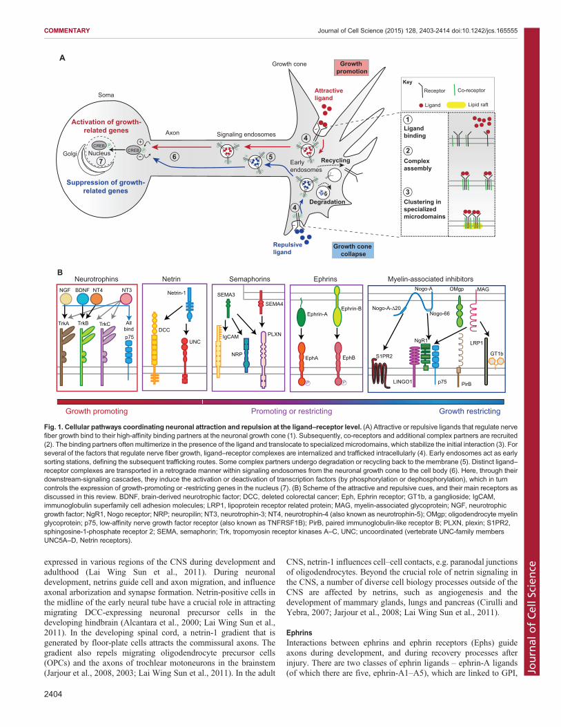
Fig. 1. Cellular pathways coordinating neuronal attraction and repulsion at the ligand–receptor level. (A) Attractive or repulsive ligands that regulate nervefiber growth bind to their high-affinity binding partners at the neuronal growth cone (1). Subsequently, co-receptors and additional complex partners are recruited(2). The binding partners often multimerize in the presence of the ligand and translocate to specialized microdomains, which stabilize the initial interaction (3). Forseveral of the factors that regulate nerve fiber growth, ligand–receptor complexes are internalized and trafficked intracellularly (4). Early endosomes act as earlysorting stations, defining the subsequent trafficking routes. Some complex partners undergo degradation or recycling back to the membrane (5). Distinct ligand–receptor complexes are transported in a retrograde manner within signaling endosomes from the neuronal growth cone to the cell body (6). Here, through theirdownstream-signaling cascades, they induce the activation or deactivation of transcription factors (by phosphorylation or dephosphorylation), which in turncontrols the expression of growth-promoting or -restricting genes in the nucleus (7). (B) Scheme of the attractive and repulsive cues, and their main receptors asdiscussed in this review. BDNF, brain-derived neurotrophic factor; DCC, deleted colorectal cancer; Eph, Ephrin receptor; GT1b, a ganglioside; IgCAM,immunoglobulin superfamily cell adhesion molecules; LRP1, lipoprotein receptor related protein; MAG, myelin-associated glycoprotein; NGF, neurotrophicgrowth factor; NgR1, Nogo receptor; NRP; neuropilin; NT3, neurotrophin-3; NT4, neurotrophin-4 (also known as neurotrophin-5); OMgp; oligodendrocyte myelinglycoprotein; p75, low-affinity nerve growth factor receptor (also known as TNFRSF1B); PirB, paired immunoglobulin-like receptor B; PLXN, plexin; S1PR2,sphingosine-1-phosphate receptor 2; SEMA, semaphorin; Trk, tropomyosin receptor kinases A–C, UNC; uncoordinated (vertebrate UNC-family membersUNC5A–D, Netrin receptors).
2404
COMMENTARY Journal of Cell Science (2015) 128, 2403-2414 doi:10.1242/jcs.165555
Journal
ofCe
llScience

and transmembrane ephrin-B ligands (of which there are three,ephrin-B1–B3). Ephrin-A ligands interact predominantly with oneof the eight EphA receptors (EphA1–A8), whereas ephrin-B ligandsinteract primarily with one of the four EphB receptors (EphB1–B4)(Grunwald and Klein, 2002). Interestingly, interactions betweenephrin ligands and Eph receptors could lead to bidirectionalsignaling – ephrins on one cell can activate Eph receptors on asecond cell (forward signaling), or Eph on the second cell can act asa ligand and activate ephrins, which function as receptors, on thefirst cell (reverse signaling) (Pasquale, 2005, 2008; Steinecke et al.,2014). During the development of spinal-motoneuron-mediatedmuscle innervation, Eph–ephrin signaling has an important role inthe correct pathfinding and target innervation by repellingoutgrowing axons from areas with high ephrin levels, and bypromoting outgrowth into regions that express low levels of ephrins(Klein and Kania, 2014; Pasquale, 2005). Retinal ganglion axonsare guided towards the superior colliculus and optic tectum area,which is one of their main targets, by ephrin-A and EphA gradients.The expression of EphA3 in the retinal ganglion cells increasesalong the nasal–temporal axis. In the optic tectum, the expression ofephrin-A2 and ephrin-A5 forms a gradient with low levels atanterior regions and high levels at posterior regions. EphA3-mediated repulsive (forward) signaling therefore prevents temporalretinal ganglion cell axons from growing into the posterior tectalregions that express high levels of ephrin-A2 and ephrin-A5. Nasalretinal ganglion cell axons that express low EphA3 levels are able togrow into the posterior optic tectum because they experience a lowerlevel of repulsion (Klein and Kania, 2014; Pasquale, 2005; Triplettand Feldheim, 2012). Ephrins and Ephs also crucially influence thecorrect formation of the corticospinal tract. Ephrin-B3 ligands,which are expressed at the midline of the brainstem and the spinalcord, interact with EphA4 receptors on corticospinal axons, and soprevent a re-crossing of the corticospinal fibers and ensure propercortical projection into the spinal cord (Dottori et al., 1998;Grunwald and Klein, 2002; Klein and Kania, 2014). Ephrin-A5-and ephrin-B1-mediated signaling in early embryonic corticalprecursors, as well as in sympathetic neurons, has been shown topromote neurite outgrowth and survival; however, the underlyingmodes of action still need to be defined (Gao et al., 2000; Zhouet al., 2001). Heparan sulfate has been identified as a modulator ofephrin-mediated signaling because ephrin-A3-dependent roundingof Chinese hamster ovary (CHO) cells and collapse of neuronalgrowth cones depends on heparan sulfate (Irie et al., 2008). Besidesthe crucial role of ephrin and Eph signaling in the nervous system,these ligand–receptor pairs have been shown to also modify manyaspects of cancer development and progression, such as the growth,migration and invasion of cancer cells (Pasquale, 2008, 2010;Surawska et al., 2004).
Myelin-associated neurite growth inhibitorsCNSmyelin, but not peripheral nervous system (PNS)myelin inhibitsaxon outgrowth (Caroni and Schwab, 1988; Schwab and Thoenen,1985). Nogo-A, myelin-associated glycoprotein (MAG) andoligodendrocyte myelin glycoprotein (OMgp, also known as OMG)are myelin-associated inhibitors of neurite outgrowth (Filbin, 2003;Schwab, 2010). Nogo-A (also known as Rtn4a in zebrafish) is amemberof the reticulon (Rtn)-familyofmembraneproteins; it has twomain extracellular inhibitory domains, Nogo-66 and Nogo-A-Δ20.These domains interact with different receptor complexes, thedownstream signals of which result in the activation of RhoA andRho kinase (ROCK, for which there are two isoforms ROCK1 andROCK2), the destabilization of the cytoskeleton and a transcription-
dependent downregulation of the neuronal growth program (Kempfand Schwab, 2013; Schwab, 2010; Schwab and Strittmatter, 2014).Nogo-A-Δ20 elicits the inhibition of neurite outgrowth and fibroblastspreading through the G-protein-coupled receptor sphingosine-1-phosphate receptor 2 (S1PR2) (Kempf et al., 2014). Interestingly,Nogo-A-Δ20 also binds to the membrane protein tetraspanin-3(TSPAN-3), and the Nogo-A–TSPAN-3 complex then co-clusterswith S1PR2.Accordingly,Nogo-A-induced growth inhibitory effectsand RhoA activation are reduced when TSPAN-3 is depletedfrom the responding cells (N.K.T.-S., Bjöern Tews, David Albrecht,Zorica Ristic, Helge Ewers and M.E.S., unpublished observation).The second active domain of Nogo-A, Nogo-66, binds to the Nogoreceptor 1 (NgR1, also known as RTN4RL1) (Fournier et al., 2001),which acts in concert with the co-receptors p75 or TROY(TNFRSF19), and LINGO1 (Mi et al., 2004; Park et al., 2005;Shao et al., 2005;Wang et al., 2002).Nogo-66 also binds to the pairedimmunoglobulin-like receptorB (PirB, also known asPILRB) (Atwalet al., 2008; Schwab, 2010).
Interestingly, the two other myelin-associated inhibitors MAG andOMgp also bind toNgR1, in complexwith the same co-receptors – i.e.p75 or TROY, and LINGO1 – althoughMAG shows a higher bindingaffinity for the Nogo-66 receptor homolog NgR2 (RTN4RL2) (Gigeret al., 2010; Schwab, 2010). MAG further interacts with gangliosides,which are involved in inhibition of MAG-induced neurite outgrowth(Vinson et al., 2001). These myelin-associated inhibitors arepredominantly thought to act as inhibitors of regeneration, circuitplasticity and functional recoveryafter injury to theCNS (Filbin, 2003;Schwab, 2004; Yiu and He, 2006). In addition to restricting axonalregeneration and structural plasticity after spinal cord injury or stroke(Schwab and Strittmatter, 2014; Wahl et al., 2014), Nogo-A, NgR1and PirB have crucial roles in restricting plasticity in the visual system,the sensory-motor cortex and the hippocampus in the uninjured adultCNS (Akbik et al., 2012; Bochner et al., 2014; Delekate et al., 2011;McGee et al., 2005; Schwab, 2010; Schwab and Strittmatter, 2014;Zemmar et al., 2014). Through its effects on cell adhesion andcytoskeletal dynamics, Nogo-A-Δ20 influences the migration ofneuronal precursors (through NgR1) and inhibits the migration ofprimary brain microvascular endothelial cells (through S1PR2), aswell as the spreading of endothelial cells and fibroblasts (Schwab,2010;Walchli et al., 2013). This indicates that specific ligand-inducedsignaling pathways and effects, such as the destabilization of thecytoskeleton, are conserved in different cell types, resulting in relatedcellular effects, such as neuronal growth cone collapse, inhibition ofmigration, or spreading of fibroblasts or neuronal precursors.
Dynamic receptor complex formation at the cell membraneNeuronal growth-promoting or -restricting molecules signal inthe developing and mature CNS through receptor complexes thatcomprise different interaction partners. The concerted interplaybetween signal-transducing receptors, their modulatory co-receptors and accessory factors is initiated by their ligands,although some binding partners can co-associate even in theabsence of the ligand in signaling-incompetent complexes. Theseligand-induced receptor complexes assemble in a dynamic mannerinto complexes that range in size from nano- to microscale andundergo structural reorganizations, giving rise to a functionalsignaling platform as discussed below (Fig. 2).
Initial events upon ligand binding – recruitment, assembly andmultimerization of binding partnersNeurotrophins were one of the first examples of ligands of multi-subunit receptor complexes to be studied in detail (Chao, 2003;
2405
COMMENTARY Journal of Cell Science (2015) 128, 2403-2414 doi:10.1242/jcs.165555
Journal
ofCe
llScience

Teng and Hempstead, 2004). Neurotrophins induce dimerization ofTrks, although it has been shown recently that inactive Trkhomodimers and also p75–TrkA heterodimers exist in the absenceof ligands (Iacaruso et al., 2011; Jing et al., 1992; Shen andMaruyama, 2011). Crystal structure analysis and biochemicalexperiments indicate a 2:2 stoichiometric binding of NGF or NT3to p75 in solution (Aurikko et al., 2005; Gong et al., 2008). Thus,the presence of dimerized NGF or NT3 promotes homodimerizationof p75, dimerization and activation of Trk, as well as formation ofheterotrimeric receptor complexes containing Trk proteins and p75.Furthermore, netrin-family members act as guidance molecules,
interacting with different partners of the receptor complex andactivating attractive- or repulsive-signaling pathways dependent
upon the respective interaction partners. Netrin-1 signaling throughDCC induces chemoattraction, but signaling through UNC-5proteins induces chemorepulsion (Lai Wing Sun et al., 2011). Ithas recently been shown that neogenin, which is structurally similarto DCC, acts as an additional receptor of netrin-1 that, incollaboration with DCC, mediates the attraction of commissuralneurons in the developing spinal cord. The binding of netrin-1 toeither DCC or neogenin results in a differential architecture of theligand–receptor complex – a 2:2 heterotetramer for the netrin-1–neogenin complex and a continuous chain-like ligand–receptorcomplex in the case of netrin-1 and DCC. These distinctarchitectures of the ligand–receptor complexes might provide themolecular basis for the different signaling events that they elicit (Xuet al., 2014). The structural insight into the binding of netrin-1 totwo DCC molecules is in line with a second crystallography studythat has defined two binding sites on netrin-1 – one that is specificfor DCC and a second that could interact either with a second DCCmolecule or UNC5. The choice between netrin-1 binding to eitherDCC or UNC5 with its second binding site is governed by thebinding of netrin-1 to one of two distinct heparan sulfate molecules(HP1 and HP2) – when netrin is bound to either HP1 or HP2, thesecond site is selective for DCC or UNC5, respectively. Netrin-1–DCC signaling induces attraction of the growth cone, whereasnetrin-1–UNC5 induces growth cone repulsion (Finci et al., 2014).Furthermore, netrin-1 interacts with other binding partners, such asthe laminin-binding integrins α6β4 and α3β1, and this has beenimplicated in the activation of adhesion and migration of pancreaticepithelial cells, which express netrin-1 (Lai Wing Sun et al., 2011;Ly et al., 2008; Moore et al., 2007; Yebra et al., 2003). Thesedifferent interactions of netrins with their partners influence the cellbiology aspects of different cell types in either an attractive orrepulsive manner.
Nogo-A also interacts with multiple binding partners. The GPI-anchored Nogo-66 receptor NgR1 is important for binding of Nogo-66, MAG or OMgp, but because it lacks a signal-transducingcytoplasmic domain, it depends on the co-receptors p75 andTROY toform a signal-transducing complex (Wang et al., 2002). LINGO1,another component of the Nogo receptor complex, forms stabletetramers, which appear to be necessary for its interaction with thereceptor-complex partners NgR1 and p75 (Mosyak et al., 2006).LINGO1 and the ectodomain of NgR1 are characterized byevolutionary conserved leucine-rich repeat domains (Filbin, 2003;Saha et al., 2014). NgR1 and p75 colocalize even in absence of theirmyelin-associated inhibitory ligands; however, presence of the ligandincreases the association between NgR1 and p75 in a concentration-dependent manner (Wang et al., 2002;Wong et al., 2002). In additionto NgR1, MAG also binds to the lipoprotein-receptor-related proteinLRP1. This binding does not depend on the presence of p75, butinduces a colocalization of LRP1 and p75, which is not detectable inabsence of the ligand (Mantuano et al., 2013; Stiles et al., 2013). Thisdynamic recruitment of p75 toMAG and/or LRP1 is followed by theactivation of RhoA (Fig. 2). An active recruitment of p75 intoMAG-bound receptor complexes has been also shown for PirB, anotherreceptor forNogo-66 andMAG(Fujita et al., 2011; Shao et al., 2005).The interaction partners of Nogo-66 and MAG assemble into afunctional complex, comprising a high-affinity binding partner(NgR1), signal-transducing receptors (p75 and TROY) and acomplex-stabilizing co-receptor (LINGO1), which cooperativelytransduces the ligand-induced inhibitory effects.
Once the initial ligand–receptor pair assembles into a smallfunctional entity, multimers of the receptor and co-receptors canbe recruited, often in a specific stoichiometry. Ligand-induced
Lipid raft microdomain
MAG
LINGO1 or
p75LRP1NgR1
GT1b
PirB Rho-GDI
RhoAGDP GTP
RhoA
Rho-GDI
ROCK
Growth cone collapseNeurite outgrowth inhibition
1. Absence of the ligand
2. Presence of the ligand
Oligodendrocyte
Neuron
Oligodendrocyte
Neuron
Fig. 2. Ligand-induced formation of receptor complexes at the cellmembrane. Illustrated here is the formation of the multi-subunit heteromericreceptor complex of myelin-associated glycoprotein (MAG) as an example of agrowth-restricting ligand. In the absence of the ligand, the binding partners ofMAG do not form a fully functional complex, although the Nogo receptor(NgR1) and p75 (also known as TNFRSF1B) partially colocalize (shown on thetop). In the presence of MAG, the receptor components assemble into a signal-transducing complex, which is stabilized by its translocation into lipid rafts(shown at the bottom). MAG presentation increases the association betweenNgR1 and p75 (or PirB) and mediates the recruitment of LRP1 into the vicinityof p75. Gangliosides such as GT1b stabilize the interaction of NgR1 with theco-receptor LINGO1. Lipid rafts scaffold the MAG receptor complex andfacilitate downstream signaling, for instance p75-mediated release of RhoGDP-dissociation inhibitors (Rho-GDI) from the GTPase RhoA. Subsequentactivation of RhoA and Rho kinase (ROCK, for which there are two isoformsROCK1 and ROCK2) results in destabilization of the cytoskeleton, followed bycollapse of the growth cone and inhibition of neurite outgrowth.
2406
COMMENTARY Journal of Cell Science (2015) 128, 2403-2414 doi:10.1242/jcs.165555
Journal
ofCe
llScience

clustering and oligomerization of receptors and co-receptorsculminates in an enhanced signaling output of the resultingreceptor cluster, as observed for ephrins, myelin-associatedgrowth inhibitors, SEMA3A and neurotrophins (Barton et al.,2004; Gong et al., 2008; Himanen et al., 2010; Janes et al., 2012;Janssen et al., 2012; Marchetti et al., 2013; Mosyak et al., 2006;Pasquale, 2005; Salaita et al., 2010; Seiradake et al., 2010;Wimmer-Kleikamp et al., 2004). In this context, the number ofligand–receptor pairs in these clusters directly correlates with thestrength and outcome of the elicited response (Janssen et al., 2012,2010; Schaupp et al., 2014). For instance, the multimer compositionof EphB2 clusters (monomers, dimers, tetramers) determines thestrength of the cellular repulsion response – the more EphB2receptor dimers that are present, the weaker the response, and themore tetramers there are, the stronger the response (Schauppet al., 2014). Although EphA and EphB subtypes preferentially bindto A- and B-type ephrins, respectively, mixed oligomers comprisingEphA and EphB exist on cells that express both Eph subtypes, andthese oligomers modulate signaling and cell retraction (Janes et al.,2011). Distinct patterns of EphA and EphB hetero-oligomerizationexist on breast, prostate and glioblastoma tumor cells, and theirmodulatory signaling might influence physiological Eph–ephrinsignaling, thereby promoting cancer progression (Al-Ejeh et al.,2014). The assembly of ephrin–Eph complexes into higher-ordersignaling clusters is mediated by the initial dimerization of thecysteine-rich domain of Ephs; at high concentrations, this mightlead to receptor clustering even in absence of the ligand (Himanenet al., 2010). The multivalent binding of a ligand (simultaneousbinding to one assembly of receptors) has recently been shown todrive receptor clustering at the nanoscale level (Conway et al.,2013). Super-resolution microscopy has been used to visualizeephrin-B2-driven clustering of the EphB4 receptor at the nanoscalelevel on adult neuronal stem cells, as ephrin-B–EphB signaling hasbeen shown to regulate neuronal stem cell differentiation (Ashtonet al., 2012; Conway et al., 2013; Vazin et al., 2009). Themultivalency of a recombinant ephrin-B2 ectodomain conjugated toa soluble biopolymer strongly enhanced the bioactivity of ligands,resulting in the induction of neuronal stem cell signaling – i.e.β-catenin expression – and stem cell differentiation (Conway et al.,2013). Other studies have shown the formation of large EphA3-signaling clusters that are induced by the multivalent ligand ephrin-A5. In those studies, the size of the EphA3 clusters exceeded that ofthe initial ephrin-A5–EphA3 contact site by several fold (Janeset al., 2012; Wimmer-Kleikamp et al., 2004). The expansion ofsignaling clusters from ‘seed’ ligand–receptor interactions leads tothe recruitment of additional Ephs in an ephrin-independentmanner. This has been described as the so-called ‘nucleation’mechanism, and crystal structure analyses provide further evidencefor it (Seiradake et al., 2010; Wimmer-Kleikamp et al., 2004). Thenucleation mechanism supports the notion of signal propagation andan increase in sensitivity to the ephrin-ligands.A single ligand can also drive distinct cellular responses depending
on the receptor it interacts with (Marchetti et al., 2013; Seiradakeet al., 2013). For instance, structure-based differences betweenEphA4 and EphA2 lead to differential binding of their ligand ephrin-A5. Presentation of ephrin-A5 to HeLa cells transfected with eitherEphA4orEphA2 results in opposing effects – i.e. in either collapse oradherence of the cell, which are associated with smaller clusters ofephrin-A5–EphA4 and larger clusters of ephrin-A5–EphA2,respectively. Therefore, a relatively small number of distinctreceptors are able to control diverse signaling pathways (Seiradakeet al., 2013). Taken together, these studies indicate that the initial
bindingof different attractive and repulsive ligands recruits additionalco-receptor and partner molecules into a signaling-competentcomplex. Further fine-tuning can occur through modulation of thespecific complex composition and the stoichiometry of the signal-transducing components present therein.
Specialized microdomains as signaling platformsThe presence of a ligand induces the dynamic assembly of multipleinteraction partners into signaling scaffolds. These scaffoldsconcentrate the specific subunits of the receptor complex intodistinct areas or microdomains at the plasma membrane, andpotentially amplify the transduction of the elicited signal. Specificplasma membrane constituents facilitate ligand-induced assembly ofthe receptor complex. In this context, lipid rafts are of particularimportance. They comprise dynamic assemblies of cholesterol andsphingolipids, and are enriched inGPI-anchoredproteins, cholesterol-or palmitoyl-linked proteins and the α-subunits of heterotrimeric G-proteins (Simons and Toomre, 2000). In neurons, the interactionbetween MAG and its receptors – NgR, and the GT1b and GD1gangliosides – occurs in lipid rafts. Therefore, it has been suggestedthat these rafts form discrete areas that support the high-aviditymultivalent binding of MAG to its interaction partners, and that theycontain the MAG receptors and the downstream effector RhoA(Vinson et al., 2003) (Fig. 2). Similar results, revealing the relevanceof lipid rafts, have been described for the interaction of Nogo-66 withits receptors p75 and NgR1; in this case, the receptors colocalize inlipid rafts, and Nogo-66 signaling is disturbed in the presence of thecholesterol-depleting substance β-methylcyclodextrin (Yu et al.,2004). Gangliosides, sialic-acid-bearing glycosphingolipids, arerequired for MAG-induced translocation of p75 to lipid rafts andsubsequent signal transduction (Fujitani et al., 2005). Furthermore,gangliosides mediate the interaction between NgR1 and its co-receptor LINGO1 (Saha et al., 2011), and it has been suggested thatthe binding of MAG to gangliosides influences the optimal axon–myelin interaction (Schnaar, 2010) (Fig. 2).
The effect of gangliosides on NGF–TrkA-signaling is not fullyunderstood. Exogenous ganglioside GM1 enhances NGF-induceddimerization of TrkA and its phosphorylation (Farooqui et al., 1997;Mutoh et al., 1995), but endogenousGM1suppressesNGFsignaling,probably by changing the intracellular localization of NGF receptors(Nishio et al., 2004). Lipid rafts have been shown to positively affectneurotrophin signaling – binding of NGF to TrkA induces itstranslocation and concentration in lipid rafts, which facilitates theformation of receptor-associated signaling complexes, theirinternalization and the activation of NGF-induced downstreamsignaling, such as phosphorylation of TrkA and activation of theextracellular signal-regulated kinase (ERK) pathway (Limpert et al.,2007). Ligand-induced receptor translocation to lipid rafts has alsobeen shown for BDNF–TrkB. In lipid rafts, TrkB-bound BDNFassociates with the lipid-raft-resident protein Fyn, which mediatesdownstream signaling of BDNF in neurons (Pereira and Chao, 2007).
An important consequence of partitioning multi-subunit receptorcomplexes into lipid rafts upon ligand presentation could be theresulting selectivity of the signals transduced (Campbell et al., 2008;Zonta and Minichiello, 2013). An example of this is the distinctmembrane compartmentalization and signaling of ephrin-A5 andephrin-B1, as studied in NIH 3T3 fibroblasts, where ephrin-A5 isfound in detergent-resistant membrane fractions, whereas ephrin-B1translocates to these membrane fractions only upon receptorbinding. In detergent-resistant membrane fractions, ephrin-A5 andephrin-B1 fractionate at different raft fractions in the living cell. Thisresults in differential regulation of the cytoskeletal architecture and
2407
COMMENTARY Journal of Cell Science (2015) 128, 2403-2414 doi:10.1242/jcs.165555
Journal
ofCe
llScience
distinct gene expression, as assessed by using microarray analysis ofNIH 3T3 fibroblasts that expressed ephrin-A5 or ephrin-B(Campbell et al., 2008; Gauthier and Robbins, 2003).In conclusion, multimeric and multi-subunit receptor complexes
that interact with and transduce attractive and repulsive axonalguidance cues frequently form in specialized plasma membranemicrodomains, the lipid rafts. The high avidity of the multivalentligand–receptor interactions facilitate, amplify and specify theresulting intracellular signals (Farooqui et al., 1997; Fujitani et al.,2005; Limpert et al., 2007; Pereira and Chao, 2007; Vinson et al.,2003; Yu et al., 2004; Zonta and Minichiello, 2013).
Translocation and internalization of receptor complexesThe ligand-induced formation of functional receptor complexes isaccompanied by the lateral motion of the individual bindingpartners and receptor components in the membrane. Subsequent totheir assembly, receptor complexes are often internalized intosignaling endosomes that contain the endocytosed ligand–receptorcomplexes and downstream effectors. Below, we review the knownpatterns of movement of attractive and repulsive multi-subunitreceptor complexes, and compare the changes associated with theseboth at the cell membrane and inside the cell.
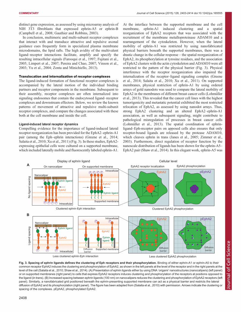
Ligand-induced lateral receptor dynamicsCompelling evidence for the importance of ligand-induced lateralreceptor reorganization has been provided for the EphA2–ephrin-A1pair (among the Eph–ephrin interactions) (Greene et al., 2014;Salaita et al., 2010; Xu et al., 2011) (Fig. 3). In these studies, EphA2-expressing epithelial cells were cultured on a supported membrane,which included laterallymobile and fluorescently labeled ephrin-A1.
At the interface between the supported membrane and the cellmembrane, ephrin-A1 induced clustering and a spatialreorganization of EphA2 receptors that was associated with therecruitment of the membrane metalloproteinase ADAM10 and arearrangement of the cytoskeleton. However, when the lateralmobility of ephrin-A1 was restricted by using nanofabricatedphysical barriers beneath the supported membranes, there was adrastic change in the cellular response – the spatial reorganization ofEphA2, its phosphorylation at tyrosine residues, and the associationof EphA2 clusters with the actin cytoskeleton andADAM10were allrestricted to the pattern of the physical barrier (Fig. 3). Physicalinterference with the receptor reorganization also impaired theinternalization of the receptor–ligand signaling complex (Greeneet al., 2014; Salaita et al., 2010; Xu et al., 2011). On supportedmembranes, physical restriction of ephrin-A1 by using orderedarrays of gold nanodots was used to compare the lateral mobility ofEphA2 in the membranes of different breast cancer cells (Lohmülleret al., 2013). This revealed that the cancer cell lines with the highesttumorigenicity and metastatic potential exhibited the most restrictedrelocation of EphA2, as assessed by using nanodot arrays. Thus,strong EphA2 clustering and an altered EphA2–ephrin-A1association, as well as subsequent signaling, might contribute topathological misregulation of processes in breast cancer cells(Lohmüller et al., 2013). The spatial coordination of ephrin-ligand–Eph-receptor pairs on apposed cells also ensures that onlyreceptor-bound ligands are released by the protease ADAM10,which cleaves ephrin in trans (Janes et al., 2005; Zimmer et al.,2003). Furthermore, direct regulation of receptor function by thenanoscale distribution of ligands has been shown for the ephrin-A5–EphA2 pair (Shaw et al., 2014). In this elegant work, ephrin-A5 was
EphA2
No
ligan
d sp
acin
g
pEphA2
Liga
nd s
paci
ng
EphA2 pEphA2
On supported membraneOn nanocaliper
Display of ephrin ligand
Intracellular
Ephrin ligand
P P P P
EphA2
Supported membraneIntracellular
Ephrin ligand
P P P P
EphA2
Nanocaliper
Intracellular
Ephrinligand
P P P
EphA2
Supported membraneDiffusion barrierIntracellular
Ephrin ligand
P P P
EphA2
Nanocaliper
Cellular level
Diffusion barrier
EphA2 receptor localization EphA2 phosphorylation
Diffusion barrier
Clustered ephrin-Eph interaction
Less clustered ephrin-Eph interaction Less clustered EphA2 phosphorylation
Clustered EphA2 phosphorylation
A
B
Fig. 3. Spacing of ephrin ligands defines the clustering of Eph receptors and their phosphorylation. Binding of either ephrin-A1 or ephrin-A5 to theircommon receptor EphA2 induces the clustering and phosphorylation of EphA2, as shown in the left panels at the level of the receptor and in the right panels at thelevel of the cell (Salaita et al., 2010; Shaw et al., 2014). (A) Presentation of ephrin ligands either by using DNA ‘origami’ nanostructures (nanocalipers) (left panel)or on supported membranes (right panel) to cells that express EphA2 receptors induces clustering and phosphorylation of the receptors at positions opposed tothe ligand (in trans). (B) Increased spacing between ephrin ligands (100 nm) on nanocalipers reduces the clustering and phosphorylation of EphA2 receptors (leftpanel). Similarly, a nanofabricated grid positioned beneath the ephrin-presenting supported membrane can act as a physical barrier and restricts the lateraldiffusion of EphA2 and its phosphorylation (right panel). The figure has been adapted from (Salaita et al., 2010) with permission. Arrows indicate the clustering orspacing of the complexes. pEphA2, phosphorylated EphA2.
2408
COMMENTARY Journal of Cell Science (2015) 128, 2403-2414 doi:10.1242/jcs.165555
Journal
ofCe
llScience

presented to EphA2-expressing cells in the form DNA ‘origami’nanostructures (nanocalipers), to which the ligand has been attachedat defined positions; there was either only one binding site or twobinding sites separated by distances of approximately 43 or 100 nm.The resulting spatial organization of ephrin-A5 at the nanoscaledetermined the degree of EphA2 receptor activation depending onthe ligand spacing used – ephrin-A5 dimers induced strongerreceptor phosphorylation than a single ephrin-A5 molecule, and theephrin-A5 dimer with the shorter distance between the binding siteswas the most efficient ligand (Shaw et al., 2014) (Fig. 3).The recent development of advanced microscopy techniques has
allowed an increased understanding of the ligand-induced formationof receptor complexes at the nanoscale level. Observing live cellswithsingle-molecule imaging techniques has enabled the analysis ofsingle steps of the formation of receptor complexes from initialbinding to signal transduction and amplification (Cui et al., 2007;Marchetti et al., 2013; Shibata et al., 2006; Tani et al., 2005), and thishas also been studied in other cellular systems, such as in the immunesystem (see Box 1). For instance, the spatio-temporal mobility offluorescently labeled NGF (NGF–Cy3) upon receptor binding hasbeen described as comprising at least two distinct states, characterizedas amobile and immobile phase. Initially,manyof the receptor-boundNGF–Cy3 molecules are mobile, and their subsequentimmobilization is associated in time with intracellular signaling.Dual live-imaging has illustrated the recruitment of intracellularsignaling components to the immobile NGF-receptor complexes(Shibata et al., 2006). Moreover, the differential biological effects ofthree different neurotrophins – NGF, NT3 and proNGF – correlatewith different patterns of ligand-induced reorganization of quantum-dot-labeled TrkA – i.e. immobile versus mobile state at the cellmembrane – and quantitative differences in the lateral mobilitypatterns and trajectories that are induced by ligand binding (Marchettiet al., 2013). Furthermore, on chicken growth cones, only 40 Cy3-conjugated NGF molecules, which bind to approximately 5% of theavailable high-affinity receptors, are necessary to initiate the motileresponses of the growth cones (Tani et al., 2005). Upon binding ofNGF–Cy3 to these receptors, the complexes are transported laterallyand unidirectionally towards the central region of the growth cone.This transport is driven by actin and is a crucial intermediate stepbefore subsequent endocytosis of the NGF-receptor complexes,which are transported to the cell body in a retrograde mannerafterwards (Tani et al., 2005). For the neurite growth inhibitorymembrane protein Nogo-A, an increased mobility of the Nogo-A co-receptor TSPAN-3 is observed upon ligand binding. TSPAN-3molecules are released from areas that had originally been confinedand exhibit increased lateral diffusion. Subsequently, the Nogo-A–TSPAN-3 complexes interact with the signal-transducing receptorS1PR2, forming large multi-subunit complexes (N.K.T.-S., BjöernTews, David Albrecht, Zorica Ristic, Helge Ewers and M.E.S.,unpublished observation). In conclusion, these studies illustrate thesensitivity of specific attractive or repulsive receptors to thepresentation of their ligand, which is reflected in changes inmobility at the nanoscale and microscale levels.
Internalization and intracellular traffickingExperiments in the early 1970s revealed the uptake of purifiedradiolabeled NGF by nerve endings of sympathetic and sensoryneurons, and its subsequent retrograde transport to the cell body(Angeletti et al., 1972; Hendry et al., 1974; Stoeckel et al., 1975).Since then, several groups have revealed crucial insights into thecellular mechanisms of the internalization, trafficking andretrograde transport of neurotrophic factors and pro-neurotrophins
(Boutilier et al., 2008; Bronfman et al., 2003; Harrington and Ginty,2013; Nykjaer et al., 2004; Zweifel et al., 2005). In particular, theuse of fluorescent labeling techniques and live imaging have made itpossible to investigate kinetic aspects, such as the time course ofneurotrophin binding, ligand concentration, and the recruitment ofinteraction partners and accessory factors relevant for correctinternalization (Bronfman et al., 2003; Gatzinsky et al., 2001;Jullien et al., 2003; Levi et al., 1980; Philippidou et al., 2011;Valdez et al., 2007; Zweifel et al., 2005). At the single-moleculelevel, the distinct progressive phases of NGF– or BDNF–receptorcomplex formation and its endocytic trafficking have beencharacterized in live cells by using fluorescent or quantum-dot-labeled NGF and BDNF. Such characterization at the single-molecule level further suggests that the dynamic regulation ofligand–receptor interaction controls the strength and duration of thedownstream signaling (Rajan et al., 2008; Vermehren-Schmaedicket al., 2014).
Recently, the dynamic control of ligand–receptor interactions hasalso been shown at the level of transcytosis between differentcellular compartments – NGF binding to the TrkA receptors at thegrowth cone induces the recruitment of additional TrkA receptorsfrom the neuronal soma surface, which are transported to the distantgrowth cones. This intracellular translocation of TrkAs from regionswith low levels of NGF to regions with higher concentrations ofNGF enables the adjustment of the neuronal sensitivity to thepresence of the ligand (Ascano et al., 2009).
In addition to neurotrophins, several other signaling complexes thatmediate attractive and repulsive axonal guidance cues also undergo
Box 1. Multi-subunit receptor complex dynamics atthe immunological synapseIn response to surface-bound antigens on antigen-presenting cells(APCs), B- and T-cell lymphocytes mediate adaptive immune responsesthrough the activation of their B- and T-cell receptors (BCRs and TCRs,respectively) (Brownlie and Zamoyska, 2013; Friedl et al., 2005; Lundand Randall, 2010). Similar to the receptor complexes that are formed byattractive and repulsive nerve fiber growth regulators, TCRs and BCRssignal in concert with several co-receptors and supplementary factors,as revealed, for example, by advanced microscopy techniques(Douglass and Vale, 2005; Grakoui et al., 1999; Monks et al., 1998).Briefly, the following events occur at an immunological synapse (theinterface between APCs and lymphocytes) (Douglass and Vale, 2005;Grakoui et al., 1999; Monks et al., 1998) – the recognition of antigensinduces the TCR co-receptor CD4 to associate with CD3 chains, asshown by fluorescent resonance energy transfer (FRET) live imaging(Zal et al., 2002). Single-molecule microscopy has also revealed thedynamic nature of specific membrane subdomains (Douglass and Vale,2005) – the co-receptor CD2 and the kinase Lck, which phosphorylatesconserved immunoreceptor tyrosine-based activation motifs (ITAMs) onCD3, are recruited to the activated TCR, together with the adapter proteinLAT. By contrast, the phosphatase CD45 (also known as PTPRC), apotential inhibitor of TCR signaling, has been found to be excluded fromthe observed TCR microdomains (Douglass and Vale, 2005). Signalinduction is followed by the recruitment of kinases and signalingmolecules, leading to the activation of the Ras–MEK–ERK pathwayand cytokine (IL-2) production, which promotes the proliferation ofactivated T-cells (Friedl et al., 2005).The formation of the BCR complex and its signaling are regulated in a
similar dynamic manner (Mattila et al., 2013; Monroe, 2006). Takentogether, the concerted signaling that is mediated by receptor complexeson lymphocytes and their partners is organized in a spatio-temporalmanner, and their misregulation might result in immune deficiencies orlymphocyte malignancies (Conley et al., 2009; Rickert, 2013; van Zelmet al., 2010).
2409
COMMENTARY Journal of Cell Science (2015) 128, 2403-2414 doi:10.1242/jcs.165555
Journal
ofCe
llScience

endocytosis. The Nogo-A inhibitory fragment Nogo-A-Δ20 isinternalized at neuronal growth cones by the same pinocyticchaperone protein Pincher [also known as EHD4, Eps15 homology(EH)-domain-containing proteins], which is also important forendocytosis of the NGF–TrkA complex (Joset et al., 2010;Philippidou et al., 2011). Internalized Nogo-A-Δ20 colocalizes withits receptor S1PR2 and its co-receptor TSPAN-3 in early endosomes(Kempf et al., 2014) (N.K.T.-S., Bjöern Tews, David Albrecht, ZoricaRistic, Helge Ewers and M.E.S., unpublished observations). Neurite
outgrowth inhibitionbyMAGdependsonLRP1-mediated endocytosis(Stiles et al., 2013), probably together with β1-integrins (Hines et al.,2010). Ephrin-mediated repulsion is regulated by endocytosis of eitherfull-length ligand–receptor complexes or ephrin-B ligands that havebeen cleaved by metalloproteinases (Marston et al., 2003; Zimmeret al., 2003). The above examples illustrate thewide-spread occurrenceof the internalization of attractive and repulsive ligand–receptorcomplexes following initial complex assembly, but this raises thequestion of its functional relevance for specific intracellular signaling.
TrkA
P
NGF
TrkA
NT3
Maturation into signaling endosomes
PP
PP
PP
PP
PP
PP
Differential sensitivity to endosomal acidification
pH 5.5
PP
PP
PP
Rac1- GTP
Cofilin
Incompetent for formation of signaling endosomes
Adapter
P P P
Ligand binding
A Neurotrophins − NGF versus NT3
S1PR2
NgR1
Nogo-A
Nogo-66Nogo-A-Δ20
LINGO1 p75
B Myelin-associated inhibitor − Nogo-A-Δ20 versus Nogo-66
Local signalingShort-term effect
Internalization into early endosomes
NeuronNeuron
Oligodendrocyte
Retrogradetrafficking
Long-term transcriptionalchanges
RhoAGDP
GTPROCK
RhoA
Nogo-A-Δ20
NGF
NT3
Cofilin
RhoA
Nogo-A-Δ20
Adapter
Maturation into signaling endosomes
Activation ofgrowth-related genes
Suppression ofgrowth-related genes
Not yet reported
pPLCγ
pERK
5
4
3
2
1
Fig. 4. Ligand-selective internalization and translocation of binding partners governs local (axonal versus global) cellular effects. Growth-promotingmolecules, such as the neurotrophins NGF and NT3 (A), or growth-restricting molecules, such as the active Nogo-A domains Nogo-A-Δ20 and Nogo-66 (B)interact at the neuronal growth conewith their respective receptors. (In vivo, full-length Nogo-Amight undergo transcytosis or proteolytic cleavage, resulting in therelease of active fragments such as Nogo-A-Δ20.) Multi-subunit receptor complexes assemble and induce downstream signaling, such as the activation ofGTPases (Rac1 for neurotrophins and RhoA for Nogo-A), which influences the local cytoskeleton of the growth cone (to promote outgrowth, turning or collapse).Formation of receptor complexes is often followed by internalization into early endosomes, i.e. in the case of NGF–TrkA, NT3–TrkA or Nogo-A-Δ20–S1PR2.Internalization of Nogo-66 and its receptors has not been demonstrated yet. Intracellular sorting sends the endosomes for degradation, recycling or retrogradetransport to the cell soma. Nogo-A-Δ20-containing endosomes and NGF–TrkA-containing endosomes undergo retrograde trafficking, whereas NT3–TrkA-containing endosomes are unable to follow this transport route. The differential recruitment of actin modifiers (such as Rac1 and cofilin-family members) results inunstable NT3–TrkA complexes within the acidic environment of early endosomes and so restricts their signaling competence to only a short distance. By contrast,NGF and Nogo-A-Δ20 continue to signal in the cell body and thus exert a long-term effect through modulating the levels of CREB phosphorylation, which affectsthe subsequent expression of neurite growth-specific proteins and transcription factors. pERK, phosphorylated ERK; pPLCγ, phosphorylated PLCγ.
2410
COMMENTARY Journal of Cell Science (2015) 128, 2403-2414 doi:10.1242/jcs.165555
Journal
ofCe
llScience

From local effects at axons to control at the cell bodyThe translocation of ligands, such as neurotrophins and Nogo-Δ20,and their receptors from distal axons to neuronal cell bodies allowscells to directly propagate signaling from the distal neurites toperinuclear regions (Harrington and Ginty, 2013; Joset et al., 2010;Winckler and Yap, 2011). Signaling endosomes that have beentransported contain the endocytosed ligand–receptor complexes andassociated downstream effectors, such as small G-proteins, Rho-GTPases, constituents of the phospholipase C-γ (PLCγ) cascade,Raf (a serine/threonine protein kinase), mitogen-activated proteinkinase kinase (MEK) and phosphatidylinositol 3-kinase (PI3K)(Ginty and Segal, 2002; Harrington et al., 2011; Joset et al., 2010;Zweifel et al., 2005). Quantitative analysis revealed that endosomesthat contain as little as a single NGF dimer can undergo retrogradetrafficking (Cui et al., 2007). Once the signaling endosomes reachthe cell body, neurotrophic factors enhance the phosphorylation oractivation of the transcription factor cyclic AMP response element-binding protein (CREB) and the expression of neurite growth-specific proteins and transcription factors, whereas Nogo-A-Δ20exerts an opposite growth-inhibitory effect (Ginty and Segal, 2002;Harrington and Ginty, 2013; Montani et al., 2009; Riccio et al.,1997; Schwab, 2010) (Fig. 4). Long-term inhibition of neuriteoutgrowth by Nogo-Δ20–S1PR2–TSPAN-3 signaling is probablylinked to cell-body-mediated effects through signaling endosomesthat have undergone retrograde transport, whereas, Nogo-66- orMAG-induced growth-cone collapse is triggered locally by theactivation of the RhoA–ROCK axis, which leads to destabilizationof the cytoskeleton in a manner that is independent of proteinsynthesis (Chivatakarn et al., 2007; Joset et al., 2010; Kempf et al.,2014; Manns et al., 2014) (Fig. 4).A local short-term effect at the axon versus a central effect at the
cell body for long-term control of neurite outgrowth has also beendescribed for the activation of TrkA by NT3 and NGF, respectively.TrkA signaling that is induced by NT3 elicits local signaling that isimportant for the finding of intermediate targets (e.g. axonextension along the vasculature) (Ascano et al., 2012; Bodmeret al., 2011). By contrast, TrkA signaling that is induced by NGFresults in its retrograde transport and the activation of calcineurin(a phosphatase that dephosphorylates the transcription factorNFAT), leading to NFAT-mediated transcriptional control ofgrowth-promoting genes (Ascano et al., 2012; Bodmer et al.,2011). Interestingly, a proteomic approach (Harrington et al., 2011)has identified the differential recruitment of actin modifiers toendosomes containing either NGF–TrkA or NT3–TrkA; theassociation of Rac1 and cofilin proteins, which occurs exclusivelyat NGF–TrkA-containing endosomes, is essential for the maturationof early-to-late endosomes that are competent for retrogradetransport. The inability of NT3–TrkA-containing endosomes toassociate with and to activate the actin modifiers Rac1 and cofilin-family members leads to unstable NT3–TrkA complexes within theacidic environment of the early endosomes and to the restriction oftheir signaling to only short-term local effects (Harrington et al.,2011). Thus, it is clear that the differential sorting of receptorcomplexes and their binding partners following internalizationpromotes spatio-temporal fine-tuning of signaling events.
ConclusionsMany important events controlling the growth, plasticity andregeneration of nerve fibers are regulated by growth factors, growthinhibitors and attractive or repulsive guidance factors, which act asligands to activate specific receptors on the membrane of theresponsive cells and the neuritic growth cones. The binding of
these ligands induces the recruitment of additional interactionpartners in the cell membrane, leading to heteromeric multi-subunitreceptor complexes. These processes modify the strength, durationand subcellular localization of the induced downstream signaling.Single ligand–receptor dimers can be sufficient for potent signaltransduction,butmicrodomainscomprisingmultimerizedcomplexesthat contain multiple ligands, receptors, adaptors, modifiers andscaffolding components seem to be a common occurrence. Thequantitative composition of these ligand–receptor clusters candetermine the strength of the cellular responses that are elicited.
Future microscopy and biochemical techniques using higher andmore sensitive resolution will help to refine our current knowledgeof the dynamics of receptor complexes, and the signalingmechanisms of growth regulators and attractive and repulsiveaxonal guidance cues, which affect the development, health anddisease of the nervous system.
AcknowledgementsWe thank all the colleagues from our group for many helpful discussions and ideas.
Competing interestsThe authors declare no competing or financial interests.
FundingThis work was supported by the Swiss National Science Foundation [grant 31003A-149315-1]; the European Research Council Advanced Grant [grant 294115,‘NogoRise’]; the National Centre for Competence in Research ‘Neural Plasticity andRepair’ of the Swiss National Science Foundation; and the Christopher and DanaReeve Foundation, USA.
ReferencesAkbik, F., Cafferty, W. B. J. and Strittmatter, S. M. (2012). Myelin associated
inhibitors: a link between injury-induced and experience-dependent plasticity.Exp. Neurol. 235, 43-52.
Alcantara, S., Ruiz, M., De Castro, F., Soriano, E. and Sotelo, C. (2000). Netrin 1acts as an attractive or as a repulsive cue for distinct migrating neurons during thedevelopment of the cerebellar system. Development 127, 1359-1372.
Al-Ejeh, F., Offenhauser, C., Lim, Y. C., Stringer, B. W., Day, B. W. and Boyd,A. W. (2014). Eph family co-expression patterns define unique clusters predictiveof cancer phenotype. Growth Factors 32, 254-264.
Angeletti, R. H., Aneletti, P. U. and Levi-Montalcini, R. (1972). Selectiveaccumulation of [125I] labelled nerve growth factor in sympathetic ganglia. BrainRes. 46, 421-425.
Ascano, M., Richmond, A., Borden, P. and Kuruvilla, R. (2009). Axonal targetingof Trk receptors via transcytosis regulates sensitivity to neurotrophin responses.J. Neurosci. 29, 11674-11685.
Ascano, M., Bodmer, D. and Kuruvilla, R. (2012). Endocytic trafficking ofneurotrophins in neural development. Trends Cell Biol. 22, 266-273.
Ashton, R. S., Conway, A., Pangarkar, C., Bergen, J., Lim, K.-I., Shah, P.,Bissell, M. and Schaffer, D. V. (2012). Astrocytes regulate adult hippocampalneurogenesis through ephrin-B signaling. Nat. Neurosci. 15, 1399-1406.
Atwal, J. K., Pinkston-Gosse, J., Syken, J., Stawicki, S., Wu, Y., Shatz, C. andTessier-Lavigne, M. (2008). PirB is a functional receptor for myelin inhibitors ofaxonal regeneration. Science 322, 967-970.
Aurikko, J. P., Ruotolo, B. T., Grossmann, J. G., Moncrieffe, M. C., Stephens, E.,Leppanen, V.-M., Robinson, C. V., Saarma, M., Bradshaw, R. A. and Blundell,T. L. (2005). Characterization of symmetric complexes of nerve growth factor andthe ectodomain of the pan-neurotrophin receptor, p75NTR. J. Biol. Chem. 280,33453-33460.
Barbacid, M. (1995). Structural and functional properties of the TRK family ofneurotrophin receptors. Ann. N. Y. Acad. Sci. 766, 442-458.
Barton, W. A., Himanen, J.-P., Antipenko, A. and Nikolov, D. B. (2004).Structures of axon guidance molecules and their neuronal receptors. Adv. ProteinChem. 68, 65-106.
Bochner, D. N., Sapp, R. W., Adelson, J. D., Zhang, S., Lee, H., Djurisic, M.,Syken, J., Dan, Y. and Shatz, C. J. (2014). Blocking PirB up-regulates spines andfunctional synapses to unlock visual cortical plasticity and facilitate recovery fromamblyopia. Sci. Transl. Med. 6, 258ra140.
Bodmer, D., Ascan o, M. and Kuruvilla, R. (2011). Isoform-specificdephosphorylation of dynamin1 by calcineurin couples neurotrophin receptorendocytosis to axonal growth. Neuron 70, 1085-1099.
2411
COMMENTARY Journal of Cell Science (2015) 128, 2403-2414 doi:10.1242/jcs.165555
Journal
ofCe
llScience

Boutilier, J., Ceni, C., Pagdala, P. C., Forgie, A., Neet, K. E. and Barker, P. A.(2008). Proneurotrophins require endocytosis and intracellular proteolysis toinduce TrkA activation. J. Biol. Chem. 283, 12709-12716.
Bronfman, F. C., Tcherpakov, M., Jovin, T. M. and Fainzilber, M. (2003). Ligand-induced internalization of the p75 neurotrophin receptor: a slow route to thesignaling endosome. J. Neurosci. 23, 3209-3220.
Brownlie, R. J. and Zamoyska, R. (2013). T cell receptor signalling networks:branched, diversified and bounded. Nat. Rev. Immunol. 13, 257-269.
Campbell, T. N., Davy, A., Liu, Y., Arcellana-Panlilio, M. and Robbins, S. M.(2008). Distinct membrane compartmentalization and signaling of ephrin-A5 andephrin-B1. Biochem. Biophys. Res. Commun. 375, 362-366.
Caroni, P. and Schwab, M. E. (1988). Two membrane protein fractions from ratcentral myelin with inhibitory properties for neurite growth and fibroblastspreading. J. Cell Biol. 106, 1281-1288.
Chao, M. V. (2003). Neurotrophins and their receptors: a convergence point formany signalling pathways. Nat. Rev. Neurosci. 4, 299-309.
Chao, M. V., Rajagopal, R. and Lee, F. S. (2006). Neurotrophin signalling in healthand disease. Clin. Sci. (Lond) 110, 167-173.
Chivatakarn, O., Kaneko, S., He, Z., Tessier-Lavigne, M. and Giger, R. J. (2007).The Nogo-66 receptor NgR1 is required only for the acute growth cone-collapsingbut not the chronic growth-inhibitory actions of myelin inhibitors. J. Neurosci. 27,7117-7124.
Cirulli, V. and Yebra, M. (2007). Netrins: beyond the brain. Nat. Rev. Mol. Cell Biol.8, 296-306.
Conley, M. E., Dobbs, A. K., Farmer, D. M., Kilic, S., Paris, K., Grigoriadou, S.,Coustan-Smith, E., Howard, V. and Campana, D. (2009). Primary B cellimmunodeficiencies: comparisons and contrasts. Annu. Rev. Immunol. 27,199-227.
Conway, A., Vazin, T., Spelke, D. P., Rode, N. A., Healy, K. E., Kane, R. S. andSchaffer, D. V. (2013). Multivalent ligands control stem cell behaviour in vitro andin vivo. Nat. Nanotechnol. 8, 831-838.
Cui, B., Wu, C., Chen, L., Ramirez, A., Bearer, E. L., Li, W.-P., Mobley, W. C. andChu, S. (2007). One at a time, live tracking of NGF axonal transport usingquantum dots. Proc. Natl. Acad. Sci. USA 104, 13666-13671.
Delekate, A., Zagrebelsky, M., Kramer, S., Schwab, M. E. and Korte, M. (2011).NogoA restricts synaptic plasticity in the adult hippocampus on a fast time scale.Proc. Natl. Acad. Sci. USA 108, 2569-2574.
Dottori, M., Hartley, L., Galea, M., Paxinos, G., Polizzotto, M., Kilpatrick, T.,Bartlett, P. F., Murphy, M., Kontgen, F. and Boyd, A. W. (1998). EphA4 (Sek1)receptor tyrosine kinase is required for the development of the corticospinal tract.Proc. Natl. Acad. Sci. USA 95, 13248-13253.
Douglass, A. D. and Vale, R. D. (2005). Single-molecule microscopy revealsplasma membrane microdomains created by protein-protein networks thatexclude or trap signaling molecules in T cells. Cell 121, 937-950.
Farooqui, T., Franklin, T., Pearl, D. K. and Yates, A. J. (1997). Ganglioside GM1enhances induction by nerve growth factor of a putative dimer of TrkA.J. Neurochem. 68, 2348-2355.
Filbin, M. T. (2003). Myelin-associated inhibitors of axonal regeneration in the adultmammalian CNS. Nat. Rev. Neurosci. 4, 703-713.
Finci, L. I., Kruger, N., Sun, X., Zhang, J., Chegkazi, M., Wu, Y., Schenk, G.,Mertens, H. D. T., Svergun, D. I., Zhang, Y. et al. (2014). The crystal structure ofnetrin-1 in complex with DCC reveals the bifunctionality of netrin-1 as a guidancecue. Neuron 83, 839-849.
Fournier, A. E., GrandPre, T. and Strittmatter, S. M. (2001). Identification of areceptor mediating Nogo-66 inhibition of axonal regeneration. Nature 409,341-346.
Friedl, P., den Boer, A. T. and Gunzer, M. (2005). Tuning immune responses:diversity and adaptation of the immunological synapse. Nat. Rev. Immunol. 5,532-545.
Fujita, Y., Takashima, R., Endo, S., Takai, T. and Yamashita, T. (2011). The p75receptor mediates axon growth inhibition through an association with PIR-B. CellDeath Dis. 2, e198.
Fujitani, M., Kawai, H., Proia, R. L., Kashiwagi, A., Yasuda, H. and Yamashita, T.(2005). Binding of soluble myelin-associated glycoprotein to specific gangliosidesinduces the association of p75NTR to lipid rafts and signal transduction.J. Neurochem. 94, 15-21.
Gao, P.-P., Sun, C.-H., Zhou, X.-F., DiCicco-Bloom, E. and Zhou, R. (2000).Ephrins stimulate or inhibit neurite outgrowth and survival as a function ofneuronal cell type. J. Neurosci. Res. 60, 427-436.
Gatzinsky, K. P., Haugland, R. P., Thrasivoulou, C., Orike, N., Budi-Santoso,A. W. and Cowen, T. (2001). p75 and TrkA receptors are both required for uptakeof NGF in adult sympathetic neurons: use of a novel fluorescent NGF conjugate.Brain Res. 920, 226-238.
Gauthier, L. R. and Robbins, S. M. (2003). Ephrin signaling: One raft to rule themall? One raft to sort them? One raft to spread their call and in signaling bind them?Life Sci. 74, 207-216.
Giger, R. J., Hollis, E. R., II and Tuszynski, M. H. (2010). Guidance molecules inaxon regeneration. Cold Spring Harb. Perspect. Biol. 2, a001867.
Ginty, D. D. and Segal, R. A. (2002). Retrograde neurotrophin signaling: Trk-ingalong the axon. Curr. Opin. Neurobiol. 12, 268-274.
Gong, Y., Cao, P., Yu, H.-J. and Jiang, T. (2008). Crystal structure of theneurotrophin-3 and p75NTR symmetrical complex. Nature 454, 789-793.
Grakoui, A., Bromley, S. K., Sumen, C., Davis, M. M., Shaw, A. S., Allen, P. M.and Dustin, M. L. (1999). The immunological synapse: a molecular machinecontrolling T cell activation. Science 285, 221-227.
Greene, A. C., Lord, S. J., Tian, A., Rhodes, C., Kai, H. and Groves, J. T. (2014).Spatial organization of EphA2 at the cell-cell interface modulates trans-endocytosis of ephrinA1. Biophys. J. 106, 2196-2205.
Grunwald, I. C. and Klein, R. (2002). Axon guidance: receptor complexes andsignaling mechanisms. Curr. Opin. Neurobiol. 12, 250-259.
Harrington, A. W. and Ginty, D. D. (2013). Long-distance retrograde neurotrophicfactor signalling in neurons. Nat. Rev. Neurosci. 14, 177-187.
Harrington, A. W., St Hillaire, C., Zweifel, L. S., Glebova, N. O., Philippidou, P.,Halegoua, S. and Ginty, D. D. (2011). Recruitment of actin modifiers to TrkAendosomes governs retrograde NGF signaling and survival. Cell 146, 421-434.
Hempstead, B. L., Martin-Zanca, D., Kaplan, D. R., Parada, L. F. and Chao, M. V.(1991). High-affinity NGF binding requires coexpression of the trk proto-oncogeneand the low-affinity NGF receptor. Nature 350, 678-683.
Hendry, I. A., Stach, R. and Herrup, K. (1974). Characteristics of the retrogradeaxonal transport system for nerve growth factor in the sympathetic nervoussystem. Brain Res. 82, 117-128.
Himanen, J. P., Yermekbayeva, L., Janes, P. W., Walker, J. R., Xu, K., Atapattu,L., Rajashankar, K. R., Mensinga, A., Lackmann, M., Nikolov, D. B. et al.(2010). Architecture of Eph receptor clusters. Proc. Natl. Acad. Sci. USA 107,10860-10865.
Hines, J. H., Abu-Rub, M. and Henley, J. R. (2010). Asymmetric endocytosis andremodeling of beta1-integrin adhesions during growth cone chemorepulsion byMAG. Nat. Neurosci. 13, 829-837.
Huang, E. J. and Reichardt, L. F. (2001). Neurotrophins: roles in neuronaldevelopment and function. Annu. Rev. Neurosci. 24, 677-736.
Iacaruso,M. F., Galli, S., Martı, M., Villalta, J. I., Estrin, D. A., Jares-Erijman, E. A.and Pietrasanta, L. I. (2011). Structural model for p75NTR–TrkA intracellulardomain interaction: a combined FRET and bioinformatics study. J. Mol. Biol. 414,681-698.
Ip,N.Y., Ibanez,C.F.,Nye,S.H.,McClain, J., Jones,P. F.,Gies,D.R.,Belluscio, L.,Le Beau, M. M., Espinosa, R., III, Squinto, S. P. et al. (1992). Mammalianneurotrophin-4: structure, chromosomal localization, tissue distribution, andreceptor specificity. Proc. Natl. Acad. Sci. USA 89, 3060-3064.
Irie, F., Okuno, M., Matsumoto, K., Pasquale, E. B. and Yamaguchi, Y. (2008).Heparan sulfate regulates ephrin-A3/EphA receptor signaling. Proc. Natl. Acad.Sci. USA 105, 12307-12312.
Janes, P. W., Saha, N., Barton, W. A., Kolev, M. V., Wimmer-Kleikamp, S. H.,Nievergall, E., Blobel, C. P., Himanen, J.-P., Lackmann, M. and Nikolov, D. B.(2005). Adam meets Eph: an ADAM substrate recognition module acts as amolecular switch for ephrin cleavage in trans. Cell 123, 291-304.
Janes, P. W., Griesshaber, B., Atapattu, L., Nievergall, E., Hii, L. L., Mensinga,A., Chheang, C., Day, B. W., Boyd, A. W., Bastiaens, P. I. et al. (2011). Ephreceptor function is modulated by heterooligomerization of A and B type Ephreceptors. J. Cell Biol. 195, 1033-1045.
Janes, P. W., Nievergall, E. and Lackmann, M. (2012). Concepts andconsequences of Eph receptor clustering. Semin. Cell Dev. Biol. 23, 43-50.
Jansen, P., Giehl, K., Nyengaard, J. R., Teng, K., Lioubinski, O., Sjoegaard,S. S., Breiderhoff, T., Gotthardt, M., Lin, F., Eilers, A. et al. (2007). Roles for thepro-neurotrophin receptor sortilin in neuronal development, aging and brain injury.Nat. Neurosci. 10, 1449-1457.
Janssen, B. J. C., Robinson, R. A., Perez-Brangulı, F., Bell, C. H., Mitchell, K. J.,Siebold, C. and Jones, E. Y. (2010). Structural basis of semaphorin–plexinsignalling. Nature 467, 1118-1122.
Janssen, B. J., Malinauskas, T., Weir, G. A., Cader, M. Z., Siebold, C. andJones, E. Y. (2012). Neuropilins lock secreted semaphorins onto plexins in aternary signaling complex. Nat. Struct. Mol. Biol. 19, 1293-1299.
Jarjour, A. A., Manitt, C., Moore, S. W., Thompson, K. M., Yuh, S. J. andKennedy, T. E. (2003). Netrin-1 is a chemorepellent for oligodendrocyte precursorcells in the embryonic spinal cord. J. Neurosci. 23, 3735-3744.
Jarjour, A. A., Bull, S.-J., Almasieh, M., Rajasekharan, S., Baker, K. A., Mui, J.,Antel, J. P., Di Polo, A. and Kennedy, T. E. (2008). Maintenance of axo-oligodendroglial paranodal junctions requires DCC and netrin-1. J. Neurosci. 28,11003-11014.
Jing, S., Tapley, P. and Barbacid, M. (1992). Nerve growth factor mediates signaltransduction through trk homodimer receptors. Neuron 9, 1067-1079.
Joset, A., Dodd, D. A., Halegoua, S. and Schwab, M. E. (2010). Pincher-generated Nogo-A endosomes mediate growth cone collapse and retrogradesignaling. J. Cell Biol. 188, 271-285.
Jullien, J., Guili, V., Derrington, E. A., Darlix, J.-L., Reichardt, L. F. and Rudkin,B. B. (2003). Trafficking of TrkA-green fluorescent protein chimerae during nervegrowth factor-induced differentiation. J. Biol. Chem. 278, 8706-8716.
Kempf, A. and Schwab, M. E. (2013). Nogo-A represses anatomical and synapticplasticity in the central nervous system. Physiology (Bethesda) 28, 151-163.
Kempf, A., Tews, B., Arzt, M. E., Weinmann, O., Obermair, F. J., Pernet, V.,Zagrebelsky, M., Delekate, A., Iobbi, C., Zemmar, A. et al. (2014). The
2412
COMMENTARY Journal of Cell Science (2015) 128, 2403-2414 doi:10.1242/jcs.165555
Journal
ofCe
llScience

sphingolipid receptor S1PR2 is a receptor for Nogo-a repressing synapticplasticity. PLoS Biol. 12, e1001763.
Klein, R. and Kania, A. (2014). Ephrin signalling in the developing nervous system.Curr. Opin. Neurobiol. 27, 16-24.
Korsching, S. (1993). The neurotrophic factor concept: a reexamination.J. Neurosci. 13, 2739-2748.
Kotlyanskaya, L., McLinden, K. A. and Giniger, E. (2013). Of proneurotrophinsand their antineurotrophic effects. Sci. Signal. 6, e6.
Kuwajima, T., Yoshida, Y., Takegahara, N., Petros, T. J., Kumanogoh, A.,Jessell, T. M., Sakurai, T. and Mason, C. (2012). Optic chiasm presentation ofsemaphorin6D in the context of plexin-A1 and Nr-CAM promotes retinal axonmidline crossing. Neuron 74, 676-690.
Lai Wing Sun, K., Correia, J. P. and Kennedy, T. E. (2011). Netrins: versatileextracellular cues with diverse functions. Development 138, 2153-2169.
Lee, R., Kermani, P., Teng, K. K. and Hempstead, B. L. (2001). Regulation of cellsurvival by secreted proneurotrophins. Science 294, 1945-1948.
Levi, A., Shechter, Y., Neufeld, E. J. and Schlessinger, J. (1980). Mobility,clustering, and transport of nerve growth factor in embryonal sensory cells and in asympathetic neuronal cell line. Proc. Natl. Acad. Sci. USA 77, 3469-3473.
Limpert, A. S., Karlo, J. C. and Landreth, G. E. (2007). Nerve growth factorstimulates the concentration of TrkA within lipid rafts and extracellular signal-regulated kinase activation through c-Cbl-associated protein. Mol. Cell. Biol. 27,5686-5698.
Lohmuller, T., Xu, Q. and Groves, J. T. (2013). Nanoscale obstacle arrays frustratetransport of EphA2-ephrin-A1 clusters in cancer cell lines. Nano Lett. 13,3059-3064.
Lund, F. E. and Randall, T. D. (2010). Effector and regulatory B cells: modulators ofCD4+ T cell immunity. Nat. Rev. Immunol. 10, 236-247.
Ly, A., Nikolaev, A., Suresh, G., Zheng, Y., Tessier-Lavigne, M. and Stein, E.(2008). DSCAM is a netrin receptor that collaborates with DCC in mediatingturning responses to netrin-1. Cell 133, 1241-1254.
Manns, R., Schmandke, A., Schmandke, A., Jareonsettasin, P., Cook, G.,Schwab, M. E., Holt, C. andKeynes, R. (2014). Protein synthesis dependence ofgrowth cone collapse induced by different Nogo-A-domains. PLoS ONE 9,e86820.
Mantuano, E., Lam, M. S. and Gonias, S. L. (2013). LRP1 assembles unique co-receptor systems to initiate cell signaling in response to tissue-type plasminogenactivator and myelin-associated glycoprotein. J. Biol. Chem. 288, 34009-34018.
Marchetti, L., Callegari, A., Luin, S., Signore, G., Viegi, A., Beltram, F. andCattaneo, A. (2013). Ligand signature in the membrane dynamics of single TrkAreceptor molecules. J. Cell Sci. 126, 4445-4456.
Marston, D. J., Dickinson, S. and Nobes, C. D. (2003). Rac-dependent trans-endocytosis of ephrinBs regulates Eph-ephrin contact repulsion. Nat. Cell Biol. 5,879-888.
Mattila, P. K., Feest, C., Depoil, D., Treanor, B., Montaner, B., Otipoby, K. L.,Carter, R., Justement, L. B., Bruckbauer, A. andBatista, F. D. (2013). The actinand tetraspanin networks organize receptor nanoclusters to regulate B cellreceptor-mediated signaling. Immunity 38, 461-474.
McGee, A. W., Yang, Y., Fischer, Q. S., Daw, N. W. and Strittmatter, S. M. (2005).Experience-driven plasticity of visual cortex limited by myelin and Nogo receptor.Science 309, 2222-2226.
Mi, S., Lee, X., Shao, Z., Thill, G., Ji, B., Relton, J., Levesque, M., Allaire, N.,Perrin, S., Sands, B. et al. (2004). LINGO-1 is a component of the Nogo-66receptor/p75 signaling complex. Nat. Neurosci. 7, 221-228.
Monks, C. R. F., Freiberg, B. A., Kupfer, H., Sciaky, N. and Kupfer, A. (1998).Three-dimensional segregation of supramolecular activation clusters in T cells.Nature 395, 82-86.
Monroe, J. G. (2006). ITAM-mediated tonic signalling through pre-BCR and BCRcomplexes. Nat. Rev. Immunol. 6, 283-294.
Montani, L., Gerrits, B., Gehrig, P., Kempf, A., Dimou, L., Wollscheid, B. andSchwab, M. E. (2009). Neuronal Nogo-A modulates growth cone motility via Rho-GTP/LIMK1/cofilin in the unlesioned adult nervous system. J. Biol. Chem. 284,10793-10807.
Moore, S. W., Tessier-Lavigne, M. and Kennedy, T. E. (2007). Netrins and theirreceptors. Adv. Exp. Med. Biol. 621, 17-31.
Mosyak, L., Wood, A., Dwyer, B., Buddha, M., Johnson, M., Aulabaugh, A.,Zhong, X., Presman, E., Benard, S., Kelleher, K. et al. (2006). The structure ofthe Lingo-1 ectodomain, a module implicated in central nervous system repairinhibition. J. Biol. Chem. 281, 36378-36390.
Mutoh, T., Tokuda, A., Miyadai, T., Hamaguchi, M. and Fujiki, N. (1995).Ganglioside GM1 binds to the Trk protein and regulates receptor function. Proc.Natl. Acad. Sci. USA 92, 5087-5091.
Nishio, M., Fukumoto, S., Furukawa, K., Ichimura, A., Miyazaki, H., Kusunoki, S.and Urano, T. (2004). Overexpressed GM1 suppresses nerve growth factor(NGF) signals by modulating the intracellular localization of NGF receptors andmembrane fluidity in PC12 cells. J. Biol. Chem. 279, 33368-33378.
Nykjaer, A., Lee, R., Teng, K. K., Jansen, P., Madsen, P., Nielsen, M. S.,Jacobsen, C., Kliemannel, M., Schwarz, E., Willnow, T. E. et al. (2004). Sortilinis essential for proNGF-induced neuronal cell death. Nature 427, 843-848.
Park, J. B., Yiu, G., Kaneko, S., Wang, J., Chang, J. and He, Z. (2005). A TNFreceptor family member, TROY, is a coreceptor with Nogo receptor in mediatingthe inhibitory activity of myelin inhibitors. Neuron 45, 345-351.
Pasquale, E. B. (2005). Eph receptor signalling casts a wide net on cell behaviour.Nat. Rev. Mol. Cell Biol. 6, 462-475.
Pasquale, E. B. (2008). Eph-ephrin bidirectional signaling in physiology anddisease. Cell 133, 38-52.
Pasquale, E. B. (2010). Eph receptors and ephrins in cancer: bidirectional signallingand beyond. Nat. Rev. Cancer 10, 165-180.
Pasterkamp, R. J. (2012). Getting neural circuits into shape with semaphorins. Nat.Rev. Neurosci. 13, 605-618.
Pereira, D. B. and Chao, M. V. (2007). The tyrosine kinase Fyn determines thelocalization of TrkB receptors in lipid rafts. J. Neurosci. 27, 4859-4869.
Philippidou, P., Valdez, G., Akmentin, W., Bowers, W. J., Federoff, H. J. andHalegoua, S. (2011). Trk retrograde signaling requires persistent, Pincher-directed endosomes. Proc. Natl. Acad. Sci. USA 108, 852-857.
Rajan, S. S., Liu, H. Y. and Vu, T. Q. (2008). Ligand-bound quantum dot probes forstudying the molecular scale dynamics of receptor endocytic trafficking in livecells. ACS Nano 2, 1153-1166.
Riccio, A., Pierchala, B. A., Ciarallo, C. L. and Ginty, D. D. (1997). An NGF-TrkA-mediated retrograde signal to transcription factor CREB in sympathetic neurons.Science 277, 1097-1100.
Rickert, R. C. (2013). New insights into pre-BCR and BCR signalling with relevanceto B cell malignancies. Nat. Rev. Immunol. 13, 701.
Saha, N., Kolev, M. V., Semavina, M., Himanen, J. and Nikolov, D. B. (2011).Ganglioside mediate the interaction between Nogo receptor 1 and LINGO-1.Biochem. Biophys. Res. Commun. 413, 92-97.
Saha, N., Kolev, M. and Nikolov, D. B. (2014). Structural features of the Nogoreceptor signaling complexes at the neuron/myelin interface. Neurosci. Res. 87,1-7.
Salaita, K., Nair, P. M., Petit, R. S., Neve, R. M., Das, D., Gray, J. W. and Groves,J. T. (2010). Restriction of receptor movement alters cellular response: physicalforce sensing by EphA2. Science 327, 1380-1385.
Schaupp, A., Sabet, O., Dudanova, I., Ponserre, M., Bastiaens, P. and Klein, R.(2014). The composition of EphB2 clusters determines the strength in the cellularrepulsion response. J. Cell Biol. 204, 409-422.
Schnaar, R. L. (2010). Brain gangliosides in axon–myelin stability and axonregeneration. FEBS Lett. 584, 1741-1747.
Schwab, M. E. (2004). Nogo and axon regeneration. Curr. Opin. Neurobiol. 14,118-124.
Schwab,M. E. (2010). Functions of Nogo proteins and their receptors in the nervoussystem. Nat. Rev. Neurosci. 11, 799-811.
Schwab, M. E. and Strittmatter, S. M. (2014). Nogo limits neural plasticity andrecovery from injury. Curr. Opin. Neurobiol. 27, 53-60.
Schwab, M. E. and Thoenen, H. (1985). Dissociated neurons regenerate intosciatic but not optic nerve explants in culture irrespective of neurotrophic factors.J. Neurosci. 5, 2415-2423.
Seiradake, E., Harlos, K., Sutton, G., Aricescu, A. R. and Jones, E. Y. (2010). Anextracellular steric seeding mechanism for Eph-ephrin signaling platformassembly. Nat. Struct. Mol. Biol. 17, 398-402.
Seiradake, E., Schaupp, A., del Toro Ruiz, D., Kaufmann, R., Mitakidis, N.,Harlos, K., Aricescu, A. R., Klein, R. and Jones, E. Y. (2013). Structurallyencoded intraclass differences in EphA clusters drive distinct cell responses. Nat.Struct. Mol. Biol. 20, 958-964.
Shao, Z., Browning, J. L., Lee, X., Scott, M. L., Shulga-Morskaya, S., Allaire, N.,Thill, G., Levesque, M., Sah, D., McCoy, J. M. et al. (2005). TAJ/TROY, anorphan TNF receptor family member, binds Nogo-66 receptor 1 and regulatesaxonal regeneration. Neuron 45, 353-359.
Shaw, A., Lundin, V., Petrova, E., Fordos, F., Benson, E., Al-Amin, A., Herland,A., Blokzijl, A., Hogberg, B. and Teixeira, A. I. (2014). Spatial control ofmembrane receptor function using ligand nanocalipers. Nat. Methods 11,841-846.
Shen, J. and Maruyama, I. N. (2011). Nerve growth factor receptor TrkA exists as apreformed, yet inactive, dimer in living cells. FEBS Lett. 585, 295-299.
Shibata, S. C., Hibino, K., Mashimo, T., Yanagida, T. and Sako, Y. (2006).Formation of signal transduction complexes during immobile phase of NGFRmovements. Biochem. Biophys. Res. Commun. 342, 316-322.
Simons, K. and Toomre, D. (2000). Lipid rafts and signal transduction. Nat. Rev.Mol. Cell Biol. 1, 31-39.
Steinecke, A., Gampe, C., Zimmer, G., Rudolph, J. and Bolz, J. (2014). EphA/ephrin A reverse signaling promotes themigration of cortical interneurons from themedial ganglionic eminence. Development 141, 460-471.
Stiles, T. L., Dickendesher, T. L., Gaultier, A., Fernandez-Castaneda, A.,Mantuano, E., Giger, R. J. and Gonias, S. L. (2013). LDL receptor-relatedprotein-1 is a sialic-acid-independent receptor for myelin-associated glycoproteinthat functions in neurite outgrowth inhibition by MAG and CNS myelin. J. Cell Sci.126, 209-220.
Stoeckel, K., Schwab, M. and Thoenen, H. (1975). Specificity of retrogradetransport of nerve growth factor (NGF) in sensory neurons: a biochemical andmorphological study. Brain Res. 89, 1-14.
2413
COMMENTARY Journal of Cell Science (2015) 128, 2403-2414 doi:10.1242/jcs.165555
Journal
ofCe
llScience

Surawska, H., Ma, P. C. and Salgia, R. (2004). The role of ephrins and Ephreceptors in cancer. Cytokine Growth Factor Rev. 15, 419-433.
Tani, T., Miyamoto, Y., Fujimori, K. E., Taguchi, T., Yanagida, T., Sako, Y. andHarada, Y. (2005). Trafficking of a ligand-receptor complex on the growth conesas an essential step for the uptake of nerve growth factor at the distal end of theaxon: a single-molecule analysis. J. Neurosci. 25, 2181-2191.
Teng, K. K. and Hempstead, B. L. (2004). Neurotrophins and their receptors:signaling trios in complex biological systems. Cell. Mol. Life Sci. 61, 35-48.
Triplett, J.W. and Feldheim, D. A. (2012). Eph and ephrin signaling in the formationof topographic maps. Semin. Cell Dev. Biol. 23, 7-15.
Valdez, G., Philippidou, P., Rosenbaum, J., Akmentin, W., Shao, Y. andHalegoua, S. (2007). Trk-signaling endosomes are generated by Rac-dependentmacroendocytosis. Proc. Natl. Acad. Sci. USA 104, 12270-12275.
van Zelm, M. C., Smet, J., Adams, B., Mascart, F., Schandene, L., Janssen, F.,Ferster, A., Kuo, C.-C., Levy, S., van Dongen, J. J. M. et al. (2010). CD81 genedefect in humans disrupts CD19 complex formation and leads to antibodydeficiency. J. Clin. Invest. 120, 1265-1274.
Vazin, T., Becker, K. G., Chen, J., Spivak, C. E., Lupica, C. R., Zhang, Y.,Worden, L. and Freed,W. J. (2009). A novel combination of factors, termed SPIE,which promotes dopaminergic neuron differentiation from human embryonic stemcells. PLoS ONE 4, e6606.
Vermehren-Schmaedick, A., Krueger, W., Jacob, T., Ramunno-Johnson, D.,Balkowiec, A., Lidke, K. A. and Vu, T. Q. (2014). Heterogeneous intracellulartrafficking dynamics of brain-derived neurotrophic factor complexes in theneuronal soma revealed by single quantum dot tracking. PLoS ONE 9, e95113.
Vinson, M., Strijbos, P. J. L. M., Rowles, A., Facci, L., Moore, S. E., Simmons,D. L. and Walsh, F. S. (2001). Myelin-associated glycoprotein interacts withganglioside GT1b: a mechanism for neurite outgrowth inhibition. J. Biol. Chem.276, 20280-20285.
Vinson, M., Rausch, O., Maycox, P. R., Prinjha, R. K., Chapman, D., Morrow, R.,Harper, A. J., Dingwall, C., Walsh, F. S., Burbidge, S. A. et al. (2003). Lipid raftsmediate the interaction between myelin-associated glycoprotein (MAG) on myelinand MAG-receptors on neurons. Mol. Cell. Neurosci. 22, 344-352.
Wahl, A. S., Omlor, W., Rubio, J. C., Chen, J. L., Zheng, H., Schroter, A., Gullo,M., Weinmann, O., Kobayashi, K., Helmchen, F. et al. (2014). Neuronal repair.Asynchronous therapy restores motor control by rewiring of the rat corticospinaltract after stroke. Science 344, 1250-1255.
Walchli, T., Pernet, V., Weinmann, O., Shiu, J.-Y., Guzik-Kornacka, A., Decrey, G.,Yuksel, D., Schneider, H., Vogel, J., Ingber, D. E. et al. (2013). Nogo-A is anegative regulator of CNS angiogenesis. Proc. Natl. Acad. Sci. USA 110,E1943-E1952.
Wang, K. C., Kim, J. A., Sivasankaran, R., Segal, R. and He, Z. (2002). P75interacts with the Nogo receptor as a co-receptor for Nogo, MAG and OMgp.Nature 420, 74-78.
Wimmer-Kleikamp, S. H., Janes, P. W., Squire, A., Bastiaens, P. I. andLackmann, M. (2004). Recruitment of Eph receptors into signaling clustersdoes not require ephrin contact. J. Cell Biol. 164, 661-666.
Winckler, B. and Yap, C. C. (2011). Endocytosis and endosomes at the crossroadsof regulating trafficking of axon outgrowth-modifying receptors. Traffic 12,1099-1108.
Wong, S. T., Henley, J. R., Kanning, K. C., Huang, K.-h., Bothwell, M. and Poo,M.-M. (2002). A p75NTRandNogo receptor complexmediates repulsive signalingby myelin-associated glycoprotein. Nat. Neurosci. 5, 1302-1308.
Xu, Q., Lin, W.-C., Petit, R. S. and Groves, J. T. (2011). EphA2 receptor activationby monomeric ephrin-A1 on supported membranes. Biophys. J. 101, 2731-2739.
Xu, K., Wu, Z., Renier, N., Antipenko, A., Tzvetkova-Robev, D., Xu, Y.,Minchenko, M., Nardi-Dei, V., Rajashankar, K. R., Himanen, J. et al. (2014).Structures of netrin-1 bound to two receptors provide insight into its axon guidancemechanism. Science 344, 1275-1279.
Yebra, M., Montgomery, A. M. P., Diaferia, G. R., Kaido, T., Silletti, S., Perez, B.,Just, M. L., Hildbrand, S., Hurford, R., Florkiewicz, E. et al. (2003). Recognitionof the neural chemoattractant Netrin-1 by integrins alpha6beta4 and alpha3beta1regulates epithelial cell adhesion and migration. Dev. Cell 5, 695-707.
Yiu, G. and He, Z. (2006). Glial inhibition of CNS axon regeneration. Nat. Rev.Neurosci. 7, 617-627.
Yoshida, Y. (2012). Semaphorin signaling in vertebrate neural circuit assembly.Front. Mol. Neurosci. 5, 71.
Yu, W., Guo, W. and Feng, L. (2004). Segregation of Nogo66 receptors into lipidrafts in rat brain and inhibition of Nogo66 signaling by cholesterol depletion. FEBSLett. 577, 87-92.
Yurchenco, P. D. andWadsworth, W. G. (2004). Assembly and tissue functions ofearly embryonic laminins and netrins. Curr. Opin. Cell Biol. 16, 572-579.
Zal, T., Zal, M. A. and Gascoigne, N. R. J. (2002). Inhibition of T cell receptor-coreceptor interactions by antagonist ligands visualized by live FRET imaging ofthe T-hybridoma immunological synapse. Immunity 16, 521-534.
Zemmar, A.,Weinmann, O., Kellner, Y., Yu, X., Vicente, R., Gullo, M., Kasper, H.,Lussi, K., Ristic, Z., Luft, A. R. et al. (2014). Neutralization of Nogo-A enhancessynaptic plasticity in the rodent motor cortex and improves motor learning in vivo.J. Neurosci. 34, 8685-8698.
Zhou, X., Suh, J., Cerretti, D. P., Zhou, R. and DiCicco-Bloom, E. (2001). Ephrinsstimulate neurite outgrowth during early cortical neurogenesis. J. Neurosci. Res.66, 1054-1063.
Zimmer, M., Palmer, A., Kohler, J. and Klein, R. (2003). EphB-ephrinB bi-directional endocytosis terminates adhesion allowing contact mediated repulsion.Nat. Cell Biol. 5, 869-878.
Zonta, B. andMinichiello, L. (2013). Synaptic membrane rafts: traffic lights for localneurotrophin signaling? Front. Synaptic Neurosci. 5, 9.
Zweifel, L. S., Kuruvilla, R. and Ginty, D. D. (2005). Functions and mechanisms ofretrograde neurotrophin signalling. Nat. Rev. Neurosci. 6, 615-625.
2414
COMMENTARY Journal of Cell Science (2015) 128, 2403-2414 doi:10.1242/jcs.165555
Journal
ofCe
llScience