ateroscleroza ar carotidiene
TRANSCRIPT

8/10/2019 Ateroscleroza ar carotidiene
http://slidepdf.com/reader/full/ateroscleroza-ar-carotidiene 1/10
1
Pathology of carotid artery atherosclerotic disease
Renu Virmani, Allen Burke, Elena Ladich and Frank D. Kolodgie
The International Registry of Pathology, Gaithersburg MD, USA
Introduction
Stroke is the third leading cause of death in the
United States, accounting for 600000 cases each
year, of which about 500 000 are first attacks
(American Heart Association, 2001; Heart and
Stroke Statistical Update. Dallas, TX, 2001). The
pathologic events leading to stroke are complex,
and involve atherosclerosis of the aorta and its
branches, especially the carotid artery, obstruction
of blood flow by increasing plaque burden, embo-
lization of plaque components, especially of throm-
botic material, and cerebrovascular factors. The
importance of plaque components that predispose
to plaque disruption, in addition to the degree of
stenosis, has relatively recently been appreciated in
relation to cerebral ischemic events. The purpose of
this chapter is to characterize atherosclerotic
carotid disease in light of our knowledge of coronary
atherosclerosis and relate carotid plaque morphol-
ogy to cerebral ischemic syndromes with special
focus on features of plaque instability. A precise
understanding of the histologic features of carotid
atherosclerosis should help target specific treat-
ments that are likely to be beneficial in the
prevention of a subsequent event.
Pathologic features of atherosclerosis,lessons learned from aortic and coronaryartery disease
The pathologic classification of atherosclerosis is
in constant evolution, and should reflect in part
variation based on the size of the artery involved.
Two types of lesions were initially described
based on gross examination of the aorta: the fatty
streak and the atheromatous plaque. The fatty
streak, as the less elevated and not prone to
thrombosis, was considered a precursor lesion to
the advanced atheromatous plaque. The fatty
streak consists of smooth muscle cells, lipid-rich
macrophages, and lymphocytes within a proteo-
glycan-collagenous matrix. The atheromatous or
fibrofatty plaque is a raised lesion having a lipid-rich necrotic core containing cholesterol and
cholesterol esters with an overlying fibrous cap.
The atheromatous plaque, unlike the fatty streak, is
prone to calcification, ulceration, thrombosis and
hemorrhage.
The American Heart Association (Stary et al .,
1994, 1995) proposed a numeric classification that
was intended to approximate the stages of plaque
progression, especially in the aorta. We recently
published a modification of the AHA classification
based on examination of over 200 cases of sudden
coronary death, tailored more to the coronary
artery (Virmani et al ., 2000). A major modification
includes the concept of thin-cap atheroma ,
which is thought to be a precursor lesion to
plaque rupture, and hence a potentially more
advanced lesion than the typical fibroatheroma
(see Table 1.1). It is characterized by a necrotic
core (25% of plaque area), and a thin fibrous
cap (<65 mm), heavily infiltrated by macrophages.
A mechanistic term for the thin-cap atheroma is
vulnerable plaque, based on the hypothetical
Carotid Disease: The Role of Imaging in Diagnosis and Management , ed. Jonathan Gillard, Martin Graves, Thomas Hatsukami and
Chun Yuan. Published by Cambridge University Press. Cambridge University Press 2007.
1
© Cambridge University Press www.cambridge.org
Cambridge University Press978-0-521-86226-4 - Carotid Disease: The Role of Imaging in Diagnosis and ManagementJonathan Gillard, Martin Graves, Thomas Hatsukami and Chun YuanExcerptMore information
8/10/2019 Ateroscleroza ar carotidiene
http://slidepdf.com/reader/full/ateroscleroza-ar-carotidiene 2/10
propensity of this lesion to rupture. Although the
importance of developing imaging modalities for
the identification of thin-cap atheroma is well
recognized in the coronary arteries, the concept
of thin-cap atheroma in the carotid circulation is
less developed.
In the coronary circulation, a less common form
of thrombosis than plaque rupture is the plaque
erosion . The precursor lesion for plaque erosion isless clearly defined than for plaque rupture, and,
based on underlying plaque morphology of acute
lesions, includes plaques with a developed necrotic
core (fibroatheroma) and those without, i.e. patho-
logic intimal thickening . The concept of eroded
plaques in the carotid artery has only been recently
described; approximately 10% of carotid thrombi in
patients with strokes or transient ischemic attacks
demonstrated plaque erosion on detailed histologic
examination of plaque removed following endar-
terectomy (Spagnoli et al ., 2004).
The ‘‘calcified nodule’’ represents the least
frequent cause of luminal thrombus accounting
for 25% of coronary thrombi (Virmani et al .,
2000). This lesion is least well understood and is
always accompanied by an underlying calcified
plate with or without bone formation and shows
multiple pieces of calcified nodules admixed with
the thrombus adjacent to the lumen. Although
calcification with nodule formation is common in
carotid plaques, thrombosis as a result of exposure
of calcified material to the luminal circulation has
not been clearly described in the carotid circula-
tion, but is likely not uncommon.
Percent stenosis and risk of stroke
It is generally accepted that the degree of luminal
compromised, as assessed by imaging, is important
in determining response to surgical treatment.
In the North American Symptomatic Carotid
Endarterectomy Trial (NASCET) endarterectomy
was efficacious in reducing the risk of stroke anddeath up to 2 years in patients with 7099%
stenosis of the ipsilateral carotid artery (North
American Symptomatic Carotid Endarterectomy
Trial Collaborators, 1991). The benefit of carotid
endarterectomy is reduced for those with 5069%
stenosis; however, for patients with less than 50%
stenosis the failure rate was similar for endarter-
ectomy or medical therapy (Barnett et al ., 1998,
2002). Subsequent studies in asymptomatic carotid
stenosis of 60% or greater among patients who are
good surgical candidates have demonstrateda reduced 5-year risk of ipsilateral stroke after
carotid endarterectomy versus medical therapy
(Endarterectomy for asymptomatic carotid artery
stenosis, 1995).
The optimal approach for managing patients
with lower degrees of stenosis than 69% remains
uncertain. The asymptomatic carotid atherosclero-
sis study (ACAS) showed that a reduction in the
aggregate risk for stroke and perioperative stroke
or death over 50 years was 53% for patients
with 60% or more carotid narrowing treated
surgically compared with those treated medically
(Endarterectomy for asymptomatic carotid artery
stenosis, 1995). Identification of asymptomatic
individuals with low-grade narrowing who would
benefit from surgical management depends on
methods of determining high-risk plaques and
stratification of carotid atherosclerosis by plaque
composition. Addressing the needs of this large
population requires an understanding of the
pathology of carotid atherosclerosis in relation to
plaque instability and thrombosis.
The NASCET study focused on luminal narrow-
ing as a primary measure for evaluating the
benefits of endarterectomy in stroke patients and
currently guides the management for patients with
symptomatic stenosis above 69% (North American
Symptomatic Carotid Endarterectomy Trial, 1991).
However, the degree of stenosis does not always
accurately predict those patients who will develop
symptomatic lesions, as low-grade stenosis may
also result in cerebrovascular events (Wasserman
et al ., 2005). Pathologic studies suggest that other
factors such as atherosclerotic plaque compositionmay represent an independent risk factor for
ischemic stroke.
2 R. Virmani et al.
© Cambridge University Press www.cambridge.org
Cambridge University Press978-0-521-86226-4 - Carotid Disease: The Role of Imaging in Diagnosis and ManagementJonathan Gillard, Martin Graves, Thomas Hatsukami and Chun YuanExcerptMore information

8/10/2019 Ateroscleroza ar carotidiene
http://slidepdf.com/reader/full/ateroscleroza-ar-carotidiene 3/10
Plaque morphology in carotidatherosclerosis
It is difficult to correlate carotid, aortic and cere-
brovascular plaque morphology at autopsy, for
technical reasons. As a result, the mechanisms by
which carotid atherosclerosis results in cerebro-
vascular symptoms are less understood than those
linking coronary disease and myocardial symp-toms. From studies of surgically excised carotid
plaques, it is apparent that occlusive thrombus
triggered by plaque rupture is relatively uncom-
mon in the carotid circulation (Carr et al ., 1996;
Golledge et al ., 2000; Chu et al ., 2004; Spagnoli
et al ., 2004). The relatively low incidence of carotid
plaque rupture is probably related to high blood
flow and tendency for ulceration and embolization
of plaque contents and mural thrombus. Unlike the
myocardial circulation, it is likely that ischemic
damage in the brain is more dependent on
embolization than static occlusion of the artery.
In the carotid artery, as in the coronary circula-
tion, plaque rupture is much more frequent in
symptomatic vs. asymptomatic patients, as are
fibrous cap thinning and infiltration of the fibrous
cap by macrophages and T cells (Carr et al ., 1996;
Golledge et al ., 2000; Chu et al ., 2004; Spagnoli
et al ., 2004). Studies in our laboratory showed that
symptomatic carotid artery disease is more fre-
quently associated with plaque rupture (74%) than
is asymptomatic disease (32%) (Carr et al ., 1996).
Our observations suggest critical differences in
plaque morphology between patients with sympto-
matic and asymptomatic disease (Table 1.2).
There have been other attempts correlating
plaque morphology, degree of stenosis, and symp-
toms in patients with carotid atherosclerosis. In a
study comparing carotid endarterectomy speci-
mens from symptomatic high-grade stenosis
lesions to asymptomatic autopsy specimens with-
out high-grade carotid artery stenosis, Bassiouny
et al . came to the conclusion that high-grade
lesions were more likely ulcerated and throm-bosed, reflecting luminal irregularity, than less
stenotic asymptomatic plaques (Bassiouny et al .,
1989). They were unable to demonstrate that
plaque composition, including collagen, DNA,
and lipid content, were associated with sympto-
matic lesions (Bassiouny et al ., 1989). However, in
a subsequent report, Bassiouny’s group studied 99
endarterectomy specimens from symptomatic and
asymptomatic patients. Plaques from symptomatic
patients had certain morphologic characteristics
more frequently than those from asymptomaticpatients. The necrotic core was twice as close to the
lumen in symptomatic plaques when compared
with asymptomatic plaques; the number of macro-
phages infiltrating the region of the fibrous cap was
three times greater in the symptomatic plaques
compared with the asymptomatic plaques; and
regions of fibrous cap disruption or ulceration were
more commonly observed in the symptomatic
plaques than in the asymptomatic plaques (32%
vs. 20%). The percent area of necrotic core or
calcification was similar for both groups (22% vs.
26% and 7% vs. 6%, respectively) (Bassiouny et al .,
1997). These observations confirm the importance
of histologic parameters, especially inflammation
and features of thin-cap atheroma, in the evolution
of symptoms associated with carotid stenoses.
A recent study by Spagnoli et al . demonstrated
that there are significant differences in the types of
surface disruption in patients with major stroke,
transient ischemic attack, and no symptoms
(Spagnoli et al ., 2004). Thrombosis was defined by
the presence of platelets or fibrin on the plaque
surface with or without interspersed red and white
blood cells. A thrombotically active plaque was
observed more frequently in patients with ipsilat-
eral major stroke, compared to patients with
transient ischemic attack and those without symp-
toms (Table 1.3). In addition, the type of thrombus
differed by patient symptoms. In patients with
major stroke, 90.1% were associated with plaque
rupture and 9.9% with luminal surface erosion.
However, erosion was seen in approximately twice
as many patients with transient ischemic attack
than with stroke. Moreover, the study demon-strated that ruptured plaques of patients affected
by stroke were characterized by the presence of
Pathology of carotid artery atherosclerotic disease 3
© Cambridge University Press www.cambridge.org
Cambridge University Press978-0-521-86226-4 - Carotid Disease: The Role of Imaging in Diagnosis and ManagementJonathan Gillard, Martin Graves, Thomas Hatsukami and Chun YuanExcerptMore information
8/10/2019 Ateroscleroza ar carotidiene
http://slidepdf.com/reader/full/ateroscleroza-ar-carotidiene 4/10
a more severe inflammatory infiltrate, constituted
by monocytes, macrophages, and T lymphocyte
cells compared with that observed in the transient
ischemic attack and asymptomatic groups
(p ¼ 0.001). These findings support other data
implicating the involvement of inflammatory cells,
cytokines, adhesion molecules, and otherinflamma-
tory mediators in the pathogenesis of ischemiccerebrovascular injury (Frijns and Kappelle, 2002)
and demonstrate a major role of carotid thrombosis
and inflammation in ischemic stroke in patients
affected by carotid atherosclerotic disease.
Effect of high flow and carotidplaque morphology
Atherosclerosis begins near branch ostia, bifurca-tions and bends, suggesting that flow dynamics
play an important role in its induction. Laminar
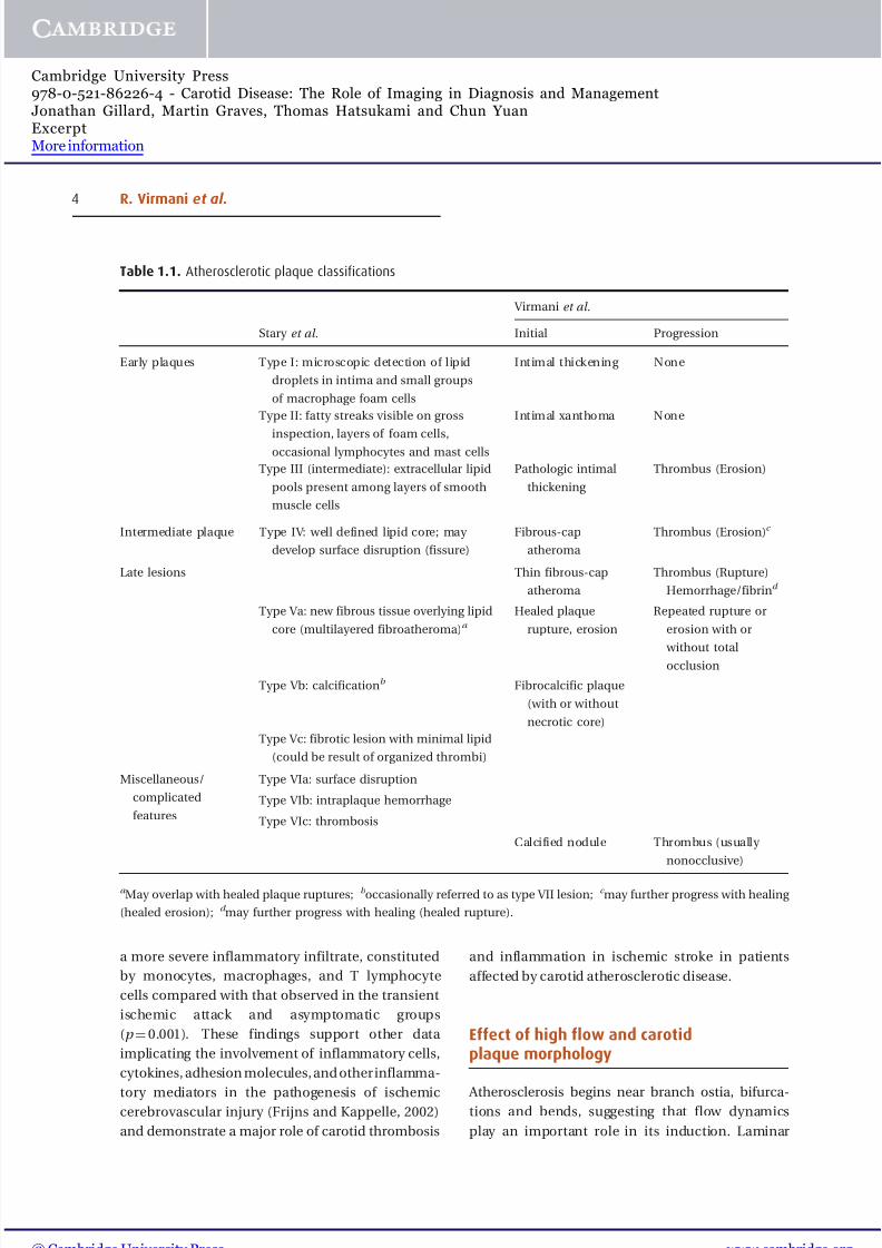
Table 1.1. Atherosclerotic plaque classifications
Virmani et al.
Stary et al. Initial Progression
Early plaques Type I: microscopic detection of lipid
droplets in intima and small groups
of macrophage foam cells
Intimal thickening None
Type II: fatty streaks visible on gross
inspection, layers of foam cells,occasional lymphocytes and mast cells
Intimal xanthoma None
Type III (intermediate): extracellular lipid
pools present among layers of smooth
muscle cells
Pathologic intimal
thickening
Thrombus (Erosion)
Intermediate plaque Type IV: well defined lipid core; may
develop surface disruption (fissure)
Fibrous-cap
atheroma
Thrombus (Erosion)c
Late lesions Thin fibrous-cap
atheroma
Thrombus (Rupture)
Hemorrhage/fibrind
Type Va: new fibrous tissue overlying lipid
core (multilayered fibroatheroma)a Healed plaque
rupture, erosion
Repeated rupture or
erosion with or
without total
occlusion
Type Vb: calcificationb Fibrocalcific plaque
(with or without
necrotic core)
Type Vc: fibrotic lesion with minimal lipid
(could be result of organized thrombi)
Miscellaneous/
complicated
features
Type VIa: surface disruption
Type VIb: intraplaque hemorrhage
Type VIc: thrombosis
Calcified nodule Thrombus (usually
nonocclusive)
a May overlap with healed plaque ruptures; b occasionally referred to as type VII lesion; c may further progress with healing
(healed erosion); d may further progress with healing (healed rupture).
4 R. Virmani et al.
© Cambridge University Press www.cambridge.org
Cambridge University Press978-0-521-86226-4 - Carotid Disease: The Role of Imaging in Diagnosis and ManagementJonathan Gillard, Martin Graves, Thomas Hatsukami and Chun YuanExcerptMore information

8/10/2019 Ateroscleroza ar carotidiene
http://slidepdf.com/reader/full/ateroscleroza-ar-carotidiene 5/10
flow is disturbed at carotid bifurcation regions,
resulting in decreased shear stress and athero-sclerotic plaque accumulation on the outer wall of
the proximal segment of the sinus of the internal
carotid artery (Zarins et al ., 1983; Anayiotos et al .,
1994; Masawa et al ., 1994). The intimal thicknessis the least on the flow divider side at the junction
of the internal and external carotid arteries
Table 1.2. Gross and microscopic plaque characteristics in symptomatic and
asymptomatic patients undergoing carotid endarterectomy
Gross morphology
Symptomatic,
% (n ¼25)
Asymptomatic,
% (n ¼17) p-value
% Stenosis(Duplex) 74±17 77±15 ns
Ulceration 94 64 0.02
Plaque hemorrhage 47 52 ns
Microscopic characteristicsPlaque rupture 74 32 0.004
Thin fibrous cap 95 48 0.003
Cap foam cells 84 44 0.006
Intraplaque fibrin 100 68 0.008
Intraplaque hemo. 84 56 0.06
Necrotic core 84 72 ns
Ulceration 11 8 ns
Calcified nodule 7 7 ns
Thrombus 63 80 ns
SMC rich area 5 0 ns
Eccentric shape 68 64 ns
Abbreviations: hemo ¼ hemorrhage; ns ¼ non significant.
Modified from Carr et al ., 1996
Table 1.3. Thrombotically active plaques, cap rupture, and cap erosion by study groups
No. of plaques % p -value
Patients with
major
ipsilateral
stroke(n ¼ 96) (%)
Patients
with TIA (n ¼ 91) (%)
Asympto-
matic
patients(n ¼82) (%)
Stroke vs.TIA
Stroke vs.
asympto-matic
TIA vs.
asympto-matic
Thrombotically
active plaque
71 (74) 32 (35.2) 12 (14.6) <0.001 <0.001 0.002
Cap rupture 64 (66.7) 21 23.1) 11 (13.4) <0.001 <0.001 0.004
Cap erosion 7 (7.3) 11 (12.1) 1 (1.2) 0.51 0.09 0.03
Abbreviation: TIA ¼ transient ischemic attack.
Reproduced with permission from Spagnoli, L. G., et al . (2004). Journal of the American Medical Association , 292:184552.
Pathology of carotid artery atherosclerotic disease 5
© Cambridge University Press www.cambridge.org
Cambridge University Press978-0-521-86226-4 - Carotid Disease: The Role of Imaging in Diagnosis and ManagementJonathan Gillard, Martin Graves, Thomas Hatsukami and Chun YuanExcerptMore information
8/10/2019 Ateroscleroza ar carotidiene
http://slidepdf.com/reader/full/ateroscleroza-ar-carotidiene 6/10
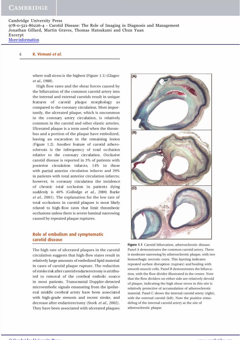
where wall stress is the highest (Figure 1.1) (Glagov
et al ., 1988).
High flow rates and the shear forces caused by
the bifurcation of the common carotid artery into
the internal and external carotids result in unique
features of carotid plaque morphology as
compared to the coronary circulation. Most impor-
tantly, the ulcerated plaque, which is uncommon
in the coronary artery circulation, is relatively common in the carotid and other elastic arteries.
Ulcerated plaque is a term used when the throm-
bus and a portion of the plaque have embolized,
leaving an excavation in the remaining lesion
(Figure 1.2). Another feature of carotid athero-
sclerosis is the infrequency of total occlusion
relative to the coronary circulation. Occlusive
carotid disease is reported in 3% of patients with
posterior circulation infarcts, 14% in those
with partial anterior circulation infarcts and 29%
in patients with total anterior circulation infarcts;
however, in coronary circulation the incidence
of chronic total occlusion in patients dying
suddenly is 40% (Golledge et al ., 2000; Burke
et al ., 2001). The explanation for the low rate of
total occlusions in carotid plaques is most likely
related to high-flow rates that limit thrombotic
occlusions unless there is severe luminal narrowing
caused by repeated plaque ruptures.
Role of embolism and symptomatic
carotid disease
The high rate of ulcerated plaques in the carotid
circulation suggests that high-flow states result in
relatively large amounts of embolized lipid material
in cases of carotid plaque rupture. The reduction
of stroke risk after carotid endarterectomy is attribu-
ted to removal of the cerebral embolic source
in most patients. Transcranial Doppler-detected
microembolic signals emanating from the ipsilat-
eral middle cerebral artery have been associated
with high-grade stenosis and recent stroke, anddecrease after endarterectomy (Stork et al ., 2002).
They have been associated with ulcerated plaques
Figure 1.1 Carotid bifurcation, atherosclerotic disease.
Panel A demonstrates the common carotid artery. There
is moderate narrowing by atherosclerotic plaque, with two
hemorrhagic necrotic cores. This layering indicates
repeated surface disruption (rupture) and healing with
smooth muscle cells. Panel B demonstrates the bifurca-
tion, with the flow divider illustrated in the center. Note
that the flow dividers on either side are relatively devoid
of plaque, indicating the high shear stress in this site is
relatively protective of accumulation of atherosclerotic
material. Panel C shows the internal carotid artery (right),
with the external carotid (left). Note the positive remo-deling of the internal carotid artery at the site of
atherosclerotic plaque.
6 R. Virmani et al.
© Cambridge University Press www.cambridge.org
Cambridge University Press978-0-521-86226-4 - Carotid Disease: The Role of Imaging in Diagnosis and ManagementJonathan Gillard, Martin Graves, Thomas Hatsukami and Chun YuanExcerptMore information

8/10/2019 Ateroscleroza ar carotidiene
http://slidepdf.com/reader/full/ateroscleroza-ar-carotidiene 7/10
as assessed by ultrasound (Valton et al ., 1998)and histologically disrupted plaques (Sitzer et al .,
1995). An association with plaque characteristics
has not been uniformly demonstrated, however
(Droste et al ., 1999; Stork et al ., 2002; Verhoeven
et al ., 2005).
Comparison of coronary and carotidatherosclerosis
In our laboratory, we have compared the histo-morphometric features of unstable coronary and
carotid atherosclerotic plaques. The mean fibrous
cap thickness in carotid plaque rupture wasnearly three times greater than coronary plaque
rupture (72 ± 15 microns vs. 23± 17 microns),
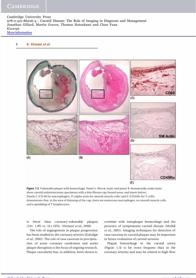
respectively (Figure 1.3). We measured carotid
vulnerable plaques (necrotic core with overly-
ing thin cap and infiltration by macrophages,
Figure 1.4) and found a mean cap thickness
of 72± 24 microns, which is greater than the
65-micron upper limit of a thin-cap fibroatheroma
in the coronary artery. In addition, there are
fewer macrophages in the fibrous cap of carotid
plaque ruptures than coronary plaque ruptures(13.5 ± 10.9% vs. 26± 20%). Similarly, in carotid-
vulnerable plaques the number of macrophages
Figure 1.2 Plaque rupture with thrombosis and ulceration. Unlike coronary arteries, in which ulcers are unusual, plaque
disruption in the carotid artery frequently results in embolization and crater formation. (A) demonstrates a routine
hematoxylin eosin section of a carotid artery with thrombus and ulcer. (B) shows the corresponding Movat pentachrome
stain, which highlights collagen (yellow) and elastic tissue (black). (CF) are immunohistochemical stains for
macrophages (Kp-1), smooth muscle cells (alpha actin), platelets (CD61) and fibrin (fibrin II). Note that at the ulcer crater,
there are abundant macrophages (C) with few smooth muscle cells (D). The thrombus itself has largely embolized;
there are residual platelets (E) and fibrin (E) at one edge of the crater.
Pathology of carotid artery atherosclerotic disease 7
© Cambridge University Press www.cambridge.org
Cambridge University Press978-0-521-86226-4 - Carotid Disease: The Role of Imaging in Diagnosis and ManagementJonathan Gillard, Martin Graves, Thomas Hatsukami and Chun YuanExcerptMore information
8/10/2019 Ateroscleroza ar carotidiene
http://slidepdf.com/reader/full/ateroscleroza-ar-carotidiene 8/10
is fewer than coronary-vulnerable plaques
(10± 1.8% vs. 14 ± 10%). (Virmani et al ., 2000)
The role of angiogenesis in plaque progression
has been studied in the coronary arteries (Kolodgie
et al ., 2003). The role of vasa vasorum in precipita-
tion of acute coronary syndromes and aorticplaque disruption is the focus of ongoing research.
Plaque vascularity has, in addition, been shown to
correlate with intraplaque hemorrhage and the
presence of symptomatic carotid disease (Mofidi
et al ., 2001). Imaging techniques for detection of
vasa vasorum in carotid plaques may be important
in future evaluation of carotid stenosis.
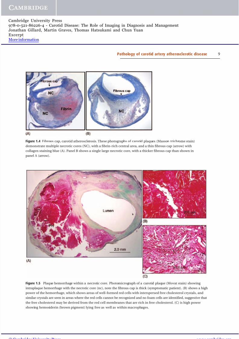
Plaque hemorrhage in the carotid artery (Figure 1.5) is far more frequent than in the
coronary arteries and may be related to high-flow
Figure 1.3 Vulnerable plaque with hemorrhage. Panel A (Movat stain) and panel B (hematoxylin-eosin stain)
show carotid endarterectomy specimens with a thin fibrous cap (boxed areas, and insets below).
Panels C (CD 68 for macrophages), D (alpha actin for smooth muscle cells) and E (CD45Ro for T-cells)
demonstrate that, in the area of thinning of the cap, there are numerous macrophages, no smooth muscle cells,
and a sprinkling of T lymphocytes.
8 R. Virmani et al.
© Cambridge University Press www.cambridge.org
Cambridge University Press978-0-521-86226-4 - Carotid Disease: The Role of Imaging in Diagnosis and ManagementJonathan Gillard, Martin Graves, Thomas Hatsukami and Chun YuanExcerptMore information
8/10/2019 Ateroscleroza ar carotidiene
http://slidepdf.com/reader/full/ateroscleroza-ar-carotidiene 9/10

8/10/2019 Ateroscleroza ar carotidiene
http://slidepdf.com/reader/full/ateroscleroza-ar-carotidiene 10/10
rates and pressures in the lumen and the vasa
vasorum. The maximum frequency of hemorrhage
is observed in arteries with 5075% cross-sectional
area luminal narrowing. We have reported in
coronary plaques that intraplaque hemorrhage is
responsible for necrotic core enlargement and
excessive foamy macrophages in the fibrous caps
(Kolodgie et al ., 2003). Red blood cell membranes
are the richest source of cholesterol as compared toany other cell in the body. The free cholesterol in
the necrotic core is believed to arise from apoptotic
cell death of foamy macrophages. However, we
have shown that free cholesterol in fibroatheromas,
thin-cap fibroatheromas and plaque ruptures is
also derived from erythrocytes that become
trapped in the necrotic core when intraplaque
hemorrhages occur (Kolodgie et al ., 2003). Takaya
et al. recently reported that patients with carotid
intraplaque hemorrhage at 18 months follow-up
had larger necrotic cores as well as accelerated
plaque progression as compared to patients with-
out intraplaque hemorrhage (Takaya et al ., 2005).
The frequency of calcification is similar in
coronary and carotid arteries, with maximum calci-
fication seen in carotid arteries narrowed greater
than 70% cross-sectional area. Calcification in the
carotid artery similar to coronary artery is at first
speckled and occurs in areas rich in smooth muscle
cells like pathologic intimal thickening and occurs
at sites of smooth muscle cell loss. The next most
frequent site is the base of the necrotic core, close
to the media (Figure 1.6). Calcium in the carotid
plaque is often fragmented and may be located
deep in the plaque or close to the surface. However,
the frequency of calcified nodules (with surface
thrombus) (Figure 1.7), a form of calcification
that results in irregular nodules of calcium, is
higher in carotid disease (approximately 67%), as
compared to coronary artery disease (12%). In
contrast, plaque erosion, while common in the
coronary circulation, is somewhat less frequent
in the carotid artery. In carotid arteries, percent
stenosis was highest in healed plaque ruptures and was greater than thin-cap atheromas and acute
plaque ruptures.
Is luminal narrowing the only determinantof vulnerability of a plaque?
Vulnerable plaque is a concept well accepted in the
coronary circulation but not so well established in
the carotid plaque. In studies carried out in the
coronary circulation it has been shown that plaque
ruptures often occur at low degrees of luminal
narrowing and that percent stenosis is a poorpredictor of plaque rupture. Ambrose et al . showed
in retrospective analysis of angiograms of patients
with acute myocardial infarction, that the median
stenosis of the initial angiogram in the artery
that caused the infarction was 48% (Ambrose
et al ., 1988). This concept subsequently has been
now repeatedly proven to be correct; we have
shown in sudden coronary death that at least 40%
of patients dying suddenly with a luminal throm-
bus have underlying plaques <75% narrowed in the
cross-sectional area (Farb et al ., 1995). Wasserman
et al . have suggested that in the carotid artery it
is time to look beyond stenosis (Figure 1.8). They
state that ‘‘although retrospective angiographic
studies of extracranial carotid atherosclerosis
and stroke have not been reported, the mech-
anism of plaque rupture may be similar to that
seen in the coronary artery’’ (Wasserman et al .,
2005).
Plaque progression through repeated
silent ruptures (Figure 1.9)
Morphologic studies of coronary plaques have
suggested that plaques beyond 50% cross-sectional
area narrowing occur through repeated ruptures,
which are most often clinically silent (Burke et al .,
2001). The sites of healed plaque ruptures can be
recognized by the presence of a necrotic core with
a discontinuous fibrous cap which is made up of
type I collagen identified by either Movat and/or
Sirius red-stained sections of the artery with an
overlying neointimal tissue that is rich in smoothmuscle cells in a proteoglycan-rich matrix and type
III collagen (Figure 1.9). In the coronary circulation
10 R. Virmani et al.
© Cambridge University Press www cambridge org
Cambridge University Press978-0-521-86226-4 - Carotid Disease: The Role of Imaging in Diagnosis and ManagementJonathan Gillard, Martin Graves, Thomas Hatsukami and Chun YuanExcerptMore information