association between melanocytic neoplasms and … · association between melanocytic neoplasms and...
TRANSCRIPT
DERMATOLOGY PRACTICAL & CONCEPTUALwww.derm101.com
Observation | Dermatol Pract Concept 2012;2(2):9 35
Association between melanocytic neoplasms and seborrheic keratosis: more than
a coincidental collision?Jennifer DeFazio, M.D.1, Iris Zalaudek, M.D.2, Klaus J. Busam, M.D.3, Carlo Cota, M.D.4,
Ashfaq Marghoob, M.D.1
1 Dermatology Service, Memorial Sloan-Kettering Cancer Center, Hauppauge, NY, USA2 Division of Dermatology, Medical University of Graz, Graz, Austria and Dermatology Unit, 1st Medical Department, IRCCS-Arcispedale Santa
Maria Nuova, Reggio Emilia, Italy3 Department of Pathology, Memorial Sloan-Kettering Cancer Center, New York, NY USA4 Dermatopathology Unit, San Gallicano Dermatological Institute, Rome, Italy
Key words: seborrheic keratosis, melanocytic nevus, cell-signalling, melanocytes, keratinocytes
Citation: DeFazio J, Zalaudek I, Busam KJ, Cota C. Marghoob A. Association between melanocytic neoplasms and seborrheic keratosis: more than a coincidental collision? Dermatol Pract Conc. 2012;2(2):9. http://dx.doi.org/10.5826/dpc.0202a09.
Received: September 20, 2011; Accepted: February 15, 2012; Published: April 30, 2012
Copyright: ©2012 DeFazio et al. This is an open-access article distributed under the terms of the Creative Commons Attribution License, which permits unrestricted use, distribution, and reproduction in any medium, provided the original author and source are credited.
Funding: None.
Competing interests: The authors have no conflicts of interest to disclose.
All authors have contributed significantly to this publication.
Corresponding author: Jennifer DeFazio, M.D., Assistant Clinical Member, Dermatology Service, Memorial Sloan-Kettering Cancer Center, 800 Veteran’s Highway, 2nd Floor, Hauppauge, NY, USA. Tel. 631.863.5150; Fax. 631.361.4694. Email: [email protected].
Introduction
The presence of a seborrheic keratoses (SK) found in asso-
ciation with a melanocytic nevus has been observed often
in our clinical practice. The utilization of dermoscopy high-
lights the specific features associated with both the nevus
component and seborrheic keratosis found in these “colli-
sion” lesions. The occurrence of this finding, which is more
readily appreciated with dermoscopy, has led us to specu-
late whether this is a coincidental finding or phenotypic
expression of underlying melanocyte cell-signaling. Here
we present five cases with histology, collected retrospec-
tively, highlighting the association between melanocytic
lesions and SK.
Clinical observations and an expanding knowledge of cell-to-cell communication have led us to specu-late that the finding of a melanocytic nevus in conjunction with a seborrheic keratosis is more than a coincidental collision of two lesions. Here we present five cases demonstrating dermoscopic features of both melanocytic lesions and seborrheic keratoses with corresponding histology. Four cases dem-onstrate dermoscopic features of a melanocytic nevus and seborrheic keratosis, and the final case a melanoma arising in association with a seborrheic keratosis.
ABSTRACT
36 Observation | Dermatol Pract Concept 2012;2(2):9
Case 1
A 31-year-old male with history of multiple nevi presented
for routine skin surveillance. On exam a lesion of the pec-
toral region was noted by the physician and examined with
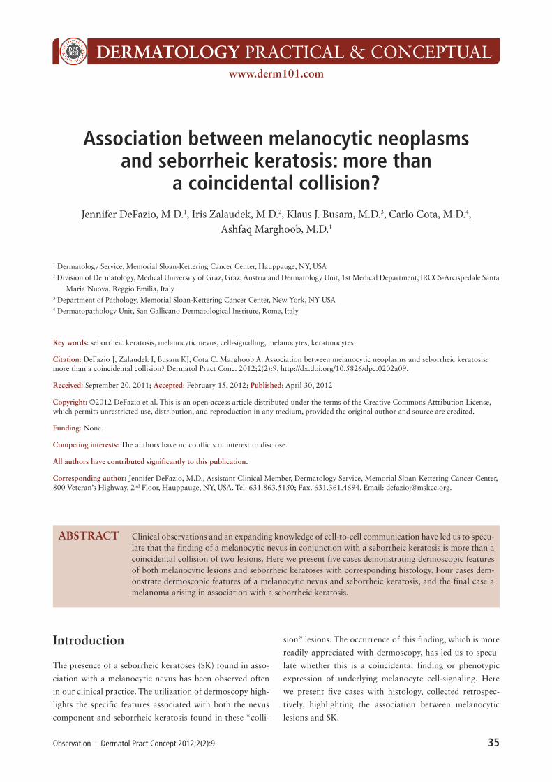
dermoscopy. Dermoscopy (Figure 1A) revealed a macular
lesion with a heavily pigmented center and peripheral reticu-
lated network, partially occluded by a second raised com-
ponent with a distinctive cerebriform appearance. A biopsy
was performed. On histology (Figure 1B), the seborrheic
keratosis showed papillomatous epidermal hyperplasia with
delicate basket-weave hyperkeratosis. A melanocytic nevus
involves part of the seborrheic keratosis. It was characterized
by a proliferation of nests of melanocytes along the dermo-
epidermal junction.
Figure 1A, B. Dermoscopy (1A) demonstrates a melanocytic nevus with reticulated network and an overlapping or colliding seborrheic
keratosis with cerebriform pattern. On histology (1B), junctional melanocytic nests are located at the tips of rete ridges of a papillo-
matous seborrheic keratosis (hematoxylin and eosin [H&E], x25). [Copyright: ©2012 DeFazio et al.]
A B
A B C
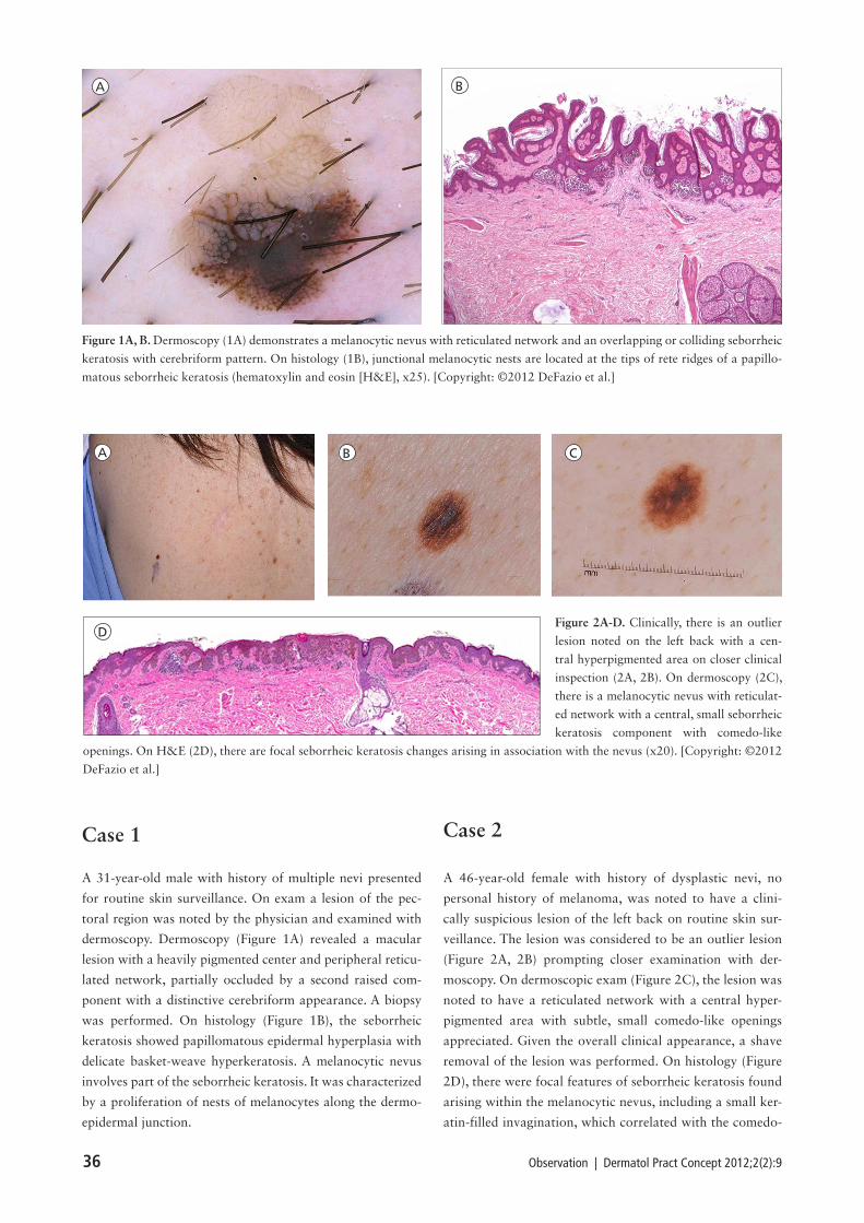
DFigure 2A-D. Clinically, there is an outlier
lesion noted on the left back with a cen-
tral hyperpigmented area on closer clinical
inspection (2A, 2B). On dermoscopy (2C),
there is a melanocytic nevus with reticulat-
ed network with a central, small seborrheic
keratosis component with comedo-like
openings. On H&E (2D), there are focal seborrheic keratosis changes arising in association with the nevus (x20). [Copyright: ©2012
DeFazio et al.]
Case 2
A 46-year-old female with history of dysplastic nevi, no
personal history of melanoma, was noted to have a clini-
cally suspicious lesion of the left back on routine skin sur-
veillance. The lesion was considered to be an outlier lesion
(Figure 2A, 2B) prompting closer examination with der-
moscopy. On dermoscopic exam (Figure 2C), the lesion was
noted to have a reticulated network with a central hyper-
pigmented area with subtle, small comedo-like openings
appreciated. Given the overall clinical appearance, a shave
removal of the lesion was performed. On histology (Figure
2D), there were focal features of seborrheic keratosis found
arising within the melanocytic nevus, including a small ker-
atin-filled invagination, which correlated with the comedo-
Observation | Dermatol Pract Concept 2012;2(2):9 37
like opening on comparison to the dermoscopic image. The
nevus was well circumscribed with junctional and dermal
melanocytic nests.
Case 3
A 38-year-old female with history of many nevi was noted by
the dermatologist to have a changed lesion on routine skin
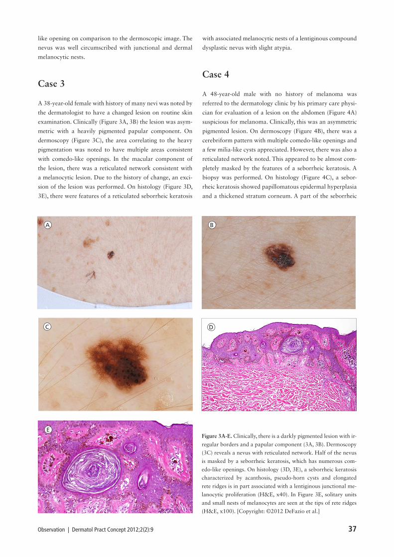
examination. Clinically (Figure 3A, 3B) the lesion was asym-
metric with a heavily pigmented papular component. On
dermoscopy (Figure 3C), the area correlating to the heavy
pigmentation was noted to have multiple areas consistent
with comedo-like openings. In the macular component of
the lesion, there was a reticulated network consistent with
a melanocytic lesion. Due to the history of change, an exci-
sion of the lesion was performed. On histology (Figure 3D,
3E), there were features of a reticulated seborrheic keratosis
with associated melanocytic nests of a lentiginous compound
dysplastic nevus with slight atypia.
Case 4
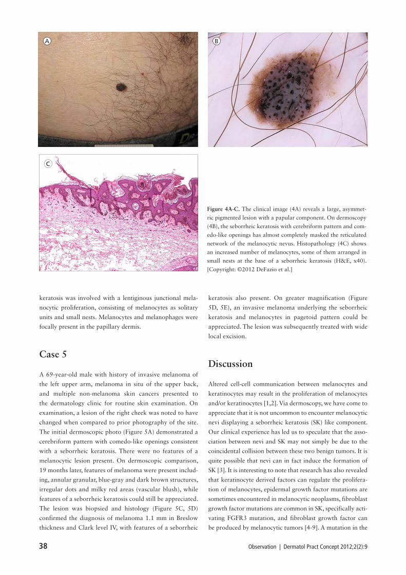
A 48-year-old male with no history of melanoma was
referred to the dermatology clinic by his primary care physi-
cian for evaluation of a lesion on the abdomen (Figure 4A)
suspicious for melanoma. Clinically, this was an asymmetric
pigmented lesion. On dermoscopy (Figure 4B), there was a
cerebriform pattern with multiple comedo-like openings and
a few milia-like cysts appreciated. However, there was also a
reticulated network noted. This appeared to be almost com-
pletely masked by the features of a seborrheic keratosis. A
biopsy was performed. On histology (Figure 4C), a sebor-
rheic keratosis showed papillomatous epidermal hyperplasia
and a thickened stratum corneum. A part of the seborrheic
A B
C D
EFigure 3A-E. Clinically, there is a darkly pigmented lesion with ir-
regular borders and a papular component (3A, 3B). Dermoscopy
(3C) reveals a nevus with reticulated network. Half of the nevus
is masked by a seborrheic keratosis, which has numerous com-
edo-like openings. On histology (3D, 3E), a seborrheic keratosis
characterized by acanthosis, pseudo-horn cysts and elongated
rete ridges is in part associated with a lentiginous junctional me-
lanocytic proliferation (H&E, x40). In Figure 3E, solitary units
and small nests of melanocytes are seen at the tips of rete ridges
(H&E, x100). [Copyright: ©2012 DeFazio et al.]
38 Observation | Dermatol Pract Concept 2012;2(2):9
keratosis was involved with a lentiginous junctional mela-
nocytic proliferation, consisting of melanocytes as solitary
units and small nests. Melanocytes and melanophages were
focally present in the papillary dermis.
Case 5
A 69-year-old male with history of invasive melanoma of
the left upper arm, melanoma in situ of the upper back,
and multiple non-melanoma skin cancers presented to
the dermatology clinic for routine skin examination. On
examination, a lesion of the right cheek was noted to have
changed when compared to prior photography of the site.
The initial dermoscopic photo (Figure 5A) demonstrated a
cerebriform pattern with comedo-like openings consistent
with a seborrheic keratosis. There were no features of a
melanocytic lesion present. On dermoscopic comparison,
19 months later, features of melanoma were present includ-
ing, annular granular, blue-gray and dark brown structures,
irregular dots and milky red areas (vascular blush), while
features of a seborrheic keratosis could still be appreciated.
The lesion was biopsied and histology (Figure 5C, 5D)
confirmed the diagnosis of melanoma 1.1 mm in Breslow
thickness and Clark level IV, with features of a seborrheic
keratosis also present. On greater magnification (Figure
5D, 5E), an invasive melanoma underlying the seborrheic
keratosis and melanocytes in pagetoid pattern could be
appreciated. The lesion was subsequently treated with wide
local excision.
Discussion
Altered cell-cell communication between melanocytes and
keratinocytes may result in the proliferation of melanocytes
and/or keratinocytes [1,2]. Via dermoscopy, we have come to
appreciate that it is not uncommon to encounter melanocytic
nevi displaying a seborrheic keratosis (SK) like component.
Our clinical experience has led us to speculate that the asso-
ciation between nevi and SK may not simply be due to the
coincidental collision between these two benign tumors. It is
quite possible that nevi can in fact induce the formation of
SK [3]. It is interesting to note that research has also revealed
that keratinocyte derived factors can regulate the prolifera-
tion of melanocytes, epidermal growth factor mutations are
sometimes encountered in melanocytic neoplasms, fibroblast
growth factor mutations are common in SK, specifically acti-
vating FGFR3 mutation, and fibroblast growth factor can
be produced by melanocytic tumors [4-9]. A mutation in the
A B
C
Figure 4A-C. The clinical image (4A) reveals a large, asymmet-
ric pigmented lesion with a papular component. On dermoscopy
(4B), the seborrheic keratosis with cerebriform pattern and com-
edo-like openings has almost completely masked the reticulated
network of the melanocytic nevus. Histopathology (4C) shows
an increased number of melanocytes, some of them arranged in
small nests at the base of a seborrheic keratosis (H&E, x40).
[Copyright: ©2012 DeFazio et al.]
Observation | Dermatol Pract Concept 2012;2(2):9 39
A B
C
D E
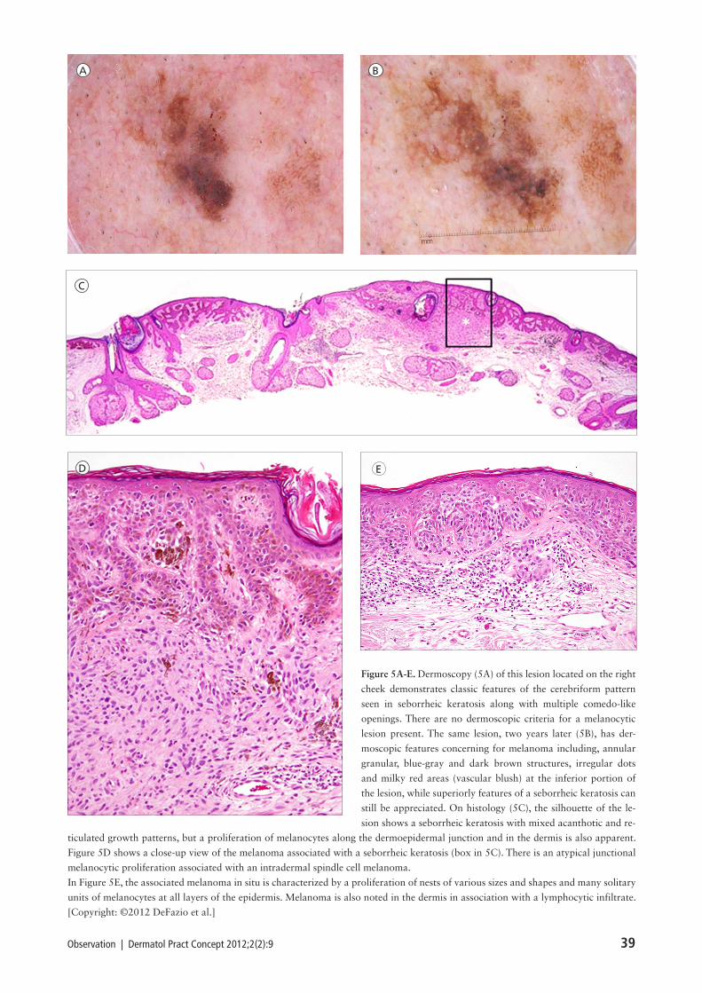
Figure 5A-E. Dermoscopy (5A) of this lesion located on the right
cheek demonstrates classic features of the cerebriform pattern
seen in seborrheic keratosis along with multiple comedo-like
openings. There are no dermoscopic criteria for a melanocytic
lesion present. The same lesion, two years later (5B), has der-
moscopic features concerning for melanoma including, annular
granular, blue-gray and dark brown structures, irregular dots
and milky red areas (vascular blush) at the inferior portion of
the lesion, while superiorly features of a seborrheic keratosis can
still be appreciated. On histology (5C), the silhouette of the le-
sion shows a seborrheic keratosis with mixed acanthotic and re-
ticulated growth patterns, but a proliferation of melanocytes along the dermoepidermal junction and in the dermis is also apparent.
Figure 5D shows a close-up view of the melanoma associated with a seborrheic keratosis (box in 5C). There is an atypical junctional
melanocytic proliferation associated with an intradermal spindle cell melanoma.
In Figure 5E, the associated melanoma in situ is characterized by a proliferation of nests of various sizes and shapes and many solitary
units of melanocytes at all layers of the epidermis. Melanoma is also noted in the dermis in association with a lymphocytic infiltrate.
[Copyright: ©2012 DeFazio et al.]
40 Observation | Dermatol Pract Concept 2012;2(2):9
PI3K pathway has been described in SK [10]. Supplemen-
tary to this, it is known that therapies targeting BRAF, which
is the most common mutation among nevi, induce eruptive
keratinocyte tumors [11].
Mature nevi, which are in a state of senescence, are stable
lesions that do not grow. However, SK developing in associa-
tion with nevi may continue to grow. It is conceivable that
the enlarging SK component may eventually mask the under-
lying nevus. In the unfortunate scenario of the development
of a nevus-associated melanoma beneath the SK, the malig-
nancy would of course eventually become visible, as may
have occurred in Case 5 presented here [12-15]. Alternative
explanations include that some of these lesions may have
been melanomas from their inception that were masquer-
ading themselves as SK [16], that normally occurring mela-
nocytes within an SK mutated into melanoma [12], or that
the initial lesion was a keratotic melanocytic nevus, which
on histology have hyperkeratotic epidermal rete ridges and
pseudohorn cysts, and clinically can have a warty appear-
ance [17] mistaken for a seborrheic keratosis. Melanoma
may arise from this lesion giving the clinical appearance of
a melanoma arising in a seborrheic keratosis or perhaps the
association between melanoma and SK is purely due to the
coincidental collision between these two entities [18].
Seborrheic keratoses in association with melanocytic nevi
and melanoma have been reported previously. In a retrospec-
tive study published by Boyd and Rapini, 69 collision tumors
were observed after assessing 40,000 cutaneous biopsies. Of
those 69, 14 were nevus and seborrheic keratosis [19]. In
a retrospective case series by Lim, over a 12-month period,
histology of 639 SK was evaluated and 85 (9%) were found
in association with other lesions. Seven melanomas (8.2%)
were reported with one found to have arisen within the seb-
orrheic keratosis and six adjacent to a seborrheic keratosis.
Thirteen melanocytic nevi (15.3%) adjacent to SK were also
reported [20]. These findings may be more than coincidental
and may suggest a not yet fully understood local phenom-
enon. This concept has been proposed previously by Brown-
stein [21], where it was suggested that nevi may interact with
the stroma to induce epithelial growths. The melanocytic
nevus may alter the local milieu and induce the development
of the adjacent seborrheic keratosis, accounting for our clini-
cal observations.
Conclusion
We have presented five cases of melanocytic lesions found
in association with SK. Nevus in association with seborrheic
keratosis has been observed frequently in our clinical expe-
rience and is more readily appreciated with dermoscopic
inspection. The frequency with which this has been noted
and an ever-increasing understanding of cell-signaling has
led us to postulate that this may be more than a chance
occurrence. Further exploration of cell-cell signaling of
melanocytes and keratinocytes is needed to truly understand
this clinical observation.
References
1. Haass NK, Smalley KS, Herlyn M. The role of altered cell-
cell communication in melanoma progression. J Mol Histol.
2004;35(3):309-18.
2. Deveci M, Gilmont RR, Terashi H, Ahmed AH, Smith DJ, Mar-
celo C. Melanocyte-conditioned medium stimulates while mela-
nocyte/keratinocyte contact inhibits keratinocyte proliferation. J
Burn Care Rehabil. 2001;22(1):9-14.
3. Betti R, Menni S, Cerri A, Vergani R, Crosti C. Seborrheic kera-
tosis with compound nevus, junctional nevus and basal cell carci-
noma in the same lesion. Dermatology. 2001;203(3):265-7.
4. Hirobe T. Role of keratinocyte-derived factors involved in regu-
lating the proliferation and differentiation of mammalian epider-
mal melanocytes. Pigment Cell Res. 2005;18(1):2-12.
5. Hirobe T, Furuya R, Akiu S, Ifuku O, Fukuda M. Keratinocytes
control the proliferation and differentiation of cultured epider-
mal melanocytes from ultraviolet radiation B-induced pigment-
ed spots in the dorsal skin of hairless mice. Pigment Cell Res.
2002;15(5):391-9.
6. James MR, Hayward NK, Dumenil T, Montgomery GW, Mar-
tin NG, Duffy DL. Epidermal growth factor gene polymor-
phism and risk of melanocytic neoplasm. J Invest Dermatol.
2004;123(4):760-2.
7. Hafner C, Hartmann A, van Oers JM, et al. FGFR3 mutations in
seborrheic keratoses are already present in flat lesions and associ-
ated with age and localization. Mod Pathol. 2007;20(8):895-903.
8. Giehl KA, Nägele U, Volkenandt M, Berking C. Protein expres-
sion of melanocyte growth factors (bFGF, SCF) and their recep-
tors (FGFR-1, c-kit) in nevi and melanomas. J Cutan Pathol.
2007;34(1):7-14.
9. Löffek S, Zigrino P, Angel P, Anwald B, Krieg T, Mauch C.
High invasive melanoma cells induce matrix metalloprotein-
ase-1 synthesis in fibroblasts by interleukin-1 and basic fibro-
blast growth factor-mediated mechanisms. J Invest Dermatol.
2005;124(3):638-43.
10. Hafner C, Lopez-Knowles E, Luis N, et al. Oncogenic PIK-
3CA mutations occur in epidermal nevi an seborrheic kerato-
ses with a characteristic mutation pattern. Proc Natl Acad Sci
USA. 2007;104(33):13450-4.
11. Flaherty KT, Puzanov I, Kim KB, et al. Inhibition of mutat-
ed, activated BRAF in metastatic melanoma. N Engl J Med.
2010;363(9):809-19.
12. Tsao H, Bevona C, Goggins W, Quinn T. The transformation
rate of moles (melanocytic nevi) into cutaneous melanoma: a
population-based estimate. Arch Dermatol. 2003;139(3):282-
8.
13. Cascajo CD, Reichel M, Sánchez JL. Malignant neoplasms
associated with seborrheic keratoses. An analysis of 54 cases.
Am J Dermatopathol. 1996;18(3):278-82.
Observation | Dermatol Pract Concept 2012;2(2):9 41
14. Thomas I, Kihiczak NI, Rothenberg J, Ahmed S, Shwartz RA.
Melanoma within the seborrheic keratosis. Dermatol Surg.
2004;30(4 Pt 1):559-61.
15. Zabel RJ, Vinson RP, McCollough ML. Malignant melano-
ma arising in a seborrheic keratosis. J Am Acad Dermatol.
2000;42(5 Pt 1):831-3.
16. Izikson L, Sober AJ, Mihm MC Jr, Zembowicz A. Prevalence
of melanoma clinically resembling seborrheic keratosis: anal-
ysis of 9204 cases. Arch Dermatol. 2002;138(12):1562-6.
17. Hornstein M, Prieto VG, Burchette JL Jr, Shea C. Keratotic
melanocytic nevus: a clinicopathologic and immunohisto-
chemical study. J Cutan Pathol. 2000;27(7):344-50.
18. 18. Jones-Caballero M, Penas PF, Buezo GF, Fraga J, Aragüés
M. Malignant melanoma appearing in a seborrheic keratosis.
Br J Dermatol. 1995;133(6):1016-8.
19. Boyd AS, Rapini RP. Cutaneous collision tumors. An analy-
sis of 69 cases and review of the literature. Am J Deramato-
pathol. 1994;16(3):253-7.
20. Lim C. Seborrhoeic keratoses with associated lesions: a
retrospective analysis of 85 lesions. Australas J Dermatol.
2006;47(2):109-13.
21. Brownstein MH, Starnik TM. Desmoplastic trichoepithelio-
ma and intradermal nevus: a combined malformation. J Am
Acad Dermatol. 1987;17(3):489-92.