assessment of the patient with a neck mass - epworth gp...
TRANSCRIPT

Assessment of the Patient with a Neck Mass
Mr Paul M Paddle, MBBS(Hons) FRACS, Laryngologist, ENT Surgeon

Introduction
} Common clinical finding } All age groups } Very complex differential diagnosis } Systematic approach essential

Anatomical Considerations } Prominent landmarks
} Bony-Cartilaginous } Midline } Lateral
} Viscera } Carotid Bulb } Ptotic Submandibular Glands

Anatomical Considerations } Triangles of the neck
} Posterior } Anterior
} Submandibular } Submental } Carotid } Muscular

Anatomical Considerations } Lymphatic Chains
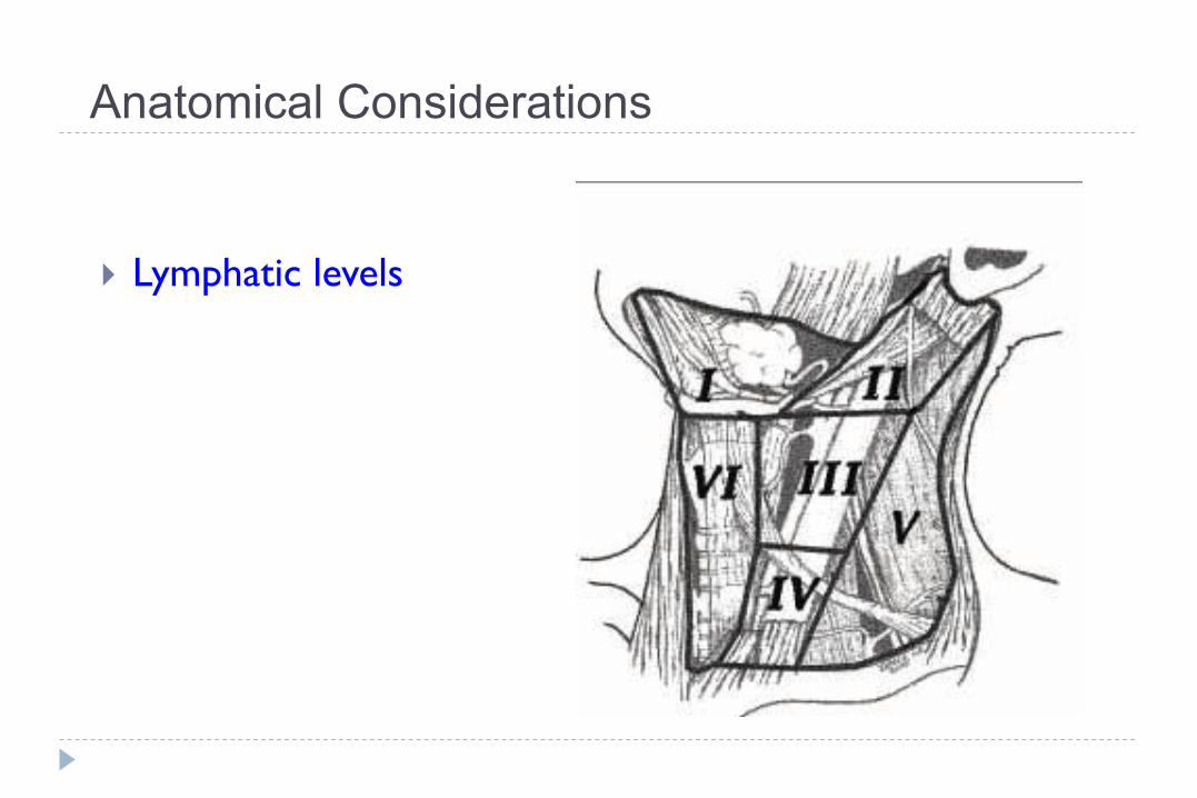
Anatomical Considerations
} Lymphatic levels

Differential Diagnosis } By Aetiology } By Age } By Location
} Ideally – consider all 3 of the above categories.

Differential Diagnosis
CONGENITAL ACQUIRED Infectious Inflammatory Neoplastic
Sebaceous cysts Branchial Cleft Thyroglossal Duct Cyst Vascular Malformations Dermoid Thymic Cyst Ectopic Thyroid Tissue Laryngocoele
Reactive Lymphadenopathy i) Viral ii) Bacterial iii) Granulomatous
Kawasaki’s Disease Benign i) Lipoma ii) Thyroid Adenoma iii) Thymoma iv) Paraganglioma
Lymphadenitis i) Viral ii) Bacterial iii) Granulomatous
a. MTB, MOTT b. Cat Scratch c. Toxoplasmosis d. Fungal
Sarcoidosis Malignant i) Primary H&N Ca
a. Carcinoma b. Sarcoma c. Melanoma
ii) Mets from H&N Primary iii) Haematological M
Sialadenitis i) Parotid ii) Submandibular
Rosai-Dorfman (SHML)
Other Connective Tissue Disease
} By Aetiology

Differential Diagnosis } Patient age
} Pediatric (0 – 15 years): } 90% benign
} Young adult (16 – 40 years): } similar to pediatric
} Late adult (>40 years): } majority neoplastic
0%
10%
20%
30%
40%
50%
60%
70%
80%
90%
100%
Paediatric (0-15) Young Adult (16-40)
Older Adult (>40)
Neoplastic
Congenital
Inflammatory

Differential Diagnosis
• Anatomical Location
LOCATION Midline Anterior Triangle Posterior Triangle
LESION Thyroglossal Duct Cyst
Lymphadenopathy / -itis
Vascular Malformations
Ectopic Cervical Thyroid
Branchial Cyst Lymphadenopathy / -itis
Teratoid Cysts (Epidermoid, Dermoid, Teratoid)
Teratoid Cysts
Laryngocoele Branchial Fistula
Thymic Cyst Vascular Malformations
Thymic Cysts
Teratoma

Metastasis Location according to Various Primary Lesions

Diagnostic Steps - History 1. History of Presenting Complaint
i) Onset: ii) Growth:
a. Slow: b. Fast
iii) Progress: a. Fluctuate – vascular malformation
iv) Airway v) GIT:
a. dysphagia, b. odynophagia
vi) Pain: a. Yes – infectious/inflammatory (except MTB/
MOTT) b. No – neoplastic
vii) Systemic Symptoms
2. Risk Factors (for the 4 groups) i) Congenital:
a. Change in size with URTI
ii) Infectious a. Infection elsewhere – e.g. skin lesions, trauma b. Dental Status c. Recent Sick Contacts – e.g. Viral URTIs, Strep,
TB d. O/S travel e. Animal Exposures – e.g. Cat Scratch,
Toxoplasmosis, Coxiella f. Travel to Rural Setting g. Immunisation status
iii) Inflammatory a. Drugs – e.g. Phenytoin, Carbamazepine b. FHx of Connective Tissue Disease
iv) Neoplastic a. Smoking, EtOH b. Lumps Elsewhere c. B symptoms
3. Family History

Diagnostic Steps - Examination 1. General 2. Neck
i) Mass ii) Cervical LNs
¨ Normal - < 2cm ¨ Abnormal > 2cm
iii) Thyroid 3. Complete ENT
i) Skin ii) Oral Cavity iii) Ear
Nose iv) Throat
4. Elsewhere: i) LN basins ii) ? Hepatosplenomegaly

Empirical Antibiotics } Inflammatory mass suspected } Two week trial of antibiotics
} Which One? } Cover Aerobes/Anaerobes*
} E.g. Augmentin Duo, Clindamycin, Azithromycin
} Follow-up for further investigation } Initial: 48-72 hours } Intermediate Term: ensure resolution
* If Oral / Periodontal Health poor

Investigations } Bloods 1. General Inflammatory Screen: FBE, +/- CRP/ESR, +/- LFT 2. Blood Culture if Febrile 3. Infectious Lymphadenopathy/itis Screen
i) EBV ii) CMV iii) Toxoplasmosis iv) Bartonella Henslae v) (Syphyilis) vi) (HIV)
4. ? MTB / MOTT i) Quantiferon Gold ii) Mantoux / TST
5. ? Sarcoid i) Serum ACE ii) Serum Corrected Ca2+

Investigations } Fine Needle Aspiration

Fine Needle Aspiration Biopsy } Standard of diagnosis (adults at least) } Indications
} Any neck mass that is not an obvious abscess } Persistence after a 2 week course of antibiotics
} Small gauge needle } Reduces bleeding } Seeding of tumor – not a concern
} No contraindications (vascular ?) } Children? Controversial
} Paediatric Malignancies are difficult to Dx on FNA } Most common malignancy is Lymphoma: FNAC often inadequate for
subtyping } Often requires a GA

Fine Needle Aspiration Biopsy } Proper Collection Required } Skilled cytopathologist
essential } On-site review best } Equipment:
i. 10-20 ml Syringe ii. 23-27-g needle iii. Alcohol Swabs iv. Gauze v. 2-4 glass slides vi. Spray Fixative or 95% EtOH vii. Balanced Salt Solution viii. Optional: flow-cytology Solution ix. Optional: Syringe Holder
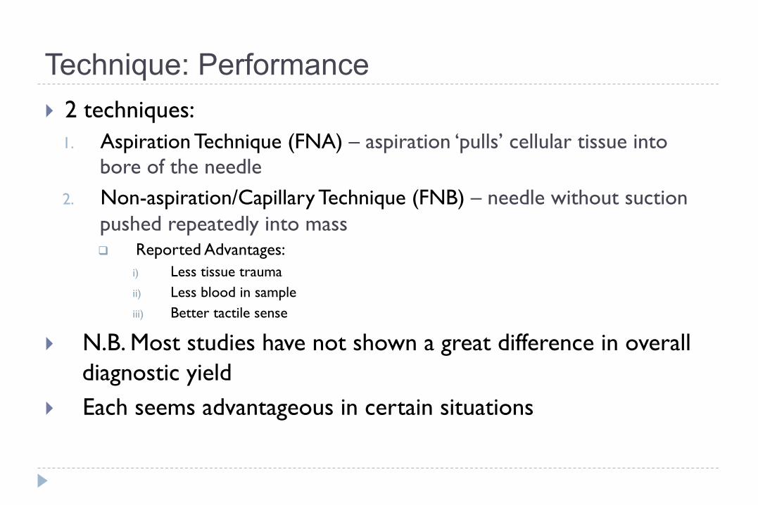
Technique: Performance } 2 techniques:
1. Aspiration Technique (FNA) – aspiration ‘pulls’ cellular tissue into bore of the needle
2. Non-aspiration/Capillary Technique (FNB) – needle without suction pushed repeatedly into mass q Reported Advantages:
i) Less tissue trauma ii) Less blood in sample iii) Better tactile sense
} N.B. Most studies have not shown a great difference in overall diagnostic yield
} Each seems advantageous in certain situations

Technique: Performance 1. Non-Aspiration Technique
(FNB) i. Cleanse skin with Alcohol wipe ii. +/- LA iii. Fixate mass with thumb/forefinger iv. 25-27g needle; No Syringe v. Once inside the mass: 10-15 short,
quick, staccato-like strokes vi. Remove needle & attach to 10-ml
syringe

Technique: Performance 2. Aspiration Technique (FNA)
i. 23-g needle on a 10ml syringe ii. 1ml air into syringe iii. Pass into mass iv. Apply 1-2ml suction pressure v. 3-5 passes (small vibratory
strokes) § 2-3 mm depth § Don’t change needle direction whilst
in mass
vi. Cease Suction as soon as material is seen in the hub
vii. Remove from mass viii. apply pressure

Technique: 3. Specimen Handling
1. Expel Material onto slides: i. Blow material onto slide using syringe ii. Bevelled edge slide-wards and touching slide surface iii. 2 slides
2. Smear: i. 2 slide surfaces against each other ii. ‘Smear’ along long-axis of slides
3. Fix / Air-dry i. Slide 1: Fixative spray @ 20-25 cm distance onto slide for Papanicolaou staining ii. Slide 2: Air-dry for Modified Romanowsky technique
4. Rinse Needle: (esp if concerned may be lymphoma) i. Use a balanced salt solution:
¨ E.g. Hanks’ Basic Salt solution or Roswell Park Memorial Institute (RPMI) medium
ii. Can be spun down for thin-layer-technique/flow/microbiologic studies

Specimen Handling

Complications 1. Haematoma
} Very uncommon } Haemorrhage causing airway compromise extremely rare
2. Needle-Track Seeding } Extremely rare } Livraghi et al: 11,700 Abdominal FNA (20-23g), 0.017% rate of tumour
seeding } Engzell et al: FNA of salivary gland adenomas – found tumour cells along the
22g needle-tract, BUT No increased recurrence @ 5 years follow up . } Largest study: 7 out of 4912 pts with papillary Carcinoma who underwent
FNAB had needle tract implant (completely excised @ surgery)
3. ‘Other’ Structure Injury } e.g. RLN injury: reported, but extremely rare
} Biggest ‘Risk’ is Non-diagnostic sample

Investigations } Imaging 1. CT Neck +/- Chest with Contrast 2. MRI 3. Ultrasonography 4. Radio-nuclide Scans / PET } ? Plain Film – Generally shouldn’t be considered in work-up of a neck
mass
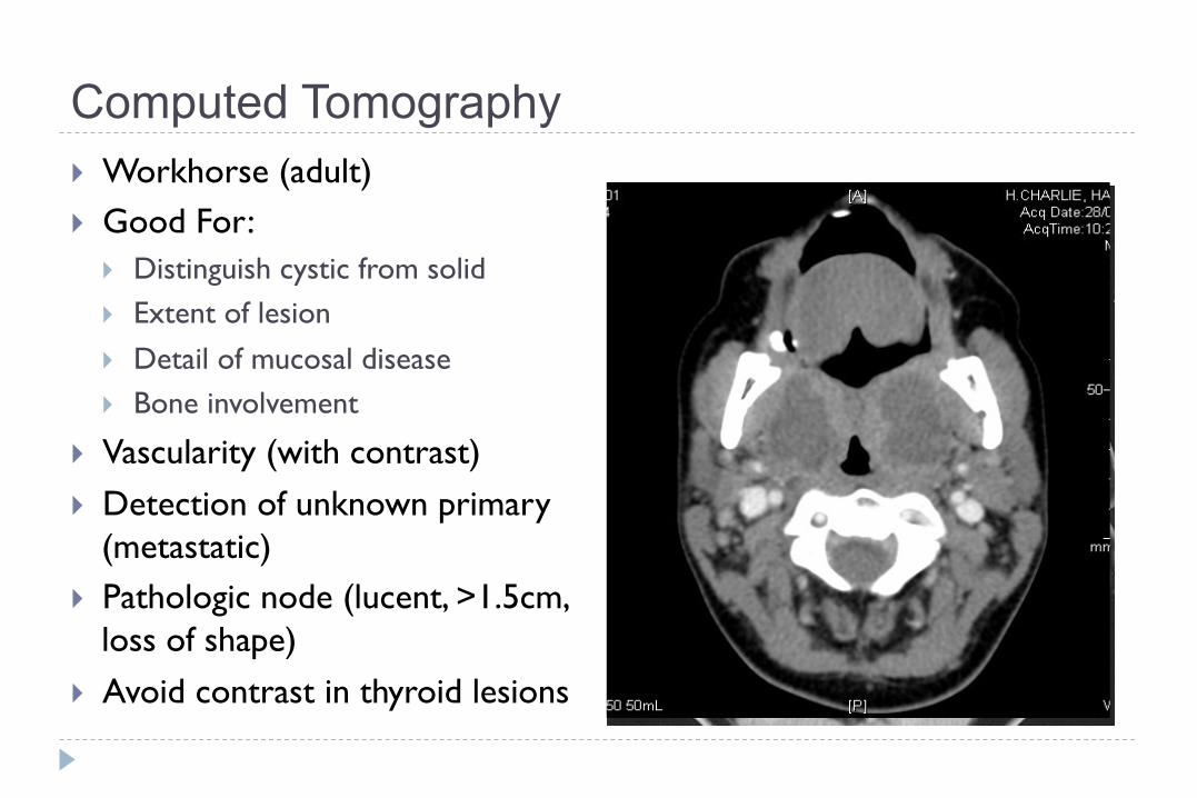
Computed Tomography } Workhorse (adult) } Good For:
} Distinguish cystic from solid } Extent of lesion } Detail of mucosal disease } Bone involvement
} Vascularity (with contrast) } Detection of unknown primary
(metastatic) } Pathologic node (lucent, >1.5cm,
loss of shape) } Avoid contrast in thyroid lesions

Magnetic Resonance Imaging } Similar information as CT } Better Soft Tissue Delineation } No Radiation exposure } Better for
} upper neck } skull base } Salivary Gland } Tongue (where dental amalgam
may obscure)
} Vascular delineation with infusion
} Nerve involvement delineation

Ultrasonography } Solid versus cystic masses } Congenital cysts from solid
nodes/tumors } In Combination with FNA –
increased accuracy } ? Children:
} Noninvasive } No-radiation } No Contrast } Inexpensive

Radionucleotide Scanning } Expanding Field:
i) PET – neoplasia, infection ii) 67-Gallium Scan (with SPECT) –
lymphoma, osteomyelitis iii) I123 - Thyroid iv) Tc-99m – Osteomyelitis, Salivary
gland v) MIBI - parathyroid vi) MIBG - paraganglioma
} Location – glandular versus extra-glandular
} Functional information } Solitary nodules } Multinodular goiter with new
increasing nodule } Hashimoto’s with new nodule

General Work-up of a Neck Mass
E
1638 Part 6 ■ +HDG�DQG�1HFN�6XUJHU\�DQG�2QFRORJ\
a neck mass is especially bloody, necrotic, or fibrotic, and specific diagnostic information may be unattainable in lesions where complex tissue architecture is necessary.7 Open biopsy of neck masses should only be considered as a last resort for diagnosis. In the case that squa-mous cell carcinoma is identified in the neck during open biopsy, the surgeon should be prepared to proceed with a comprehensive neck dissection. This possibility should be part of the patient’s preoperative counseling.
Once the diagnosis is made, it will be necessary to determine whether the particular diagnosis warrants further investigations. The best example of this is the case of an unknown primary malignancy of the neck. A detailed diagnostic protocol must be adhered to in the workup of an unknown primary malignancy in the neck. A variety of pathologic entities can manifest with the primary presentation being an enlarged metastatic cervical lymph node. Among these, adenocarci-noma, thyroid malignancies, poorly differentiated carcinoma, and melanoma are seen on occasion, but squamous cell carcinoma is the most common.3,5 The detailed workup required in the evaluation of the unknown primary squamous cell carcinoma of the neck is discussed later in this chapter.
Unknown Neck Mass: Differential Diagnosis
,QÁDPPDWRU\�1HFN�0DVVHVInflammatory neck masses represent the most common and ubiquitous neck masses across all age groups.
Lymphadenopathy/LymphadenitisInflammatory lymphadenopathy can occur in response to any infec-tious or inflammatory process in the head and neck and is typically a self-limited process lasting days to weeks. Lymph nodes will enlarge and become tender in the face of infection or inflammation, then typically subside without treatment. Occasionally lymph nodes will become necrotic in the face of bacterial or viral infection, and an abscess forms. These nodes can be differentiated by ultrasound or CT. Staphy-lococcus and Streptococcus are the organisms most commonly cultured from neck abscesses.3,4,10 Diffuse lymphadenopathy is common in patients with human immunodeficiency virus, but a growing or domi-nant mass should raise suspicion for lymphoma.11
Granulomatous DiseasesThe clinician should keep a multitude of granulomatous processes in mind when evaluating a neck mass. Diagnosis of most of these will rely on associated signs and symptoms, history of exposure or travel, and, of course, FNA biopsy. These entities include tuberculosis, atypical mycobacterial infections, actinomycosis, cat-scratch disease, and syphi-lis. FNA is the preferred biopsy technique because open procedures may create nonhealing wounds.
Sialadenitis/SialolithiasisInflammation, infection, and/or blockage of the parotid or submandib-ular glands or their ducts can produce neck masses. The infected parotid or submandibular gland will present as a warm, tender, enlarged gland. Systemic symptoms such as fevers and chills may be present. Often, the examiner will be able to express purulent material from the duct draining the gland. This diagnosis typically does not require imaging, and treatment is with antibiotics, hydration, warm com-presses, massage, and sialagogues.
Patients with sialolithiasis will experience periodically enlarged and painful glands, often in association with eating. Ultrasound imaging will typically detect salivary stones either within the glands or the ducts. Chronic sialolithiasis can lead to salivary stasis and eventually sialadenitis. Treatment is with hydration, massage, and sialagogues.
&RQJHQLWDO�1HFN�0DVVHVThe list of congenital causes may be considered in the differential diagnosis both for children and adults with neck masses. Caution, however, should be exercised before assuming a cystic mass in a patient in the older adult population is of congenital origin. In these instances, malignancy must be considered first and foremost, and congenital
antibiotic trial, the mass progresses or the patient develops new symp-toms, the management paradigm should be reconsidered as well. Imaging of the neck mass is a good starting point to better define the mass and the structures it involves.
If a patient has failed an antibiotic trial, or in any case of an adult with a new neck mass, biopsy should always be considered as an essen-tial step in making a diagnosis. The gold-standard biopsy modality in the workup of a neck mass is fine-needle aspiration (FNA) (Fig. 116-2). The sensitivity and specificity of FNA for both pediatric and adult head and neck masses have been reported to be approximately 97% when diagnostic material is obtained.7-9 FNA should always be done before the consideration of any open procedures. FNA can be used for both cytology and culture (in cases in which a suspected infectious neck mass does not respond to conventional antibiotic therapy). If an FNA is unsuccessful or if sufficient information is not obtained from an initial FNA, the FNA should be repeated before open biopsy. Multiple aspi-rations using a thin needle (usually 25-gauge) are necessary. The FNA should be performed in the presence of an experienced cytopathologist to ensure ideal slide preparation and thus useful diagnostic informa-tion.8,9 Studies comparing the sufficiency, accuracy, and diagnostic outcome of FNAs performed by cytopathologists versus those per-formed by clinicians have actually shown that cytopathologists’ FNA attempts yield significantly more useful diagnostic information than those of clinicians.8 Cells obtained by FNA can be analyzed for micro-biologic, molecular, and cytogenetic properties if these techniques are useful in the diagnosis.5,8,9 If an FNA has been unsuccessful or has failed to reveal useful results after several attempts, a core needle biopsy should be considered.3,5,9 FNA may miss the true lesion in cases where
NoYes
Yes
Vascular origin?(paraganglioma,
hemangioma)
Do imagingcharacteristics fitwith congenital
origin(thyroglossal,branchial cleft,
etc.)?
Older adult(>40 years)
Pediatric(0-15 years)
Resolution?
Infectioussymptoms?
Orderimaging
New neck mass
Young adult(16-40 years)
Orderimaging
Yes
NoYes Yes
Antibiotictrial
No
Cystic?
No
FNA
No
Figure 116-2. Diagnostic schema for a new neck mass.
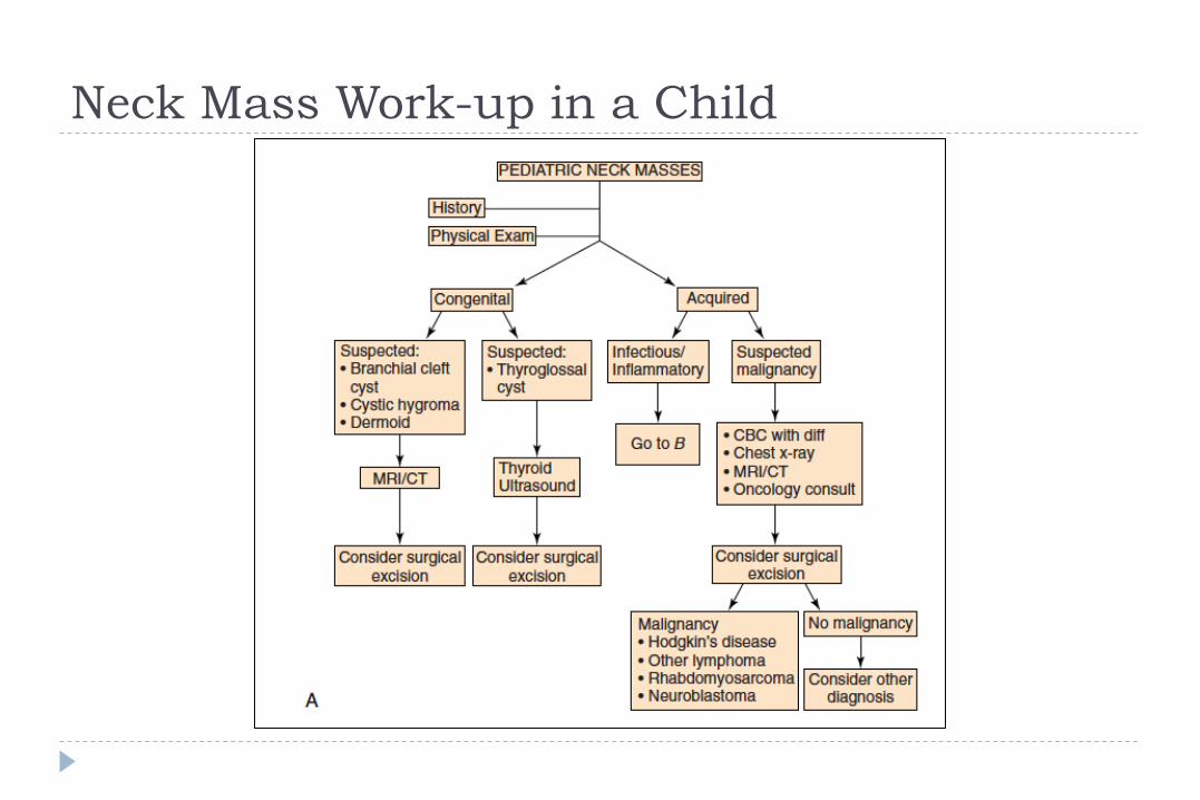
Neck Mass Work-up in a Child
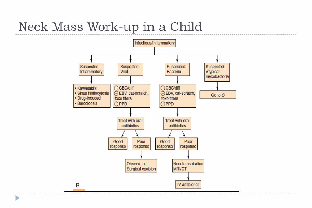
Neck Mass Work-up in a Child
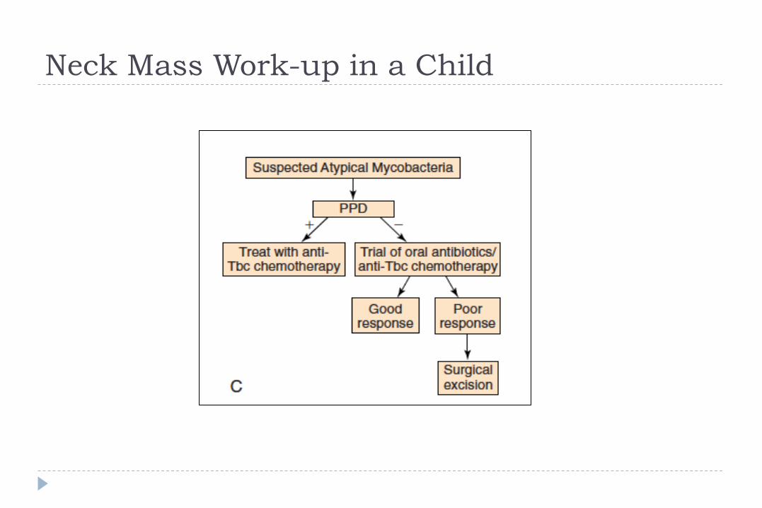
Neck Mass Work-up in a Child

Nodal Mass Workup in the Adult } Any solid asymmetric mass MUST be considered a metastatic
neoplastic lesion until proven otherwise } Asymptomatic cervical mass – 12% of cancer
} ~ 80% of these are SCCa
} Any New Cystic mass (> 40 y.o.a.) must be considered a cystic metastatic lesion until proven otherwise } Thyroid } SCC } Melanoma

Neck mass Work-up in an Adult

Thyroid Nodule Work-Up in an Adult So Which Nodules detected on Ultrasound Need an FNA?

Thyroid Nodule Work-Up in an Adult So How do I interpret and Action the various Cytology Results from Thyroid FNA
RESULT Frequency Features Interpreta4on / DDx Ac4on 1. Inadequate / Non-‐
diagnos4c 15% • Fail to meet criteria for
cytologic Adequacy: • ≥ 6 follicular cell groups • each containing 10-‐15 cells
• Sampling from cys$c, haemorrhagic, hyper-‐vascular, hypo-‐cellular colloid nodules
• 1-‐4% malignancy rate
• Repeat FNAC with U/S guidance (reveals malignancy in 4% of women, 29% of Men)
• Hemithyroidectomy if con;nues to be Non-‐diagnos4c – esp. if a solid nodule • 7% of nodules remain Non-‐
diagnos$c on FNA
2. Benign 60-‐90% • Syncy;al groups +/-‐ dis;nct micro-‐follicles
• Increased cellularity; scanty colloid
• Uniform cells with round nuclei, inconspicuous nucleoli, and well-‐defined borders
• Cytoplasmic features vary from scant to oxyphilic
o 0-‐3 % malignancy rate o Sampling from a:
• Macrofollicular neoplasm • Colloid Nodule
o False Nega4ve Rate (i.e. missed malignancy): 1-‐6%
1. Observe • Repeat U/S in 6-‐18/12 • If Stable in size: then U/S
every 3-‐5 years o Stable: a. <50% increase in
volume OR b. <20% increase in at
least 2 dimensions (solid por;on)
2. Repeat Bx – if: • é in size (SEE ABOVE)
3. Consider Surgery – if: i) Symptoma4c ii) Cosme4c Concerns

Thyroid Nodule Work-Up in an Adult So How do I interpret and Action the various Cytology Results from Thyroid FNA
RESULT Frequency Features Interpreta4on / DDx Ac4on
3. Indeterminate ~15-‐30% o Atypia of undetermined significance
o (FLUS) ‘Abnormal’ Follicular epithelium with varying degrees of Atypia
o Focal features sugges4ve of papillary carcinoma in an otherwise predominantly benign-‐appearing sample
o +/-‐ Hürthle (Oxyphilic) Cells
o 5-‐15% Malignancy Rate o Sampling from a:
i) ? Follicular Adenoma ii) ? Follicular Carcinoma iii) ? Hürthle Cell Adenoma iv) ? Hürthle Cell Carcinoma v) ? Thyroidi$s (esp.
Hashimoto’s) vi) ? Papillary Carcinoma vii) ? Micro-‐follicular Lesion
o Carcinoma Rate (Follicular/Hurthle): 20% of Indeterminate FNAs harbor Carcinoma
1. ? Hürthle Neoplasm -‐ Surgery 2. ? TSH
• Normal/High – Surgery • Low – Radio-‐nuclide Scan
3. ? Radio-‐nuclide Scan: if ? follicular i) Cold – Surgery ii) Hot – Evaluate & Treat
for Thyrotoxicosis 4. ? Molecular Markers: e.g. BRAF,
RAS, RET/PTC, Pax8-‐PPARϒ, galec$n-‐3 • Recent studies demonstrated
ability to improve pre-‐opera;ve diagnos;c accuracy

Thyroid Nodule Work-Up in an Adult So How do I interpret and Action the various Cytology Results from Thyroid FNA
RESULT Frequency Features Interpreta4on / DDx Ac4on
4. Follicular Neoplasm or Suspicious of Follicular Neoplasm
o Suspicious for malignancy including papillary/medullary/other
15-‐30% Malignancy rate 1. Surgical Lobectomy
5. Suspicious o Papillary/medullary/metasta4c/lymphoma
60-‐75% Malignancy rate 1. Near-‐Total Thyroidectomy or surgical lobectomy
6. Malignant 5% o Papillary Carcinoma: • Papillary forma$ons,
with complex branching and a central vascular core
• Characteris;c Nuclear features: intra-‐nuclear inclusions, nuclear grooves, nuclear crowding
• Psammoma bodies • Mul$-‐nucleated cells
o Poorly Differen4ated Carcinoma o Medullary Thyroid Carcinoma o +/-‐ Immunohistochemistry to
detect Calcitonin
• Carcinoma Rate: 95% at surgery • Sampling from a:
i) Papillary Carcinoma ii) Anaplas$c Carcinoma iii) Medullary Carcinoma iv) Lymphoma
• False Posi4ve Rate: <5% o Arises from difficulty
interpre;ng Cytology on B/G of Hashimoto’s Thyroidi$s, Grave’s Disease, Toxic Nodules.
1. Surgery – Total Thyroidectomy +/-‐ Neck Dissec$on (Level VI +/-‐ Lateral)
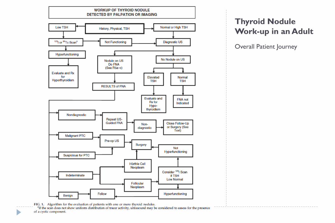
Thyroid Nodule Work-up in an Adult
Overall Patient Journey
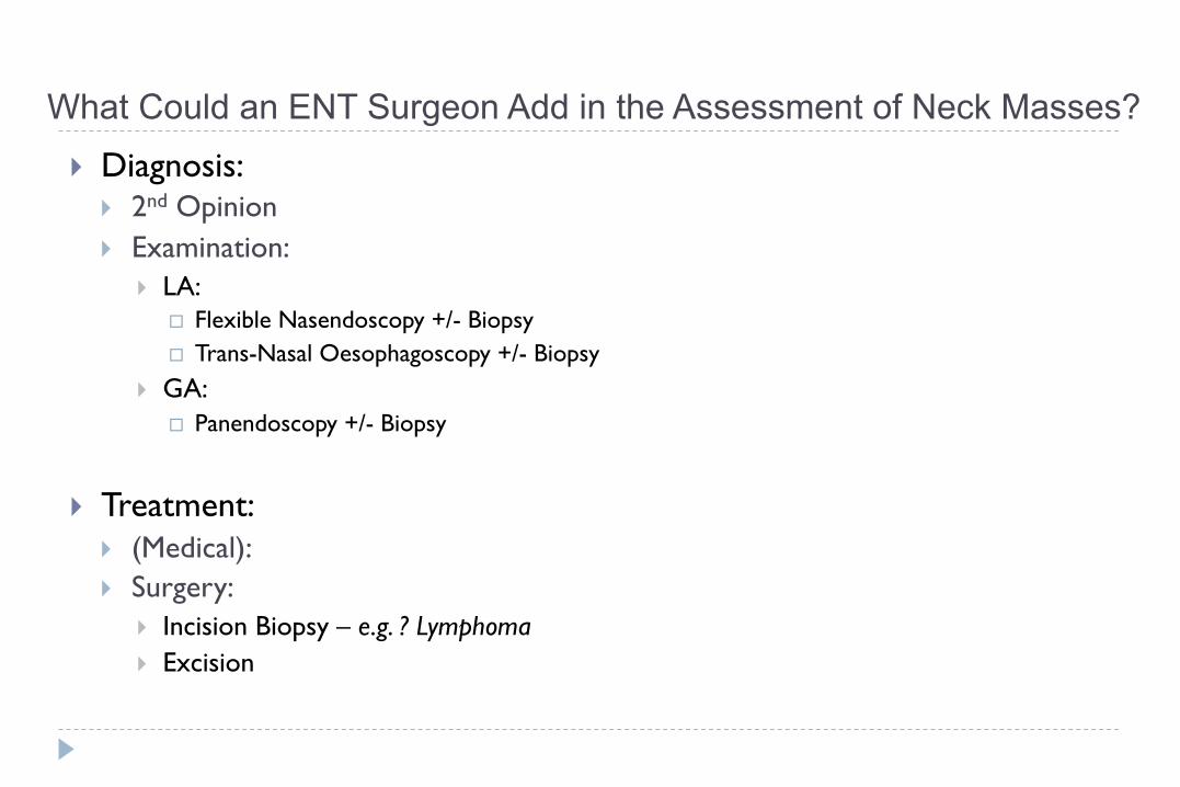
What Could an ENT Surgeon Add in the Assessment of Neck Masses? } Diagnosis:
} 2nd Opinion } Examination:
} LA: ¨ Flexible Nasendoscopy +/- Biopsy ¨ Trans-Nasal Oesophagoscopy +/- Biopsy
} GA: ¨ Panendoscopy +/- Biopsy
} Treatment: } (Medical): } Surgery:
} Incision Biopsy – e.g. ? Lymphoma } Excision
} Ipsilateral otalgia with normal otoscopy – direct attention to tonsil, tongue base, supraglottis and hypopharynx
} Unilateral serous otitis – direct examination of nasopharynx
What Could an ENT Surgeon Add in the Assessment of Neck Masses?

} Panendoscopy } FNAB positive with no primary on repeat exam } FNAB equivocal/negative in high risk patient
} Directed Biopsy } All suspicious mucosal lesions } Areas of concern on CT/MRI } None observed – nasopharynx, tonsil (ipsilateral tonsillectomy for
jugulodigastric nodes), base of tongue and piriforms
} Synchronous primaries (10 to 20%)
What Could an ENT Surgeon Add in the Assessment of Neck Masses?
} Unknown primary } University of Florida (August, 2001) } Detected primary in 40% } Without suggestive findings on CT or panendoscopy yield dropped to
20% } Tonsillar fossa in 80%
What Could an ENT Surgeon Add in the Assessment of Neck Masses?

Nodal Mass Workup in the Adult } Open excisional biopsy
} Only if complete workup negative } Occurs in ~5% of patients } Be prepared for a complete neck dissection } Frozen section results (complete node excision)
} Inflammatory or granulomatous – culture } Lymphoma or adenocarcinoma – close wound

Additional Possible Referrals } Infectious Diseases Consultation } Medical Oncologist } Rheumatologist } Endocrinologist

Thank You Questions

Primary Tumors
} Thyroid mass } Lymphoma } Salivary tumors } Lipoma
} Carotid body and glomus tumors
} Neurogenic tumors

Thyroid Masses } Leading cause of anterior neck masses } Children
} Most common neoplastic condition } Male predominance } Higher incidence of malignancy
} Adults } Female predominance } Mostly benign
Thyroid Masses
} Lymph node metastasis } Initial symptom in 15% of papillary carcinomas } 40% with malignant nodules } Histologically (microscopic) in >90%
} FNAB has replaced USG and radionucleotide scanning } Decreases # of patients with surgery } Increased # of malignant tumors found at surgery } Doubled the # of cases followed up } Unsatisfactory aspirate – repeat in 1 month

Thyroid Masses

Lymphoma
} More common in children and young adults } Up to 80% of children with Hodgkin’s have a neck mass } Signs and symptoms
} Lateral neck mass only (discrete, rubbery, nontender) } Fever } Hepatosplenomegaly } Diffuse adenopathy

Lymphoma } FNAB – first line diagnostic test } If suggestive of lymphoma – open biopsy } Full workup – CT scans of chest, abdomen, head and neck; bone
marrow biopsy

Lymphoma

Salivary Gland Tumors } Enlarging mass anterior/inferior to ear or at the mandible angle is
suspect } Benign
} Asymptomatic except for mass
} Malignant } Rapid growth, skin fixation, cranial nerve palsies
Salivary Gland Tumors
} Diagnostic tests } Open excisional biopsy (submandibulectomy or parotidectomy)
preferred } FNAB
} Shown to reduce surgery by 1/3 in some studies } Delineates intra-glandular lymph node, localized sialadenitis or benign
lymphoepithelial cysts } May facilitate surgical planning and patient counseling } Accuracy >90% (sensitivity: ~90%; specificity: ~80%)
} CT/MRI – deep lobe tumors, intra vs. extra-parotid } Be prepared for total parotidectomy with possible
facial nerve sacrifice

Salivary Gland Tumors

Carotid Body Tumor
} Rare in children } Pulsatile, compressible mass } Mobile medial/lateral not superior/inferior } Clinical diagnosis, confirmed by angiogram or CT } Treatment
} Irradiation or close observation in the elderly } Surgical resection for small tumors in young patients
} Hypotensive anesthesia } Preoperative measurement of catecholamines

Carotid Body Tumor

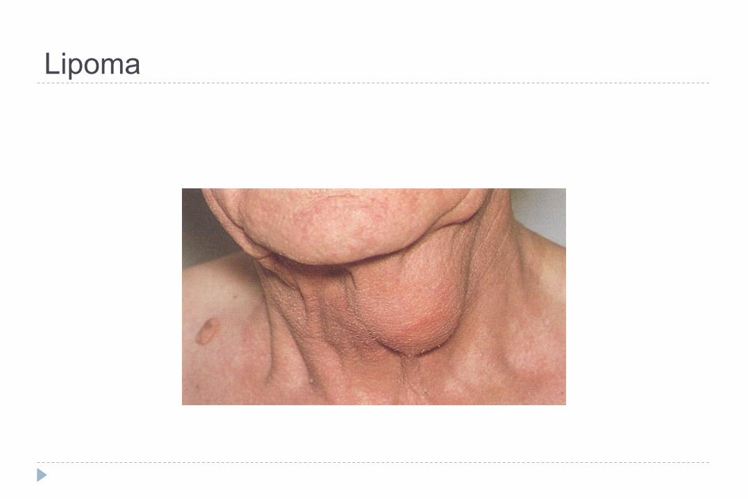
Lipoma } Soft, ill-defined mass } Usually >35 years of age } Asymptomatic } Clinical diagnosis – confirmed by excision
Lipoma

Neurogenic Tumors } Arise from neural crest derivatives } Include schwannoma, neurofibroma, and malignant peripheral
nerve sheath tumor } Increased incidence in NF syndromes } Schwannoma most common in head & neck

Schwannoma } Sporadic cases mostly } 25 to 45% in neck when extracranial } Most commonly between 20 and 50 years } Usually mid-neck in poststyloid compartment } Signs and symptoms
} Medial tonsillar displacement } Hoarseness (vagus nerve) } Horner’s syndrome (sympathetic chain)

Schwannoma
Congenital and Developmental Mass } Epidermal and sebaceous cysts } Branchial cleft cysts } Thyroglossal duct cyst } Vascular tumors

Epidermal and Sebaceous Cysts } Most common congenital/developmental mass } Older age groups } Clinical diagnosis
} Elevation and movement of overlying skin } Skin dimple or pore
} Excisional biopsy confirms

Epidermal and Sebaceous Cysts

Branchial Cleft Cysts } Branchial cleft anomalies } 2nd cleft most common (95%) – tract medial to cnXII between
internal and external carotids } 1st cleft less common – close association with facial nerve
possible } 3rd and 4th clefts rarely reported } Present in older children or young adults often following URI
Branchial Cleft Cysts } Most common as smooth, fluctuant mass underlying the SCM } Skin erythema and tenderness if infected } Treatment
} Initial control of infection } Surgical excision, including tract
} May necessitate a total parotidectomy (1st cleft)

Branchial Cleft Cysts

Thyroglossal Duct Cyst } Most common congenital neck mass (70%) } 50% present before age 20 } Midline (75%) or near midline (25%) } Usually just inferior to hyoid bone (65%) } Elevates on swallowing/protrusion of tongue } Treatment is surgical removal (Sis trunk) after resolution of any
infection

Thyroglossal Duct Cyst

Vascular Tumors } Lymphangiomas and hemangiomas } Usually within 1st year of life } Hemangiomas often resolve spontaneously, while lymphangiomas
remain unchanged } CT/MRI may help define extent of disease

Vascular Tumors } Treatment
} Lymphangioma – surgical excision for easily accessible or lesions affecting vital functions; recurrence is common
} Hemangiomas – surgical excision reserved for those with rapid growth involving vital structures or associated thrombocytopenia that fails medical therapy (steroids, interferon)

Vascular Tumors (lymphangioma)

Vascular Tumors (hemangioma)

Inflammatory Disorders } Lymphadenitis } Granulomatous lymphadenitis

Lymphadenitis
} Very common, especially within 1st decade } Tender node with signs of systemic infection } Directed antibiotic therapy with follow-up } FNAB indications (pediatric)
} Actively infectious condition with no response } Progressively enlarging } Solitary and asymmetric nodal mass } Supraclavicular mass (60% malignancy) } Persistent nodal mass without active infection

Lymphadenopathy
} Equivocal or suspicious FNAB in the pediatric nodal mass requires open excisional biopsy to rule out malignant or granulomatous disease
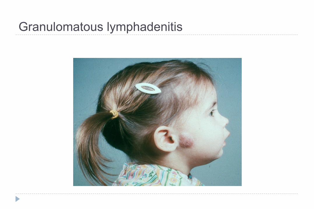
Granulomatous lymphadenitis } Infection develops over weeks to months } Minimal systemic complaints or findings } Common etiologies
} TB, atypical TB, cat-scratch fever, actinomycosis, sarcoidosis
} Firm, relatively fixed node with injection of skin
Granulomatous lymphadenitis
} Typical M. tuberculosis } more common in adults } Posterior triangle nodes } Rarely seen in our population } Usually responds to anti-TB medications } May require excisional biopsy for further workup
Granulomatous lymphadenitis
} Atypical M. tuberculosis } Pediatric age groups } Anterior triangle nodes } Brawny skin, induration and pain } Usually responds to complete surgical excision or curettage
Granulomatous lymphadenitis
} Cat-scratch fever (Bartonella) } Pediatric group } Preauricular and submandibular nodes } Spontaneous resolution with or without antibiotics
Granulomatous lymphadenitis

Summary } Extensive differential diagnosis } Age of patient is important } Accurate history and complete exam essential } FNAB – invaluable diagnostic tool } Possibility for malignancy in any age group } Close follow-up and aggressive approach is best for favorable
outcomes