assessment of fetal wellbeing in pregnancy and labour
TRANSCRIPT
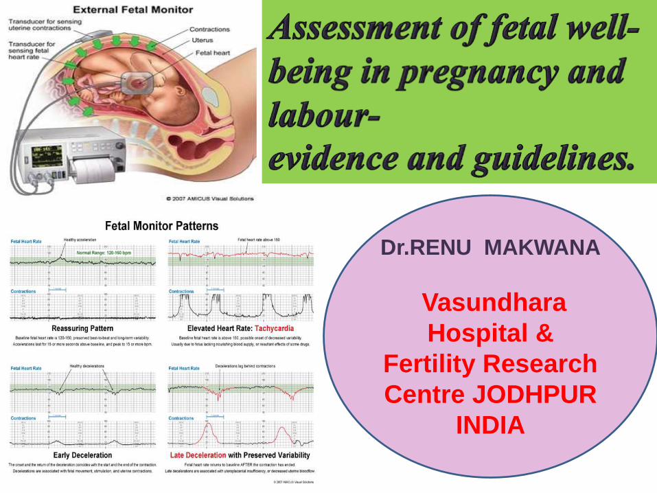
Dr.RENU MAKWANA
Vasundhara
Hospital &
Fertility Research
Centre JODHPUR
INDIA

Antepartum fetal monitoring
• Prevent fetal injury and death.
• Improve long-term neurologic outcome through optimal timing of delivery
• Avoiding unnecessary intervention, such as cesarean delivery or preterm delivery.
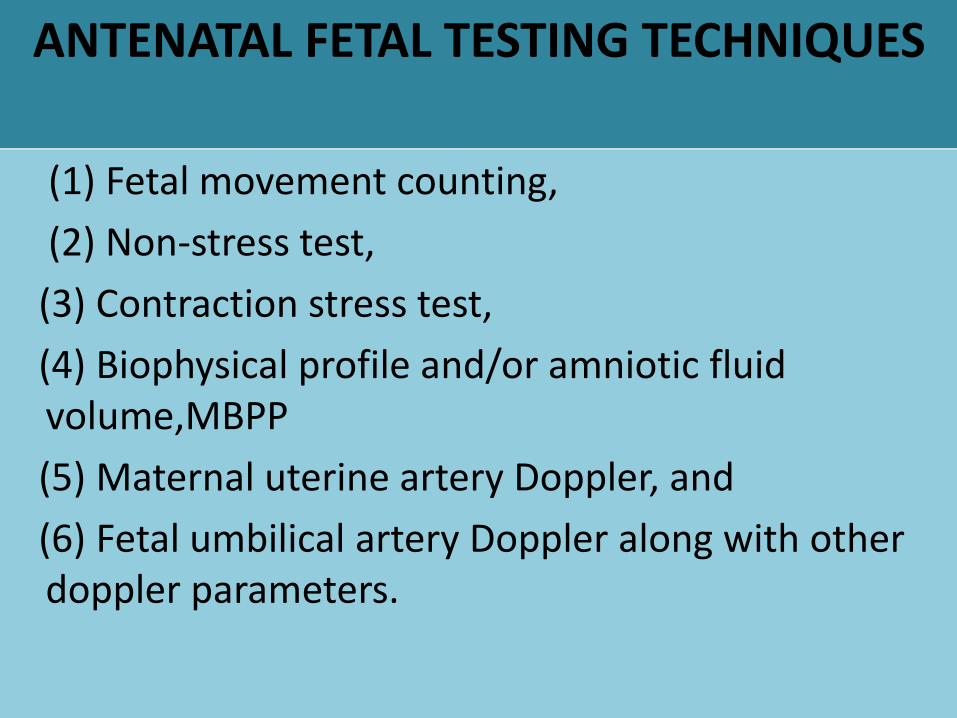
ANTENATAL FETAL TESTING TECHNIQUES
(1) Fetal movement counting,
(2) Non-stress test,
(3) Contraction stress test,
(4) Biophysical profile and/or amniotic fluid volume,MBPP
(5) Maternal uterine artery Doppler, and
(6) Fetal umbilical artery Doppler along with other doppler parameters.

DFMC
The only antenatal surveillance technique recommended for all pregnant women, with and without risk factors, is maternal awareness of fetalmovements.
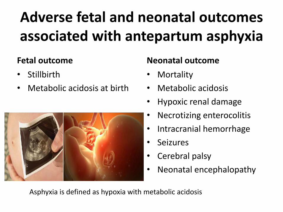
Adverse fetal and neonatal outcomesassociated with antepartum asphyxia
Fetal outcome
• Stillbirth
• Metabolic acidosis at birth
Neonatal outcome
• Mortality
• Metabolic acidosis
• Hypoxic renal damage
• Necrotizing enterocolitis
• Intracranial hemorrhage
• Seizures
• Cerebral palsy
• Neonatal encephalopathy
Asphyxia is defined as hypoxia with metabolic acidosis
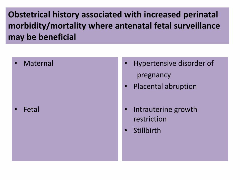
• Maternal
• Fetal
• Hypertensive disorder of
pregnancy
• Placental abruption
• Intrauterine growth restriction
• Stillbirth
Obstetrical history associated with increased perinatalmorbidity/mortality where antenatal fetal surveillance may be beneficial
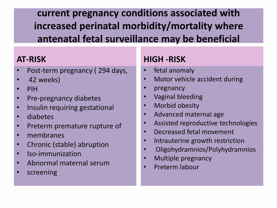
current pregnancy conditions associated with increased perinatal morbidity/mortality where antenatal fetal surveillance may be beneficial
AT-RISK• Post-term pregnancy ( 294 days,• 42 weeks)• PIH• Pre-pregnancy diabetes• Insulin requiring gestational• diabetes• Preterm premature rupture of• membranes• Chronic (stable) abruption• Iso-immunization• Abnormal maternal serum• screening
HIGH -RISK• fetal anomaly• Motor vehicle accident during• pregnancy• Vaginal bleeding• Morbid obesity• Advanced maternal age• Assisted reproductive technologies• Decreased fetal movement• Intrauterine growth restriction• Oligohydramnios/Polyhydramnios• Multiple pregnancy• Preterm labour

DFMC
• Sadovsky and Yaffee (1973) pre-eclamptic patients noticed decreased fetal movement prior to fetaldemise.
• Women perceive most movement when lying down fewer when sitting and least while standing.
• Busy pregnant women: not concentrating on fetalactivity: often report a misperception of RFM.
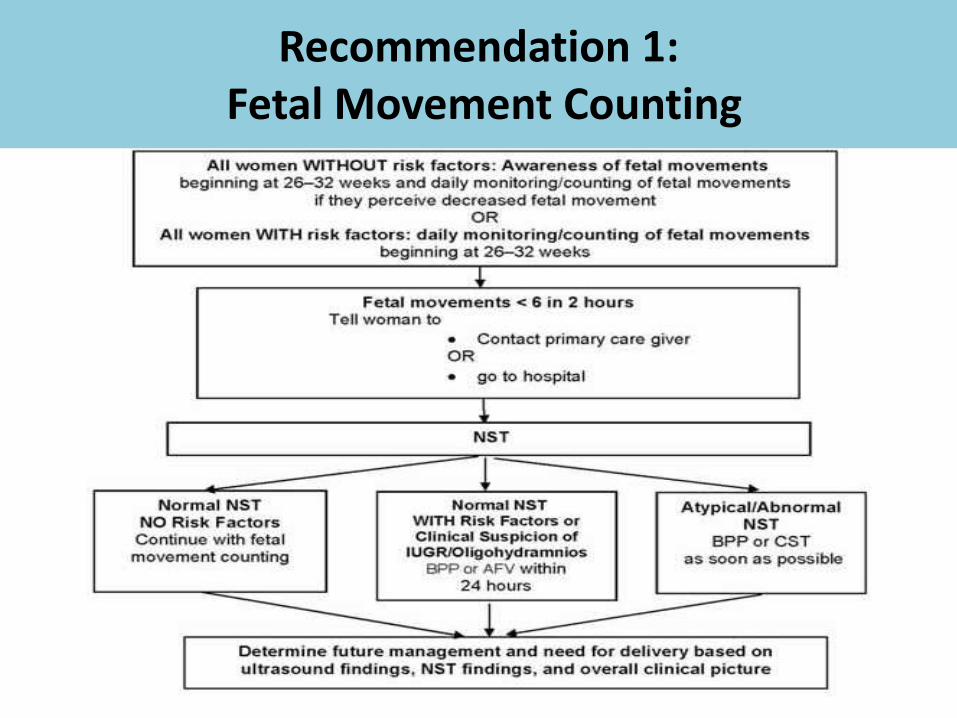
Recommendation 1:Fetal Movement Counting
Non-Stress Test
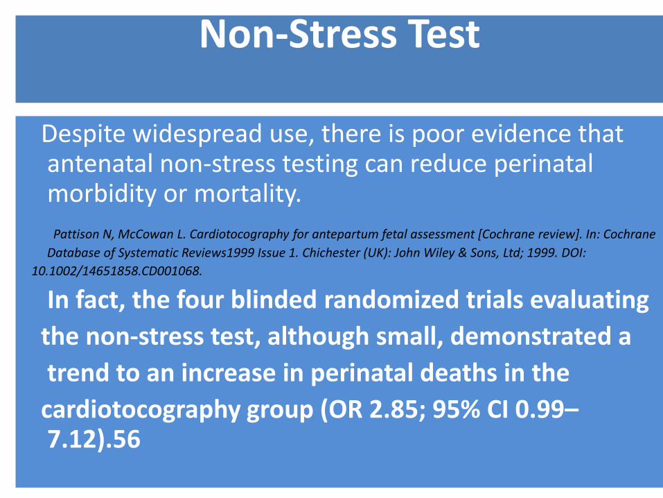
Despite widespread use, there is poor evidence that antenatal non-stress testing can reduce perinatalmorbidity or mortality.
Pattison N, McCowan L. Cardiotocography for antepartum fetal assessment [Cochrane review]. In: Cochrane
Database of Systematic Reviews1999 Issue 1. Chichester (UK): John Wiley & Sons, Ltd; 1999. DOI:
10.1002/14651858.CD001068.
In fact, the four blinded randomized trials evaluating
the non-stress test, although small, demonstrated a
trend to an increase in perinatal deaths in the
cardiotocography group (OR 2.85; 95% CI 0.99–7.12).56

A negative predictive value of the test for
fetal and neonatal death is 99% within one week of testing
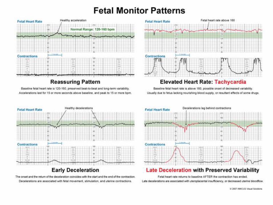
• Baseline FHR: 110-150 b/m
• Baseline variability: 10-25 b/m
• At least 2 accelerations (>15 beats for> 15 sec in 20 min)
• No decelerations
Electronic fetal heart rate monitoring:
research guidelines for interpretation.
National Institute of Child Health and Human Development Research
Planning Workshop. Am J Obstet Gynecol1997;177(6):1385–90.

Recommendation : Non-Stress Test
• 1. Antepartum non-stress testing may be considered when risk factors for adverse perinatal outcome are present. (III-B)
• 2. In the presence of a normal non-stress test, usual fetal movement patterns, and absence of suspected oligohydramnios, it is not necessary to conduct a biophysical profile or contraction stress test. (III-B)
• 3. A normal non-stress test should be classified and documented by an appropriately trained and designated individual as soon as possible, (ideally within 24 hours).


• Ray M, Freeman R, Pine S, Hesselgesser R. Clinical experience with the oxytocin challenge test. Am J Obstet Gynecol 1972;114(1):1–9.
• . Lagrew DC. The contraction stress test. Clin Obstet Gynecol 1995;38(1):11–25.
• Creasy R, Reznik R, Iams J. Maternal fetal medicine principles and practice 5th ed. Philadelphia: W.B. Saunders; 2003.
Contraction Stress Test
To unmask poor
placental function
Biophysical profile/Modified BPP and Doppler interrogation of uterine or fetal
vessels

Recommendation 3: Contraction Stress Test
1. The contraction stress test should be considered in thepresence of an atypical non-stress test as a proxy for theadequacy of intrapartum uteroplacental function and,together with the clinical circumstances, will aid in decisionmaking about timing and mode of delivery. (III-B)
2. The contraction stress test should not be performed when vaginal delivery is contraindicated. (III-B)
3. The contraction stress test should be performed in a setting where emergency Caesarean section is available. (III-B)

Sonographic Assessment of Fetal Behaviourand/or Amniotic Fluid Volume
Components of fetal biophysical profile
Inclusion of NST brings the maximum possible score to 10 when the NST is normal
The modified BPP consists of a non-stress test and an AFI (> 5 cm is considered adequate)
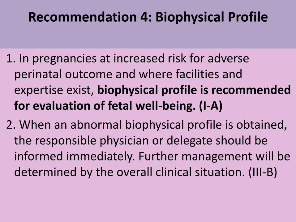
Recommendation 4: Biophysical Profile
1. In pregnancies at increased risk for adverse perinatal outcome and where facilities and expertise exist, biophysical profile is recommended for evaluation of fetal well-being. (I-A)
2. When an abnormal biophysical profile is obtained, the responsible physician or delegate should be informed immediately. Further management will be determined by the overall clinical situation. (III-B)


Recommendation 5: Uterine Artery Doppler
1. Where facilities and expertise exist, uterine artery Doppler may be performed at the time of the 17 to 22 weeks’ gestation during detailed anatomical ultrasound scan in women with the following factors for adverse perinatal outcome. (II-A)2. Women with a positive uterine artery Doppler screen should have the following second uterine artery Doppler at 24 to 26 weeks.
.
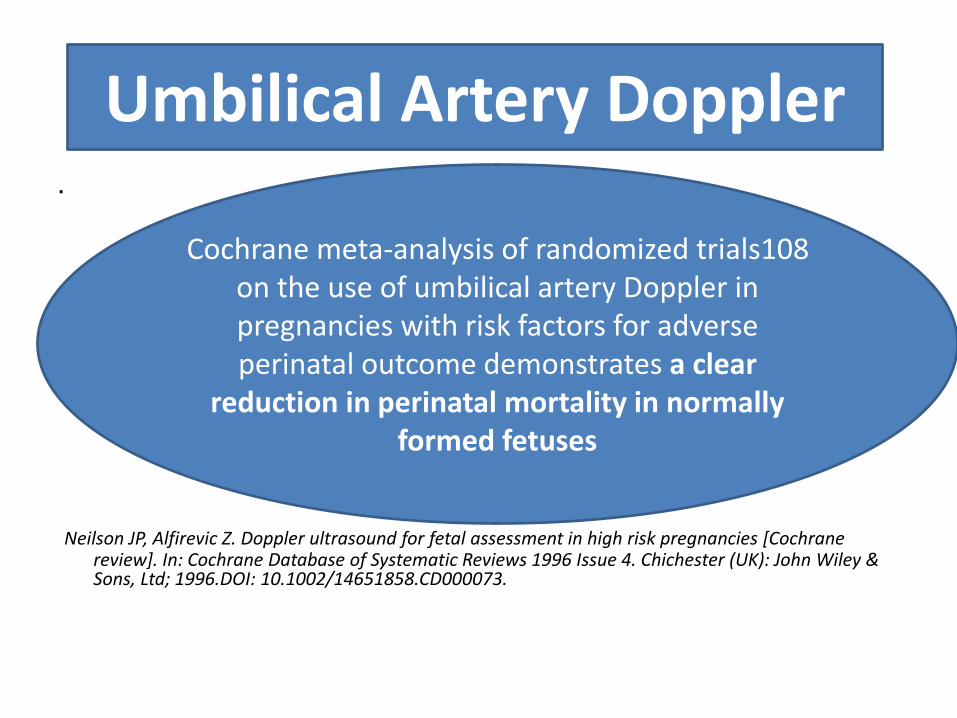
Neilson JP, Alfirevic Z. Doppler ultrasound for fetal assessment in high risk pregnancies [Cochrane review]. In: Cochrane Database of Systematic Reviews 1996 Issue 4. Chichester (UK): John Wiley & Sons, Ltd; 1996.DOI: 10.1002/14651858.CD000073.
Umbilical Artery Doppler
Cochrane meta-analysis of randomized trials108 on the use of umbilical artery Doppler in pregnancies with risk factors for adverse perinatal outcome demonstrates a clear
reduction in perinatal mortality in normally formed fetuses
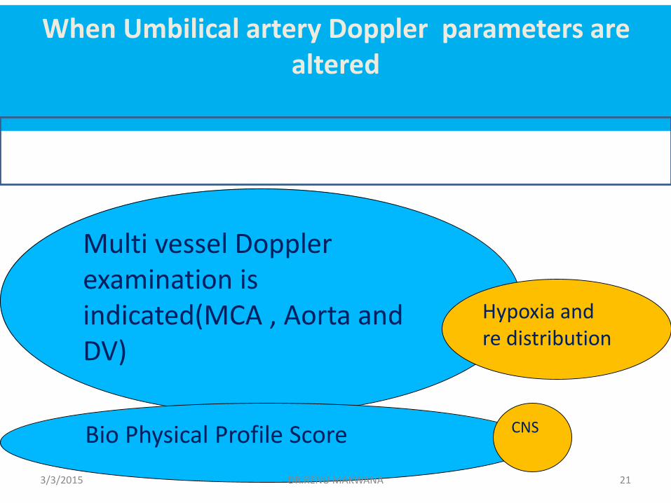
Multi vessel Doppler examination is indicated(MCA , Aorta and DV)
Bio Physical Profile Score CNS
Hypoxia and re distribution
3/3/2015 DR.RENU MAKWANA 21
When Umbilical artery Doppler parameters are altered
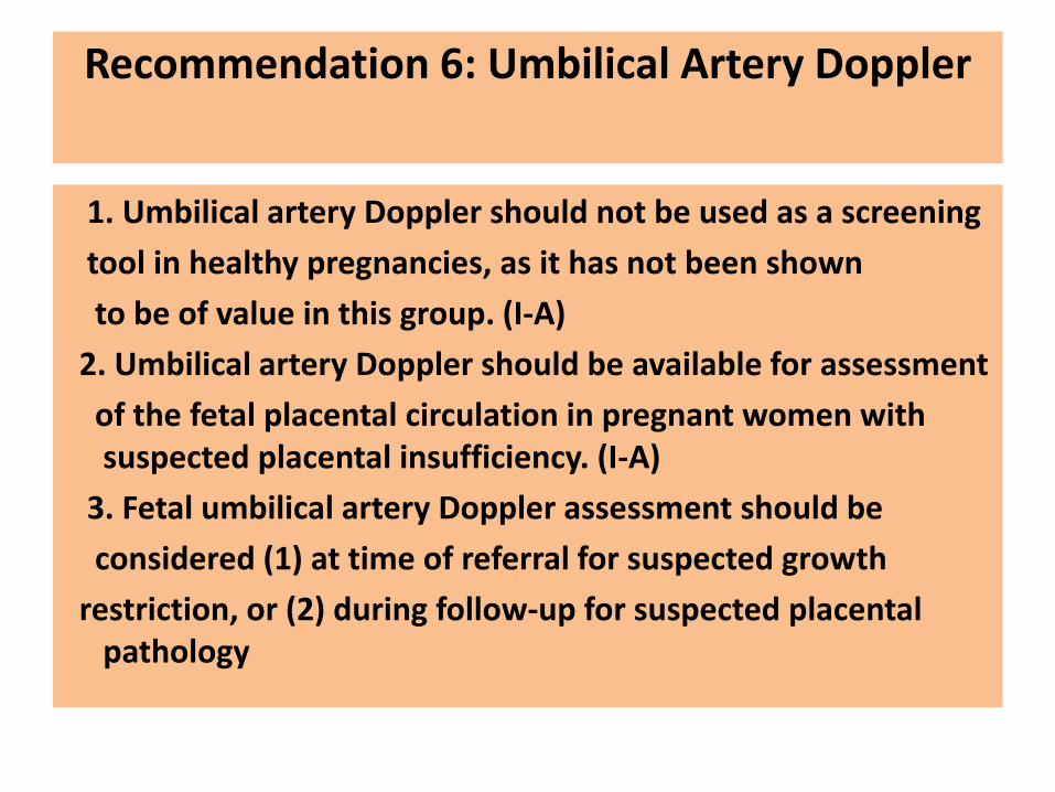
Recommendation 6: Umbilical Artery Doppler
1. Umbilical artery Doppler should not be used as a screening
tool in healthy pregnancies, as it has not been shown
to be of value in this group. (I-A)
2. Umbilical artery Doppler should be available for assessment
of the fetal placental circulation in pregnant women with suspected placental insufficiency. (I-A)
3. Fetal umbilical artery Doppler assessment should be
considered (1) at time of referral for suspected growth
restriction, or (2) during follow-up for suspected placental pathology

Recommendation 7: Labour Support During Active Labour
Women in active labour should receive continuous close support from an appropriately trained person (I-A)
Recommendation 8: Professional One-to-One Care and
Intrapartum Fetal Surveillance
Intensive fetal surveillance by intermittent auscultation or electronic fetal monitoring requires the continuous presence of nursing or midwifery staff (III-C)
INTRAPARTUM FETAL MONITORING
• To detect potential fetal decompensation• To allow timely and effective intervention
Changes in fetal heart rate precede brain injury constitutes the rationale for FH monitoring
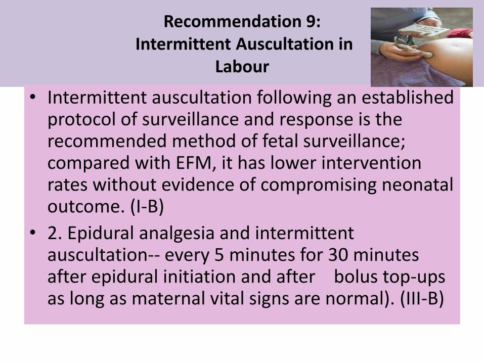
Recommendation 9:Intermittent Auscultation in
Labour
• Intermittent auscultation following an established protocol of surveillance and response is the recommended method of fetal surveillance; compared with EFM, it has lower intervention rates without evidence of compromising neonatal outcome. (I-B)
• 2. Epidural analgesia and intermittent auscultation-- every 5 minutes for 30 minutes after epidural initiation and after bolus top-ups as long as maternal vital signs are normal). (III-B)

What is not assessed?- Baseline variability and classification ofdecelerations
Benefits Limitations
• Difficult to hear a fetalheart rate in very large women when Some women may feel the technique is more intrusive because of the frequency of assessment
Less costly, less constrictingFreedom of movement is increased Assessments of the fetal heart rate can be done with the woman immersed in water

Recommended frequency of auscultation

Common indications/contraindications for intermittent auscultation
Preterm labour
Postdated labour
Epidural analgesia
VBACNeilson J. Electronic fetal monitoring plus scalp sampling vs intermittent auscultation
in labour[Revised May 1994]. In: Keirse M, Renfrew MJ, Neilson J, Crowther C, editors. Cochrane Collaborative Issue 2. Oxford; 1995.
Incidence of other pathologies is increased ….
So EFM
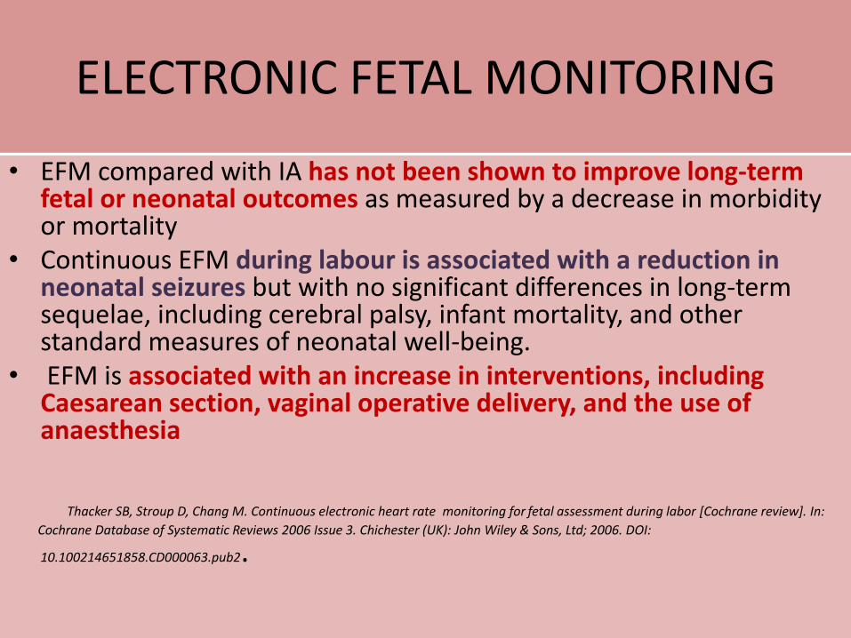
ELECTRONIC FETAL MONITORING
• EFM compared with IA has not been shown to improve long-term fetal or neonatal outcomes as measured by a decrease in morbidity or mortality
• Continuous EFM during labour is associated with a reduction in neonatal seizures but with no significant differences in long-term sequelae, including cerebral palsy, infant mortality, and other standard measures of neonatal well-being.
• EFM is associated with an increase in interventions, including Caesarean section, vaginal operative delivery, and the use of anaesthesia
Thacker SB, Stroup D, Chang M. Continuous electronic heart rate monitoring for fetal assessment during labor [Cochrane review]. In:
Cochrane Database of Systematic Reviews 2006 Issue 3. Chichester (UK): John Wiley & Sons, Ltd; 2006. DOI:
10.100214651858.CD000063.pub2.
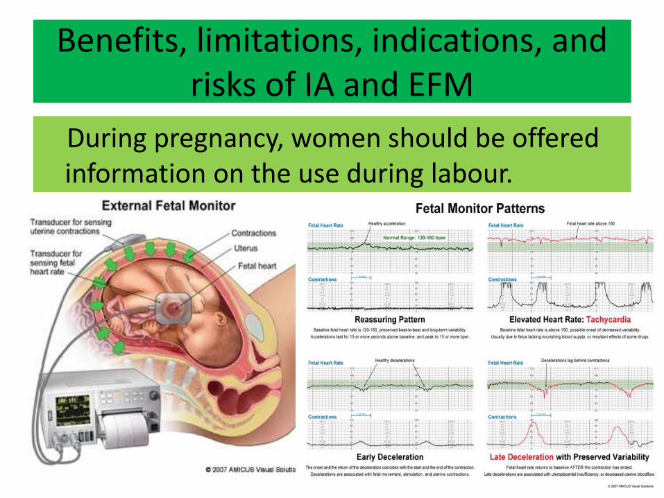
Benefits, limitations, indications, and risks of IA and EFM
During pregnancy, women should be offered information on the use during labour.
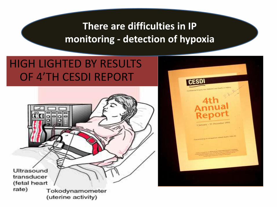
HIGH LIGHTED BY RESULTS OF 4’TH CESDI REPORT
There are difficulties in IP monitoring - detection of hypoxia

LACK OF KNOWLEDGE TO INTERPRET TRACESFAILURE TO INCORPORATE CLINICAL PICTUREDELAY IN INTERVENTIONCOMMUNICATION / COMMON SENSE ISSUES
EFM – Difficulties in IP EFM & decision making
CAN WE DETECT HYPOXIA IN TIME?
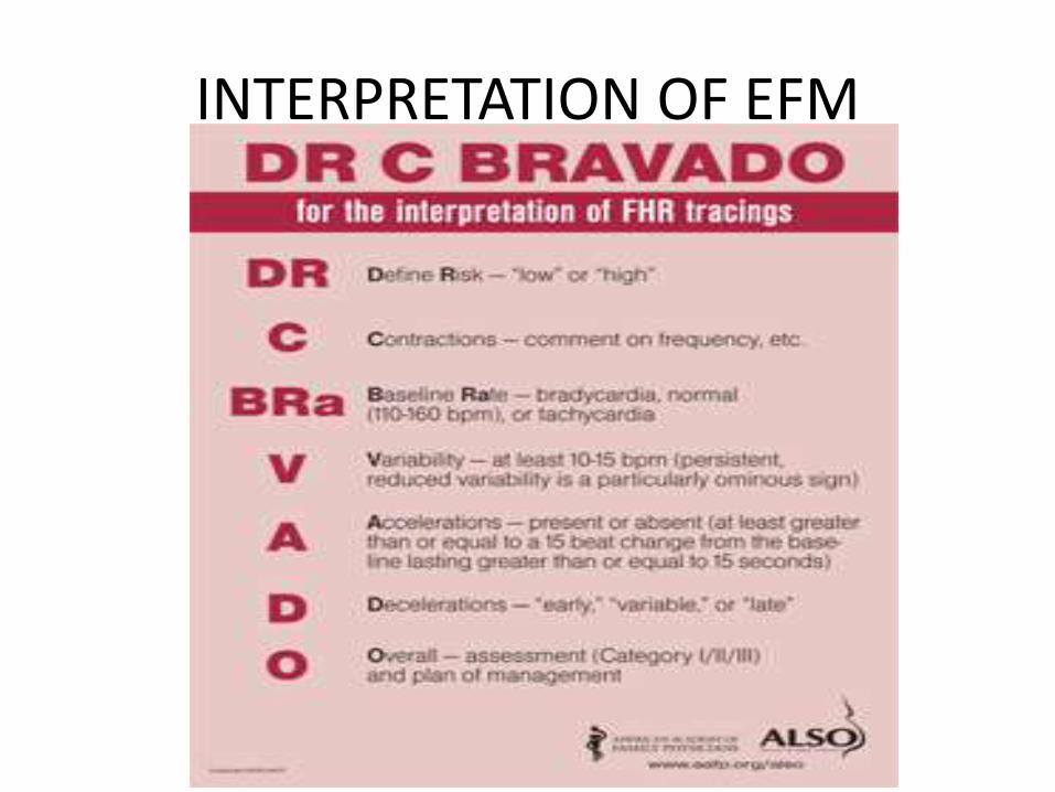
INTERPRETATION OF EFM

If CTG is reactive and shows cycling the fetus is unlikely to be acidotic or to have previous insult
more chances that the fetus may be born acidotic

Most CTG abnormalities do not result in fetal acidosis
R. W. Beard, et al. The significance of the changes in the continuous foetal heart rate
in the first stage of labour. J Obstet Gynaecol Br Commonw 78:865-881, 1971.
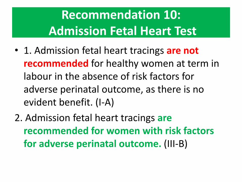
Recommendation 10:Admission Fetal Heart Test
• 1. Admission fetal heart tracings are not recommended for healthy women at term in labour in the absence of risk factors for adverse perinatal outcome, as there is no evident benefit. (I-A)
2. Admission fetal heart tracings are recommended for women with risk factors for adverse perinatal outcome. (III-B)

Recommendation 11: Intrapartum Fetal Surveillance for Women with Risk Factors for
Adverse Perinatal Outcome
EFM is recommended for pregnancies at risk of adverse perinataloutcome. (II-A)
Meconium,abnormal doppler
Antenatal risk factors FGR,BreechMultiple,
PProm
IntrapartumRisk facors
Document every15-30 min

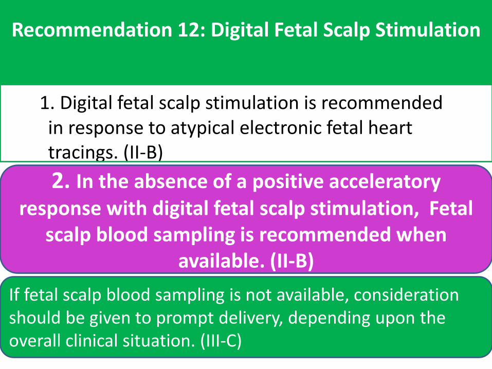
Recommendation 12: Digital Fetal Scalp Stimulation
1. Digital fetal scalp stimulation is recommended in response to atypical electronic fetal heart tracings. (II-B)
2. In the absence of a positive acceleratory response with digital fetal scalp stimulation, Fetal
scalp blood sampling is recommended when available. (II-B)
If fetal scalp blood sampling is not available, consideration should be given to prompt delivery, depending upon the overall clinical situation. (III-C)
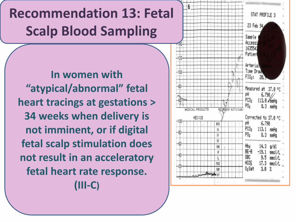
Recommendation 13: FetalScalp Blood Sampling
In women with “atypical/abnormal” fetal
heart tracings at gestations > 34 weeks when delivery is not imminent, or if digital
fetal scalp stimulation does not result in an acceleratory
fetal heart rate response. (III-C)
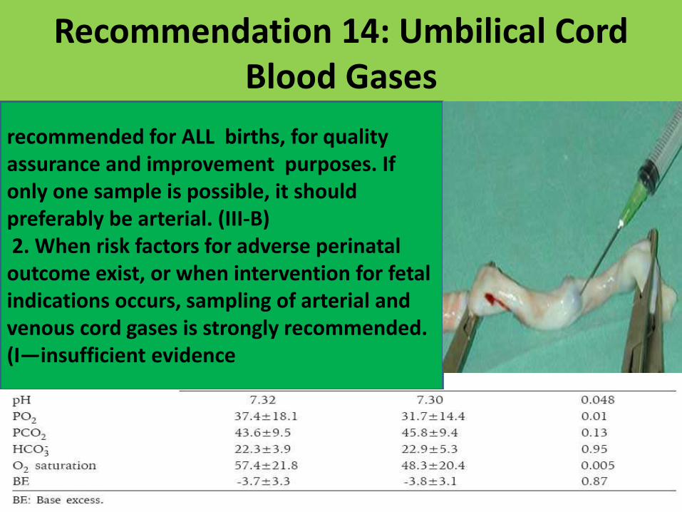
Recommendation 14: Umbilical Cord Blood Gases
recommended for ALL births, for quality assurance and improvement purposes. If only one sample is possible, it should preferably be arterial. (III-B)2. When risk factors for adverse perinataloutcome exist, or when intervention for fetalindications occurs, sampling of arterial and venous cord gases is strongly recommended. (I—insufficient evidence
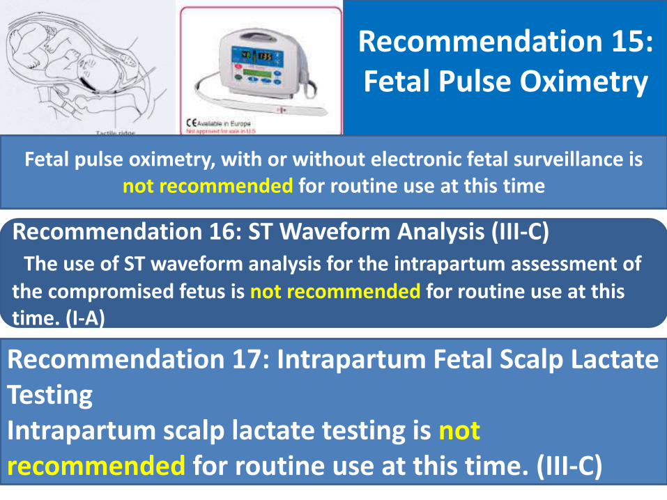
Recommendation 15: Fetal Pulse Oximetry
Fetal pulse oximetry, with or without electronic fetal surveillance is not recommended for routine use at this time
Recommendation 16: ST Waveform Analysis (III-C) The use of ST waveform analysis for the intrapartum assessment of
the compromised fetus is not recommended for routine use at this time. (I-A)
Recommendation 17: Intrapartum Fetal Scalp Lactate TestingIntrapartum scalp lactate testing is not recommended for routine use at this time. (III-C)
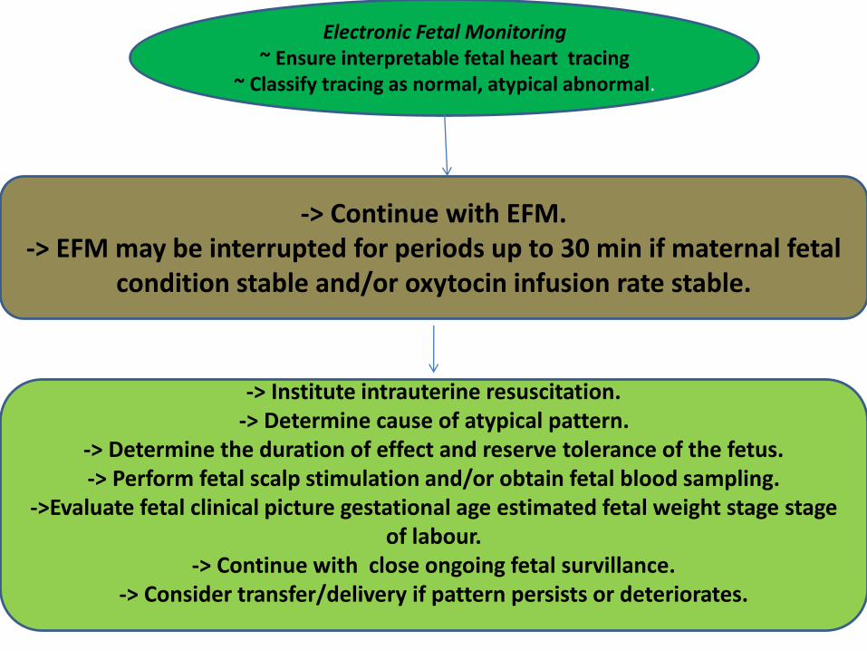
Electronic Fetal Monitoring~ Ensure interpretable fetal heart tracing
~ Classify tracing as normal, atypical abnormal.
-> Continue with EFM.-> EFM may be interrupted for periods up to 30 min if maternal fetal
condition stable and/or oxytocin infusion rate stable.
-> Institute intrauterine resuscitation.-> Determine cause of atypical pattern.
-> Determine the duration of effect and reserve tolerance of the fetus.-> Perform fetal scalp stimulation and/or obtain fetal blood sampling.
->Evaluate fetal clinical picture gestational age estimated fetal weight stage stageof labour.
-> Continue with close ongoing fetal survillance.-> Consider transfer/delivery if pattern persists or deteriorates.
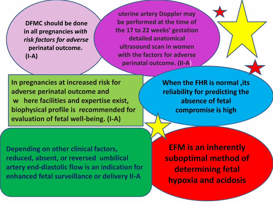
DFMC should be done in all pregnancies with risk factors for adverse
perinatal outcome.(I-A)
uterine artery Doppler may be performed at the time of
the 17 to 22 weeks’ gestation detailed anatomical
ultrasound scan in women with the factors for adverse
perinatal outcome. (II-A)
In pregnancies at increased risk for adverse perinatal outcome andw here facilities and expertise exist, biophysical profile is recommended for evaluation of fetal well-being. (I-A)
EFM is an inherently suboptimal method of
determining fetal hypoxia and acidosis
Depending on other clinical factors, reduced, absent, or reversed umbilical artery end-diastolic flow is an indication for enhanced fetal surveillance or delivery II-A
When the FHR is normal ,its reliability for predicting the
absence of fetal compromise is high
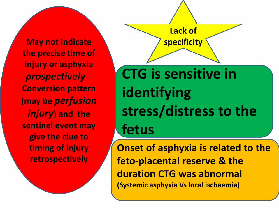
May not indicate the precise time of injury or asphyxia
prospectively –Conversion pattern
(may be perfusion injury) and the
sentinel event may give the clue to timing of injury retrospectively
Onset of asphyxia is related to the feto-placental reserve & the duration CTG was abnormal (Systemic asphyxia Vs local ischaemia)
Lack of specificity
CTG is sensitive in identifying stress/distress to the fetus