assessment of alterations in gene expression in...
TRANSCRIPT
CLINICOPATHOLOGICAL STUDIES
Assessment of Alterations in Gene Expression in RecurrentMalignant Glioma after Radiotherapy Using Complementary
Deoxyribonucleic Acid Microarrays
Tatsuhiro Joki, M.D., Ph.D., Rona S. Carroll, Ph.D.,Ian F. Dunn, B.S., Jianping Zhang, M.S.,
Toshiaki Abe, M.D., Ph.D.,Peter McL. Black, M.D., Ph.D.
Brain Tumor Laboratory (TJ, RSC, IFD, JZ, PMcLB), Department of Neurosurgery,Brigham and Women’s Hospital, Boston, Massachusetts; Children’s Hospital (TJ, RSC,IFD, JZ, PMcLB), Harvard Medical School, Boston, Massachusetts; and Department of
Neurosurgery (TJ, TA), Jikei University School of Medicine, Tokyo, Japan
OBJECTIVE: We used complementary deoxyribonucleic acid expression microarrays to assess the effects of radio-therapy on gene expression in glioblastoma multiforme. We hypothesized that postradiation recurrent tumorsmay demonstrate alterations in gene expression from the primary tumor specimen.
METHODS: Patients were diagnosed with glioblastoma multiforme at resection of the initial tumor, and theyreceived 60 Gy of fractionated radiotherapy before recurrence. Ribonucleic acid samples from both the primaryand the postradiation recurrent tumor in each patient were screened and compared using complementarydeoxyribonucleic acid expression arrays and Northern blot analysis.
RESULTS: Messenger ribonucleic acid levels of growth factors participating in paracrine loops, such as vascular endo-thelial growth factor and platelet-derived growth factor receptor b, were decreased in postradiation recurrent tumorsas compared with primary tumors in three of four patients. However, messenger ribonucleic acid levels of growthfactors involved in autocrine loops, such as epidermal growth factor receptor, platelet-derived growth factor a,platelet-derived growth factor A, and basic fibroblast growth factor, were decreased in two of four, two of four, threeof four, and three of four patients’ recurrent tumors, respectively. Microvessel counts demonstrated that blood vesselgrowth was decreased significantly in postradiation recurrent tumor specimens.
CONCLUSION: After radiotherapy of glioblastoma multiforme, levels of paracrine-acting growth factors arediminished in correspondence with the reduction in vascular density. In contrast, growth factors that participatein autocrine loops demonstrate elevated levels of gene expression. These results suggest that maintenance ofautocrine loops may be important in tumor regrowth after radiotherapy. (Neurosurgery 48:195–202, 2001)
Key words: Complementary deoxyribonucleic acid microarray, Gene expression, Glioblastoma, Radiotherapy, Recurrence
Current data indicate that approximately 18,000 primarymalignant brain tumors are diagnosed each year andthat approximately 13,000 deaths annually are attrib-
utable to malignant brain tumors (2, 16). The prognosis forpatients with malignant brain cancer is very poor; the recur-rence rate is greater than 90%, and the 5-year survival rate isless than 10% (19). Radiotherapy is a vital component ofmultimodal treatment for malignant brain tumors. Deliveredat sufficient doses and in conjunction with surgery, radiationtreatment has been demonstrated to prolong survival (21, 22).
Questions remain, however, regarding its effects on recurrenttumor phenotype. For unknown reasons, recurrent high-grade gliomas seem to behave more aggressively after radio-therapy than their primary counterparts, which suggests thatradiotherapy may be at least partly responsible for theseobserved changes. Radiotherapy seems to affect significantlythe plasma levels of a number of growth factors measured inpatients before and directly after radiotherapy (9).
Although the short-term effects of high- and low-dose irra-diation on gene expression have been studied in glioblastoma
195Neurosurgery, Vol. 48, No. 1, January 2001

multiforme (GBM) cells in vitro and in an animal model (17),the specific effects of long-term radiotherapy on gene expres-sion have not been examined. The purpose of this study wasto investigate the effects of radiotherapy on malignant glio-mas. We assessed patterns of gene expression in primarytumor tissue and recurrent tumor tissue taken from patientswhose treatment consisted solely of surgery and adjuvantradiotherapy. We compared the levels of gene expression inpatients’ primary and postradiation recurrent tumor speci-mens using complementary deoxyribonucleic acid (cDNA)microarray analysis, and we confirmed the differences inexpression in a subset of genes by Northern blot analysis.
PATIENTS AND METHODS
Patients
Four patients with recurrent GBM (three men and onewoman) were included in the study. Their mean age was59.25 years, and the mean interval from primary resection toresection of recurrent tumor after radiotherapy was 7.25months (Table 1). The Brain Tumour Tissue Bank of the Na-tional Cancer Institute of Canada, London Regional CancerCentre, provided the primary tumor and recurrent tumorspecimens from each patient. All patients were diagnosedwith GBM when the primary tumor was resected, and subse-quently they received 2.0 Gy of fractionated radiotherapy perday for 5 consecutive days each week during a 6-week period,for a total dose of 60 Gy. Patients received conventional x-rayphoton radiation using a linear accelerator.
Isolation of RNA
Total ribonucleic acid (RNA) was isolated from tumor spec-imens as described by Chirgwin et al. (4). Tissue samples wereplaced in 4 mol/L guanidinium isothiocyanate and were ho-mogenized with a polytron. Samples were then centrifugedfor 10 minutes at 20°C and 3000 rpm to remove debris, and thesupernatant was layered over 5.7 mol/L cesium chloride. Thesamples were ultracentrifuged for 16 hours at 22°C. The re-sulting RNA pellet was dissolved in 0.3 mol/L sterile sodiumacetate, followed by ethanol precipitation of the RNA. Be-cause of the sample tissue size, only 20 mg of total RNA was
obtained from each tumor. This amount was sufficient foronly one Northern blot analysis.
cDNA filters
Atlas human cDNA expression array membranes (ClontechLaboratories, Inc., Palo Alto, CA) were used to perform high-throughput analysis of each of the four patients’ primary andpostradiation recurrent tumor specimens to assess the effectof radiotherapy on gene expression. Each membrane is apositively charged nylon membrane on which 588 humancDNAs are immobilized. The cDNAs are grouped into ninecategories on each membrane: 1) oncogenes and tumor sup-pressor genes, 2) cell-cycle control proteins, 3) modulators/effectors/intracellular transducers, 4) stress response pro-teins, 5) apoptosis-associated proteins, 6) DNA synthesis/repair/recombination proteins, 7) DNA-binding proteins/transcription factors, 8) cell receptors and cell surfaceantigens, and 9) extracellular cell-signaling proteins.
cDNA synthesis and hybridization32P-labeled cDNA was synthesized from total RNA isolated
from both primary tumor specimens and postradiation recur-rent tumor tissue by reverse transcriptase in the presence of[a-32P]deoxyadenosine triphosphate. In brief, 10 mg of totalRNA was denatured at 75°C for 10 minutes in the presence of8 pmol of deoxythymidine mixture. After the denaturationstep, cDNAs were synthesized by incubation at 42°C for 45minutes in a master mixture containing 3 ml of deoxynucleo-side triphosphate, 5 ml of [a-32P]deoxyadenosine triphosphate(3000 Ci/mmol; NEN Life Science Products, Boston, MA), and1600 U of Moloney murine leukemia virus reverse transcrip-tase (GIBCO/BRL, Rockville, MD). The reaction was termi-nated by heating for 15 minutes at 70°C, and unincorporatednucleotides were removed by spin column purification(Chroma Spin-200; Clontech). For each reaction, 2 3 107 cpmwere incorporated into the final product. After purification,labeled cDNAs were denatured by boiling for 5 minutes, andthey were then hybridized to Atlas human cDNA array blots(Clontech) in 10 ml ExpressHyb hybridization solution (2 3106 cpm/ml) (Clontech). Membranes were prehybridized at68°C for at least 1 hour before probe addition. Hybridizationwas performed at 68°C in a rolling bottle overnight. After twowashes with 23 standard sodium citrate (SSC) (0.30 mol/LNaCl, 30 mmol/L sodium citrate, pH 7.0) and 0.1% sodiumdodecyl sulfate (SDS) at 68°C for 20 minutes, the membraneswere washed twice in 0.13 SSC (0.15 mol/L NaCl, 0.015mol/L sodium citrate, pH 7) and 0.5% SDS for 30 minutes perwash at 68°C. Membranes were then exposed to BioMax x-rayfilm (Kodak, Rochester, NY) for 1 or 3 days at 270°C. Thedensity of each signal was determined using an ImageQuantPhosphorImager (Molecular Dynamics, Sunnyvale, CA).
Northern blot analysis
Northern blot analysis was performed to confirm differ-ences in gene expression between primary and postradiation
TABLE 1. Patient Profilesa
PatientNo.
Age (yr)/Sex DiagnosisInterval(mo)b
RadiationDose
1 46/M GBM 6 60 Gy2 60/M GBM 13 60 Gy3 69/M GBM 5 60 Gy4 62/F GBM 5 60 Gy
a GBM, glioblastoma multiforme.b The elapsed period of time between initial resection of the primary
tumor and resection of the recurrent tumor.
196 Joki et al.
Neurosurgery, Vol. 48, No. 1, January 2001

recurrent tumors demonstrated by a cDNA microarray forvascular endothelial growth factor (VEGF), platelet-derivedgrowth factor receptor b (PDGFRb), platelet-derived growthfactor receptor a (PDGRFa), and platelet-derived growth fac-tor A (PDGF A). From each tumor sample, 20 mg of total RNAwas isolated and subjected to electrophoresis. The RNA wastransferred and ultraviolet-crosslinked to Duralon-UV nylonmembranes (Stratagene, La Jolla, CA). The blots were prehy-bridized for 2 hours at 42°C in a mixture of 50% formamideand 53 SSC, 103 Denhardt’s solution, 50 nmol/L Na3PO4, 1%SDS, 10 mg/ml free acid, and degraded herring sperm DNA.The blots were hybridized overnight at 42°C in a mixture of50% formamide, 53 SSC, 13 Denhardt’s solution, 20 mmol/LNa3PO4, 0.5% SDS, 5% dextran sulfate, and 20 mg/ml of32P-labeled cDNA probe. Northern blots were hybridized se-quentially to detect basic fibroblast growth factor (bFGF),PDGF A, PDGFRa, PDGFRb, VEGF mRNA, and b-actin. Blotswere washed in 13 SSC/1% SDS at room temperature for 15minutes, followed by 0.53 SSC/0.5% SDS at room tempera-ture for 15 minutes. Blots were then washed twice in 0.13SSC/0.1% SDS at room temperature, followed by a final washin 0.13 SSC/0.1% SDS for 30 minutes at 50°C. After washing,blots were subjected to autoradiography, and band densitieswere determined by using laser densitometry. The cDNAprobes used were bFGF (provided by Dr. Judith Abraham),PDGF A (provided by Dr. Tucker Collins), PDGFRa (provid-ed by Dr. Daniel Pope-Bowen), PDGFRb (American TypeCulture Collection, Manassas, VA), and VEGF (provided byDr. N. Ferrara). Probes were labeled with 32P to achieve aspecific activity of 0.5 to 1 3 109 cpm/mg DNA with anOligolabeling kit (Amersham, Piscataway, NJ). According tothe Duralon-UV Instruction Manual (Stratagene), this mem-brane could be probed only seven times with reliable results.
Immunohistochemistry
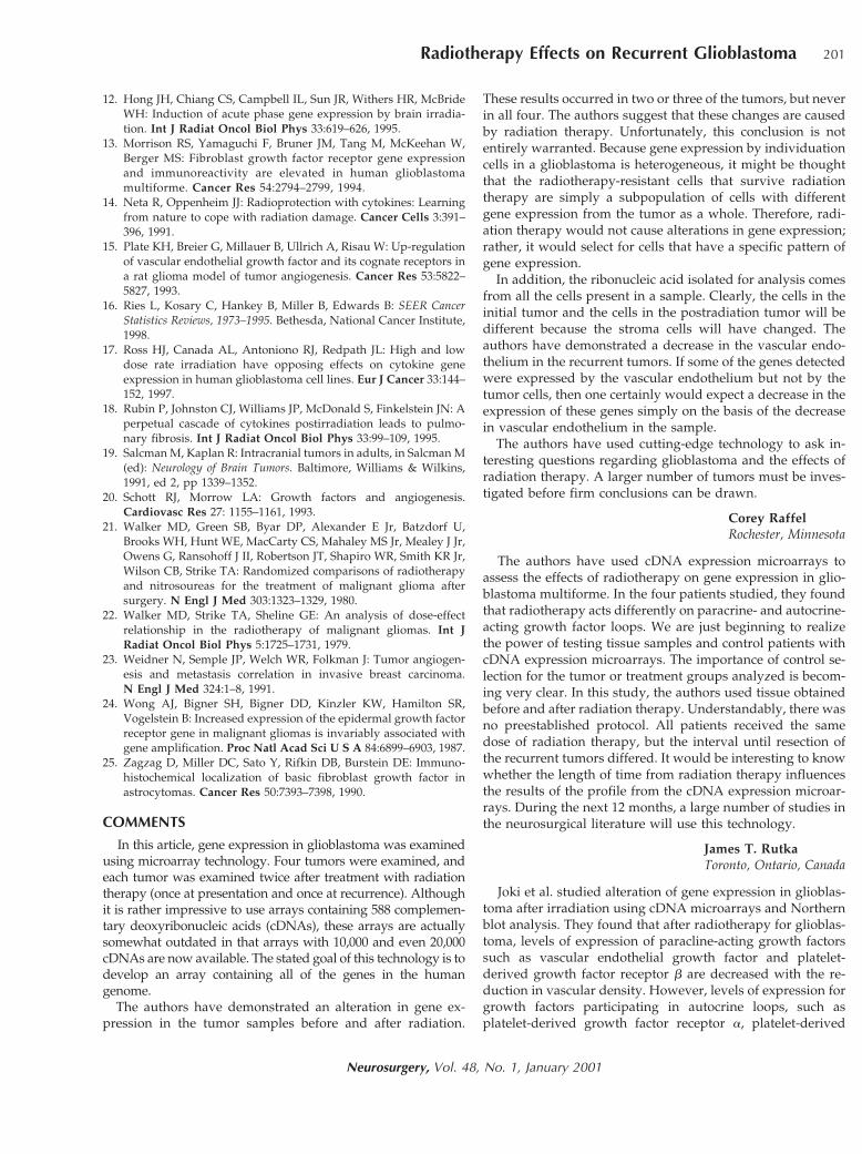
Immunohistochemistry was performed using 4- to 6-mmparaffin tissue sections of tumor specimens for an antibodyagainst von Willebrand factor (vWF) to assess vascular den-sity. Slides were deparaffinized, and endogenous peroxidaseactivity was blocked by incubation in 1% H2O2 in 13phosphate-buffered saline for 30 minutes. Antigen retrievalwas performed by incubating slides in 10 mmol/L sodiumcitrate, pH 6.0, at 100°C for 30 minutes. A mouse monoclonalantibody against vWF (DAKO, Carpinteria, CA) was thenapplied at a 1:250 dilution overnight at 4°C. After the slideswere rinsed with 13 PBS, a biotinylated secondary immuno-globulin G antibody was applied for 30 minutes at roomtemperature. Immunoperoxidase staining was performed us-ing the VECTASTAIN ABC Elite kit (Vector Laboratories,Burlingame, CA). Hematoxylin (Sigma Diagnostics, St. Louis,MO) was used as a counterstain. Slides stained in parallel,which were incubated with horse serum instead of primaryantibody, served as negative controls.
Microvessel count
Microvessel counts were performed as described previ-ously (23). In brief, the blood vessels were counted in three
areas in each tumor section in 1.0 mm2 2003 microscope fieldsusing an Olympus BH2 microscope (Olympus Optical Co.,Tokyo, Japan) on vWF-stained tissue sections. Vascular den-sity was determined by calculating an average number ofvessels in the three most vascular areas. A neuropathologistperformed all microvessel counts.
RESULTS
cDNA microarrays
We assessed levels of gene expression in primary tumorand postradiation recurrent tumor specimens from patientswho had not received chemotherapy. Of the 588 cDNA frag-ments spotted on the Atlas human expression microarray, 172were detectable (Fig. 1); the remaining 416 were undetectableeven after exposure to BioMax film for 72 hours. We nar-rowed our analysis of the 172 detectable genes to the 13 genesfor which the levels of expression varied in at least two of fourpatients (Table 2). As indicated in Table 2, the levels of expres-sion in 12 of these 13 genes were decreased in the postradia-tion recurrent tumors as compared with the primary tumors.Of these 12 genes, two are oncogenes (angiopoietic 1 receptorprecursor, proto-oncogene r-A multidrug resistance protein),one is a signal transduction modulator (ras-related proteinRAB-5A), and three are transcription factors (SNF2L1, DB1,FUSE-binding protein). The remaining six genes in whichlevels of expression were decreased in recurrent tumors wereeither growth factors or growth factor receptors: PDGFRa,pro-PDGFRb, VEGF, pleiotrophin precursor/osteoblast-specific factor 1, and migration-inhibitory factor-related pro-tein 8. Only 2 of 13 genes demonstrated higher levels ofexpression in recurrent tumors as compared with primarytumors: PDGFRa and epidermal growth factor receptor. Ad-ditional studies using Northern blot analysis were performedon genes that have been demonstrated to be involved inglioma etiology and biology.
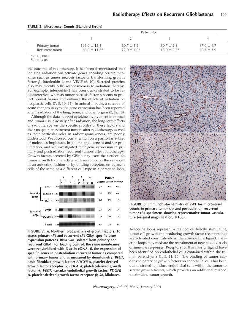
Northern blot analysis
Northern blot analysis was performed to confirm differ-ences in gene expression between primary and postradiationrecurrent tumors demonstrated by cDNA microarray forVEGF, PDGFRb, PDGFRa, and PDGF A. bFGF has beenstrongly implicated in glioma biology, but it is not repre-sented on the cDNA array membrane. Northern blot analysisalso was performed to assess the levels of expression of thisgrowth factor (Fig. 2). A marked decrease in VEGF and PDGFRbexpression in postradiation recurrent tumors as compared withprimary tumors was observed in three of four patients and intwo of four patients, respectively. In the same patients, similarresults in the expression of these genes were demonstrated bycDNA microarray analysis. Differences in expression ofPDGFRa in primary and postradiation recurrent tumors as dem-onstrated by Northern blot analysis also mirrored the resultsfrom microarray analysis; mRNA levels were decreased in pos-tradiation recurrent tumors in Patients 1 and 2, and they wereincreased from primary to recurrent tumors in Patients 3 and 4.Levels of PDGF A, as determined by Northern blot analysis,
Radiotherapy Effects on Recurrent Glioblastoma 197
Neurosurgery, Vol. 48, No. 1, January 2001

were increased in recurrent tumors as compared with primarytumors in three of four patients; no difference was noted in onepatient. A significant increase in bFGF expression in postradia-tion specimens was observed on Northern blot analysis in threeof four patients.
Microvessel count
We performed microvessel counts on 4- to 6-mm paraffinvWF-stained tissue sections (Fig. 3) from each patient to assessthe effect of radiation on blood vessel growth in postradiationrecurrent tumors. In three of four patients, microvessel countswere significantly lower in recurrent tumors after radiother-apy as compared with primary tumors (Patients 1 and 3, P #0.001; Patient 2, P # 0.005) (Table 3). Microvessel counts alsowere decreased, although not significantly, in Patient 4.
DISCUSSION
In this study, we used cDNA microarray technology toinvestigate the biological differences between primary tumorsand postradiation recurrent tumors after radiotherapy. Thistechnique permitted the screening of a large number of genessimultaneously. The ability to assess global levels of geneexpression in primary and postradiation recurrent tumors inparallel fashion served as a powerful comparative tool. Al-though this analytical approach is useful in the number ofcDNAs that may be screened, the method requires moresensitivity to lower levels of tissue mRNA. In addition, ouranalysis was limited by the low quantities of mRNA availablefor these studies; very few patients fit the inclusion criteria forthis study, because the overwhelming majority of brain tumorpatients receive chemotherapy as well as radiotherapy.
A variety of growth factors and their receptors that areimportant for GBM development have been identified (1, 5,11, 13, 15, 24, 25). These include proteins such as endothelialgrowth factor, bFGF, and VEGF, which not only can promoteneoplastic growth (20) but also may play significant roles in
FIGURE 1. Expression patterns of genes in primary GBM (A)and postradiation recurrent GBM tissue (B) of Patient 3. Differ-ential hybridization of two identical human cDNA expressionarrays was performed as described in Patients and Methods. A,the expression array membrane hybridized with cDNA fromprimary GBM. B, the expression array membrane hybridizedwith cDNA from postradiation recurrent GBM in the samepatient. Classes of genes and selected individual genes on themembranes are indicated. The expression of housekeepinggenes such as ubiquitin and b-actin served as control. 1, onco-genes and tumor suppressors; 2, cell-cycle control proteins; 3,modulators/effectors/intracellular transducers; 4, stress responseproteins; 5, apoptosis-associated proteins; 6, DNA synthesis/repair/recombination proteins; 7, DNA-binding proteins/transcription factors; 8, cell receptors and cell surface antigens;and 9, extracellular cell-signaling proteins. EGFR, epidermalgrowth factor receptor; PDGFR-a, platelet-derived growth fac-tor receptor a; PDGFR-b, platelet-derived growth factor recep-tor b; VEGF, vascular endothelial growth factor; PDGF-A;platelet-derived growth factor A.
TABLE 2. Selected Genes That Were Differentially Expressedin at Least Two of Four Patients
Gene Decrease Increase
Angiopoietin 1 2/4Proto-oncogene r-A multidrug
resistance protein3/4
Ras-related protein RAB-5A 2/4Protein-tyrosine phosphatase z
precursor2/4
Global transcription activatorSNF2L1
2/4
Putative transcription activator DB1 2/4FUSE-binding protein 2/4Platelet-derived growth factor
receptor a2/4 2/4
Platelet-derived growth factorreceptor b precursor
2/4
Heparin-binding vascularendothelial growth factor
3/4
Pleiotrophin precursor 1osteoblast-specific factor 1
2/4
Migration-inhibitory factor-relatedprotein 8
2/4
Epidermal growth factor receptor 2/4
198 Joki et al.
Neurosurgery, Vol. 48, No. 1, January 2001
the outcome of radiotherapy. It has been demonstrated thationizing radiation can activate genes encoding certain cyto-kines such as tumor necrosis factor a, transforming growthfactor b, interleukin-1, and VEGF (6, 10). Secreted proteinsalso may modify cells’ responsiveness to radiation therapy.For example, interleukin-1 has been demonstrated to be ra-dioprotective, whereas tumor necrosis factor a seems to pro-tect normal tissues and enhance the effects of radiation onneoplastic cells (7, 8, 10, 14). In animal models, a cascade ofacute changes in cytokine gene expression has been reportedafter irradiation of the lung, brain, and other organs (3, 12, 18).
Although the data support cytokine involvement in normaland tumor tissue acutely after radiation, the long-term effectsof radiotherapy on the specific profiles of these factors andtheir receptors in recurrent tumors after radiotherapy, as wellas their particular roles in radioresponsiveness, are poorlyunderstood. We focused our attention on a particular subsetof molecules implicated in glioma angiogenesis and/or pro-liferation, and we investigated their gene expression in pri-mary and postradiation recurrent tumors after radiotherapy.Growth factors secreted by GBMs may exert their effects ontumor growth by interacting with receptors on the same cellin an autocrine fashion or by binding receptors on adjacentcells of the same or a different cell type in a paracrine loop.
Autocrine loops represent a method of directly stimulatingtumor cell growth and producing growth factor receptors thatare activated constitutively in the absence of a ligand. Para-crine loops may mediate the recruitment of new blood vesselsor immune responses. Receptors for this class of ligand havebeen identified on endothelial cells contained within the tu-mor parenchyma (1, 5, 11, 15). The binding of tumor cell-derived paracrine growth factors on endothelial cells has beendemonstrated to induce endothelial cells within the tumor tosecrete growth factors, which provides an additional methodto stimulate tumor growth.
FIGURE 2. A, Northern blot analysis of growth factors. Toassess primary (P ) and recurrent (R ) GBM-specific geneexpression patterns, RNA was isolated from primary andrecurrent GBM. For loading control, the same membraneswere rehybridized with b-actin cDNA. B, the expression ofspecific genes in postradiation recurrent tumor as comparedwith primary tumor and as measured by densitometry. BFGF,basic fibroblast growth factor; PDGFR a, platelet-derivedgrowth factor receptor a; PDGF A; platelet-derived growthfactor A; VEGF, vascular endothelial growth factor; PDGFRb, platelet-derived growth factor receptor b; kb, kilobases.
FIGURE 3. Immunohistochemistry of vWF for microvesselcounts in primary tumor (A) and postradiation recurrenttumor (B ) specimens showing representative tumor vascula-ture (original magnification, 3100).
TABLE 3. Microvessel Counts (Standard Errors)
Patient No.
1 2 3 4
Primary tumor 196.0 6 12.1 60.7 6 1.2 80.7 6 2.3 87.0 6 4.7Recurrent tumor 66.0 6 11.6a 22.0 6 4.9b 15.0 6 2.6a 70.3 6 3.9
a P # 0.001.b P # 0.005.
Radiotherapy Effects on Recurrent Glioblastoma 199
Neurosurgery, Vol. 48, No. 1, January 2001

Among the four patients studied, the average time of re-currence after radiotherapy was 7.25 months, which allowedus to investigate long-term rather than acute effects of radia-tion on the expression of particular genes in malignant glio-mas. Of interest, growth factors and/or receptors that exerttheir biological effects in a paracrine fashion were associatedwith decreased levels of expression in recurrent postradiationtumors as compared with primary tumors. In three of fourpatients, VEGF levels as determined by cDNA microarray andNorthern blot analysis were decreased in recurrent tumors ascompared with primary tumors. In two of four patients,cDNA microarray analysis demonstrated that the expressionof PDGFRb decreased from primary to recurrent tumors andwas unchanged in the remaining patients. Northern blot anal-ysis revealed that PDGFRb levels decreased in three of fourpatients, and the levels of expression remained the same inprimary and postradiation recurrent tumors in one patient.
Although paracrine factors such as VEGF and PDGFRbdemonstrated decreased levels of expression in tumor speci-mens after radiotherapy, proteins that participate in autocrineloops were associated with increased levels of expression inpostradiation recurrent tumors as compared with primarytumors. Northern blot analysis revealed that PDGF A, whichwas undetectable by cDNA microarray analysis, was up-regulated in postradiation recurrent tumors in three of fourpatients, as was bFGF. PDGFRa also was up-regulated in twoof four patients as demonstrated by Northern blot and cDNAmicroarray analysis. Furthermore, epidermal growth factorreceptor, which has been strongly implicated in GBM patho-genesis, was expressed more strongly in postradiation recur-rent tumors, with levels of expression that were increased intwo of four patients and unchanged in the remaining twopatients.
CONCLUSION
The differences in microvessel density between primaryand postradiation recurrent tumors mirrored the decreasedexpression of paracrine-acting growth factors. In all patients,microvessel count was decreased. Taken together, these datasuggest that radiotherapy of primary GBMs is capable of induc-ing long-term changes in the expression of particular genes.Furthermore, our results suggest that although autocrine- andparacrine-acting growth factors are intact in the primary tumor,they are differentially affected by radiotherapy. In recurrenttumors after radiotherapy, proteins participating in autocrineloops were up-regulated, indicating growth and proliferation ofradioresistant tumor cells. However, paracrine factors weredown-regulated or unchanged, which may be explained by aconcomitant decrease in a sizable fraction of tumor along withvulnerable endothelial cells. Consistent with a down-regulationof paracrine factors such as VEGF and PDGFRb in recurrenttumors, microvessel growth, a process fueled by intratumoralparacrine action, also was significantly reduced. Therefore, re-currence and the clinical response to radiation in GBMs may bedetermined, at least in part, by tumor-derived cytokines thatenhance growth in an autocrine manner, as well as by intrinsictumor radiosensitivity.
ACKNOWLEDGMENTS
We thank the Brain Tumour Tissue Bank of the NationalCancer Institute of Canada, London Regional Cancer Centre,for supplying glioblastoma tissue and Dr. Rebecca Folkerthfor serving as the independent neuropathologist on thisproject. We thank Dr. Judith Abraham, Dr. Tucker Collins, Dr.Daniel Pope-Bowen, and Dr. N. Ferrara for providing cDNAprobes. This work was supported by a grant from the BostonNeurosurgical Foundation. TJ is a recipient of a fellowshipfrom Jikei University School of Medicine.
Received, March 15, 2000.Accepted, September 11, 2000.Reprint requests: Rona S. Carroll, Ph.D., Brigham and Women’sHospital, Division of Neurosurgery, Harvard Medical School, 221Longwood Avenue, LMRC Room 121, Boston, MA 02115. Email:[email protected]
REFERENCES
1. Berkman RA, Merrill MJ, Reinhold WC, Monacci WT, Saxena A,Clark WC, Robertson JT, Ali IU, Oldfield EH: Expression of thevascular permeability factor/vascular endothelial growth factorgene in central nervous system neoplasms. J Clin Invest 91:153–159, 1993.
2. Central Brain Tumor Registry of the United States: CBTRUS 1997Annual Report. The Central Brain Tumor Registry of the UnitedStates, 1998.
3. Chiang CS, McBride WH, Withers HR: Radiation-inducedastrocytic and microglial responses in mouse brain. RadiotherOncol 29:60–68, 1993.
4. Chirgwin JM, Przybyla AE, MacDonald RJ, Rutter WJ: Isolationof biologically active ribonucleic acid from sources enriched inribonuclease. Biochemistry 18:5294–5299, 1979.
5. Fleming TP, Saxena A, Clark WC, Robertson JT, Oldfield EH,Aaronson SA, Ali IU: Amplification and/or overexpression ofplatelet-derived growth factor receptors and epidermal growthfactor receptor in human glial tumors. Cancer Res 52:4550–4553,1992.
6. Gorski DH, Beckett MA, Jaskowiak NT, Calvin DP, Mauceri HJ,Salloum RM, Seetharam S, Koons A, Hari DM, Kufe DW,Weichselbaum RR: Blockage of the vascular endothelial growthfactor stress response increases the antitumor effects of ionizingradiation. Cancer Res 59:3374–3378, 1999.
7. Gridley DS, Glisson WC, Uhm JR: Interaction of tumour necrosisfactor-alpha and radiation against human colon tumour cells.Ther Immunol 1:25–31, 1994.
8. Gridley DS, Hammond SN, Liwnicz BH: Tumor necrosis factor-alpha augments radiation effects against human colon tumorxenografts. Anticancer Res 14:1107–1112, 1994.
9. Gridley DS, Loredo LN, Slater JD, Archambeau JO, Bedros AA,Andres ML, Slater JM: Pilot evaluation of cytokine levels inpatients undergoing radiotherapy for brain tumor. Cancer DetectPrev 22:20–29, 1998.
10. Hallahan DE, Haimovitz-Friedman A, Kufe DW, Fuks Z,Weichselbaum RR: The role of cytokines in radiation oncology.Important Adv Oncol 71–80, 1993.
11. Hermansson M, Nister M, Betsholtz C, Heldin CH, Westermark B,Funa K: Endothelial cell hyperplasia in human glioblastoma:Coexpression of mRNA for platelet-derived growth factor(PDGF) B chain and PDGF receptor suggests autocrine growthstimulation. Proc Natl Acad Sci U S A 85:7748–7752, 1988.
200 Joki et al.
Neurosurgery, Vol. 48, No. 1, January 2001
12. Hong JH, Chiang CS, Campbell IL, Sun JR, Withers HR, McBrideWH: Induction of acute phase gene expression by brain irradia-tion. Int J Radiat Oncol Biol Phys 33:619–626, 1995.
13. Morrison RS, Yamaguchi F, Bruner JM, Tang M, McKeehan W,Berger MS: Fibroblast growth factor receptor gene expressionand immunoreactivity are elevated in human glioblastomamultiforme. Cancer Res 54:2794–2799, 1994.
14. Neta R, Oppenheim JJ: Radioprotection with cytokines: Learningfrom nature to cope with radiation damage. Cancer Cells 3:391–396, 1991.
15. Plate KH, Breier G, Millauer B, Ullrich A, Risau W: Up-regulationof vascular endothelial growth factor and its cognate receptors ina rat glioma model of tumor angiogenesis. Cancer Res 53:5822–5827, 1993.
16. Ries L, Kosary C, Hankey B, Miller B, Edwards B: SEER CancerStatistics Reviews, 1973–1995. Bethesda, National Cancer Institute,1998.
17. Ross HJ, Canada AL, Antoniono RJ, Redpath JL: High and lowdose rate irradiation have opposing effects on cytokine geneexpression in human glioblastoma cell lines. Eur J Cancer 33:144–152, 1997.
18. Rubin P, Johnston CJ, Williams JP, McDonald S, Finkelstein JN: Aperpetual cascade of cytokines postirradiation leads to pulmo-nary fibrosis. Int J Radiat Oncol Biol Phys 33:99–109, 1995.
19. Salcman M, Kaplan R: Intracranial tumors in adults, in Salcman M(ed): Neurology of Brain Tumors. Baltimore, Williams & Wilkins,1991, ed 2, pp 1339–1352.
20. Schott RJ, Morrow LA: Growth factors and angiogenesis.Cardiovasc Res 27: 1155–1161, 1993.
21. Walker MD, Green SB, Byar DP, Alexander E Jr, Batzdorf U,Brooks WH, Hunt WE, MacCarty CS, Mahaley MS Jr, Mealey J Jr,Owens G, Ransohoff J II, Robertson JT, Shapiro WR, Smith KR Jr,Wilson CB, Strike TA: Randomized comparisons of radiotherapyand nitrosoureas for the treatment of malignant glioma aftersurgery. N Engl J Med 303:1323–1329, 1980.
22. Walker MD, Strike TA, Sheline GE: An analysis of dose-effectrelationship in the radiotherapy of malignant gliomas. Int JRadiat Oncol Biol Phys 5:1725–1731, 1979.
23. Weidner N, Semple JP, Welch WR, Folkman J: Tumor angiogen-esis and metastasis correlation in invasive breast carcinoma.N Engl J Med 324:1–8, 1991.
24. Wong AJ, Bigner SH, Bigner DD, Kinzler KW, Hamilton SR,Vogelstein B: Increased expression of the epidermal growth factorreceptor gene in malignant gliomas is invariably associated withgene amplification. Proc Natl Acad Sci U S A 84:6899–6903, 1987.
25. Zagzag D, Miller DC, Sato Y, Rifkin DB, Burstein DE: Immuno-histochemical localization of basic fibroblast growth factor inastrocytomas. Cancer Res 50:7393–7398, 1990.
COMMENTS
In this article, gene expression in glioblastoma was examinedusing microarray technology. Four tumors were examined, andeach tumor was examined twice after treatment with radiationtherapy (once at presentation and once at recurrence). Althoughit is rather impressive to use arrays containing 588 complemen-tary deoxyribonucleic acids (cDNAs), these arrays are actuallysomewhat outdated in that arrays with 10,000 and even 20,000cDNAs are now available. The stated goal of this technology is todevelop an array containing all of the genes in the humangenome.
The authors have demonstrated an alteration in gene ex-pression in the tumor samples before and after radiation.
These results occurred in two or three of the tumors, but neverin all four. The authors suggest that these changes are causedby radiation therapy. Unfortunately, this conclusion is notentirely warranted. Because gene expression by individuationcells in a glioblastoma is heterogeneous, it might be thoughtthat the radiotherapy-resistant cells that survive radiationtherapy are simply a subpopulation of cells with differentgene expression from the tumor as a whole. Therefore, radi-ation therapy would not cause alterations in gene expression;rather, it would select for cells that have a specific pattern ofgene expression.
In addition, the ribonucleic acid isolated for analysis comesfrom all the cells present in a sample. Clearly, the cells in theinitial tumor and the cells in the postradiation tumor will bedifferent because the stroma cells will have changed. Theauthors have demonstrated a decrease in the vascular endo-thelium in the recurrent tumors. If some of the genes detectedwere expressed by the vascular endothelium but not by thetumor cells, then one certainly would expect a decrease in theexpression of these genes simply on the basis of the decreasein vascular endothelium in the sample.
The authors have used cutting-edge technology to ask in-teresting questions regarding glioblastoma and the effects ofradiation therapy. A larger number of tumors must be inves-tigated before firm conclusions can be drawn.
Corey RaffelRochester, Minnesota
The authors have used cDNA expression microarrays toassess the effects of radiotherapy on gene expression in glio-blastoma multiforme. In the four patients studied, they foundthat radiotherapy acts differently on paracrine- and autocrine-acting growth factor loops. We are just beginning to realizethe power of testing tissue samples and control patients withcDNA expression microarrays. The importance of control se-lection for the tumor or treatment groups analyzed is becom-ing very clear. In this study, the authors used tissue obtainedbefore and after radiation therapy. Understandably, there wasno preestablished protocol. All patients received the samedose of radiation therapy, but the interval until resection ofthe recurrent tumors differed. It would be interesting to knowwhether the length of time from radiation therapy influencesthe results of the profile from the cDNA expression microar-rays. During the next 12 months, a large number of studies inthe neurosurgical literature will use this technology.
James T. RutkaToronto, Ontario, Canada
Joki et al. studied alteration of gene expression in glioblas-toma after irradiation using cDNA microarrays and Northernblot analysis. They found that after radiotherapy for glioblas-toma, levels of expression of paracline-acting growth factorssuch as vascular endothelial growth factor and platelet-derived growth factor receptor b are decreased with the re-duction in vascular density. However, levels of expression forgrowth factors participating in autocrine loops, such asplatelet-derived growth factor receptor a, platelet-derived
Radiotherapy Effects on Recurrent Glioblastoma 201
Neurosurgery, Vol. 48, No. 1, January 2001

growth factor receptor A, and basic fibroblast growth factor,were increased in postradiation recurrent tumors as com-pared with the primary tumor.
It is our clinical experience, as well as that of others, thatrecurrent glioblastomas are different from primary tumorswith regard to growth behavior and response to therapy. Ithas been speculated that postoperative radiotherapy and che-motherapy induce genetic alterations in the tumor. It also hasbeen suggested that radiotherapy facilitates malignant trans-
formation of low-grade gliomas. This study beautifully dem-onstrates that radiotherapy is responsible for the biologicalalteration of glioblastomas. Although difficult to carry out,this type of study is important to understanding the patho-physiology of recurrent glioblastoma and improving the treat-ment of this malignant tumor.
Yukitaka UshioKumamoto, Japan
202 Joki et al.