ascites associate professor dr meltem ergun yeditepe university department of gastroenterology
TRANSCRIPT
AscitesAssociate Professor Dr Meltem Ergun
Yeditepe University
Department of Gastroenterology
Points of this lecture
What is ascites? Etiologies of ascites Clinical presentations Pyhsical examination findings Laboratory tests Prognosis Spontaneous bacterial peritonitis
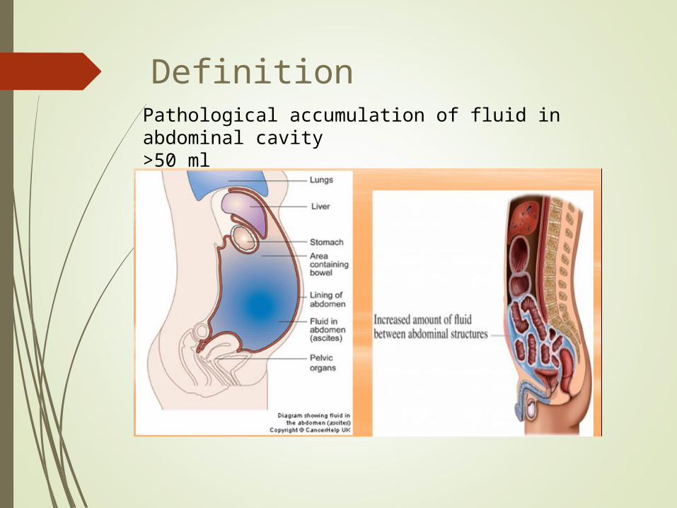
Pathological accumulation of fluid in abdominal cavity>50 ml
Definition
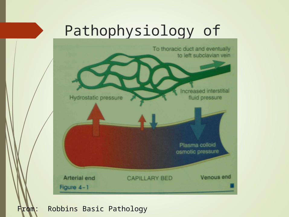
Pathophysiology of Ascites
From: Robbins Basic Pathology
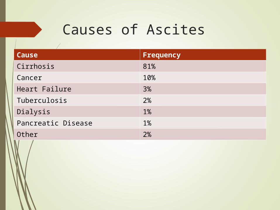
Causes of Ascites
Cause Frequency
Cirrhosis 81%
Cancer 10%
Heart Failure 3%
Tuberculosis 2%
Dialysis 1%
Pancreatic Disease 1%
Other 2%
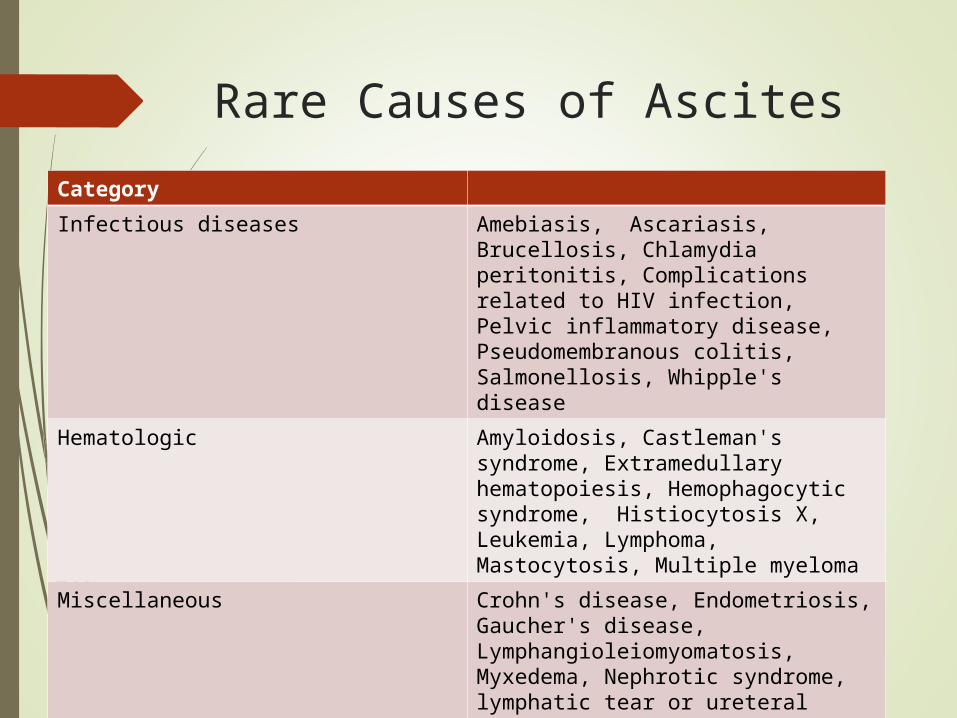
Rare Causes of Ascites
Category
Infectious diseases Amebiasis, Ascariasis, Brucellosis, Chlamydia peritonitis, Complications related to HIV infection, Pelvic inflammatory disease, Pseudomembranous colitis, Salmonellosis, Whipple's disease
Hematologic Amyloidosis, Castleman's syndrome, Extramedullary hematopoiesis, Hemophagocytic syndrome, Histiocytosis X, Leukemia, Lymphoma, Mastocytosis, Multiple myeloma
Miscellaneous Crohn's disease, Endometriosis, Gaucher's disease, Lymphangioleiomyomatosis, Myxedema, Nephrotic syndrome, lymphatic tear or ureteral injury. Ovarian hyperstimulation
Imaging
Ultrasound with Dopplers
Easily confirms ascites
May see nodularity of cirrhosis
Evaluate patency of vasculature
No radiation, contrast
CT / MRI
Evaluation for malignancy
Serum to Ascites Albumin Gradient
Is portal hypertension present?
97% accurate
SAAG > 1.1 g/dL Portal HTN
SAAG < 1.1 g/dL Other causes
The serum-ascites albumin gradient is superior to the exudate-transudate concept in the differential diagnosis of ascites. Runyon BA; Montano AA; Akriviadis EA; Antillon MR; Irving MA; McHutchison Ann Intern Med 1992 Aug 1;117(3):215-20.
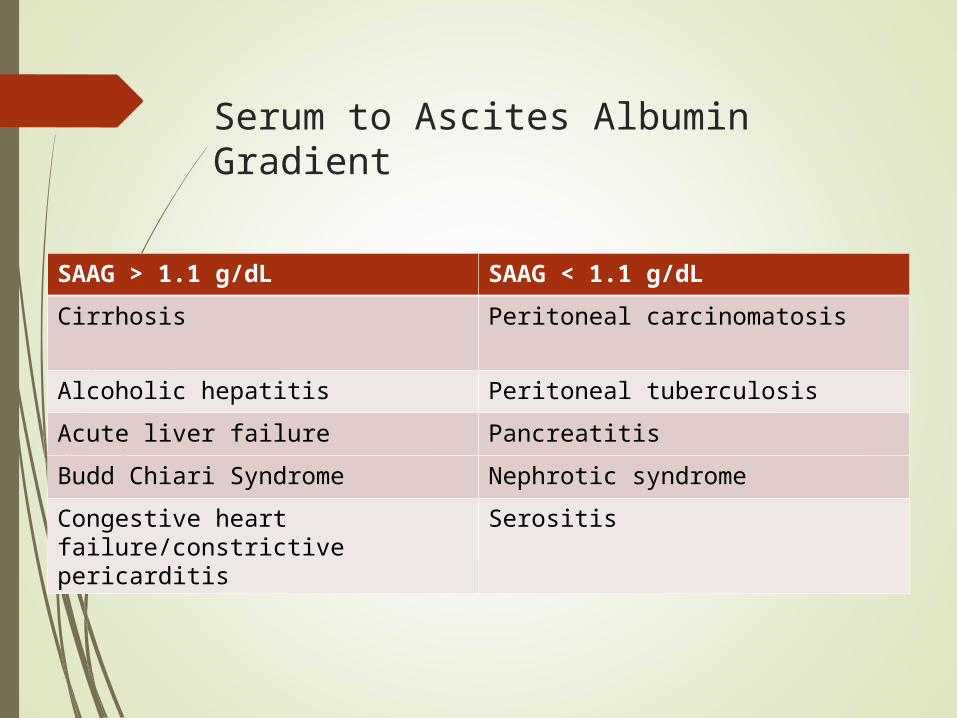
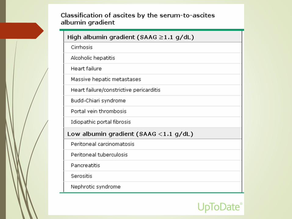
Serum to Ascites Albumin Gradient
SAAG > 1.1 g/dL SAAG < 1.1 g/dL
Cirrhosis Peritoneal carcinomatosis
Alcoholic hepatitis Peritoneal tuberculosis
Acute liver failure Pancreatitis
Budd Chiari Syndrome Nephrotic syndrome
Congestive heart failure/constrictive pericarditis
Serositis
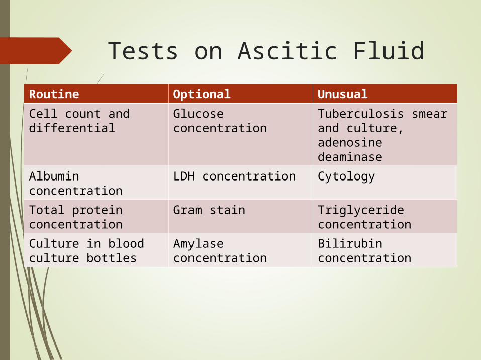
Tests on Ascitic Fluid
Routine Optional Unusual
Cell count and differential
Glucose concentration
Tuberculosis smear and culture, adenosine deaminase
Albumin concentration
LDH concentration Cytology
Total protein concentration
Gram stain Triglyceride concentration
Culture in blood culture bottles
Amylase concentration
Bilirubin concentration
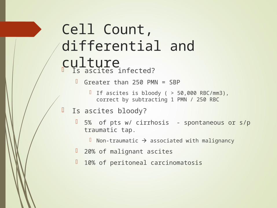
Cell Count, differential and culture
Is ascites infected?
Greater than 250 PMN = SBP
If ascites is bloody ( > 50,000 RBC/mm3), correct by subtracting 1 PMN / 250 RBC
Is ascites bloody?
5% of pts w/ cirrhosis - spontaneous or s/p traumatic tap.
Non-traumatic associated with malignancy
20% of malignant ascites
10% of peritoneal carcinomatosis
Total Protein
Exudate ( > 2.5 g/dL) or Transudate?
Supplanted by SAAG
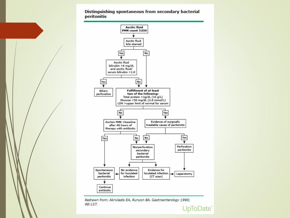
Is there gut perforation? (vs SBP)
Total protein >1 g/dL
Glucose <50 mg/dL (2.8 mmol/L)
LDH greater than serum ULN
Glucose and LDH
Consistent with infection or malignancy?
Infection and cancer consume glucoselow
LDH is a larger molecule than glucose, enters ascitic fluid with difficulty.
Ascitis/Serum LDH ratio
~ 0.4 in cirrhotic ascites
Approaches 1.0 in SBP
>1.0, usually infection or tumor
Other tests Amylase
Uncomplicated cirrhotic ascites
About 40 IU/L. The AF/S ratio is about 0.4
Pancreatic ascites
About 2000 IU/L. The AF/S ratio is about 6
Triglycerides — run on milky fluid.
Chylous ascites - TG > 200 mg/dL, usually 1000 mg/dL
Bilirubin — run on brown ascites.
Biliary perforation – AF Bili > serum Bili
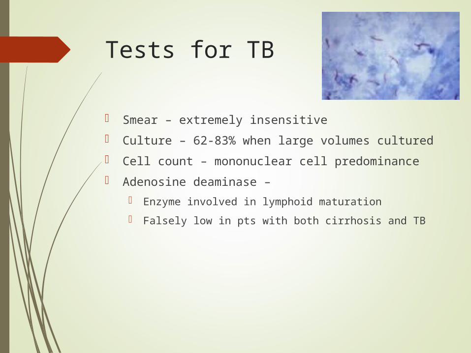
Tests for TB
Smear – extremely insensitive
Culture – 62-83% when large volumes cultured
Cell count – mononuclear cell predominance
Adenosine deaminase –
Enzyme involved in lymphoid maturation
Falsely low in pts with both cirrhosis and TB
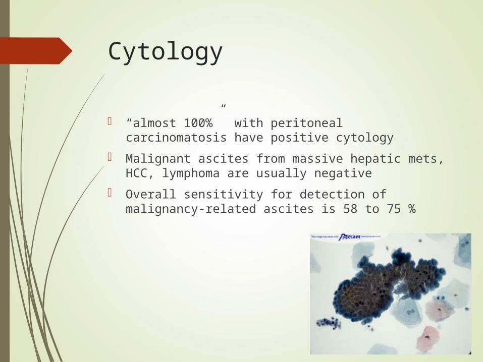
Cytology
“almost 100%” with peritoneal carcinomatosis have positive cytology
Malignant ascites from massive hepatic mets, HCC, lymphoma are usually negative
Overall sensitivity for detection of malignancy-related ascites is 58 to 75 %
Not helpful
“Some tests of ascitic fluid appear to be useless. These include pH, lactate, and ‘humoral tests of malignancy’ such as fibronectin, cholesterol, and many others”
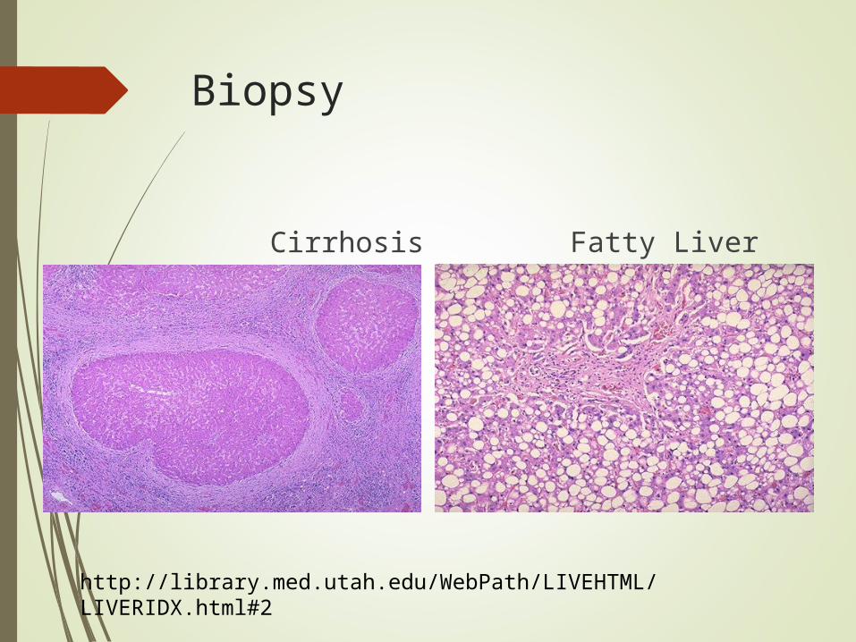
Biopsy
Cirrhosis Fatty Liver
http://library.med.utah.edu/WebPath/LIVEHTML/LIVERIDX.html#2
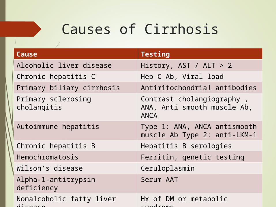
Causes of Cirrhosis
Cause Testing
Alcoholic liver disease History, AST / ALT > 2
Chronic hepatitis C Hep C Ab, Viral load
Primary biliary cirrhosis Antimitochondrial antibodies
Primary sclerosing cholangitis Contrast cholangiography , ANA, Anti smooth muscle Ab, ANCA
Autoimmune hepatitis Type 1: ANA, ANCA antismooth muscle Ab Type 2: anti-LKM-1
Chronic hepatitis B Hepatitis B serologies
Hemochromatosis Ferritin, genetic testing
Wilson’s disease Ceruloplasmin
Alpha-1-antitrypsin deficiency Serum AAT
Nonalcoholic fatty liver disease Hx of DM or metabolic syndrome
Malignant Ascites
Definition: abnormal accumulation of fluid in the peritoneal cavity as a consequence of cancer.
Commonly caused by cancers of:
Breast, bronchus, ovary, stomach, pancreas, colon
20% of cases have tumors of unknown primary
Survival poor – usually less than 3 months
Becker, G. Malignant ascites: Systematic review and guideline for treatment. European Journal of Cancer 42 (2006) 589 - 597
Malignant Ascites: Pathophysiology
Obstruction of lymphatics by tumor
Prevents absorption of fluid and protein
Alteration in vascular permeability
Hormonal mechanisms (VEGF, IL2, TNF alpha)
Decreased circulating blood volume
Activates RAAS leading to Na retention
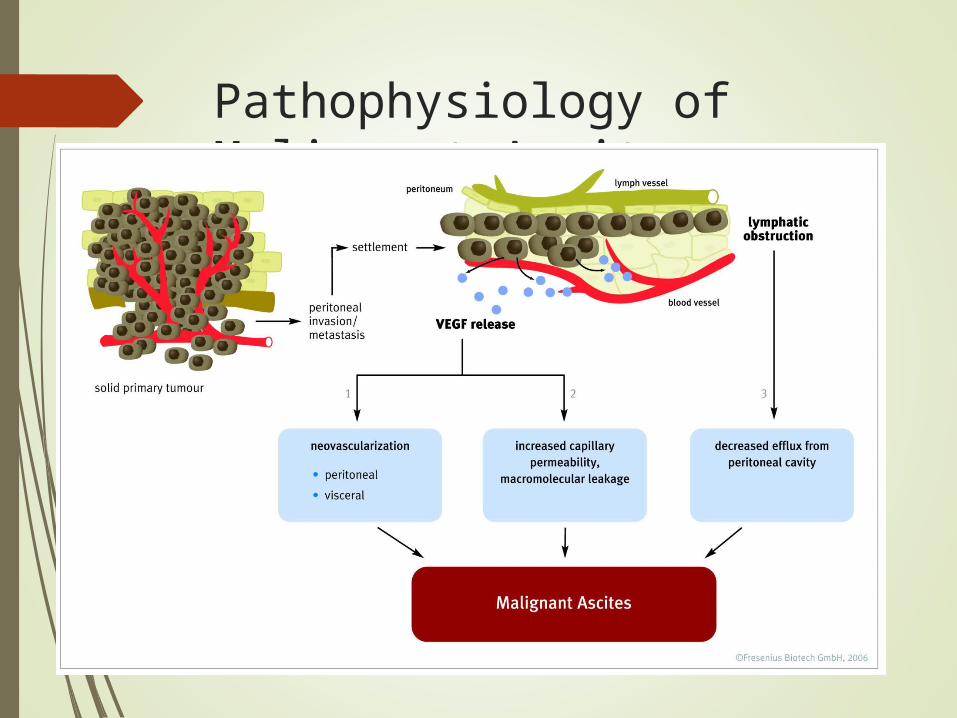
Pathophysiology of Malignant Ascites
Management of Malignant Ascites
Therapeutic paracentesis
Removing up to 5L appears safe
No good data on role of volume expanders
Diuretics
Equivocal evidence of efficacy
May be helpful for portal HTN
Less/minimally useful when no portal HTN
Drainage Catheters
Peritoneovenous shunts
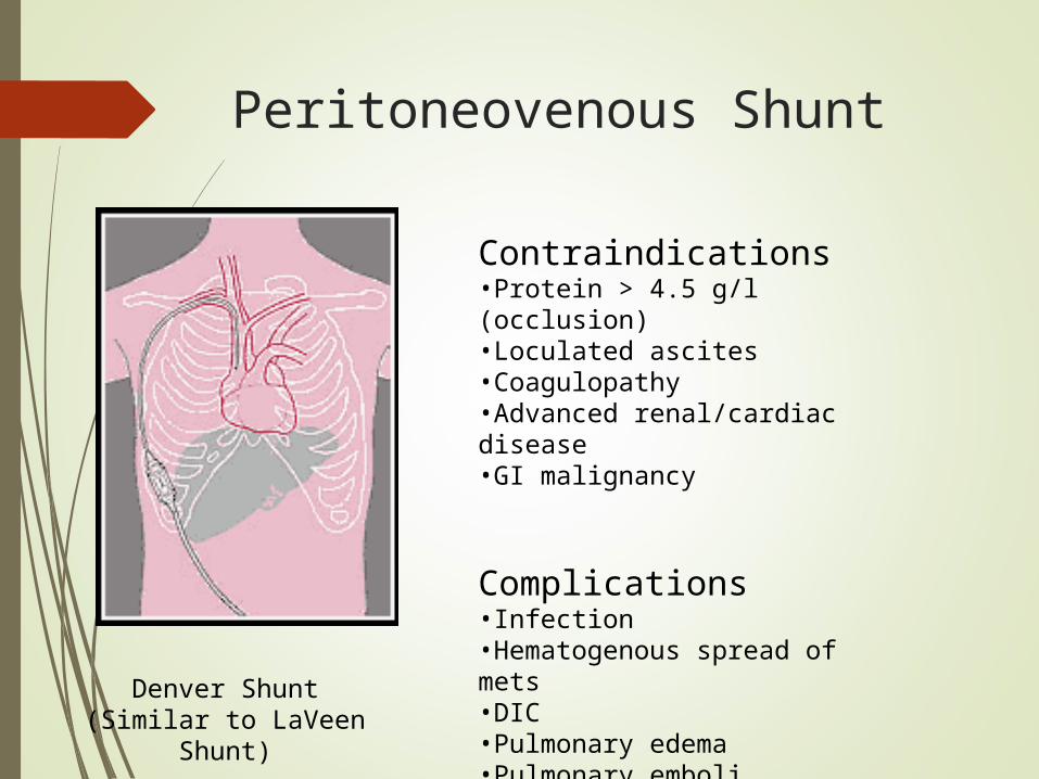
Peritoneovenous Shunt
Denver Shunt(Similar to LaVeen Shunt)
Contraindications•Protein > 4.5 g/l (occlusion)•Loculated ascites•Coagulopathy•Advanced renal/cardiac disease•GI malignancy
Complications•Infection•Hematogenous spread of mets•DIC•Pulmonary edema•Pulmonary emboli
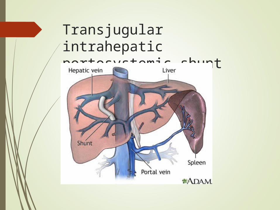
Transjugular intrahepatic portosystemic shunt (TIPS)
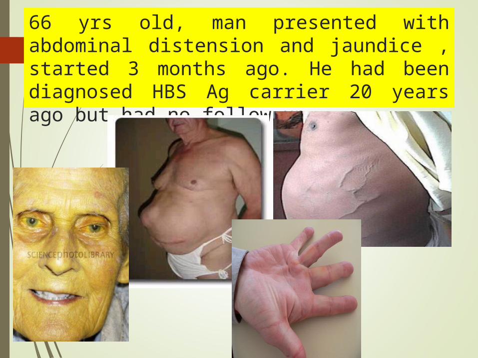
66 yrs old, man presented with abdominal distension and jaundice , started 3 months ago. He had been diagnosed HBS Ag carrier 20 years ago but had no follow up.
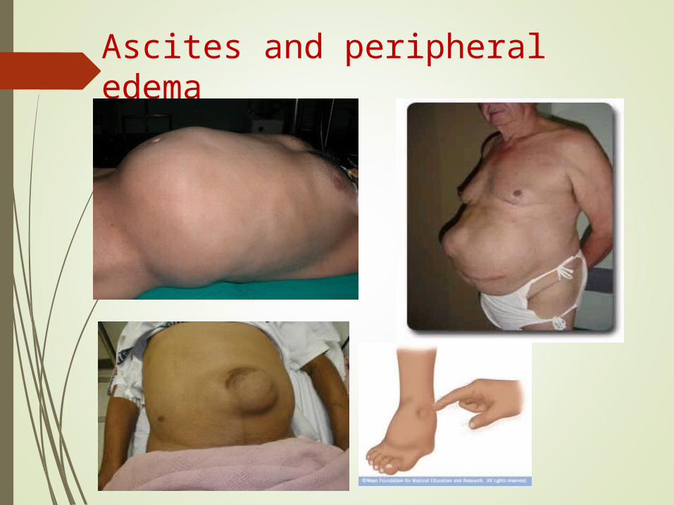
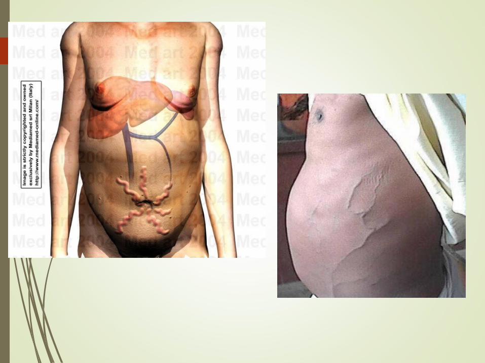
Ascites and peripheral edema
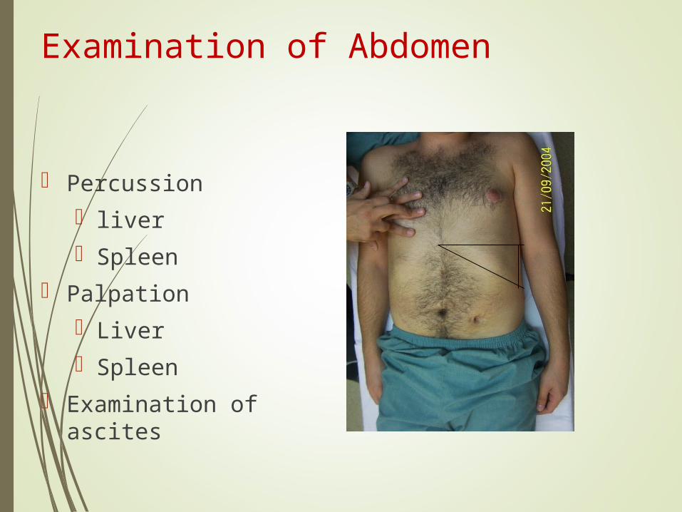
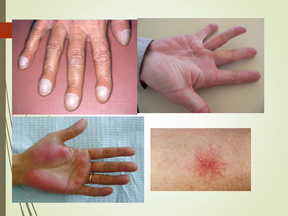
Examination of Abdomen
Percussion liver Spleen
Palpation Liver Spleen
Examination of ascites
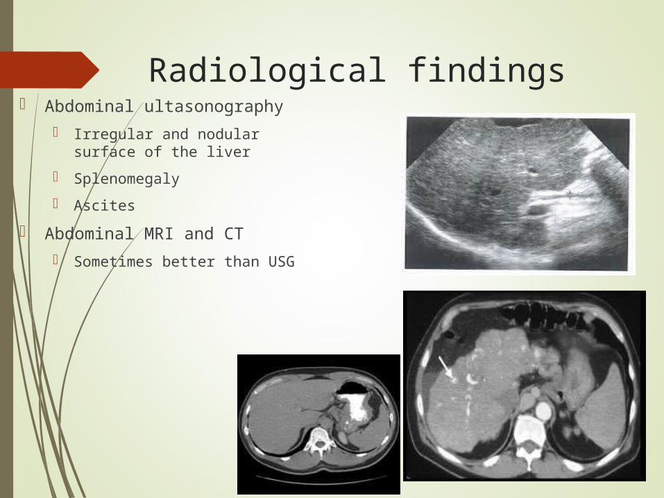
Radiological findings Abdominal ultasonography
Irregular and nodular surface of the liver
Splenomegaly
Ascites
Abdominal MRI and CT
Sometimes better than USG
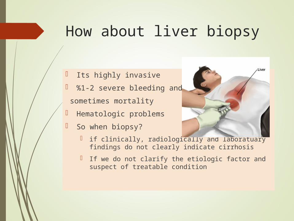
How about liver biopsy
Its highly invasive
%1-2 severe bleeding and
sometimes mortality
Hematologic problems
So when biopsy?
if clinically, radiologically and laboratuary findings do not clearly indicate cirrhosis
If we do not clarify the etiologic factor and suspect of treatable condition
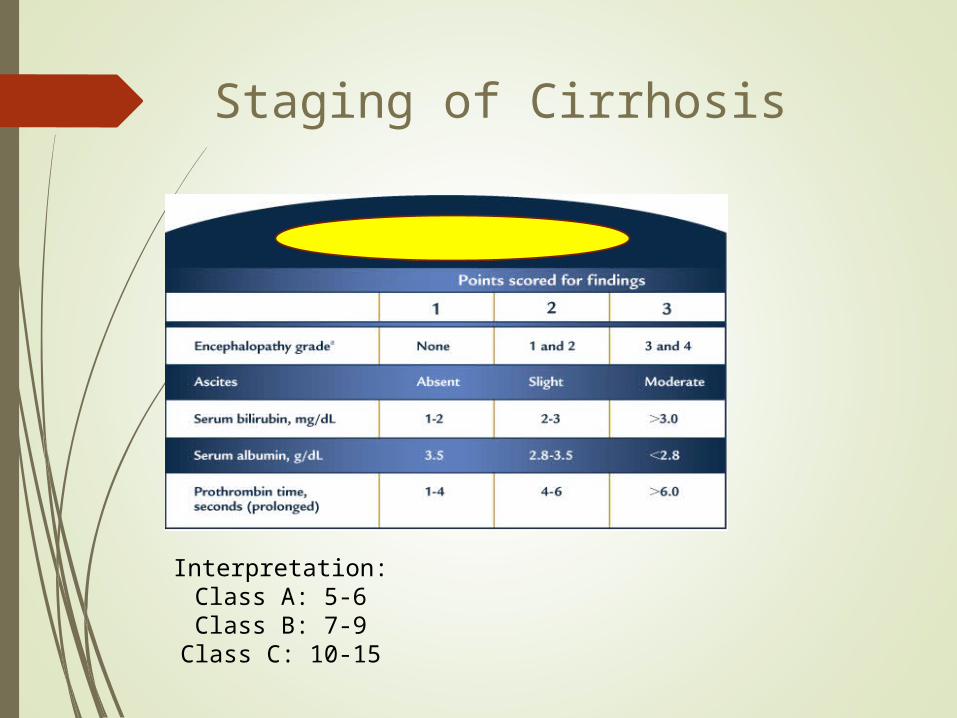
Staging of Cirrhosis
Interpretation:Class A: 5-6Class B: 7-9
Class C: 10-15
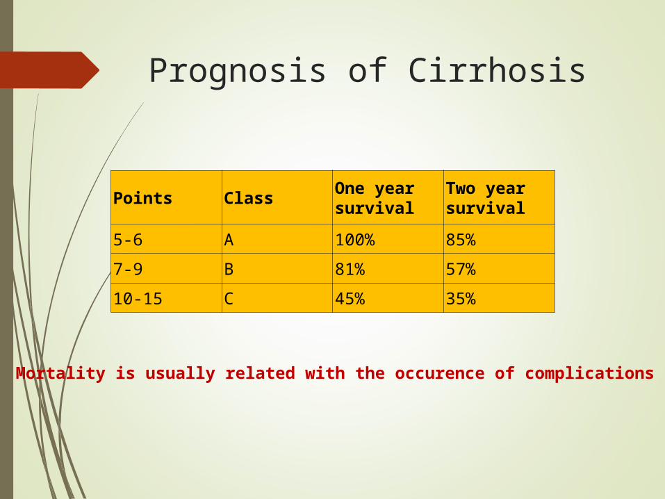
Prognosis of Cirrhosis
Points ClassOne year survival
Two year survival
5-6 A 100% 85%
7-9 B 81% 57%
10-15 C 45% 35%
Mortality is usually related with the occurence of complications
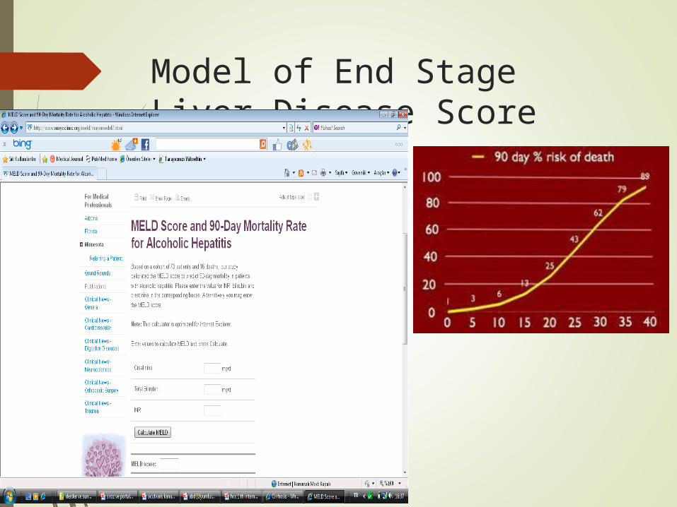
Model of End Stage Liver Disease Score (MELD)
Treatment At Early Stage Early treatment may affect prognosis
Stop alcohol drinking
Eradication of viruses
Treatment of the cause
– Autoimmune
– Wilson
– Hemachromatosis
– Sclerozing Cholangitis
Very good followup
Prevention of complications
Low Na Diet
Treatment for Ascites Diuretics (Spironolactone, furosemide)
Low sodium diet
Therapeutic parasentesis
Transplantation

Ascites; usually the first complication !!
Sometimes no symptom Abdominal distension Feeling abdominal
tenderness Treatment
Na restriction Dıuretics Terapeutic parasentesis
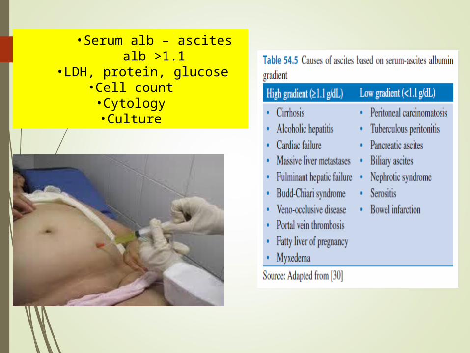
•Serum alb – ascites alb >1.1•LDH, protein, glucose
•Cell count•Cytology•Culture
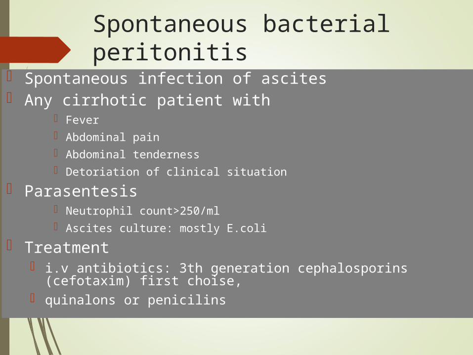
Spontaneous bacterial peritonitis
Spontaneous infection of ascites Any cirrhotic patient with
Fever Abdominal pain Abdominal tenderness Detoriation of clinical situation
Parasentesis Neutrophil count>250/ml Ascites culture: mostly E.coli
Treatment i.v antibiotics: 3th generation cephalosporins (cefotaxim) first
choise, quinalons or penicilins
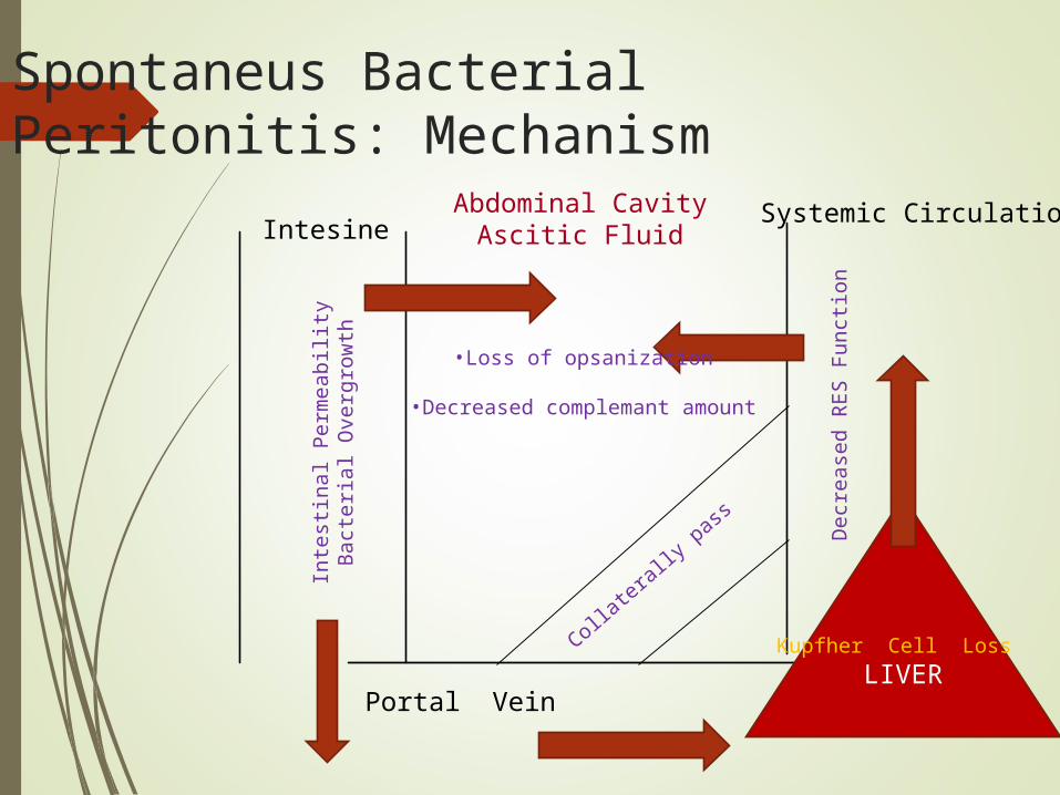
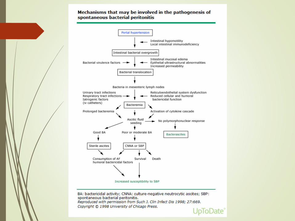
Spontaneus Bacterial Peritonitis: Mechanism
IntesineAbdominal Cavity
Ascitic Fluid
Portal VeinLIVER
Systemic Circulation
Inte
stin
al P
erm
eab
ility
Bac
teria
l Ove
rgro
wth •Loss of opsanization
•Decreased complemant amount
Kupfher Cell Loss
Dec
reas
ed R
ES
Fun
ctio
n
Collatera
lly pass
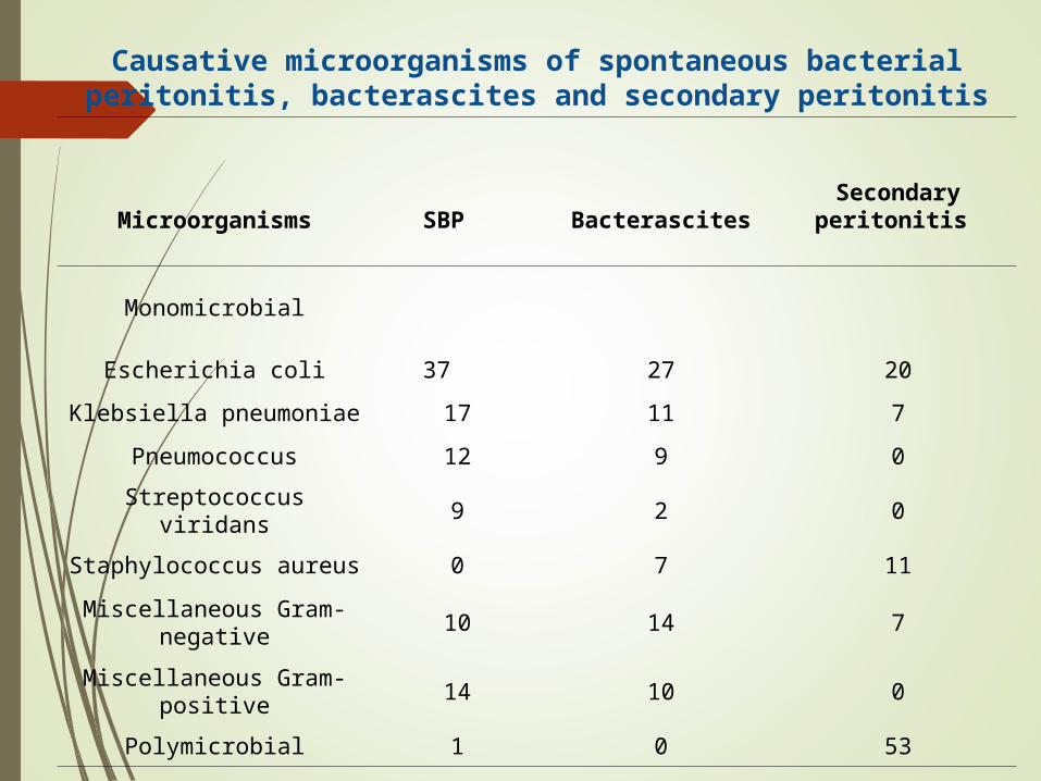
Causative microorganisms of spontaneous bacterial peritonitis, bacterascites and secondary peritonitis
Microorganisms SBP BacterascitesSecondary peritonitis
Monomicrobial
Escherichia coli 37 27 20
Klebsiella pneumoniae 17 11 7
Pneumococcus 12 9 0
Streptococcus viridans 9 2 0
Staphylococcus aureus 0 7 11
Miscellaneous Gram-negative
10 14 7
Miscellaneous Gram-positive
14 10 0
Polymicrobial 1 0 53
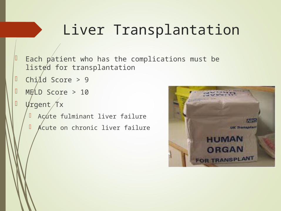
Liver Transplantation
Each patient who has the complications must be listed for transplantation
Child Score > 9
MELD Score > 10
Urgent Tx
Acute fulminant liver failure
Acute on chronic liver failure
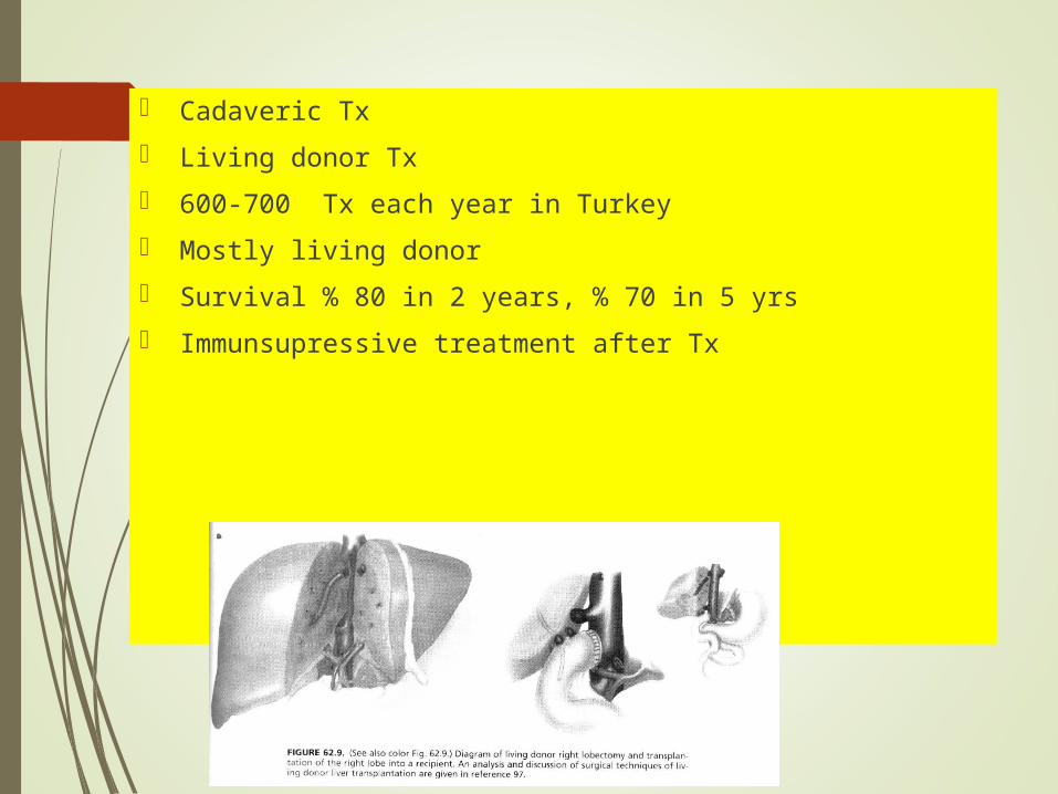
Cadaveric Tx
Living donor Tx
600-700 Tx each year in Turkey
Mostly living donor
Survival % 80 in 2 years, % 70 in 5 yrs
Immunsupressive treatment after Tx

You are free now !!!