article - university of california, los angeles · h3n2 iav viral rnas. iav/beijing/353/89 virus...
TRANSCRIPT

Article
MAP kinase p38a regulates type III interferon(IFN-l1) gene expression in human
monocyte-derived dendritic cells in responseto RNA stimulation
Miao Jiang,*,†,‡,1 Pamela Osterlund,* Riku Fagerlund,§ Diana N. Rios,§ Alexander Hoffmann,§,{
Minna M. Poranen,‡ Dennis H. Bamford,†,‡ and Ilkka Julkunen*,‖,1
*Virology Unit, Department of Infectious Disease Surveillance and Control, National Institute for Health and Welfare, Helsinki,Finland; §Department of Chemistry and Biochemistry, University of California, San Diego, La Jolla, California, USA; {Department of
Microbiology, Immunology, and Molecular Genetics, Institute for Quantitative and Computational Biosciences, University ofCalifornia, Los Angeles, California, USA; †Institute of Biotechnology and ‡Department of Biosciences, University of Helsinki,
Finland; and ‖Department of Virology, University of Turku, Finland
RECEIVED JANUARY 29, 2014; REVISED OCTOBER 22, 2014; ACCEPTED OCTOBER 23, 2014. DOI: 10.1189/jlb.2A0114-059RR
ABSTRACT
Recognition of viral nucleic acids leads to type I and type
III IFN gene expression and activation of host antiviral
responses. At present, type III IFN genes are the least
well-characterized IFN types. Here, we demonstrate that
the p38 MAPK signaling pathway is involved in regulating
IFN-l1 gene expression in response to various types of
RNA molecules in human moDCs. Inhibition of p38 MAPK
strongly reduced IFN gene expression, and overexpres-
sion of p38a MAPK enhanced IFN-l1 gene expression in
RNA-stimulated moDCs. The regulation of IFN gene
expression by p38 MAPK signaling was independent of
protein synthesis and thus, a direct result of RNA
stimulation. Moreover, the RIG-I/MDA5-MAVS-IRF3
pathway was required for p38a MAPK to up-regulate
IFN-l1 promoter activation, whereas the MyD88-IRF7
pathway was not needed, and the regulation was not
involved directly in IRF7-dependent IFN-a1 gene expres-
sion. The stimulatory effect of p38a MAPK on IFN-l1
mRNA expression in human moDCs did not take place
directly via the activating TBK1/IKK« complex, but rather,
it occurred through some other parallel pathways.
Furthermore, mutations in ISRE and NF-kB binding sites
in the promoter region of the IFN-l1 gene led to a signif-
icant reduction in p38a MAPK-mediated IFN responses
after RNA stimulation. Altogether, our data suggest that
the p38a MAPK pathway is linked with RLR signaling
pathways and regulates the expression of early IFN
genes after RNA stimulation cooperatively with IRF3
and NF-kB to induce antiviral responses further.
J. Leukoc. Biol. 97: 307–320; 2015.
IntroductionInnate immune responses constitute the first-line defense systemagainst invading microbial pathogens, and they also play anessential role in regulating antigen-specific adaptive-immuneresponses. The first step in the activation of innate immuneresponses is the recognition of pathogen-associated molecularpatterns by PRRs [1]. The IFN system, including type I and typeIII IFN, is activated as a host response to viral infection, and itforms an important part of innate antiviral responses byinterfering directly with the replication of viruses and activatingdownstream IFN-stimulated genes to elicit subsequent antiviralresponses [2–5]. As sentinel cells of the host, DCs are the keymediators linking innate and adaptive immunity. They areprofessional APCs, playing a pivotal role in initiation, regulation,and maintenance of innate and antigen-specific adaptive-immune responses against viruses [6]. Human DCs expressmultiple RNA-sensing PRRs that recognize viral genomic RNA orRNA replication intermediates during infection [7]. RLRs, suchas RIG-I and MDA5, reside in the cytoplasm, where theypreferentially recognize uncapped RNA molecules containing 59-triphosphate moieties and base-paired regions or long dsRNAs,respectively [8–12]. In contrast to RLRs, TLR3 senses viral dsRNAon the plasma membrane or in endosomes, depending on thecell type, whereas TLR7 and TLR8 sense ssRNA in endosomes[13, 14]. Upon binding with RNAs, RLRs and TLRs activate theirdownstream signaling pathways via different adaptors.
1. Correspondence: M.J., Mannerheimintie 166, 00300, Helsinki, Finland.E-mail: [email protected] or [email protected]; I.J., University ofTurku, FIN-20520 Turku, Finland. E-mail: [email protected] or [email protected]
Abbreviations: D = change/difference, ATF = activating transcription factor,
CHX = cycloheximide, Ct = threshold cycle, DC = dendritic cell, dnp38 =
dominant-negative form of p38a , HEK293 = human embryonic kidney cell
293, IAV = influenza A virus, IKK = IkB kinase, IRF = IFN-regulatory factor, ISRE =
IFN-stimulated response element, KO = knockout, MAPKAPK = MAPK-
activated protein kinase, MAVS = mitochondrial antiviral-signaling protein,
(continued on next page)
The online version of this paper, found at www.jleukbio.org, includessupplemental information.
0741-5400/15/0097-307 © Society for Leukocyte Biology Volume 97, February 2015 Journal of Leukocyte Biology 307

Mitochondria-associated adaptor molecule IFN-b promoterstimulator 1 (also called MAVS, Cardif, or virus-induced signalingadaptor) is recruited by activated RIG-I or MDA5 [15–18],whereas TRIF is recruited by TLR3 activation. In addition, theadaptor protein MyD88 is mediating TLR7- and TLR8-dependentsignaling [19]. The stimulation of both RLR and TLR signalingpathways results in the activation of different transcription factors,such as NF-kB and IRF3 and -7, leading to the expression of IFNgenes and activation of subsequent antiviral responses [14, 20–23].MAPKs are ubiquitously expressed serine/threonine kinases
that are engaged in regulating multiple cellular functions,including gene expression, cell proliferation, differentiation, andapoptosis [24]. The MAPK family includes 3 major groups: 1)ERKs, which are stimulated by growth factors and mainly regulatecell proliferation, 2) JNKs, and 3) p38 MAPKs, which areactivated by cytokines and cell stress and mediate cellular stressresponses and apoptosis [25–28]. The p38 MAPK family consistsof 4 isoforms of p38 kinases: 1) p38a [29], 2) p38b [30], 3) p38d[31] and 4) p38g [32], among which p38a MAPK is ubiquitouslyexpressed at high levels in almost all cell types [33]. Moreover,previous studies have shown that the p38 MAPK pathway isactivated by type I IFNs, and it is also involved in regulating type IIFN-dependent antiviral responses [34, 35].Activation of IFN responses is regulated by at least 3 groups of
transcription factors: 1) ATF/c-jun, 2) IRFs, and 3) NF-kB. Theybind to specific sites on promoters upstream of IFN genes andcooperatively activate the transcription [36]. The promoterregions of all type I and type III IFN genes contain PRDI/III-likemotifs, which function as IRF-specific binding sites, indicatingthat IRFs play an important role in transcriptional regulation oftype I and III IFN genes. Among 9 IRF family members, IRF3 andIRF7 are essential for regulating IFN gene expression [37–39].Besides PRDI/III-like motifs, the promoter regions of IFN-b, IFN-l1, and IFN-l3 genes also contain a NF-kB-binding motif,suggesting that these genes are regulated by IRFs and NF-kB [40].The mechanism of induction and regulation of type I IFN
(IFN-a/b) signaling have been investigated extensively duringthe last decades. Our previous study also showed that RNAs ofdifferent sizes and forms induce differential IFN gene expressionvia different signaling pathways in human moDCs [41]. Thedivergent cellular signaling pathways activated by various types ofRNAs may lead to differential activation of transcription factorsand IFN promoters. Relatively little is known about themechanisms by which signaling pathways, apart from thoseleading to the activation of IRFs and NF-kB, are involved inregulating IFN gene expression. Moreover, less attention hasbeen paid to the structure of promoter regions of type III IFNgenes, such as IFN-l1. In the current study, by use ofa pharmacological approach to inhibit different signaling
pathways, we show that several signaling pathways are involved inregulating IFN-l1 gene expression after stimulation with differenttypes of RNAs in human moDCs. Furthermore, with theexperimental approach of nucleofection of the p38a MAPKexpression plasmid in human moDCs and overexpression ofp38a MAPK or the dnp38 MAPK in HEK293 cells, our studystrongly suggests that p38a MAPK positively regulates theinduction of the IFN-l1 gene. We also obtained evidence that theRIG-I/MDA5-MAVS-IRF3 signaling pathway but not the MyD88-IRF7 pathway is essential for IFN gene regulation by p38a MAPK.Moreover, p38a MAPK appears to regulate IFN gene expressionvia a pathway apart from the activation of TBK1/IKKe. With theuse of mouse 3T3 IRF3/IRF7 and NF-kB KO cells, we observedthat these transcription factors are essential in regulating Ifn-b1gene expression, and overexpression of p38a MAPK could notcompensate for the lack of IRFs or NF-kB transcription factors.The DNA affinity-binding assay in human moDCs and thepromoter mutation assay in HEK cells further confirmed thatISRE and NF-kB1 binding sites of the IFN-l1 promoter areessential for p38 MAPK to regulate IFN-l1 gene expression.Altogether, our data suggest that p38a MAPK positively regulatesthe induction of the IFN-l1 gene cooperatively with IRF3 or NF-kBtranscription factors in RNA-stimulated human moDCs.
MATERIALS AND METHODS
Cell culturesThe HEK293 cell line (ATCC CLR 1573; American Type Culture Collection,Manassas, VA, USA) was maintained with continuous growth in Eagle’s MEM(Sigma-Aldrich, St. Louis, MO, USA).
The primary WT and Irf32/2Irf72/2 MEFs and the RelA2/2c-Rel2/2Nfkb12/2
(NfkB2/2) 3T3 cell lines were generated from E12.5-14.5 embryos andmaintained as described previously [42]. Primary MEFs were immortalized bya rigorous passaging protocol to obtain WT and IRF3/7 KO 3T3 cell lines. TheMEFs and 3T3 cell lines were maintained with continuous growth in DMEM(Sigma-Aldrich).
Human primary monocytes were purified from freshly collected, leukocyte-rich buffy coats, obtained from healthy blood donors (Finnish Red CrossBlood Transfusion Service, Helsinki, Finland), as described previously [43].Monocytes were differentiated into immature DCs for 6 d in the presence of10 ng/ml human rGM-CSF (BioSource International, Camarillo, CA, USA),and 20 ng/ml human rIL-4 (R&D Systems, Minneapolis, MN, USA) inRPMI-1640 medium.
All media were supplemented with 0.6 mg/ml penicillin, 60 mg/mlstreptomycin, 2 mM L-glutamine, 20 mM HEPES, and 10% FCS (Integro,Englewood, CO, USA). All cells were maintained at 37°C in a humidifiedatmosphere in the presence of 5% CO2.
RNA preparationsEnzymatic synthesis of ssRNA and dsRNA molecules. ssRNAs and dsRNAs
were produced by in vitro transcription and replication with bacteriophage T7DNA-dependent RNA polymerase and bacteriophage f6 RNA-dependentRNA polymerase, as described previously [41]. Enzymatically synthesizedssRNA and dsRNA molecules were purified with TRIzol (Invitrogen, Carlsbad,CA, USA)/chloroform (5:1) extraction, followed by preparative agarose gelelectrophoresis and precipitation with 3 M sodium acetate (88 nt RNAs) orstepwise lithium chloride precipitation (1800 nt RNAs). All RNAs were washedwith 70% ethanol, dissolved in sterile water, and precipitated with 0.75 MNH4Ac–70% ethanol. The pellets were suspended in sterile water and desaltedwith NAP-5 columns (GE Healthcare, Pittsburgh, PA, USA) before HPLCpurification (Gen-Pak FAX; Waters, Milford, MA, USA).
(continued from previous page)
MDA5 = melanoma differentiation-associated gene 5, MEF = mouse
embryonic fibroblast, moDC = monocyte-derived dendritic cell, pcDNA =
plasmid construct DNA, PRD = positive regulatory domain, PRR = pattern
recognition receptor, PVDF = polyvinylidene difluoride, RIG-I = retinoic acid-
inducible gene I, RLR = retinoic acid-inducible gene-like receptor, RSV =
Rous sarcoma virus, TBK = TNFR-associated factor family member-
associated NF-kB activator-binding kinase, TRIF = Toll/IL-1R domain-
containing adaptor-inducing IFN-b, WT = wild-type
308 Journal of Leukocyte Biology Volume 97, February 2015 www.jleukbio.org
H3N2 IAV viral RNAs. IAV/Beijing/353/89 virus was concentrated bysedimentation though a 30% sucrose cushion by ultracentrifugation at26,000 rpm in an SW32 rotor for 90 min (Beckman Coulter, Brea, CA, USA).Viral RNA was isolated from virions by use of the QIAmp viral RNA extractionkit (Qiagen, Valencia, CA, USA).
RNAs isolated from H3N2 IAV-infected moDCs. moDCs from 4 differentblood donors were infected separately with H3N2 IAV. The infected moDCsfrom different blood donors were harvested and pooled, and total cellularRNA was isolated by use of the RNeasy Mini Kit (Qiagen), including DNasedigestion (RNase-free DNase kit; Qiagen).
Expression plasmid of p38a MAPK, dnp38 MAPK, andIFN-l1 promoter mutantsThe cDNA encoding for p38aMAPK was amplified from total cellular RNA isolatedfrom human moDCs and cloned into the BamHI site of the pcDNA3.1(+)expression vector by use of primers (upstream) 59 TCGGGATCCATGTCTCAG-GAGAGGCCCACGT 39 and (downstream) 59 TCGGGATCCTTATCAGGACTC-CATCTCTTCTTGGTC 39 (BamHI site underlined; initiation codon in bold). Thisconstruct served as the template to generate further the plasmid of dnp38 MAPKby replacing threonine 180 and tyrosine 182 with alanine and phenylalanine,respectively. The oligonucleotides used to create the dnp38 MAPK construct are59-CAGATGATGAAATGGCAGGCTTCGTGGCCACTAGG-39 and 59-CCTAGT-GGCCACGAAGCCTGCCATTTCATCATCTG-39. Mutations were incorporatedby use of the QuikChange Site-Directed Mutagenesis Kit (Stratagene, La Jolla,CA, USA).
Plasmid pGL3-IFN-l1-luc [40] was applied as a template in the generationof the single-site mutations at the ISRE, NF-kB1, or NF-kB2 sites of the IFN-l1promoter region. The construct harboring the mutation at the ISRE site of theIFN-l1 promoter region was used further as a template to generate an ISRE/NF-kB1 double mutant, in which the ISRE and NF-kB1 sites were mutated.The oligonucleotides used to generate the promoter mutants are describedelsewhere [44]. Mutations were incorporated by use of the QuikChange Site-Directed Mutagenesis Kit. All plasmids were maintained and propagatedin Escherichia coli strain DH5a.
Stimulation of cells with RNA moleculesmoDC stimulations. All experiments were performed with moDCs obtained
from 4 blood donors, and the cells were stimulated separately. Cells were firsttreated with different pharmacological signaling inhibitors (10 mM) or CHX(10 mg/ml), 0.5 h before RNA transfection. Enzymatically synthesized RNAs,viral RNAs isolated from H3N2 IAV virions, or RNAs isolated from H3N2 IAV-infected moDCs (50 ng/ml) were transfected with Lipofectamine 2000(Invitrogen) into moDCs, according to the manufacturer’s instructions. Fourhours after transfection, cells from different donors were collected and pooled.As there is a considerable individual variation in the responses in different blooddonors (see Supplemental Fig. 2C), we used pooled cellular RNA specimens toobtain a more global view of RNA-stimulated responses in moDCs.
HEK293 cell stimulations. HEK293 cells were plated on 12-well cultureplates (5 3 105 cells/well), 1 d before transfection, after which, theexpression plasmids of p38a MAPK, dnp38, RIG-I, or empty vectors (pcDNA)were transfected into cells by use of TransIT-LT1 transfection reagent (MirusBio, Madison, WI, USA). Sixteen hours after the plasmid transfection, 88 bpdsRNA (200 ng/ml) molecules was transfected into the cells with Lipofectamine2000 (Invitrogen). Three hours after RNA transfection, cells were used forisolation of total cellular RNA or lysed for protein samples with Passive Lysisbuffer (Dual Glo kit; Promega, Madison, WI, USA).
Mouse 3T3 cell stimulations. 3T3 WT, IRF3/IRF7 KO 3T3, or NF-kB KO3T3 cells were plated on 12-well culture plates (5 3 105 cells/well), 1 dbefore transfection, after which, the expression plasmids of p38a MAPK,RIG-I, or empty vectors (pcDNA) were transfected into cells by use of TransIT-LT1 transfection reagent (Mirus Bio). Five hours after the plasmid trans-fection, 88 bp dsRNA (100 ng/ml) molecules were transfected into the cellswith Lipofectamine 2000 (Invitrogen). Three hours after RNA transfection,cells were used for isolation of total cellular RNA or lysed for protein sampleswith Passive Lysis buffer (Dual Glo kit; Promega).
Nucleofection in moDCsMonocytes were differentiated into immature DCs for 6 d in the presenceof 10 ng/ml human rGM-CSF (BioSource International) and 20 ng/mlhuman rIL-4 (R&D Systems) in RPMI-1640 medium, as described above. Cells(2 3 106) were then collected and resuspended in 100 ml nucleofectorsolution and combined with 5 mg plasmid, followed by the nucleofection,according to the manual of the nucleofector kit (Amaxa Human DendriticCell Nucleofector kit; Lonza, Cologne, Germany). Transfected cells were thenplated onto a 12-well plate and incubated in a humidified 37°C/5% CO2
incubator for a further 20 h, followed by RNA stimulation.
Luciferase reporter assayHEK293 cells were plated in 96-well culture plates (3 3 104 cells/well), 1 dbefore transfection. Cells were first transfected with IFN-l1/b/a1 promoteror IFN-l1 mutant promoter-reporter constructs that harbor the fireflyluciferase gene under the IFN gene promoter [40, 45] together withexpression plasmids (see above) by use of TransIT-LT1 transfectionreagent (Mirus Bio). Renilla luciferase plasmid (Promega) was used tocontrol transfection efficiency. RNAs (20 ng/well) were transfected withLipofectamine 2000 (Invitrogen), 4 h after initial transfection with reporter andexpression plasmids, according to the manufacturer’s instructions. Luciferaseassays were performed 20 h after RNA stimulation with a Dual Glo kit(Promega), according to the manufacturer’s instructions, and the results werenormalized to the intensity of Renilla luciferase (Promega).
Quantitative RT-PCRTotal cellular RNA was isolated from moDCs derived from 4 pooled donorsor from HEK293 cells by use of the RNeasy Mini Kit. DNase-treated totalcellular RNA (1 mg) was reverse transcribed into cDNA by use of the TaqManRT kit (Applied Biosystems, Carlsbad, CA, USA). cDNA samples were thenamplified by use of a TaqMan Universal PCR Master Mix buffer (AppliedBiosystems) and the commercial gene expression system assay (AppliedBiosystems) with primers and probes for human IFN-a1 (Hs00256882_s1),IFN-b (Hs00277188_s1), IFN-l1 (Hs00601677_g1), and b-ACTIN(Hs99999903_ml) or mouse Ifn-b1 (Mm00439552-s1) and Gapd (4352339E-1207040). Each cDNA sample was amplified in duplicate with an Mx3005PQPCR System (Stratagene). The relative amount of cytokine mRNAs wascalculated with the DDCt method by use of human b-ACTIN or mouse GapdmRNA in standardization.
Western blot analysisWhole-cell lysates were prepared from moDCs, derived from 4 pooled donorsor from HEK293 cells in Passive Lysis buffer of the Dual Luciferase assay kit(Promega) containing 1 mM Na3VO4. Protein aliquots of whole-cell lysates(30 mg) were separated on 10% SDS polyacrylamide gels by use of theLaemmli buffer system [46]. Proteins were transferred onto Immobilon-Pmembranes, followed by blocking with 5% milk in PBS (or in TBS for cell-signaling anti-phospho-protein antibodies). Rabbit anti-IRF3 antibody, guineapig anti-RIG-I, and anti-IRF7 were used, as described previously [41, 47].Antibodies against the phosphorylated forms of IRF3 (Ser396), thephosphorylated forms of p38 MAPK (T180/Y182), TRIF, and p38 MAPK werefrom Cell Signaling Technology (Beverly, MA, USA). Rabbit anti-MyD88 andanti-MAVS antibodies were prepared by immunizing rabbits with baculovirus-expressed, preparative SDS-PAGE (Bio-Rad Laboratories, Hercules, CA, USA)-purified proteins (4 immunizations, 20 mg/immunization). HRP-conjugatedgoat anti-rabbit or rabbit anti-guinea pig antibodies (DakoCytomation,Carpinteria, CA, USA) were used in secondary staining. Antibody binding wasvisualized by the ECL system on HyperMax films (GE Healthcare). SoftwareImageJ was used for densitometry analysis of Western blots.
DNA affinity-binding assaydsRNA-induced activation of transcription factors was studied by use of theDNA affinity-binding method, as described previously [47]. The used, putativeISRE and NF-kB1 binding-site oligonucleotide sequences from the IFN-l1
Jiang et al. p38 MAPK regulates IFN-l1 gene expression
www.jleukbio.org Volume 97, February 2015 Journal of Leukocyte Biology 309

promoters were described elsewhere [40, 47]. The bound proteins wereseparated on SDS-PAGE and visualized by use of specific antibodies against thephosphorylated form of IRF3 (Ser396) and against p65 and p50 proteins(Santa Cruz Biotechnology, Santa Cruz, CA, USA).
Statistical analysisAll experiments are presented as means 6 SD. Statistical analyses wereperformed by use of unpaired, two-tailed Student’s t-test. The differenceswere considered to be statistically significant when P , 0.05.
RESULTS
Effects of MAPK, PI3K, and NF-kB inhibitors onRNA-induced IFN-l1 gene expression in human moDCsIn human moDCs, different types of RNAs can induce type IIIIFN gene expression, such as IFN-l1, through multiple PRRsignaling pathways with different efficiency [41]. To investigatefurther the role of multiple signaling pathways in regulating IFN-l1 gene expression after RNA stimulation, we analyzed the roleof several pharmacological inhibitors on the key component ofselected signaling pathways. moDCs were transfected with varioustypes of enzymatically synthesized ds/ssRNAs in the presence orabsence of inhibitors PD98059 (ERK inhibitor), SB202190 (p38MAPK inhibitor), SP600125 (JNK inhibitor), Ly294002 (PI3Kinhibitor), and pyrrolidine dithiocarbamate (NF-kB inhibitor).The expression of IFN-l1, IFN-b, and IFN-a1 was studied atthe mRNA level at 4 h after RNA stimulation (Fig. 1A andSupplemental Fig. 1A and B). Consistent with our previous study[41], short dsRNA and ssRNA molecules induced higher levels ofIFN-l1 mRNA expression compared with long RNA molecules.We observed that all studied inhibitors suppressed the up-regulation of IFN-l1 mRNA after RNA stimulation to a varyingdegree. Notably, the production of IFN-l1 mRNA was mostdramatically and significantly inhibited by the p38 MAPKinhibitor, SB202190 (Fig. 1A and Supplemental Fig. 2B), and thisinhibition was clearly dose dependent (Supplemental Fig. 2A).Similar results were seen with the IFN-b and IFN-a1 genes(Supplemental Fig. 1A and B). The explicit inhibition of IFN-l1gene expression by the p38 MAPK inhibitor, SB202190 was alsoevident in cells from all different blood donors that wereanalyzed separately (Supplemental Fig. 2C). To study the abilityof real viral RNA to induce IFN-l1 mRNA expression, viral RNAisolated from purified IAV virions or RNA isolated from IAV-infected moDCs was used to stimulate moDCs, in the presence orabsence of signaling inhibitors (Fig. 1B). Both RNA preparationsinduced the expression of IFN-l1 (Fig. 1B) and IFN-a1(Supplemental Fig. 1C) mRNAs. Furthermore, the p38 MAPKinhibitor suppressed the expression of IFN-l1 (Fig. 1B) andIFN-a1 (Supplemental Fig. 1C) mRNAs after stimulation withviral RNA or cellular RNAs from IAV-infected moDCs.
p38 MAPK is regulating IFN-l1 gene expression inRNA-stimulated human moDCsTo investigate further the role of p38 MAPK in regulating IFN-l1gene expression in RNA-stimulated human moDCs, we analyzedthe time-dependent kinetics of 2 p38 MAPK inhibitors SB203580and SB202190 on regulating IFN-l1 mRNA expression. moDCswere transfected with 88 bp dsRNAs in the presence or absence
of inhibitors, and IFN-l1 mRNA expression was studied at 1, 2, 4,and 8 h after RNA stimulation. Both inhibitors suppressed theup-regulation of IFN-l1 mRNA expression in a similar fashion,and inhibition was already observed at 2 h after RNA stimulation,reaching the highest level at 4 h and reducing within 8 h afterstimulation (Fig. 2A).It is well known that multiple IFN types are regulated by IRF3
[5, 40]. Next, we investigated whether the p38 MAPK wasinvolved in dsRNA-induced phosphorylation of IRF3 in humanmoDCs. The expression of phosphorylated IRF3, phosphorylatedp38, total cellular IRF3, and p38 was analyzed by Western blotting
Figure 1. Analysis of host cell-signaling pathways regulating IFN-l1mRNAexpression in RNA-stimulated human moDCs. moDCs from 4 differentblood donors were grown separately on 24-well plates. Variouspharmacological signaling inhibitors (10 mM) were added into thegrowth media, 0.5 h before RNA transfection with different in vitro-synthesized RNAs (50 ng/ml; A) or with viral RNA preparationsoriginating from H3N2 IAV virions or RNA extracted from H3N2 IAV-infected moDCs (B). Cells were collected at 4 h after RNA transfection.Total cellular RNA was isolated, and relative expression of IFN-l1 mRNAwas measured by quantitative RT-PCR. The values were normalizedagainst human b-ACTIN mRNA, and relative expression levels werecalculated with the DDCt method by use of untreated cells as a calibrator.The means (6SD) of 3 parallel determinations are shown. The data arerepresentative of 3 individual experiments. Results were consideredstatistically significant when *P , 0.05 compared with the uninhibitedsamples (boxed bars).
310 Journal of Leukocyte Biology Volume 97, February 2015 www.jleukbio.org

at different time-points after 88 bp dsRNA stimulation in thepresence or absence of SB203580 and SB202190 (Fig. 2B).Consistent with the kinetics of IFN-l1 mRNA expression (Fig.2A), the amount of phosphorylated IRF3 increased up to the 8 htime-point in the absence of p38 MAPK inhibitors (Fig. 2B).Neither one of the inhibitors decreased the phosphorylation ofIRF3, whereas the presence of p38 inhibitors led to increasedphosphorylation level of p38 at 4 h and 8 h in RNA-stimulatedand unstimulated moDCs (Fig. 2B). The total amount of IRF3and p38 remained relatively constant.
As off-target effects of p38 MAPK inhibitors have beendescribed in some studies [48, 49], we investigated whether p38MAPK is involved in regulating IFN-l1 mRNA expression inhuman moDCs by nucleofection expression of cDNA for thehuman p38a isoform in human moDCs. Among the 4 isoforms ofp38 MAPK, p38a MAPK is the one that is ubiquitously expressedin different cell types [33] and is involved in regulating IFNsignaling [35]. The overexpression of p38a MAPK increasedIFN-l1 mRNA expression in RNA-stimulated moDCs (Fig. 2C).Western blot analysis showed that the protein amount of
Figure 2. The role of p38 MAPK in regulating IFN-l1 mRNA expression in short dsRNA-stimulated human moDCs. moDCs from 4 different blooddonors were grown separately on 24-well plates. (A) Cells remained untreated or were pretreated with p38 MAPK inhibitor SB203580 or SB202190(10 mM), 0.5 h before transfection with 88 bp dsRNA (50 ng/ml). Cells were collected at indicated time-points after RNA transfection, total cellular RNAwas isolated, and IFN-l1 mRNA expression was determined by quantitative RT-PCR. (C) IFN-l1 mRNA expression in short dsRNA-stimulated humanmoDCs after nucleofection of p38a MAPK. Cells (2 3 106 cells/well) were un-nucleofected or nucleofected with an empty pcDNA or a p38a MAPK-expressing plasmid (5 mg/well), 20 h before transfection of 88 bp dsRNA (50 ng/ml); cells were collected 4 h after RNA transfection; total cellular RNAwas isolated; and relative expression of IFN-l1 mRNA was measured by quantitative RT-PCR. The values were normalized against b-ACTIN, and relativeIFN-l1 mRNA levels were calculated with the DDCt method by use of untreated cells as a calibrator. The means (6SD) of 3 parallel determinations areshown. Data are representative of 3 individual experiments. Results were considered statistically significant when *P , 0.05 compared with uninhibitedand pcDNA-transfected cellular samples, respectively (boxed bars). (B and D) Western blot analysis for the expression of phospho (P)-IRF3 and totalIRF3 protein and phospho (P)-p38 MAPK and total p38 protein in short dsRNA-stimulated human moDCs. Cells were collected at 1, 2, 4, and 8 h afterRNA transfection, with or without the presence of p38 MAPK inhibitors (B) or 4 h after RNA transfection (D), with or without the nucleofection of thep38 MAPK expression plasmid, and whole-cell lysates were prepared. Cellular proteins (30 mg/lane) were separated on 10% SDS-PAGE, followed byelectrophoretic transfer of the proteins onto PVDF membranes and detection of the transferred proteins by antibodies that are specific forphospho-IRF3, IRF3, phospho-p38, and p38, as indicated. The data of 1 representative experiment of 3 independent experiments are shown.
Jiang et al. p38 MAPK regulates IFN-l1 gene expression
www.jleukbio.org Volume 97, February 2015 Journal of Leukocyte Biology 311
phosphorylated and unphosphorylated p38 MAPK slightly in-creased after nucleofection of p38a MAPK in moDCs, and thephosphorylation level of IRF3 was increased after 88 bp dsRNAstimulation, whereas it remained approximately the same afterp38a MAPK overexpression (Fig. 2D).
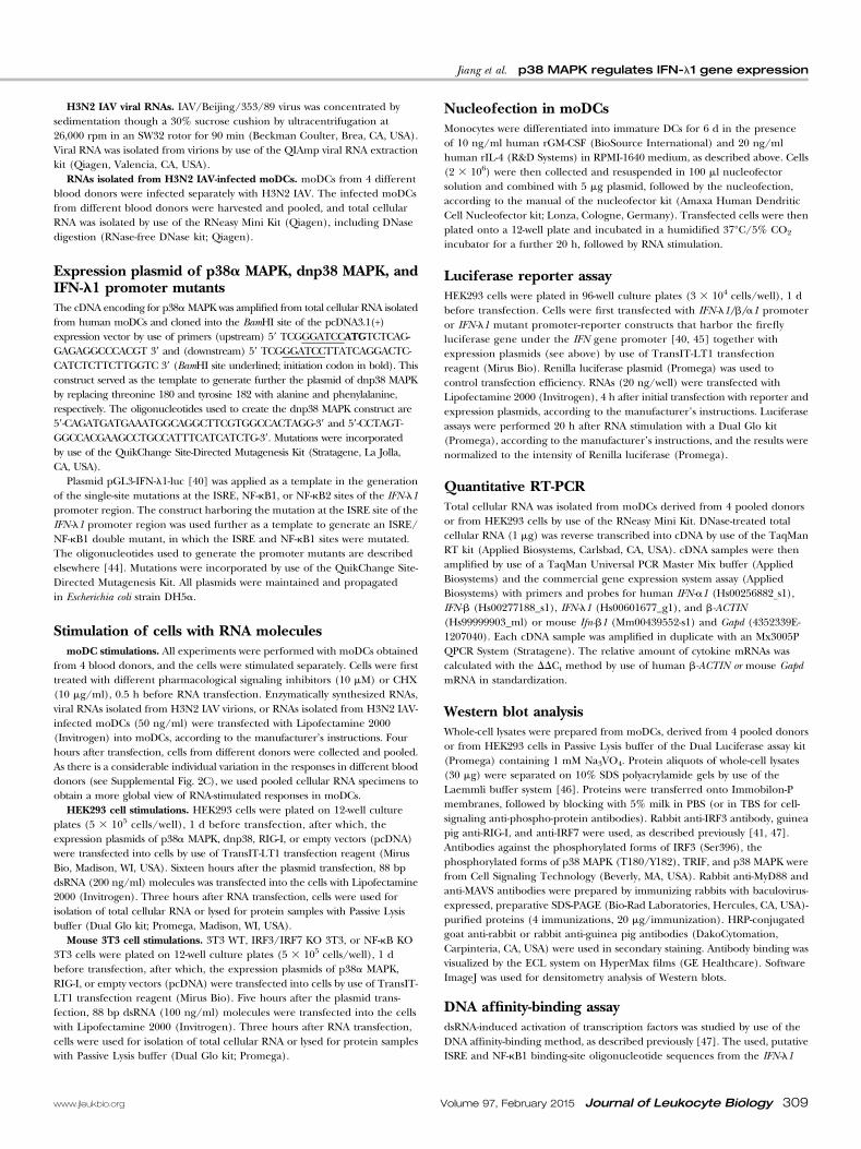
dsRNA-induced IFN-l1 gene expression is regulated byp38 MAPK in the absence of protein synthesisTo analyze whether ongoing protein synthesis was needed forRNA-induced IFN-l1 gene expression via the p38 MAPKpathway, we used CHX to block protein synthesis during thestimulation. As CHX is toxic to the cells, we selected a relativelyearly time-point of 3 h for our stimulation experiment. HumanmoDCs were stimulated with 88 bp dsRNA, and IFN-l1 mRNAexpression was analyzed by quantitative PCR (Fig. 3). We foundthat 88 bp dsRNA was capable of inducing IFN-l1 mRNAexpression in the presence and absence of CHX, and CHX didnot affect SB202190-mediated inhibition of IFN-l1 expression,indicating that the p38 MAPK pathway is involved directly inthe regulation of IFN-l1 gene expression. Similar results werealso obtained for the IFN-a1 gene (Supplemental Fig. 3).However, CHX treatment led to superinduction of IFN-a1mRNA expression (Supplemental Fig. 3), which is a previouslyidentified phenomenon related to the accumulation of mRNAsas a result of blocking protein synthesis or of removal ofa negative-feedback mechanism of cytokine mRNAdegradation.
The p38 MAPK signaling pathway regulates IFN-l1 geneexpression through RLR/TLR3 signaling pathways indsRNA-stimulated HEK293 cellsIt is well known that RLRs and TLR3 are the main cellulardsRNA-recognizing receptors that activate IFN gene expressionand antiviral responses [14, 50]. Having seen that the p38 MAPKsignaling pathway is involved in regulating IFN-l1 gene expres-sion in human moDCs after RNA stimulation (Figs. 1 and 2 andSupplemental Figs. 1 and 2), we transiently expressed differentRLR/TLR receptors in HEK293 cells to identify which signalingpathways are positively regulated by p38 MAPK. For thesestimulation experiments, the expression construct of p38aMAPKand dnp38 together with 88 bp dsRNA was used to study the roleof p38a MAPK in dsRNA-regulated IFN gene expression. InRIG-I-, MDA5-, and TLR3-overexpressing HEK293 cells,SB202190 inhibited the activation of the IFN-l1 gene promotertriggered by 88 bp dsRNA (Fig. 4A), indicating a positive role forp38a MAPK in RLR/TLR-stimulated gene expression. Over-expression of human p38a MAPK enhanced the activation of theIFN-l1 promoter in a dose-dependent fashion, whereas over-expression of dnp38 decreased the promoter activation (Fig. 4A),further demonstrating the positive role of p38a MAPK in RLR/TLR signaling. Especially, the inhibition of IFN-l1 promoteractivation by SB202190 and overexpression of dnp38 and theenhancement of promoter activation by overexpression of p38aMAPK were more significant in RIG-I- or MDA5-expressingHEK293 cells (Fig. 4A), indicating that the regulation of IFN-l1promoter activation by p38a MAPK is more likely through RLRsignaling. SB202190 was also able to inhibit dsRNA-inducedactivation of IFN-b and IFN-a1 promoters (Fig. 4B). The positive
regulatory role of overexpressed p38a MAPK was more modeston IFN-b and IFN-a1 promoters compared with that of the IFN-l1promoter, but IFN-b promoter activation was still sensitive to theinhibitory effect by SB202190, and IFN-a1 promoter activationwas reduced by overexpression of dnp38 (Fig. 4B).We also analyzed dsRNA-induced expression of IFN-l1 in
HEK293 cells in the presence of SB202190 or during over-expression of p38a MAPK and dnp38 (Fig. 5A). Stimulationof HEK293 cells with 88 bp dsRNA led to the induction ofendogenous IFN-l1 mRNA expression, and this induction wasenhanced with ectopic expression of RIG-I, whereas overexpres-sion of p38aMAPK or dnp38 could not enhance the induction ofIFN-l1 mRNA expression (Fig. 5A). Moreover, co-overexpressionof RIG-I and p38a MAPK significantly enhanced the induction ofIFN-l1 mRNA expression, whereas co-overexpression of RIG-Iand dnp38 decreased the induction of the IFN-l1 mRNAcompared with cells that were transfected only with the RIG-I-expression plasmid (Fig. 5A). Again, in RIG-I-overexpressingcells, the induction of IFN-l1 mRNA expression by 88 bp dsRNAwas inhibited by SB202190 (Fig. 5A). Western blot analysisshowed that the amount of phosphorylated p38 was increasedafter transfection with 88 bp dsRNA and decreased in dnp38-overexpressing cells (Fig. 5B, 1st row). Moreover, after 88 bpdsRNA stimulation, the phosphorylation level of IRF3 wasincreased in RIG-I overexpressing HEK293 cells compared withcells that were not overexpressing RIG-I, and co-overexpressionof RIG-I together with p38a MAPK or dnp38 did not affect thephosphorylation of IRF3 compared with RIG-I-overexpressing
Figure 3. Inhibition of IFN-l1 mRNA expression by the p38 MAPKinhibitor in human moDCs after stimulation with short dsRNA molecules,with or without inhibition of protein biosynthesis by CHX. moDCs from 4different blood donors were treated separately with CHX (10 mg/ml)and/or 6 SB202190 (SB; 10 mM), 1 or 0.5 h before RNA transfection,respectively, as indicated in the figure. The cells were subsequentlytransfected with 88 bp dsRNA molecules (50 ng/ml) for 3 h. Cells werecollected, total cellular RNA was isolated, and relative expression ofIFN-l1 mRNA was measured by quantitative RT-PCR. The values werenormalized against b-ACTIN mRNA, and the relative expression levelswere calculated with the DDCt method by use of untreated cells asa calibrator. The means (6SD) of 3 parallel determinations are shown.The data are representative of 3 individual experiments. Results wereconsidered statistically significant when *P , 0.05 compared with thedsRNA-stimulated and uninhibited sample (boxed bar).
312 Journal of Leukocyte Biology Volume 97, February 2015 www.jleukbio.org

cells (Fig. 5B, 3rd row), even though the IFN-l1 mRNAexpression level was greatly changed after co-overexpression (Fig.5A). It is of interest to note that the amount of total p38 MAPKwas increased slightly in HEK293 cells co-overexpressing RIG-Iand p38a MAPK or RIG-I and dnp38 (Fig. 5B, 2nd row),whereas the expression level of IRF3 remained approximatelythe same in all conditions (Fig. 5B, bottom row).
ISRE and NF-kB1 sites are essential for p38aMAPK-dependent activation of IFN-l1 gene expressionTo investigate further the mechanism how the IFN-l1 gene isactivated and regulated by p38a MAPK, we analyzed the role ofISRE and NF-kB promoter elements in the IFN-l1 promoteractivation after stimulation by different RNA molecules. Tocharacterize the role of these transcription factor binding sites onIFN-l1 gene activation, different single and double mutationswere introduced into the promoter region of IFN-l1 (Fig. 6A). InRIG-I-overexpressing cells, the WT IFN-l1 promoter was wellactivated by 88 bp dsRNA, but the activation level was reduced ifISRE and NF-kB1 sites of the promoters were mutated (Fig. 6B).However, mutations at the NF-kB2 site did not change the levelof promoter activation in response to RNA stimulation,suggesting that the NF-kB2 site is not as important in theactivation of the IFN-l1 promoter as the NF-kB1 site (Fig. 6B).Moreover, if both the ISRE and NF-kB1 sites were mutated, theIFN-l1 promoter did not respond to RNA stimulation in RIG-I-overexpressing cells (Fig. 6B), suggesting that both of these sitesare required for maximal activation of the promoter. Similarresults were also found in MDA5-overexpressing HEK cells(Supplemental Fig. 4).
To study further the role of the p38a MAPK signaling pathwayon ISRE- and NF-kB1-dependent IFN-l1 gene activation, theeffect of SB202190 and overexpression of p38a MAPK wereanalyzed on WT, ISRE, or NF-kB1 mutant IFN-l1 promoteractivation. Overexpression of an active form of RIG-I, DRIG-I,as well as MAVS and TRIF activated the WT IFN-l1 promoter,whereas treatment with SB202190 decreased this activation(Fig. 7A, 1st bar chart). In addition, overexpression of p38aMAPK together with other signaling components (DRIG-I,MAVS, or TRIF) led to clearly enhanced activation of the IFN-l1promoter. Moreover, mutations at ISRE and NF-kB1 sites on theIFN-l1 promoter resulted in reduced promoter activation andhigh sensitivity to SB202190 (Fig. 7A, 2nd and 3rd bar charts).Interestingly, SB202190 treatment and overexpression of p38aMAPK did not affect the activation of the WT IFN-l1 promoter orthe promoter containing mutated ISRE or NF-kB1 sites if thecells were stimulated by cotransfection with MyD88 and IRF7expression constructs (Fig. 7A).It has been shown that besides being regulated by IRF3, the
human IFN-a1 gene is mainly regulated by IRF7 [39, 51, 52]. Todemonstrate further that the IRF7 signaling pathway is notessential for p38a MAPK to regulate the IFN-a1 gene, theactivation of the IFN-a1 promoter was measured after stimulationby IRF7 expression or coexpression of IRF7 and MyD88 in thepresence or absence of SB202190 or overexpression of p38aMAPK. Consistent with the data above (Fig. 7A), SB202190 didnot inhibit the activation of the IFN-a1 promoter stimulated byexpression of IRF7 alone or by coexpression of IRF7 and MyD88(Fig. 7B), whereas the protein expression level of MyD88 or IRF7remained the same (Fig. 7C). The overexpression of p38a MAPK
Figure 4. Regulation of IFN gene promoteractivity by the p38 MAPK signaling pathway inHEK293 cells overexpressing dsRNA receptorsafter stimulation with short dsRNA molecules.HEK293 cells (in 96-well plates; 3 3 104 cells/well)remained untreated or were pretreated withthe p38 MAPK inhibitor SB202190 (10 mM),0.5 h before transfection with pcDNA (emptyvector) or RIG-I, MDA5, or TLR3 (20 ng/well)expression plasmids, a construct containing thefirefly luciferase gene under the IFN-l1 pro-moter (20 ng/well) and RSV-Renilla luciferaseplasmids (5 ng/well). Selected transfectionswere supplemented further with the p38aMAPK or dnp38 expression plasmid as indicated(A). Alternatively, cells were transfected withthe pcDNA or RIG-I (20 ng/well) expressionplasmid together with a construct-containing fire-fly luciferase gene under the IFN-l1, IFN-b, orIFN-a1 promoter (20 ng/well) and RSV-Renillaluciferase plasmids (5 ng/well), with or withoutthe p38a MAPK or dnp38 expression plasmid(10 ng/well; B). After 4 h of incubation, cellswere stimulated by transfection with 88 bpdsRNAs (20 ng/well) for an additional 20 h.Cells were collected, and luciferase assays werecarried out to determine the relative promoteractivity. The means (6SD) of 3 parallel determi-
nations are shown. The data are representative of 3 individual experiments. Results were considered statistically significant when *P , 0.05compared with the IFN promoter activity after RNA stimulation alone (black bars).
Jiang et al. p38 MAPK regulates IFN-l1 gene expression
www.jleukbio.org Volume 97, February 2015 Journal of Leukocyte Biology 313

also failed to enhance the promoter activation effectively underthese conditions (Fig. 7B), whereas overexpression of p38aMAPK reduced the expression of MyD88 or IRF7 proteins tosome extent (Fig. 7C). Altogether, our data suggest that theMyD88-IRF7 pathway is not essential for p38a MAPK to regulateIFN promoter activity.
IRF3/IRF7 and NF-kB are essential for p38aMAPK-dependent regulation of Ifn-b1 gene expressionin mouse 3T3 cellsAs described previously, IRFs regulate IFN-l1 gene expression,mainly through binding to the ISRE site, whereas the regulationby NF-kB takes place through its binding to cognate NF-kB sites.Having observed that ISRE and NF-kB1 sites at the IFN-l1promoter are essential for p38a MAPK-dependent activation ofIFN-l1 transcription, we further studied the role of IRF3, IRF7,and NF-kB in the regulation of Ifn-b1 gene expression by p38aMAPK in mouse 3T3 cells. As mice lack the human counterpartof IFN-l1, we concentrated on analyzing mouse Ifn-b1 geneexpression in IRF3/IRF7 KO and NF-kB KO mouse 3T3 cells.Consistent with the data on IFN-l1 gene expression in HEK293cells (Fig. 5A), mouse Ifn-b1 mRNA expression was activated bystimulation with 88 bp dsRNA in WT mouse 3T3 cells, and the
Figure 5. Regulation of IFN-l1 mRNA expression and phospho-IRF3expression by p38a MAPK in RIG-I-expressing HEK293 cells afterstimulation with short dsRNA molecules. (A) HEK293 cells (in 12-wellplates; 5 3 105 cells/well) remained untreated, or they were pretreatedwith the p38 MAPK inhibitor SB202190 (10 mM), 0.5 h beforetransfection with indicated expression plasmids (100 ng/ml). After 16 hof incubation, cells were stimulated by transfection with 88 bp dsRNAs(200 ng/ml) for an additional 3 h, followed by cell collection, RNAisolation, and quantitative RT-PCR analysis. The values were normalizedagainst b-ACTIN mRNA, and the relative IFN-l1 mRNA level wascalculated with the DDCt method by use of untreated cells as a calibrator.The means (6SD) of 3 parallel determinations are shown. Data arerepresentative of 3 individual experiments. IFN-l1 gene expression resultswere considered statistically significant when *P , 0.05 compared withthe experimentally relevant stimulatory conditions. (B) Western blotanalysis for the expression of phospho-IRF3, total IRF3, phospho-p38MAPK, and total p38a MAPK proteins in RNA-stimulated HEK293 cells,which were collected at 3 h after 88 bp dsRNA transfection, and whole-cell lysates were prepared. Cellular proteins (30 mg/lane) were separatedon 10% SDS-PAGE, followed by electrophoretic transfer of the proteinsonto PVDF membranes and visualization of the transferred proteins byprotein-specific antibodies, as indicated. The data of 1 representativeexperiment of 3 independent experiments are shown.
Figure 6. Promoter structure of the IFN-l1 gene and the differencesbetween IFN-l1mutated and natural promoter activation in RIG-I-expressingHEK cells after different RNA stimulations. (A) The promoter structureof the human IFN-l1 gene. Computer analysis was performed to elucidate4 putative transcription factor binding sites on the promoter region ofIFN-l1 near the start codon. The putative binding sites for IRFs (ISRE andPRDI) and NF-kB (gray boxes), their sequences, and relative positionswith respect to the start codon (arrow) are shown. (B) HEK293 cells (in96-well plates; 3 3 104 cells/well) were transfected with pcDNA or RIG-I(20 ng/well) expression plasmid and together with RSV-Renilla luciferaseplasmids (5 ng/well) and constructs containing the firefly luciferase geneunder the WT IFN-l1 promoter or IFN-l1 ISRE or/and NF-kB mutant(mt) promoters, as indicated (20 ng/well). After 4 h of incubation, cellswere stimulated by transfection with 88 bp dsRNAs (20 ng/well) for anadditional 20 h. Cells were collected, and luciferase assays were carriedout to determine the relative promoter activity. The means (6SD) of 3parallel determinations are shown. The data are representative of 3individual experiments. Results were considered statistically significantwhen *P , 0.05 compared with the WT IFN-l1 promoter activity(black bars).
314 Journal of Leukocyte Biology Volume 97, February 2015 www.jleukbio.org

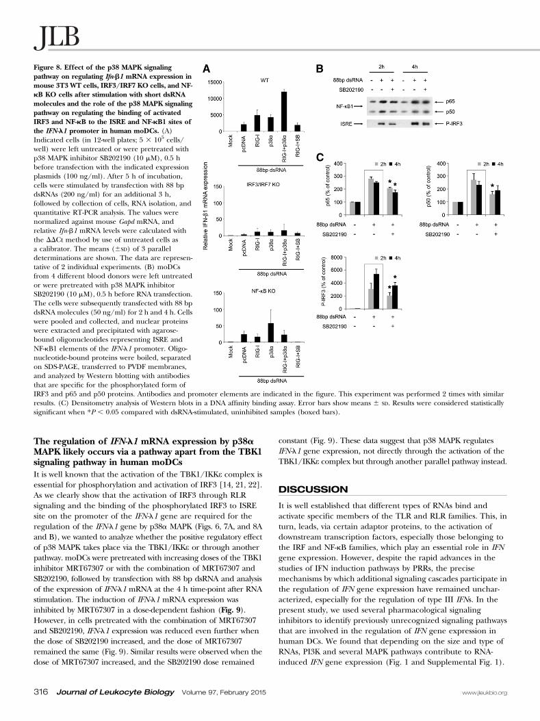
activation was weakly enhanced by overexpression of RIG-I orp38a MAPK (Fig. 8A, 1st bar chart). However, coexpressionof RIG-I and p38a MAPK led to a clearly detectable increase inIfn-b1 mRNA expression. SB202190 efficiently inhibited Ifn-b1mRNA expression in RIG-I-overexpressing WT 3T3 cells.However, in IRF3/IRF7 KO and NF-kB KO cells, 88 bp dsRNA-induced expression of Ifn-b1 mRNA was decreased significantlycompared with the WT mouse 3T3 cells, and coexpression ofRIG-I and p38a MAPK failed to enhance the Ifn-b1 mRNAexpression (Fig. 8A, 2nd and 3rd bar charts). This data suggestthat IRF3/IRF7 and NF-kB are the key transcription factorsthat work in concert with p38a MAPK to regulate early IFNgene expression, such as those of mouse Ifn-b and humanIFN-l1 genes.
p38a MAPK is involved in the binding of IRF3 andNF-kB to the IFN-l1 promoterTo characterize further the role of the p38a MAPK signalingpathway on regulating the binding of IRF3 and NF-kB to theIFN-l1 promoter, DNA affinity-binding assays were carried out.moDCs were transfected with 88 bp dsRNA in the presence orabsence of SB202190, cells were collected at 2 h and 4 h after RNAstimulation, and nuclear proteins were extracted and precipitatedby use of the IFN-l1 gene promoter ISRE- and NF-kB1-siteoligonucleotides. The binding of phosphorylated IRF3 to the ISREsite and p65 and p50 to the NF-kB1 site was clearly enhancedalready at 2 h after RNA stimulation, whereas the presence ofSB202190 reduced the binding of both transcription factors, especiallyat the early stage—2 h after RNA stimulation (Fig. 8B and C).
Figure 7. Effects of the p38 MAPK signaling pathway on IFN-l1 promoter activity in HEK293 cells expressing different components of the RLR/TLRsignaling pathways. HEK293 cells (in 96-well plates; 3 3 104 cells/well) were left untreated or were pretreated with p38 MAPK inhibitor SB202190 (10 mM),0.5 h before transfection with pcDNA or indicated expression plasmids for different signaling components (20 ng/well), RSV-Renilla luciferase plasmids(5 ng/well) and constructs containing the firefly luciferase gene under the WT IFN-l1 promoter (prom.) or mutated IFN-l1 promoters (mut-prom.; A) orunder the IFN-a1 promoter (B; 20 ng/well). After 24 h incubation, cells were collected, and luciferase assays were carried out to determine the relativepromoter activity. The means (6SD) of 3 parallel determinations are shown. The data are representative of 3 individual experiments. Results wereconsidered statistically significant when *P , 0.05 compared with the black bar of the control IFN-l1 promoter activity. (C) Western blot analysis for theexpression of RIG-I, MAVS, TRIF, IRF7, MyD88, and p38a MAPK in HEK cells. HEK293 cells (in 12-well plates, 3 3 105 cells/well) were left untreated orwere pretreated with the p38 MAPK inhibitor SB202190 (10 mM). 0.5 h before transfection with pcDNA or indicated expression plasmids for differentsignaling components (200 ng/well). The whole-cell lysates were prepared, and proteins (20 mg/lane) were separated on 10% SDS-PAGE, followed byelectrophoretic transfer of the proteins onto PVDF membranes and detection of the transferred proteins by antibodies that are specific for RIG-I, MAVS,TRIF, IRF7, MyD88, and p38a MAPK, as indicated. The data of 1 representative experiment of 3 independent experiments are shown.
Jiang et al. p38 MAPK regulates IFN-l1 gene expression
www.jleukbio.org Volume 97, February 2015 Journal of Leukocyte Biology 315
The regulation of IFN-l1 mRNA expression by p38aMAPK likely occurs via a pathway apart from the TBK1signaling pathway in human moDCsIt is well known that the activation of the TBK1/IKKe complex isessential for phosphorylation and activation of IRF3 [14, 21, 22].As we clearly show that the activation of IRF3 through RLRsignaling and the binding of the phosphorylated IRF3 to ISREsite on the promoter of the IFN-l1 gene are required for theregulation of the IFN-l1 gene by p38a MAPK (Figs. 6, 7A, and 8Aand B), we wanted to analyze whether the positive regulatory effectof p38 MAPK takes place via the TBK1/IKKe or through anotherpathway. moDCs were pretreated with increasing doses of the TBK1inhibitor MRT67307 or with the combination of MRT67307 andSB202190, followed by transfection with 88 bp dsRNA and analysisof the expression of IFN-l1 mRNA at the 4 h time-point after RNAstimulation. The induction of IFN-l1 mRNA expression wasinhibited by MRT67307 in a dose-dependent fashion (Fig. 9).However, in cells pretreated with the combination of MRT67307and SB202190, IFN-l1 expression was reduced even further whenthe dose of SB202190 increased, and the dose of MRT67307remained the same (Fig. 9). Similar results were observed when thedose of MRT67307 increased, and the SB202190 dose remained
constant (Fig. 9). These data suggest that p38 MAPK regulatesIFN-l1 gene expression, not directly through the activation of theTBK1/IKKe complex but through another parallel pathway instead.
DISCUSSION
It is well established that different types of RNAs bind andactivate specific members of the TLR and RLR families. This, inturn, leads, via certain adaptor proteins, to the activation ofdownstream transcription factors, especially those belonging tothe IRF and NF-kB families, which play an essential role in IFNgene expression. However, despite the rapid advances in thestudies of IFN induction pathways by PRRs, the precisemechanisms by which additional signaling cascades participate inthe regulation of IFN gene expression have remained unchar-acterized, especially for the regulation of type III IFNs. In thepresent study, we used several pharmacological signalinginhibitors to identify previously unrecognized signaling pathwaysthat are involved in the regulation of IFN gene expression inhuman DCs. We found that depending on the size and type ofRNAs, PI3K and several MAPK pathways contribute to RNA-induced IFN gene expression (Fig. 1 and Supplemental Fig. 1).
Figure 8. Effect of the p38 MAPK signalingpathway on regulating Ifn-b1 mRNA expression inmouse 3T3 WT cells, IRF3/IRF7 KO cells, and NF-kB KO cells after stimulation with short dsRNAmolecules and the role of the p38 MAPK signalingpathway on regulating the binding of activatedIRF3 and NF-kB to the ISRE and NF-kB1 sites ofthe IFN-l1 promoter in human moDCs. (A)Indicated cells (in 12-well plates; 5 3 105 cells/well) were left untreated or were pretreated withp38 MAPK inhibitor SB202190 (10 mM), 0.5 hbefore transfection with the indicated expressionplasmids (100 ng/ml). After 5 h of incubation,cells were stimulated by transfection with 88 bpdsRNAs (200 ng/ml) for an additional 3 h,followed by collection of cells, RNA isolation, andquantitative RT-PCR analysis. The values werenormalized against mouse Gapd mRNA, andrelative Ifn-b1 mRNA levels were calculated withthe DDCt method by use of untreated cells asa calibrator. The means (6SD) of 3 paralleldeterminations are shown. The data are represen-tative of 2 individual experiments. (B) moDCsfrom 4 different blood donors were left untreatedor were pretreated with p38 MAPK inhibitorSB202190 (10 mM), 0.5 h before RNA transfection.The cells were subsequently transfected with 88 bpdsRNA molecules (50 ng/ml) for 2 h and 4 h. Cellswere pooled and collected, and nuclear proteinswere extracted and precipitated with agarose-bound oligonucleotides representing ISRE andNF-kB1 elements of the IFN-l1 promoter. Oligo-nucleotide-bound proteins were boiled, separatedon SDS-PAGE, transferred to PVDF membranes,and analyzed by Western blotting with antibodiesthat are specific for the phosphorylated form ofIRF3 and p65 and p50 proteins. Antibodies and promoter elements are indicated in the figure. This experiment was performed 2 times with similarresults. (C) Densitometry analysis of Western blots in a DNA affinity binding assay. Error bars show means 6 SD. Results were considered statisticallysignificant when *P , 0.05 compared with dsRNA-stimulated, uninhibited samples (boxed bars).
316 Journal of Leukocyte Biology Volume 97, February 2015 www.jleukbio.org

Interestingly, the p38 MAPK inhibitors were very efficient ininhibiting IFN gene expression (Figs. 1 and 2A), and thisinhibition took place also in the presence of protein synthesisinhibitor (Fig. 3 and Supplemental Fig. 3), indicating that p38MAPK is contributing to the very early transcriptional eventsregulating type I and type III IFN gene expression. Phosphory-lation of p38 MAPK increased after 88 bp dsRNA stimulation(Figs. 2B and D and 5B), suggesting that short dsRNA moleculescan also activate the p38 MAPK signaling pathway in addition tothe classic PRR signaling pathways. Nucleofection of p38a MAPKin dsRNA-stimulated moDCs enhanced IFN-l1 mRNA expression(Fig. 2C), further demonstrating the positive role of p38 MAPKin IFN-l1 gene regulation. The expression of phosphorylatedforms of IRF3 and p38 MAPK was increased after 88 bp dsRNAstimulation (Fig. 2B and D), suggesting that these 2 signalingpathways cooperatively regulate the induction of the IFN-l1 gene.In addition, overexpression studies with p38a MAPK, dnp38, andsome TLR/RLR family members suggest that the RIG-I and IRF3pathway, but not the MyD88 and IRF7 pathway, is required forthe regulation of IFN gene expression by p38a MAPK (Figs. 4, 5,and 7). Moreover, the effects of mutations in the IFN-l1promoter (Figs. 6 and 7), DNA affinity-binding experiments inhuman moDCs (Fig. 8B), and experiments in mouse IRF3/IRF7and NF-kB KO cells (Fig. 8A) suggest that the IRF and NF-kBtranscription factors and their respective binding sites ISRE and
NF-kB1 in the IFN-l1 promoter are essential in RNA-regulatedIFN gene expression. Furthermore, in these experiments, p38aMAPK had a positive regulatory role (Figs. 7 and 8). In addition,the inhibitor-combination experiments in human moDCs suggestthat the regulation of IFN-l1 mRNA expression by p38a MAPKdoes not take place directly through activation of the TBK1/IKKecomplex (Fig. 9), and Western blot analysis also showed that theaddition of inhibitors of p38 MAPK and overexpression of p38aMAPK or dnp38 could not significantly affect the phosphorylationof IRF3 (Figs. 2B and D and 5B), further suggesting that p38MAPK regulates IFN-l1 gene expression, not directly throughactivation of IRF3. Therefore, our data strongly indicate thatp38a MAPK positively regulates early IFN-l1 gene expressionby cooperating with the transcription factors IRF3 and NF-kBthrough some parallel signaling pathway after RNA stimula-tion (Fig. 10).It has been shown previously that p38 MAPK plays a central
role in inflammatory responses induced by environmental stress,growth factors, and inflammatory factors [53–55]. Furthermore,the p38 MAPK signaling pathway was shown to be involved in IFNsignaling and regulation of antiviral responses [34]. However,less is known about the role of p38 MAPK in regulating IFN geneexpression. It has been reported that the activation of RIG-Iduring Sendai virus infection leads to the activation of p38MAPK, which contributes to type I IFN-mediated antiviralresponses and the activation of conventional DCs [56]. It hasalso been found that 2 p38a MAPKAPKs—MAPKAPK2 andMAPKAPK3—regulate the expression of IFN-inducible genes andantiviral responses [35]. The present study demonstrates thatbesides the regulation of IFN signaling and antiviral response[34, 35, 56], p38 MAPK is activated by short dsRNA stimulation(Figs. 2B and D and 5B) and is taking part in the regulation ofRNA-induced early IFN gene expression in human moDCs,especially those of IFN-l1 and IFN-b genes (Figs. 1 and 2 andSupplemental Figs. 1 and 2). Our data are also consistent witha previous study, which showed that in human macrophages,IFN induction by the infection with an RNA virus—IAV—ispredominantly regulated by IRF3 and p38 MAPK [57]. ThePI3K inhibitor also decreased the expression of the IFN-l1 gene,especially in response to stimulation with short 88 nt ssRNA andlong 1.8 kb dsRNA molecules (Fig. 1), suggesting someselectivity of the PI3K pathway in RNA-induced innate immuneresponse. However, further studies are needed to elucidatethe potential role of these pathways in regulating IFN geneexpression.To investigate further the mechanism by which p38 MAPK
regulates the expression of the IFN gene in response to RNAstimulation, the relationship of different RNA receptors wasstudied. Overexpression of p38a or dnp38 MAPK regulated theactivation of the IFN-l1 promoter positively or negatively,respectively, through RLR (RIG-I and MDA5) and TLR (TLR3)pathways (Fig. 4A), indicating a broader role for p38a MAPK inintracellular signaling. Moreover, co-overexpression of RIG-I andp38a MAPK enhanced the IFN-l1 promoter activity at higherlevels compared with IFN-b and IFN-a1 promoters (Fig. 4B),suggesting that the promoter of type III IFN genes can beregulated by a broader scale of transcription factors than type IIFN genes. Even though the precise site of action of p38a MAPK
Figure 9. The role of the p38 MAPK inhibitor and TBK1 inhibitor onregulating IFN-l1 mRNA expression in short dsRNA-stimulated humanmoDCs. moDCs from 4 different blood donors were grown separately on24-well plates. Cells remained untreated or were pretreated with anindicated amount of p38 MAPK inhibitor SB202190 or TBK1 inhibitorMRT67307 (MRT), 0.5 h before transfection with 88 bp dsRNA (50 ng/ml).Cells were collected 4 h after RNA transfection, and total cellularRNA was isolated, and IFN-l1 mRNA expression was determined byquantitative RT-PCR. The values were normalized against b-ACTIN, andrelative IFN-l1 mRNA levels were calculated with the DDCt method byuse of untreated cells as a calibrator. The means (6SD) of 3 paralleldeterminations are shown. Data are representative of 3 individualexperiments.
Jiang et al. p38 MAPK regulates IFN-l1 gene expression
www.jleukbio.org Volume 97, February 2015 Journal of Leukocyte Biology 317
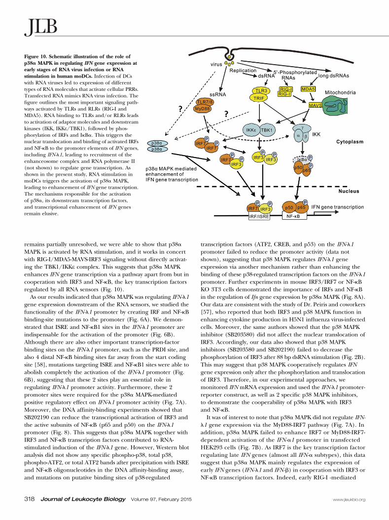
remains partially unresolved, we were able to show that p38aMAPK is activated by RNA stimulation, and it works in concertwith RIG-I/MDA5-MAVS-IRF3 signaling without directly activat-ing the TBK1/IKKe complex. This suggests that p38a MAPKenhances IFN gene transcription via a pathway apart from but incooperation with IRF3 and NF-kB, the key transcription factorsregulated by all RNA sensors (Fig. 10).As our results indicated that p38a MAPK was regulating IFN-l1
gene expression downstream of the RNA sensors, we studied thefunctionality of the IFN-l1 promoter by creating IRF and NF-kBbinding-site mutations to the promoter (Fig. 6A). We demon-strated that ISRE and NF-kB1 sites in the IFN-l1 promoter areindispensable for the activation of the promoter (Fig. 6B).Although there are also other important transcription-factorbinding sites on the IFN-l1 promoter, such as the PRDI site, andalso 4 distal NF-kB binding sites far away from the start codingsite [58], mutations targeting ISRE and NF-kB1 sites were able toabolish completely the activation of the IFN-l1 promoter (Fig.6B), suggesting that these 2 sites play an essential role inregulating IFN-l1 promoter activity. Furthermore, these 2promoter sites were required for the p38a MAPK-mediatedpositive regulatory effect on IFN-l1 promoter activity (Fig. 7A).Moreover, the DNA affinity-binding experiments showed thatSB202190 can reduce the transcriptional activation of IRF3 andthe active subunits of NF-kB (p65 and p50) on the IFN-l1promoter (Fig. 8). This suggests that p38a MAPK together withIRF3 and NF-kB transcription factors contributed to RNA-stimulated induction of the IFN-l1 gene. However, Western blotanalysis did not show any specific phospho-p38, total p38,phospho-ATF2, or total ATF2 bands after precipitation with ISREand NF-kB oligonucleotides in the DNA affinity-binding assay,and mutations on putative binding sites of p38-regulated
transcription factors (ATF2, CREB, and p53) on the IFN-l1promoter failed to reduce the promoter activity (data notshown), suggesting that p38 MAPK regulates IFN-l1 geneexpression via another mechanism rather than enhancing thebinding of these p38-regulated transcription factors on the IFN-l1promoter. Further experiments in mouse IRF3/IRF7 or NF-kBKO 3T3 cells demonstrated the importance of IRFs and NF-kBin the regulation of Ifn gene expression by p38a MAPK (Fig. 8A).Our data are consistent with the study of Dr. Peiris and coworkers[57], who reported that both IRF3 and p38 MAPK function inenhancing cytokine production in H5N1 influenza virus-infectedcells. Moreover, the same authors showed that the p38 MAPKinhibitor (SB203580) did not affect the nuclear translocation ofIRF3. Accordingly, our data also showed that p38 MAPKinhibitors (SB203580 and SB202190) failed to decrease thephosphorylation of IRF3 after 88 bp dsRNA stimulation (Fig. 2B).This may suggest that p38 MAPK cooperatively regulates IFNgene expression only after the phosphorylation and translocationof IRF3. Therefore, in our experimental approaches, wemonitored IFN mRNA expression and used the IFN-l1 promoter-reporter construct, as well as 2 specific p38 MAPK inhibitors,to demonstrate the cooperability of p38a MAPK with IRF3and NF-kB.It was of interest to note that p38a MAPK did not regulate IFN-
l1 gene expression via the MyD88-IRF7 pathway (Fig. 7A). Inaddition, p38a MAPK failed to enhance IRF7 or MyD88-IRF7-dependent activation of the IFN-a1 promoter in transfectedHEK293 cells (Fig. 7B). As IRF7 is the key transcription factorregulating late IFN genes (almost all IFN-a subtypes), this datasuggest that p38a MAPK mainly regulates the expression ofearly IFN genes (IFN-l1 and IFN-b) in cooperation with IRF3 orNF-kB transcription factors. Indeed, early RIG-I -mediated
Figure 10. Schematic illustration of the role ofp38a MAPK in regulating IFN gene expression atearly stages of RNA virus infection or RNAstimulation in human moDCs. Infection of DCswith RNA viruses led to expression of differenttypes of RNA molecules that activate cellular PRRs.Transfected RNA mimics RNA virus infection. Thefigure outlines the most important signaling path-ways activated by TLRs and RLRs (RIG-I andMDA5). RNA binding to TLRs and/or RLRs leadsto activation of adaptor molecules and downstreamkinases (IKK, IKKe/TBK1), followed by phos-phorylation of IRFs and IkBa. This triggers thenuclear translocation and binding of activated IRFsand NF-kB to the promoter elements of IFN genes,including IFN-l1, leading to recruitment of theenhanceosome complex and RNA polymerase II(not shown) to regulate gene transcription. Asshown in the present study, RNA stimulation inmoDCs triggers the activation of p38a MAPK,leading to enhancement of IFN gene transcription.The mechanisms responsible for the activationof p38a, its downstream transcription factors,and transcriptional enhancement of IFN genesremain elusive.
318 Journal of Leukocyte Biology Volume 97, February 2015 www.jleukbio.org

promoter activities of human IFN-l1, IFN-b, and IFN-a1 wereinhibited by SB202190 and enhanced by p38a MAPK over-expression (Fig. 4B). Furthermore, RNA-induced IFN-l1 orIFN-a1 expression was inhibited by SB202190, also in thepresence of protein synthesis inhibitor CHX, indicating thatthe regulation of IFN mRNA expression by p38 MAPK takesplace directly after RNA stimulation and does not require denovo protein synthesis. Our data are also consistent with thestudy of Dr. Ludwig and coworkers [59], who reported thatinhibition of p38 MAPK by the p38-specific inhibitor SB202190led to reduced expression of IFN-b and other cytokines inH5N1 and H7N7 virus-infected cells. The studies by us andothers indicate a clear role of p38 MAPK in regulating IFNgene expression at early stages of RNA stimulation or viralinfection.Our data, as concluded from multiple experimental
approaches, strongly suggest that p38a MAPK, together withIRF3 and NF-kB, regulates the expression of early IFN genes,especially that of the type III IFN-l1 gene. This suggests that p38MAPK is not only involved in IFN signaling and induction of anantiviral response, but it also contributes to transcriptionalregulation of IFN genes in RNA-stimulated cells. Our study thusprovides new mechanistic evidence for the function of p38aMAPK in the early antiviral signaling pathways and providesfurther evidence for the complexity of signal transductionpathways and the fine-tuning of IFN responses.
AUTHORSHIP
M.J. designed the study, performed laboratory experiments,analyzed the results, and wrote the paper. P.O. and I.J. designedthe study and wrote the paper. R.F. performed laboratoryexperiments. D.N.R. performed laboratory experiments andwrote the paper. A.H., M.M.P., and D.H.B wrote the paper.
ACKNOWLEDGMENTS
This work was supported by the Sigrid Juselius Foundation (to I.J.,M.M.P., and D.H.B.), Finnish Foundation for Research on ViralDiseases (to M.J.), Research Council for Health of the Academy ofFinland (Grants 252252 and 256159 to I.J. and 256197 to D.H.B.),and Research Council for Biosciences and Environment of theAcademy of Finland (Grants 250113, 256069, and 272507 toM.M.P.and 255342 and 256518 to D.H.B.). The authors thank HannaValtonen and Teija Aalto for their excellent technical assistance incell experiments and protein expression assays.
DISCLOSURES
The authors declare no conflict of interest.
REFERENCES
1. Janeway, Jr., C. A. (1989) Approaching the asymptote? Evolution andrevolution in immunology. Cold Spring Harb. Symp. Quant. Biol. 54, 1–13.
2. Dupuis, S., Jouanguy, E., Al-Hajjar, S., Fieschi, C., Al-Mohsen, I. Z., Al-Jumaah, S., Yang, K., Chapgier, A., Eidenschenk, C., Eid, P., AlGhonaium, A., Tufenkeji, H., Frayha, H., Al-Gazlan, S., Al-Rayes, H.,
Schreiber, R. D., Gresser, I., Casanova, J. L. (2003) Impaired response tointerferon-alpha/beta and lethal viral disease in human STAT1deficiency. Nat. Genet. 33, 388–391.
3. Takeuchi, O., Akira, S. (2009) Innate immunity to virus infection.Immunol. Rev. 227, 75–86.
4. Ank, N., Iversen, M. B., Bartholdy, C., Staeheli, P., Hartmann, R., Jensen,U. B., Dagnaes-Hansen, F., Thomsen, A. R., Chen, Z., Haugen, H.,Klucher, K., Paludan, S. R. (2008) An important role for type IIIinterferon (IFN-lambda/IL-28) in TLR-induced antiviral activity. J.Immunol. 180, 2474–2485.
5. Ank, N., West, H., Paludan, S. R. (2006) IFN-lambda: novel antiviralcytokines. J. Interferon Cytokine Res. 26, 373–379.
6. Palucka, K., Banchereau, J. (2002) How dendritic cells and microbesinteract to elicit or subvert protective immune responses. Curr. Opin.Immunol. 14, 420–431.
7. Iwasaki, A., Medzhitov, R. (2010) Regulation of adaptive immunity by theinnate immune system. Science 327, 291–295.
8. Bruns, A. M., Horvath, C. M. (2012) Activation of RIG-I-like receptorsignal transduction. Crit. Rev. Biochem. Mol. Biol. 47, 194–206.
9. Yoneyama, M., Kikuchi, M., Natsukawa, T., Shinobu, N., Imaizumi, T.,Miyagishi, M., Taira, K., Akira, S., Fujita, T. (2004) The RNA helicaseRIG-I has an essential function in double-stranded RNA-induced innateantiviral responses. Nat. Immunol. 5, 730–737.
10. Kawai, T., Akira, S. (2006) Innate immune recognition of viral infection.Nat. Immunol. 7, 131–137.
11. Takeuchi, O., Akira, S. (2008) MDA5/RIG-I and virus recognition. Curr.Opin. Immunol. 20, 17–22.
12. McCartney, S. A., Colonna, M. (2009) Viral sensors: diversity in pathogenrecognition. Immunol. Rev. 227, 87–94.
13. Mogensen, T. H., Paludan, S. R. (2005) Reading the viral signature byToll-like receptors and other pattern recognition receptors. J. Mol. Med.(Berl). 83, 180–192.
14. Akira, S., Uematsu, S., Takeuchi, O. (2006) Pathogen recognition andinnate immunity. Cell 124, 783–801.
15. Kawai, T., Takahashi, K., Sato, S., Coban, C., Kumar, H., Kato, H., Ishii,K. J., Takeuchi, O., Akira, S. (2005) IPS-1, an adaptor triggering RIG-I-and Mda5-mediated type I interferon induction. Nat. Immunol. 6,981–988.
16. Seth, R. B., Sun, L., Ea, C. K., Chen, Z. J. (2005) Identification andcharacterization of MAVS, a mitochondrial antiviral signaling proteinthat activates NF-kappaB and IRF 3. Cell 122, 669–682.
17. Meylan, E., Curran, J., Hofmann, K., Moradpour, D., Binder, M.,Bartenschlager, R., Tschopp, J. (2005) Cardif is an adaptor protein in theRIG-I antiviral pathway and is targeted by hepatitis C virus. Nature 437,1167–1172.
18. Xu, L. G., Wang, Y. Y., Han, K. J., Li, L. Y., Zhai, Z., Shu, H. B. (2005)VISA is an adapter protein required for virus-triggered IFN-betasignaling. Mol. Cell 19, 727–740.
19. O’Neill, L. A., Bowie, A. G. (2007) The family of five: TIR-domain-containing adaptors in Toll-like receptor signalling. Nat. Rev. Immunol. 7,353–364.
20. Ghosh, S., May, M. J., Kopp, E. B. (1998) NF-kappa B and Rel proteins:evolutionarily conserved mediators of immune responses. Annu. Rev.Immunol. 16, 225–260.
21. Mogensen, T. H. (2009) Pathogen recognition and inflammatorysignaling in innate immune defenses. Clin. Microbiol. Rev. 22, 240–273(Table of Contents.).
22. Hiscott, J. (2007) Triggering the innate antiviral response through IRF-3activation. J. Biol. Chem. 282, 15325–15329.
23. Hiscott, J. (2007) Convergence of the NF-kappaB and IRF pathways inthe regulation of the innate antiviral response. Cytokine Growth Factor Rev.18, 483–490.
24. Kyriakis, J. M., Avruch, J. (1996) Sounding the alarm: protein kinasecascades activated by stress and inflammation. J. Biol. Chem. 271,24313–24316.
25. Schaeffer, H. J., Weber, M. J. (1999) Mitogen-activated protein kinases:specific messages from ubiquitous messengers. Mol. Cell. Biol. 19,2435–2444.
26. Chang, L., Karin, M. (2001) Mammalian MAP kinase signalling cascades.Nature 410, 37–40.
27. Platanias, L. C. (2003) Map kinase signaling pathways and hematologicmalignancies. Blood 101, 4667–4679.
28. Davis, R. J. (2000) Signal transduction by the JNK group of MAP kinases.Cell 103, 239–252.
29. Lee, J. C., Laydon, J. T., McDonnell, P. C., Gallagher, T. F., Kumar, S.,Green, D., McNulty, D., Blumenthal, M. J., Heys, J. R., Landvatter, S. W.,Strickler, J. E., McLaughlin, M. M., Siemens, I. R., Fisher, S. M., Livi, G. P.,White, J. R., Adams, J. L., Young, P. R. (1994) A protein kinase involved inthe regulation of inflammatory cytokine biosynthesis. Nature 372,739–746.
30. Jiang, Y., Chen, C., Li, Z., Guo, W., Gegner, J. A., Lin, S., Han, J. (1996)Characterization of the structure and function of a new mitogen-activated protein kinase (p38beta). J. Biol. Chem. 271, 17920–17926.
Jiang et al. p38 MAPK regulates IFN-l1 gene expression
www.jleukbio.org Volume 97, February 2015 Journal of Leukocyte Biology 319

31. Jiang, Y., Gram, H., Zhao, M., New, L., Gu, J., Feng, L., Di Padova, F.,Ulevitch, R. J., Han, J. (1997) Characterization of the structure andfunction of the fourth member of p38 group mitogen-activated proteinkinases, p38delta. J. Biol. Chem. 272, 30122–30128.
32. Li, Z., Jiang, Y., Ulevitch, R. J., Han, J. (1996) The primary structure ofp38 gamma: a new member of p38 group of MAP kinases. Biochem.Biophys. Res. Commun. 228, 334–340.
33. Schieven, G. L. (2009) The p38alpha kinase plays a central role ininflammation. Curr. Top. Med. Chem. 9, 1038–1048.
34. Platanias, L. C. (2003) The p38 mitogen-activated protein kinase pathwayand its role in interferon signaling. Pharmacol. Ther. 98, 129–142.
35. Li, Y., Sassano, A., Majchrzak, B., Deb, D. K., Levy, D. E., Gaestel, M.,Nebreda, A. R., Fish, E. N., Platanias, L. C. (2004) Role of p38alphaMap kinase in type I interferon signaling. J. Biol. Chem. 279,970–979.
36. Carey, M. (1998) The enhanceosome and transcriptional synergy. Cell 92,5–8.
37. Yoneyama, M., Suhara, W., Fukuhara, Y., Fukuda, M., Nishida, E., Fujita,T. (1998) Direct triggering of the type I interferon system by virusinfection: activation of a transcription factor complex containing IRF-3and CBP/p300. EMBO J. 17, 1087–1095.
38. Sato, M., Hata, N., Asagiri, M., Nakaya, T., Taniguchi, T., Tanaka, N.(1998) Positive feedback regulation of type I IFN genes by the IFN-inducible transcription factor IRF-7. FEBS Lett. 441, 106–110.
39. Honda, K., Yanai, H., Negishi, H., Asagiri, M., Sato, M., Mizutani, T.,Shimada, N., Ohba, Y., Takaoka, A., Yoshida, N., Taniguchi, T. (2005)IRF-7 is the master regulator of type-I interferon-dependent immuneresponses. Nature 434, 772–777.
40. Osterlund, P. I., Pietila, T. E., Veckman, V., Kotenko, S. V., Julkunen, I.(2007) IFN regulatory factor family members differentially regulate theexpression of type III IFN (IFN-lambda) genes. J. Immunol. 179, 3434–3442.
41. Jiang, M., Osterlund, P., Sarin, L. P., Poranen, M. M., Bamford, D. H.,Guo, D., Julkunen, I. (2011) Innate immune responses in humanmonocyte-derived dendritic cells are highly dependent on the size andthe 59 phosphorylation of RNA molecules. J. Immunol. 187, 1713–1721.
42. Hoffmann, A., Levchenko, A., Scott, M. L., Baltimore, D. (2002) TheIkappaB-NF-kappaB signaling module: temporal control and selectivegene activation. Science 298, 1241–1245.
43. Veckman, V., Miettinen, M., Pirhonen, J., Siren, J., Matikainen, S.,Julkunen, I. (2004) Streptococcus pyogenes and Lactobacillus rhamnosusdifferentially induce maturation and production of Th1-type cytokinesand chemokines in human monocyte-derived dendritic cells. J. Leukoc.Biol. 75, 764–771.
44. Nousiainen, L., Sillanpaa, M., Jiang, M., Thompson, J., Taipale, J.,Julkunen, I. (2013) Human kinome analysis reveals novel kinasescontributing to virus infection and retinoic-acid inducible gene I-inducedtype I and type III IFN gene expression. Innate Immun. 19, 516–530.
45. Lin, R., Genin, P., Mamane, Y., Hiscott, J. (2000) Selective DNAbinding and association with the CREB binding protein coactivatorcontribute to differential activation of alpha/beta interferon genesby interferon regulatory factors 3 and 7. Mol. Cell. Biol. 20,6342–6353.
46. Laemmli, U. K. (1970) Cleavage of structural proteins during theassembly of the head of bacteriophage T4. Nature 227, 680–685.
47. Osterlund, P., Veckman, V., Siren, J., Klucher, K. M., Hiscott, J.,Matikainen, S., Julkunen, I. (2005) Gene expression and antiviral activityof alpha/beta interferons and interleukin-29 in virus-infected humanmyeloid dendritic cells. J. Virol. 79, 9608–9617.
48. Menon, M. B., Kotlyarov, A., Gaestel, M. (2011) SB202190-induced celltype-specific vacuole formation and defective autophagy do not dependon p38 MAP kinase inhibition. PLoS ONE 6, e23054.
49. Lavoie, H., Thevakumaran, N., Gavory, G., Li, J. J., Padeganeh, A., Guiral,S., Duchaine, J., Mao, D. Y., Bouvier, M., Sicheri, F., Therrien, M. (2013)Inhibitors that stabilize a closed RAF kinase domain conformationinduce dimerization. Nat. Chem. Biol. 9, 428–436.
50. Kawai, T., Akira, S. (2009) The roles of TLRs, RLRs and NLRs inpathogen recognition. Int. Immunol. 21, 317–337.
51. Levy, D. E., Marie, I., Smith, E., Prakash, A. (2002) Enhancement anddiversification of IFN induction by IRF-7-mediated positive feedback.J. Interferon Cytokine Res. 22, 87–93.
52. Genin, P., Lin, R., Hiscott, J., Civas, A. (2009) Differential regulation ofhuman interferon A gene expression by interferon regulatory factors 3and 7. Mol. Cell. Biol. 29, 3435–3450.
53. Kumar, S., Boehm, J., Lee, J. C. (2003) p38 MAP kinases: key signallingmolecules as therapeutic targets for inflammatory diseases. Nat. Rev. DrugDiscov. 2, 717–726.
54. Cuadrado, A., Nebreda, A. R. (2010) Mechanisms and functions of p38MAPK signalling. Biochem. J. 429, 403–417.
55. Rincon, M., Davis, R. J. (2009) Regulation of the immune response bystress-activated protein kinases. Immunol. Rev. 228, 212–224.
56. Mikkelsen, S. S., Jensen, S. B., Chiliveru, S., Melchjorsen, J., Julkunen, I.,Gaestel, M., Arthur, J. S., Flavell, R. A., Ghosh, S., Paludan, S. R. (2009)RIG-I-mediated activation of p38 MAPK is essential for viral induction ofinterferon and activation of dendritic cells: dependence on TRAF2 andTAK1. J. Biol. Chem. 284, 10774–10782.
57. Hui, K. P., Lee, S. M., Cheung, C. Y., Ng, I. H., Poon, L. L., Guan, Y., Ip,N. Y., Lau, A. S., Peiris, J. S. (2009) Induction of proinflammatorycytokines in primary human macrophages by influenza A virus (H5N1) isselectively regulated by IFN regulatory factor 3 and p38 MAPK. J.Immunol. 182, 1088–1098.
58. Thomson, S. J., Goh, F. G., Banks, H., Krausgruber, T., Kotenko, S. V.,Foxwell, B. M., Udalova, I. A. (2009) The role of transposable elements inthe regulation of IFN-lambda1 gene expression. Proc. Natl. Acad. Sci. USA106, 11564–11569.
59. Borgeling, Y., Schmolke, M., Viemann, D., Nordhoff, C., Roth, J.,Ludwig, S. (2014) Inhibition of p38 mitogen-activated protein kinaseimpairs influenza virus-induced primary and secondary host generesponses and protects mice from lethal H5N1 infection. J. Biol. Chem.289, 13–27.
KEY WORDS:
RIG-I-like receptors • signaling • transcription • IRF3 • NF-kB
320 Journal of Leukocyte Biology Volume 97, February 2015 www.jleukbio.org