article enzyme replacement therapy with pabinafusp alfa
TRANSCRIPT

Article
Enzyme Replacement Therapy with Pabinafusp Alfa for Neu-
ronopathic Mucopolysaccharidosis II: an Integrated Analysis of
Preclinical and Clinical Data
Roberto Giugliani1, Ana Maria Martins2, Torayuki Okuyama3, Yoshikatsu Eto4, Norio Sakai5, Kimitoshi Naka-
mura6, Hideto Morimoto7, Kohtaro Minami7, Tatsuyoshi Yamamoto7, Mariko Yamaoka7, Toshiaki Ikeda7, Sairei
So7, Kazunori Tanizawa7, Hiroyuki Sonoda7, Mathias Schmidt7 and Yuji Sato7*
1 Hospital de Clínicas de Porto Alegre, Universidade Federal do Rio Grande do Sul; [email protected]
2 Universidade Federal de São Paulo [email protected]
3 National Centre for Child Health and Development; [email protected]
4 Advanced Clinical Research Centre & Asian Lysosome Storage Disorder Centre, Institute of Neurological
Disorders; [email protected]
5 Osaka University Graduate School of Medicine; [email protected]
6 Kumamoto University Graduate School of Medical Science; [email protected]
7 JCR Pharmaceuticals; [email protected], [email protected], [email protected],
[email protected], [email protected], [email protected], [email protected],
[email protected], [email protected]
* Correspondence: [email protected], Tel.: +81-797-32-8591
Abstract: (179 < 200)
Enzyme replacement therapy (ERT) improves the somatic manifestations in mucopolysaccharidoses
(MPS)).However, because intravenously administered enzymes cannot cross the blood brain bar-
rier (BBB), ERT is ineffective against the progressive neurodegeneration and resultant severe central
nervous system (CNS) symptoms observed in patients with neuronopathic MPS. Attempts to sur-
mount this problem have been made with intrathecal and intracerebroventricular ERT intend to
achieve CNS effects, but the burdens on patients are inimical to long-term multiple administrations.
However, after pabinafusp alfa, a human iduronate-2-sulfatase fused with a BBB-crossing anti-
transferrin receptor antibody, showed both central and peripheral efficacy in mice model, subse-
quent clinical trials in a total of 62 patients with MPS-II (Hunter syndrome) in Japan and Brazil
substantiated this dual efficacy and provided an acceptable safety profile. To date, pabinafusp alfa
is the only approved intravenous ERT effective against both the somatic and CNS symptoms of
patients with MPS-II. This article summarizes the hitherto obtained preclinical and clinical evidence
associated with this drug, and discusses the preclinical, translational and clinical challenges of eval-
uating, ameliorating and preventing the neurodegeneration in patients with MPS-II.
Preprints (www.preprints.org) | NOT PEER-REVIEWED | Posted: 10 September 2021
© 2021 by the author(s). Distributed under a Creative Commons CC BY license.
Preprints (www.preprints.org) | NOT PEER-REVIEWED | Posted: 10 September 2021 doi:10.20944/preprints202109.0192.v1
© 2021 by the author(s). Distributed under a Creative Commons CC BY license.
Keywords: neuronopathic mucopolysaccharidosis; Hunter syndrome; mucopolysaccharidosis II,
iduronate-2-sulfatase4;enzyme replacement therapy; neurodegeneration; neurocognitive impair-
ment; pabinafusp afa, blood brain barrier
(List three to ten pertinent keywords specific to the article yet reasonably common within the sub-
ject discipline.)
1. Introduction
A number of treatment modalities have been developed to the benefit of patients with
mucopolysaccharidoses (MPS). Chief among them is enzyme replacement therapy (ERT)
that compensates for the specific genetic deficiencies of enzymes and thereby ameliorates
most of the somatic symptomscaused by the systemic accumulation of glycosaminogly-
cans (GAGs) and related events [1,2]. However, as large molecules cannot penetrate the
blood brain barrier (BBB), intravenously (IV) administered enzymes are prevented from
reaching the brain parenchyma and catabolizing the GAGs therein. The GAG accumula-
tion that then form in the central nervous system (CNS) initiates a complex neurodegen-
erative process [3] that culminates in multifaceted progressive CNS symptoms in patients
with MPS I, II, III, and VII (also known as neuronopathic MPS), often leading to early
mortality[4].
Various efforts have been made to address the debilitating CNS manifestations in
patients with neuronopathic MPS, including hematopoietic stem cell transplantation,
gene therapy, and ERT via intrathecal (IT) and intracerebroventricular (ICV) administra-
tion. IT and ICV ERT are intended to circumvent the BBB and deliver enzymes directly
into the brain, and they have been reported to show positive CNS efficacy [5-7]. However,
they invariably involve invasive procedures that are not conducive to long-term repeated
administrations, and because they are ineffective against the somatic symptoms, patients
are faced with the additional onus of concomitant IV ERT.
Other attempts to address the CNS symptoms have been made by utilizing the insu-
lin [8, 9] and transferrin [10] receptors located on the cerebrovascular endothelial cells, so
that the modified enzymes can traverse the BBB through these receptors and exert their
effects in the brain parenchyma.
Pabinafusp alfa (JR-141), developed by JCR Pharmaceuticals, consists of human idu-
ronate-2-sulfatase (IDS), the enzyme that is deficient in patients with MPS-II (Hunter syn-
drome), fused to the C-terminus of the heavy chain of an anti-human transferrin receptor
Preprints (www.preprints.org) | NOT PEER-REVIEWED | Posted: 10 September 2021 Preprints (www.preprints.org) | NOT PEER-REVIEWED | Posted: 10 September 2021 doi:10.20944/preprints202109.0192.v1

(hTfR) antibody. Its successful delivery across the BBB into the CNS by way of TfR-medi-
ated transcytosis has been demonstrated in animal models, along with the resultant effects
of decreasing heparan sulfate (HS) accumulations in the brain [11, 12]. These promising
preclinical data have prompted the first-in-human phase I/II study in Japan involving pa-
tients with MPS-II, which also produced encouraging results [13]. A subsequent phase II
study in Brazil [14] and a phase II/III study in Japan [15] have further substantiated the
somatic / peripheral and central efficacy of pabinafusp alfa, leading to its regulatory ap-
proval for general use in Japan in March 2021 as the first BBB-crossing ERT. This article
reports an integrated analysis of the hitherto accumulated preclinical and clinical data,
including the latest long-term efficacy and safety data from the ongoing extension studies
in the two countries , and some of the methodological and scientific challenges that had
to be overcome in preclinical and clinical evaluations of the drug’s efficacy against com-
plex progressive neurodegeneration.
2. Results
2.1. Preclinical safety and efficacy results
2.1.1. Mechanism of action: cellular uptake and BBB penetration
Pabinafusp alfa has mannose-6-phosphate (M6P) residues that possess binding affinity
against M6P receptors, and it is taken up by target cells through M6P receptor-mediated
endocytosis; the TfR-mediated pathway also facilitates cellular uptake [11].
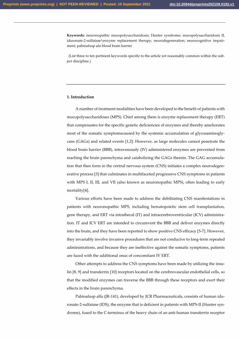
The major advantage of pabinafusp alfa is the fusion of anti-hTfR antibody to IDS, which
gives it the ability to pass the BBB to reach CNS tissues [17, 18]. Figure 1 shows the results
of an immunohistochemical analysis in which pabinafusp alfa, when administered intra-
venously to cynomolgus monkeys and hTfR-expressing mice, was found in neuronal cells
in the different brain regions [11]. These results indicate that , thanks to its TfR-binding
ability, pabinafusp alfa reaches the brain parenchyma by crossing the BBB via TfR-medi-
ated transcytosis.
Preprints (www.preprints.org) | NOT PEER-REVIEWED | Posted: 10 September 2021 Preprints (www.preprints.org) | NOT PEER-REVIEWED | Posted: 10 September 2021 doi:10.20944/preprints202109.0192.v1
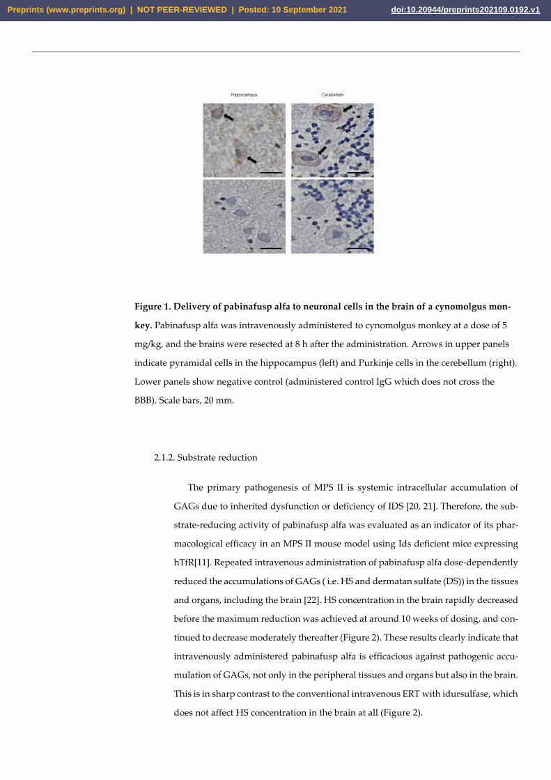
Figure 1. Delivery of pabinafusp alfa to neuronal cells in the brain of a cynomolgus mon-
key. Pabinafusp alfa was intravenously administered to cynomolgus monkey at a dose of 5
mg/kg, and the brains were resected at 8 h after the administration. Arrows in upper panels
indicate pyramidal cells in the hippocampus (left) and Purkinje cells in the cerebellum (right).
Lower panels show negative control (administered control IgG which does not cross the
BBB). Scale bars, 20 mm.
2.1.2. Substrate reduction
The primary pathogenesis of MPS II is systemic intracellular accumulation of
GAGs due to inherited dysfunction or deficiency of IDS [20, 21]. Therefore, the sub-
strate-reducing activity of pabinafusp alfa was evaluated as an indicator of its phar-
macological efficacy in an MPS II mouse model using Ids deficient mice expressing
hTfR[11]. Repeated intravenous administration of pabinafusp alfa dose-dependently
reduced the accumulations of GAGs ( i.e. HS and dermatan sulfate (DS)) in the tissues
and organs, including the brain [22]. HS concentration in the brain rapidly decreased
before the maximum reduction was achieved at around 10 weeks of dosing, and con-
tinued to decrease moderately thereafter (Figure 2). These results clearly indicate that
intravenously administered pabinafusp alfa is efficacious against pathogenic accu-
mulation of GAGs, not only in the peripheral tissues and organs but also in the brain.
This is in sharp contrast to the conventional intravenous ERT with idursulfase, which
does not affect HS concentration in the brain at all (Figure 2).
Preprints (www.preprints.org) | NOT PEER-REVIEWED | Posted: 10 September 2021 Preprints (www.preprints.org) | NOT PEER-REVIEWED | Posted: 10 September 2021 doi:10.20944/preprints202109.0192.v1

Figure 2. Substrate reducing efficacy of pabinafusp alfa in the brain of MPS II mice. Pabin-
afusp alfa was intravenously administered to the mice at a dose of 2 mg/kg once per
week for 1, 4, 8, 12, or 36 weeks. The dose of idursulfase was 0.5 mg/kg/week. Data
are from independent experiments (mean with S.D. bars, n = 3-5).
2.1.3. Prevention of neuroinflammation and subsequent neurodegeneration
The neurodegenerative processes in MPS II mice were preceded by activation of glial
cells [23, 24]. For instance, the intensity of glial fibrillary acidic protein (GFAP) signals
increased in the astroglia, as did the number of CD68-positive microglia in the brain cor-
tex (Figure 3). These histopathological changes were suppressed by chronic intravenous
treatment with pabinafusp alfa (Figure 3). The relief from neuroinflammation afforded
by pabinafusp alfa further prevented morphological abnormalities and neuronal death
in the brain of untreated MPS II mice (Figure 3), whereas idursulfase was ineffective
against these pathological changes in the brain [22].
Preprints (www.preprints.org) | NOT PEER-REVIEWED | Posted: 10 September 2021 Preprints (www.preprints.org) | NOT PEER-REVIEWED | Posted: 10 September 2021 doi:10.20944/preprints202109.0192.v1

Figure 3. Prevention of neuroinflammation and neurodegeneration by pabinafusp alfa in
MPS II mouse brains. Specimens from the brain cortex were stained with GFAP (top),
CD68 (middle), and hematoxylin/eosin (bottom). Data are from the 36-week study. Ar-
rows indicate vacuolation of neuronal cells. Scale bars, 50 m.
2.1.4. Prevention of neurocognitive abnormalities
In MPS II mice, progressive neurocognitive impairments manifest themselves as loss
of spatial learning ability that can be assessed with the Morris water maze test [25]. When
normal healthy mice were subjected to the test, the time to reach the platform ( goal latency
) became shorter day by day, whereas untreated MPS II mice showed difficulty learning
how to reach the platform [22] (Figure 4). The mice receiving chronic treatment with pabin-
afusp alfa maintained their spatial learning ability, unlike the wild-type animals (Figure
4). Idursulfase failed to prevent the loss of learning ability, so the attenuation of neurocog-
nitive abnormalities observed in the pabinafusp alfa-treated MPS II mice can be primarily
attributed to the clearance of HS deposited in the brain. In other words, HS concentration
in the brain can be viewed as a good predictor of neurodegeneration as well as a marker
of drug efficacy in patients with neuronopathic MPS II.
Preprints (www.preprints.org) | NOT PEER-REVIEWED | Posted: 10 September 2021 Preprints (www.preprints.org) | NOT PEER-REVIEWED | Posted: 10 September 2021 doi:10.20944/preprints202109.0192.v1

Figure 4. Maintenance of spatial learning abilities in MPS II mice receiving chronic treat-
ment with pabinafusp alfa. After 36 weeks of treatment, spatial learning ability was
assessed with the Morris water maze test. The time to reach the platform (goal la-
tency) was measured 3 times per day and the means were calculated within each day
for individual animals. Values are presented as the mean with S.E. for each group (n
= 12-15). Paired t-test, **P < 0.01 (Day 1 vs. Day 5). EW, every week; EoW, every other
week.
2.1.5. Identification of biomarker for CNS efficacy
Thanks to the weakness of the barrier between the brain parenchyma and the cere-
brospinal fluid (CSF) [26], HS concentrations in the brain are considered to be directly
correlated with those in the CSF, as demonstrated by the high correlations we found be-
tween the intracerebral and CSF HS concentrations in the MPS II mice treated with pabin-
afusp alfa (Figure 5). Thus, HS concentrations in the CSF are a useful and practical sur-
rogate biomarker to monitor drug efficacy in patients with neuronopathic MPS II, be-
cause the HS concentrations in the brain cannot be measured in clinical settings.
Preprints (www.preprints.org) | NOT PEER-REVIEWED | Posted: 10 September 2021 Preprints (www.preprints.org) | NOT PEER-REVIEWED | Posted: 10 September 2021 doi:10.20944/preprints202109.0192.v1
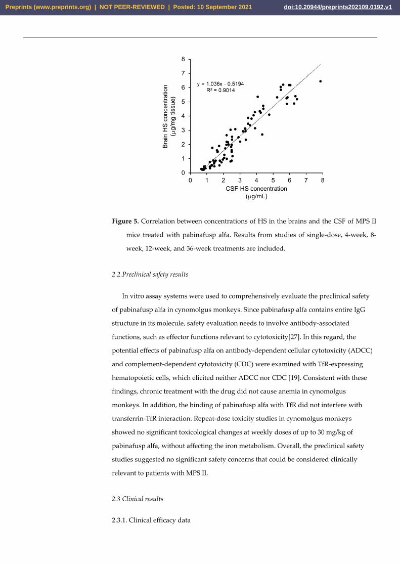
Figure 5. Correlation between concentrations of HS in the brains and the CSF of MPS II
mice treated with pabinafusp alfa. Results from studies of single-dose, 4-week, 8-
week, 12-week, and 36-week treatments are included.
2.2.Preclinical safety results
In vitro assay systems were used to comprehensively evaluate the preclinical safety
of pabinafusp alfa in cynomolgus monkeys. Since pabinafusp alfa contains entire IgG
structure in its molecule, safety evaluation needs to involve antibody-associated
functions, such as effector functions relevant to cytotoxicity[27]. In this regard, the
potential effects of pabinafusp alfa on antibody-dependent cellular cytotoxicity (ADCC)
and complement-dependent cytotoxicity (CDC) were examined with TfR-expressing
hematopoietic cells, which elicited neither ADCC nor CDC [19]. Consistent with these
findings, chronic treatment with the drug did not cause anemia in cynomolgus
monkeys. In addition, the binding of pabinafusp alfa with TfR did not interfere with
transferrin-TfR interaction. Repeat-dose toxicity studies in cynomolgus monkeys
showed no significant toxicological changes at weekly doses of up to 30 mg/kg of
pabinafusp alfa, without affecting the iron metabolism. Overall, the preclinical safety
studies suggested no significant safety concerns that could be considered clinically
relevant to patients with MPS II.
2.3 Clinical results
2.3.1. Clinical efficacy data
Preprints (www.preprints.org) | NOT PEER-REVIEWED | Posted: 10 September 2021 Preprints (www.preprints.org) | NOT PEER-REVIEWED | Posted: 10 September 2021 doi:10.20944/preprints202109.0192.v1

1) Substrate reduction in the CSF
On the basis of the preclinical findings(2.1.4), HS levels in the CSF were stipulated as
the primary efficacy surrogate endpoint in the three clinical trials conducted so far.
Figure 6 shows the baseline HS levels in the CSF of the 29 patients in the phase I/II and
III studies in Japan, which correlated with the disease severity that are ascribed to each
patient by his physicians based on their clinical judgment. The HS levels in the CSF
serves as an accurate indicator of neurodegenerative severity as well as a predictor of
clinical outcomes in terms of CNS manifestations. Most patients with attenuated
subtypes show HS levels below 4000 ng/ml, which may well indicate that this level is the
threshold below which CNS manifestations seldom, or only very slowly, develop.
Therefore, it may be useful as a tentative treatment goal for ERT in reducing or
maintaining HS levels.
Figure 6. Baseline HS concentrations in the CSF of the 29 patients in the phase II/III clinical trial of pabinafusp alfa
Figure 7 shows the changes in the HS levels in the CSF of all 39 patients in the phase
II/III study in Japan, the phase II study in Brazil and the respective extention studies.
The data are presented with respect to their MPS subtypes ( severe or atttenuated) and
all patients had received pabinafusp alfa for 104 weeks at either 2.0 or 4.0mg/kg. In the
Preprints (www.preprints.org) | NOT PEER-REVIEWED | Posted: 10 September 2021 Preprints (www.preprints.org) | NOT PEER-REVIEWED | Posted: 10 September 2021 doi:10.20944/preprints202109.0192.v1
2.0 mg/kg group in the phase II and phase II/III studies, HS concentrations in the CSF
significantly decreased between week 1 and week 26 (p<0.001), with a difference of
−3366±1923 ng/mL (relative changes from week 1 to week 26: −57.655±11.500%). HS
concentrations in the CSF decreased in all subjects in both groups. Notably, the
treatement reduced the CSF HS levels in the majority of the patients to below the
threshold level of 4000 ng/ml.
Figure 7. Reductons in the HS concentrations in the CSF of patients with the severe
and attenuated subtypes of MPS II in the phase II and II/III studies.
2) Neurocognitive efficacy
In the studies carried out in Japan, neurocognitive develoment was evaluated
according to the Kyoto Scale of Psychological Development (KSPD). This corresponds to
the Bayley scales of infant and toddler development, third edition (BSID-III), which was
employed for the patients in Brazil with developmental ages younger than 42 months
(age-equivalent [AE] scores), while the Kaufman Assessment Battery for Children, 2nd
edition (KABCII), was used for the older patients.
Figure 8 shows the changes in AE scores in the patients with the severe subtype of
MPS-II in the phase II and II/III studies, overlayed onto those from the natural history
data on Japanese patients with the severe subtype [28]. Almost all of the patients in
Brazil showed marked improvement in AE scores over 104 weeks, while most of the
Japanese patients showed stabilization, along with improvement in some.
Preprints (www.preprints.org) | NOT PEER-REVIEWED | Posted: 10 September 2021 Preprints (www.preprints.org) | NOT PEER-REVIEWED | Posted: 10 September 2021 doi:10.20944/preprints202109.0192.v1
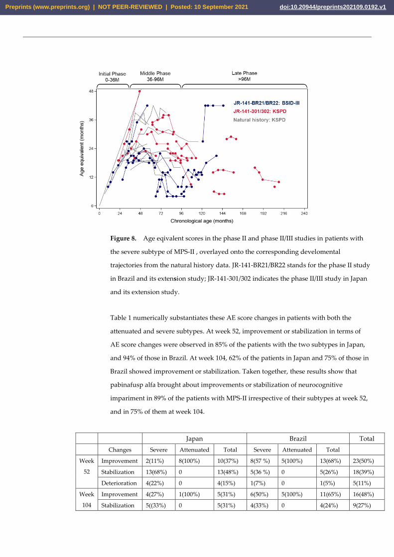
Figure 8. Age eqivalent scores in the phase II and phase II/III studies in patients with
the severe subtype of MPS-II , overlayed onto the corresponding develomental
trajectories from the natural history data. JR-141-BR21/BR22 stands for the phase II study
in Brazil and its extension study; JR-141-301/302 indicates the phase II/III study in Japan
and its extension study.
Table 1 numerically substantiates these AE score changes in patients with both the
attenuated and severe subtypes. At week 52, improvement or stabilization in terms of
AE score changes were observed in 85% of the patients with the two subtypes in Japan,
and 94% of those in Brazil. At week 104, 62% of the patients in Japan and 75% of those in
Brazil showed improvement or stabilization. Taken together, these results show that
pabinafusp alfa brought about improvements or stabilization of neurocognitive
impariment in 89% of the patients with MPS-II irrespective of their subtypes at week 52,
and in 75% of them at week 104.
Japan Brazil Total
Changes Severe Attenuated Total Severe Attenuated Total
Week
52
Improvement 2(11%) 8(100%) 10(37%) 8(57 %) 5(100%) 13(68%) 23(50%)
Stabilization 13(68%) 0 13(48%) 5(36 %) 0 5(26%) 18(39%)
Deterioration 4(22%) 0 4(15%) 1(7%) 0 1(5%) 5(11%)
Week
104
Improvement 4(27%) 1(100%) 5(31%) 6(50%) 5(100%) 11(65%) 16(48%)
Stabilization 5((33%) 0 5(31%) 4(33%) 0 4(24%) 9(27%)
Preprints (www.preprints.org) | NOT PEER-REVIEWED | Posted: 10 September 2021 Preprints (www.preprints.org) | NOT PEER-REVIEWED | Posted: 10 September 2021 doi:10.20944/preprints202109.0192.v1

Deterioration 6(40%) 0 6(38%) 2(17%) 0 2(12%) 8(24%)
Table 1. Changes in AE scores on KSPD (for Japan), BSID-III and KABCII (for Brazil).
Changes in AE scores exceeding 3 months were defined as improvement, while those ± 3
months as stabilization, and those below 3 months as deterioration, respectively.
In addition to the standardized neurocognitive assessments, clinical behavioral
observations by the subjects’ families and investigators were collected in order to
register subtle but potentially meaningful behavioral changes that the standardized
assessments might fail to capture, in particular in patients with the advanced severe
subtype (detailed tabulated reports of the narrative records are published elsewhere[14,
15]).
In both the phase II/III and II studies, similar behavioral changes were observed
across 3 major areas (speech, motor functions and liveliness/expression). Positive
behavioral changes in speech included increased utterances, better verbal
responsiveness and resumption of singing. In terms of liveliness/expression, stable
mood, less agitation and more smiling were often reported. These positive changes were
observed across all ages and subtypes, although the younger subjects showed more
marked improvements than the older ones. Notably, even among the subjects without
marked improvements in speech or motor functions (especially the adult subjects with a
long history of disease), positive changes in important attachment behaviors ( e.g.
smiling) [29] were still recognizable.
The fact that these subjective, non-standardized observations are in accordance with
the objective findings from the neurodevelopmental scales seems to further buttresses
the neurocognitive efficacy of pabinafusp alfa across all patient populations with
different ages and subtypes.
3) Somatic efficacy
As a measure of the efficacy of pabinafusp alfa against the somatic symptoms of MPS
II, liver and spleen volumes are reported here as representative efficacyl endpoints,
because progressive hepatosplenomegaly is a prominent clinical feature of the disease.
In evaluating the changes in organomegaly, attention needs to be paid to the large
variability in organ volumes in pediatric subjects, so individual subjects’ relative volume
changes at week 52, with the baseline volume taken as 100%. The liver and spleen
volumes significantly decreased in the naïve patients without prior ERT, and they also
decreased by about 5 % in the patients who were switched from conventional ERT to
pabinafusp alfa (Table 2, Figure 9).
Preprints (www.preprints.org) | NOT PEER-REVIEWED | Posted: 10 September 2021 Preprints (www.preprints.org) | NOT PEER-REVIEWED | Posted: 10 September 2021 doi:10.20944/preprints202109.0192.v1

Figure 9. Relative changes in the liver and spleen volumes from baseline to week 52. The
blue dotted lines are suggested thresholds of clinically significant changes (10% for liver
volume and 20% for spleen volume), and values below these can be interpreted as
either stabilization or improvement of hepatosplenomegaly.
ERT status N Mean (SD)
Median
[min—max] 95% CI
Liver volume Switched
subjects 28 -5.4 (13.3)
-6.0
[-38.0— 23.4] -10.6 — -0.2
Naïve subjects 4 -30.8 (6.5)
-31.1
[-38.4 — -
22.6]
-41.2 — -20.4
Spleen volume Switched
subjects 28 -4.3 (19.7)
-3.7
[-62.2 — 31.6] -11.9 — 3.4
Naïve subjects 4 -32.3 (6.7)
-33.3
[-38.7 — -
23.8]
-42.9 — -21.6
Table 2. Liver and spleen volumes
Liver volume
Spleen volume
Preprints (www.preprints.org) | NOT PEER-REVIEWED | Posted: 10 September 2021 Preprints (www.preprints.org) | NOT PEER-REVIEWED | Posted: 10 September 2021 doi:10.20944/preprints202109.0192.v1

These results, along with other somatic efficacy data (e.g. changes in serum HS and DS
concentrations and cardiac function [14, 15]), suggest that the efficacy of pabinafusp alfa
against somatic symptoms is comparable to that ofconventional idursulfase.
2.3.2 Clinical safety data
The safety of pabinafusp alfa was evaluated on the basis of the results of the phase
I/II, II, and II/III studies, and safety was confirmed in the patients in the 1.0, 2.0, and 4.0
mg/kg groups. The optimal weekly dose of pabinafusp alfa was considered to be 2.0
mg/kg, because most of the adverse drug reactions (ADRs) in this group were mild and
all duly managed without patients having to withdraw from the study. A summary of
the clinical safety of pabinafusp alfa is shown in Table 3. Of note is the fact that no dose-
limiting toxicities were observed at 4.0 mg/kg in the phase II study in Brazil, even
though the infusion-associated reaction (IARs) were observerd most frequently at this
dose.
Phase I/II study in Japan Phase II/III study in Japan Phase II study in Brazil All
N
Proportion
(%)
Number
of events N
Proportion
(%)
Number
of events N
Proportion
(%)
Number
of events N
Proportion
(%)
Number
of events
Number of subjects 14 -- -- 28 -- -- 20 -- -- 62 -- --
Adverse events 9 64.3 20 28 100.0 340 20 100.0 202 57 91.9 562
Serious adverse events 1 7.1 1 5 17.9 10 7 35.0 7 13 21.0 18
(Deaths) 0 0.0 0 1 3.6 2 1 5.0 1 2 3.2 3
Significant adverse events 4 28.6 8 17 60.7 61 11 55.0 47 32 51.6 116
(Infusion associated reacion) 4 28.6 8 14 50.0 51 10 50.0 45 28 45.2 104
Adverse drug reactions 7 50.0 11 15 53.6 59 11 55.0 46 33 53.2 116
Serious adverse drug reactions 1 7.1 1 0 0.0 0 0 0.0 0 1 1.6 1
(Deaths) 0 0.0 0 0 0.0 0 0 0.0 0 0 0.0 0
Significant adverse drug reac-
tions 4 28.6 8 14 50.0 51 10 50.0 45 28 45.2 104
(Infusion associated reacion) 4 28.6 8 14 50.0 51 10 50.0 45 28 45.2 104
Table 3. Summary of adverse events and adverse drug reactions for pabinafusp alfa in
the phase II/III and II studies
There were 11 non-fatal serious adverse events in the three studies, none of which
were related to pabinafusp alfa. Both of the two reported deaths were due to respiratory
Preprints (www.preprints.org) | NOT PEER-REVIEWED | Posted: 10 September 2021 Preprints (www.preprints.org) | NOT PEER-REVIEWED | Posted: 10 September 2021 doi:10.20944/preprints202109.0192.v1
failure as an exacerbation of MPS-II and unrelated to the drug. In summary, pabinafasup
alfa was found to be generally well tolerated at doses of up to 4mg/kg, and its long-term
safety up to 104 weeks has been confirmed as acceptable.
Discussion
To date, pabinafusp alfa is the first and only recombinant enzyme that can be suc-
cessfully delivered via intravenous administration across the BBB. It also demonstrated
clinically meaningful efficacy against the CNS symptoms of MPS-II, whilst also showing
efficacy against the somatic symptoms. Establishment of this novel IV ERT with both cen-
tral and peripheral efficacy has overcome formidable challenges.
Despite the fairly straightforward basic pathophysiology of neuronopathic MPS
which starts with a genetic enzyme deficiency that leads to the accumulation of uncatab-
olized substrates in the CNS and progressive neurodegeneration, details of the functional
and structural neuronal damages it causes remain yet to be elucidated [3]. To try to un-
ravel the complexities behind this pathogenesis and progression, we took a three-pronged
approaches. First, we evaluated the initial component of the pathogenesis (i.e. substrate
accumulation) by measuring HS concentrations in the brain and the CSF. Second, we car-
ried out histopathological evaluations to investigate the neurodegeneration, the second
component. And third, we examined behavioral abnormalities as representing CNS man-
ifestations, thereby capturing the final component of the neurodegenerative events.
The systemic GAG accumulations in our MPS II mouse model were reduced dose-
dependently by intravenous administration of pabinafusp alfa (2.1.2), which then duly
suppressed neuroinflammation and other neuropathological abnormalities (2.1.3), lead-
ing to normalization of impaired spatial learning abilities (2.1.4). These preclinical find-
ings corroborated the efficacy of pabinafusp alfa through all three components of the path-
ogenesis of the neuronopathy, and encouraged translation of these findings into clinical
studies.
We found that the extent of damages to the CNS and its manifestations in MPS II
mice were not solely determined by the HS concentrations in the brain, but also by the
duration of HS elevation [22], suggesting a cumulative pathogenicity of intracerebral HS
accumulation. It was clear, therefore, that temporal factors must be taken into account to
better address the onset and progression of neurodegeneration (Figure 10) [22]. This un-
derpins the importance of early introduction of ERT for patients with neuronopathic MPS-
Preprints (www.preprints.org) | NOT PEER-REVIEWED | Posted: 10 September 2021 Preprints (www.preprints.org) | NOT PEER-REVIEWED | Posted: 10 September 2021 doi:10.20944/preprints202109.0192.v1
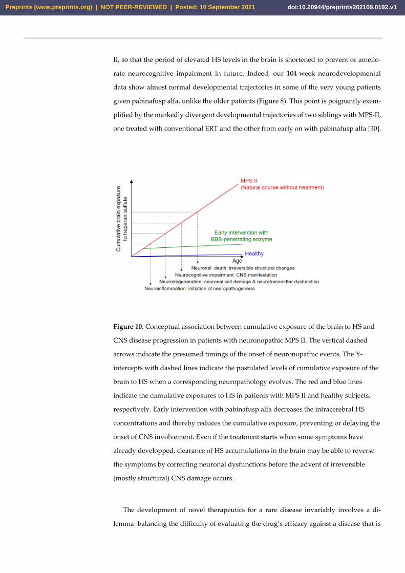
II, so that the period of elevated HS levels in the brain is shortened to prevent or amelio-
rate neurocognitive impairment in future. Indeed, our 104-week neurodevelopmental
data show almost normal developmental trajectories in some of the very young patients
given pabinafusp alfa, unlike the older patients (Figure 8). This point is poignantly exem-
plified by the markedly divergent developmental trajectories of two siblings with MPS-II,
one treated with conventional ERT and the other from early on with pabinafusp alfa [30].
Figure 10. Conceptual association between cumulative exposure of the brain to HS and
CNS disease progression in patients with neuronopathic MPS II. The vertical dashed
arrows indicate the presumed timings of the onset of neuronopathic events. The Y-
intercepts with dashed lines indicate the postulated levels of cumulative exposure of the
brain to HS when a corresponding neuropathology evolves. The red and blue lines
indicate the cumulative exposures to HS in patients with MPS II and healthy subjects,
respectively. Early intervention with pabinafusp alfa decreases the intracerebral HS
concentrations and thereby reduces the cumulative exposure, preventing or delaying the
onset of CNS involvement. Even if the treatment starts when some symptoms have
already developped, clearance of HS accumulations in the brain may be able to reverse
the symptoms by correcting neuronal dysfunctions before the advent of irreversible
(mostly structural) CNS damage occurs .
The development of novel therapeutics for a rare disease invariably involves a di-
lemma: balancing the difficulty of evaluating the drug’s efficacy against a disease that is
Preprints (www.preprints.org) | NOT PEER-REVIEWED | Posted: 10 September 2021 Preprints (www.preprints.org) | NOT PEER-REVIEWED | Posted: 10 September 2021 doi:10.20944/preprints202109.0192.v1
perhaps not fully understood and with few patients available for clinical trials on the one
hand with the requirement to expedite development to meet urgent medical needs on the
other. MPS-II is known to be a heterogeneous yet progressive, debilitating, and often fatal
disease. While long-term functional and structural assessments would have provided
more robust efficacy data, we had to make a realistic compromise had to be made to ad-
vance the development of pabinafusp alfa by capturing both the biochemical surrogate
endpoints and the clinical endpoints reflecting CNS manifestations. In other words, this
study examined both the initial process of neurodegeneration and, at the same time, some
of the clinical neuropsychiatric manifestations as the final outcome of the long and com-
plex pathological process. Limitations in the reported clinical studies, in particular in re-
spect to the long-term neurodevelopmental data, need to be addressed in post-marketing
studies in Japan and the planned phase III global trial, which will provide further evidence
of the dual efficacy of pabinafusp alfa against both somatic and CNS symptoms in patients
with MPS-II.
4. Materials and Methods
4.1. Preclinical studies
4.1.1. Animals
hTfR-KI/Ids-KO mice, a mouse model of MPS II, were established as described pre-
viously [11], and C57BL/6 mice (Charles River, Japan) were used as a normal control. The
cynomolgus monkeys (Macaca fascicularis) used had all been purpose-bred for research.
All animal experiments were conducted under the approval of the Animal Care and Use
Committees of Shin Nippon Biomedical Laboratories, Nihon Bioresearch, and JCR Phar-
maceuticals.
4.1.2. BBB penetration
Brain delivery of pabinafusp alfa by BBB penetration was determined by immuno-
histochemical analysis in cynomolgus monkeys that had received intravenous infusion
of the drug at 5 mg/kg. Pabinafusp alfa was detected with an HRP-labeled human IgG
antibody. Detailed methods are described elsewhere [11].
4.1.3. Substrate reduction
Preprints (www.preprints.org) | NOT PEER-REVIEWED | Posted: 10 September 2021 Preprints (www.preprints.org) | NOT PEER-REVIEWED | Posted: 10 September 2021 doi:10.20944/preprints202109.0192.v1
To evaluate the efficacy of pabinafusp alfa in reducing substrate accumulations, the
drug was administered to MPS II mice through the tail vein at a 2 mg/kg once a week for
1, 4, 8, 12, or 36 weeks. Control mice were given idursulfase at 0.5 mg/kg once a week.
One week after the final dosing, tissues and organs including the brain and CSF were
collected so that HS concentrations could be measured by liquid chromatography-tan-
dem mass spectrometry [12].
4.1.4. Evaluation of neuroinflammation and neurodegeneration
Pabinafusp alfa was intravenously administered to MPS II mice at 2 mg/kg once a
week for 36 weeks. One week after the final dosing, tissues and organs were collected
and the brains were subjected to histopathological analysis [22]. Expression of GFAP
was used as a marker for activation of the astrocytes, and expression of CD68 as a maker
for the activation of microglial cells. Staining with hematoxylin and eosin was per-
formed to detect morphological changes in neuronal cells.
4.1.5. Evaluation of neurocognitive abnormalities
The Morris water maze test was used to examine spatial learning ability was per-
formed after 36-week treatment. Briefly, each mouse was placed with its head facing the
wall of a circular pool equipped with a transparent acrylic resin platform, and the time
taken to reach the platform (goal latency) was measured [22].
4.1.6. Safety
Safety evaluations using in vitro assays in cynomolgus monkeys were carried out as
previously described [19].
4.2. Clinial studies
The study designs, procedures, outcomes, statistical analyses and other l details of
the three clinical trials of pabinafusp alfa are summarised in Appendix A.
5. Conclusions
Preprints (www.preprints.org) | NOT PEER-REVIEWED | Posted: 10 September 2021 Preprints (www.preprints.org) | NOT PEER-REVIEWED | Posted: 10 September 2021 doi:10.20944/preprints202109.0192.v1

This article summarizes and updates our preclinical and clinical evidence of the dual
efficacy of pabinafusp alfa against both the central and peripheral/somatic symptoms of
neuronopathic MPS-II. The drug’s mechanism of action has been highlighted: intrave-
nously administered IDS is delivered to the body via M6P- and TfR-mediated transcyto-
ses, and into the brain parenchyma via TfR-mediated transcytosis. By reducing HS accu-
mulations in the brain, pabinafusp alfa prevents or alleviates neurodegeneration. Further
long-term data on more patients is expected to provide further evidence of the benefits of
this novel drug, and we hope that its mechanism of action can in due course be applied to
treat other neuronopathic lysosomal storage diseases, so that their hitherto unaddressed
CNS manifestations can also be better managed.
Author Contributions:
Conceptualization, K.T and H.S.; methodology, H.M.,K.M., T.Y. and S.S.; formal analysis, M.Y. and
T.I; investigation, R.G., A.M.M., T.O., Y.E., N.S.,K.N. and K.M.; writing—original draft preparation,
K.M. and Y.S.; writing—review and editing, Y.S. and M.S..; supervision, K.T.,H.S. and M.S.; project
administration, T.Y. and S.S.. All authors have read and agreed to the published version of the man-
uscript.
Funding:
This research was funded by JCR Pharmaceuticals.
Institutional Review Board Statement:
The study was conducted according to the guidelines of the Declaration of Helsinki, and approved
by the Institutional Review Boards of the 21 participating investigational sites. The names of the
sites and the IRBs, identification code of the study, and the approved dates are given in Appendix
B
Informed Consent Statement:
All patients or their legal guardians submitted a signed, informed consent form prior to enrolment
in the study. The blank informed consent documents for the studies are also attached as Appendix
C.
Data Availability Statement:
The data presented in this study may be available on request from the corresponding author. The
data are not publicly available due to the intellectual property rights for pabinafusp alfa.
Acknowledgments:
Preprints (www.preprints.org) | NOT PEER-REVIEWED | Posted: 10 September 2021 Preprints (www.preprints.org) | NOT PEER-REVIEWED | Posted: 10 September 2021 doi:10.20944/preprints202109.0192.v1
The authors are grateful to all investigators, sub-investigators, study coordinators, the clinical re-
search team, and patients involved for their contribution and commitment to the study. The helpful
suggestions and guidance of Elsa Shapiro of the University of Minnesota, Minneapolis, on the neu-
rodevelopmental assessment are greatly appreciated. We also thank Maiko Kokado, Naoko Ta-
kasao, Minako Kobayashi, Ryo Ibaraki, Ami Kamitani, Kohtaro Hamauchi, Yukimichi Nakano and
Saki Yasui of JCR Pharmaceuticals for their support at various stages of the study. Special thanks
are due to Timothy Minton of Keio University, Tokyo, for his immense editorial help.
Conflicts of Interest:
R.G. has been an investigator, consultant, and/or speaker within the last 12 months for Abeona,
Allevex, Amicus, Azafaros, BioMarin, Chiesi, Cyclo, DASA/GENEONE, Denali, Idorsia, Inventiva,
JCR, Lysogene, Novartis, Paradigm, PassageBio, PTC, RegenxBio, Sanofi-Genzyme, Sigilon, Sobi,
Takeda, and Ultragenyx. A.M.M. has received honoraria and support for travelling to congresses
from BioMarin, Sanofi Genzyme, Takeda, and Ultragenyx. A.M.M. has received research fundings
from Alexion, BioMarin, Sanofi Genzyme, and Takeda. T.O. has conducted consultancy for JCR
Pharmaceuticals and reports research grants from BioMarin Pharmaceutical, Green Cross, Sanofi,
Takeda, and JCR Pharmaceuticals. Y.E. has conducted consultancy for JCR Pharmaceuticals, and he
has been awarded grants and research support from Actelion, BioMarin Pharmaceutical, and Sanofi;
he has also received honoraria from Actelion, BioMarin Pharmaceutical, Sanofi, Takeda, and Dain-
ippon Sumitomo Pharma. N.S. has conducted consultancy for JCR Pharmaceuticals, and he has been
awarded grant/research support from Sanofi and Dainippon Sumitomo Pharma and honoraria from
Actelion, BioMarin Pharmaceutical, Sanofi, Shire, and Dainippon Sumitomo Pharma. K.N. has con-
ducted consultancy for JCR Pharmaceuticals. H.M., K.M., T.Y., M.Y., T.I., S.S., K.T. and Y.S. are em-
ployed by JCR Pharmaceuticals, of which H.S. and M.S. are board members.
Appendix A (attached)
Appendix B (attached)
Appendix C (attached)
References
1. Bigger, B.W.; Begley, D.J.; Virgintino, D.; Pshezhetsky, A.V. Anatomical changes and pathophysiology of the brain in mucopol-
ysaccharidosis disorders. Mol. Genet. Metab. 2018, 125, 322–331. doi: 10.1016/j.ymgme.2018.08.003
Preprints (www.preprints.org) | NOT PEER-REVIEWED | Posted: 10 September 2021 Preprints (www.preprints.org) | NOT PEER-REVIEWED | Posted: 10 September 2021 doi:10.20944/preprints202109.0192.v1
2. Jakobkiewicz-Banecka, J.; Gabig-Ciminska, M.; Kloska, A.; Malinowska, M.; Piotrowska, E.; Banecka-Majkutewicz, Z.; Banecki,
B.; Wegrzyn, A.; Wegrzyn, G. Glycosaminoglycans and mucopolysaccharidosis type III. Front. Biosci. 2016, 21, 1393–1409. doi:
10.2741/4463.
3. Sato, Y.; Okuyama, T. Novel Enzyme Replacement Therapies for Neuropathic Mucopolysaccharidoses. Int. J. Mol. Sci., 2020, 21,
400. doi: 10.3390/ijms21020400.
4. Burton BK, Jego V, Mikl J etal. (2017) Survival in idursulfase-treated and untreated patients with
mucopolysaccharidosis type II: data from the Hunter Outcome Survey (HOS). J Inherit Metab Dis. 2017, 40, 867-874.
doi: 10.1007/s10545-017-0075-x
5. Muenzer, J.; Hendriksz, C.J.; Fan, Z.; Vijayaraghavan, S.; Perry, V.; Santra, S.; Solanki, G.A.; Mascelli, M.A.; Pan, L.; Wang, N.;
et al. A phase I/II study of intrathecal idursulfase-IT in children with severe mucopolysaccharidosis II. Genet Med 2016, 18, 73–
81. doi: 10.1038/gim.2015.36.
6. Eisengart, J.B.; Pierpont, E.I.; Kaizer, A.M.; Rudser, K.D.; King, K.E.; Pasquali, M.; Polgree, L.E.; Dickson, P.I.; Le, S.Q.; Miller,
W.P.; Tolar, J.; Orchard, P.J. Lund TC. Intrathecal enzyme replacement for Hurler syndrome: Biomarker association with neu-
rocognitive outcomes. Genet. Med. 2019, 21, 2552–2560. doi: 10.1038/s41436-019-0522-1.
7. Seo, J.H.; Kosuga, M.; Hamazaki, T.; Shintaku, H.; Okuyama, T. Impact of intracerebroventricular enzyme replacement therapy
in patients with neuronopathic mucopolysaccharidosis type II. Mol Ther Methods Clin Dev. 2021, 21, 67-75. doi:
10.1016/j.omtm.2021.02.018.
8. Boado, R.J.; Hui, E.K-W.; Lu, J.Z.; Pardridge, W.M. Insulin receptor antibody-iduronate 2-sulfatase fusion protein: pharmacoki-
netics, anti-drug antibody, and safety pharmacology in Rhesus monkeys. Biotechnol Bioeng 2014, 111, 2317–2325. doi:
10.1002/bit.25289.
9. Giugliani, R.; Giugliani, L.; de Oliveira Poswar, F.; Donis, K.C.; Corte, A.D.: Schmidt, M.; Boado, R.J.; Nestrasil, I.; Nguyen, C.;
Chen, S.; Pardridge, W.M. l. Neurocognitive and somatic stabilization in pediatric patients with severe Mucopolysaccharidosis
Type I after 52 weeks of intravenous brain-penetrating insulin receptor antibody-iduronidase fusion protein (valanafusp alpha):
an open label phase 1-2 trial. Orphanet J Rare Dis 2018, 13,110. doi: 10.1186/s13023-018-0849-8.
10. Couch, J.A.; Yu, Y.J.; Zhang, Y.; Tarrant, J.M.; Fuji, R.N.; Meilandt, W.J.; Solanoy, H.; Tong, R.K.; Hoyte, K.; Luk ,W.; Lu, Y.;
Gadkar, K.; Prabhu, S.; Ordonia, B.A.; Nguyen, Q.; Lin, Y.; Lin, Z.; Balazs, M.; Scearce-Levie, K.; Ernst, J.A.; Dennis, M.S.; Watts,
R.J. Addressing safety liabilities of TfR bispecific antibodies that cross the blood-brain barrier. Sci Transl Med 2013, 5, 183ra57.
doi: 10.1126/scitranslmed.3005338.
11. Sonoda, H.; Morimoto, H.; Yoden, E.; Koshimura, Y.; Kinoshita, M.; Golovina, G.;Takagi, H.; Yamamoto, R.; Minami, K.; Mizo-
guchi, A.; Tachibana, K.; Hirato, T.; Takahashi, K.. A Blood-Brain-Barrier-Penetrating Anti-human Transferrin Receptor Anti-
body Fusion Protein for Neuronopathic Mucopolysaccharidosis II. Mol Ther 2018, 26, 1366–1374. doi:
10.1016/j.ymthe.2018.02.032.
12. Tanaka, N.; Kida, S.; Kinoshita, M.; Morimoto, H.; Shibasaki, T.; Tachibana, K.; Yamamoto, R. Evaluation of cerebrospinal fluid
heparan sulfate as a biomarker of neuropathology in a murine model of mucopolysaccharidosis type II using high-sensitivity
LC/MS/MS. Mol Genet Metab 2018, 125, 53-58. doi: 10.1016/j.ymgme.2018.07.013.
13. Okuyama, T.; Eto, Y.; Sakai, N.; Minami, K.; Yamamoto, T.; Sonoda, H.; Yamaoka, M.; Tachibana, K.; Hirato, T.; and Sato, Y.
Iduronate-2-Sulfatase with Antihuman Transferrin Receptor Antibody for Neuropathic Mucopolysaccharidosis II: A Phase
1/2 Trial. Mol Ther 2019, 27, 456–464. doi: 10.1016/j.ymthe.2018.12.005.
Preprints (www.preprints.org) | NOT PEER-REVIEWED | Posted: 10 September 2021 Preprints (www.preprints.org) | NOT PEER-REVIEWED | Posted: 10 September 2021 doi:10.20944/preprints202109.0192.v1
14. Giugliani, R.; Martins, A.M.; So, S.; Yamamoto, T.; Yamaoka, M.; Ikeda, T.; Tanizawa, K.; Sonoda, H.; Schmidt, M.; Sato, Y.
Iduronate-2-sulfatase fused with anti-hTfR antibody, pabinafusp alfa, for MPS-II: A phase 2 trial in Brazil. Mol Ther 2021, 29,
2378-2386. doi: 10.1016/j.ymthe.2021.03.019.
15. Okuyama, T.; Eto, Y.; Sakai, N.; Nakamura, K.;Yamamoto, T.; Yamaoka, M.; Ikeda, T.; So, S.; Tanizawa, K.; Sonoda, H.; Sato, Y.
(2021). A phase 2/3 trial of pabinafusp alfa, IDS fused with anti-human transferrin receptor antibody, targeting neurodegener-
ation in MPS-II. Mol Ther 2021, 29, 671–679. doi: 10.1016/j.ymthe.2020.09.039.
16. Tomanin, R.; Zanetti, A.; Zaccariotto, E.; D’Avanzo, F.; Bellettato, C.M.; Scarpa, M. Gene therapy approaches for lysosomal
storage disorders, a good model for the treatment of mendelian diseases. Acta Paediatr. Int J Paediatr 2012, 101, 692–701. doi:
10.1111/j.1651-2227.2012.02674.x.
17. Pardridge, W.M. Receptor-mediated peptide transport through the bloodbrain barrier. Endocr Rev 1986, 7, 314–330. doi:
10.1210/edrv-7-3-314
18. Pardridge, W.M.; Eisenberg, J.; Yang, J. Human blood-brain barrier transferrin receptor. Metabolism 1987, 36, 892–895. oi:
10.1016/0026-0495(87)90099-0.
19. Yamanoto, R.; Yoden, E.; Tanaka, N.; Kinoshita, M.; Imakiire, A.; Hirato, T.; Minami, K. Nonclinical safety evaluation of pabin-
afusp alfa, an anti-human transferrin receptor antibody and iduronate-2-sulfatase fusion protein, for the treatment of neu-
ronopathic mucopolysaccharidosis type II. Mol Genet Metab Rep 2021, 27, 100758. doi: 10.1016/j.ymgmr.2021.100758.
20. Neufeld, E.F.; Muenzer, J. The Mucopolysaccharidoses. In The Metabolic & Molecular Bases of Inherited Disease, Scriver, C.R.,
Beaudet, A.L.; Sly, W.S.; Valle, D.; Eds.; McGraw Hill, New York, U.S.A, 2001; pp. 3421–3452.
21. Tylki-Szymanska, A. Mucopolysaccharidosis type II, Hunter’s syndrome. Pediatr Endocrinol Rev 2014, 12 (Suppl 1 ), 107–113.
22. Morimoto, H.; Kida, S.; Yoden, E.; Kinoshita, M.; Tanaka, N.; Yamamoto, R.; Koshimura, Y.; Takagi, H.; Takahashi, K.; Hirato,
T.; Minami, K.; Sonoda, H. Clearance of heparan sulfate in the brain prevents neurodegeneration and neurocognitive impair-
ment in MPS II mice. Mol Ther 2021, 29, 1853–1861. doi: 10.1016/j.ymthe.2021.01.027.
23. Fusar Poli, E.; Zalfa, C.; D’Avanzo, F.; Tomanin, R.; Carlessi, L.; Bossi, M.; Nodari, L.R.; Binda, E.; Marmiroli, P.; Scarpa, M.;
Delia, D. ; Vescovi, A.L. ; De Filippis, L. Murine neural stem cells model Hunter disease in vitro: glial cell-mediated neurodegen-
eration as a possible mechanism involved. Cell Death Dis 2013, 4, e906. doi: 10.1038/cddis.2013.430.
24. Zalfa, C.; Verpelli, C.; D’Avanzo, F.; Tomanin, R.; Vicidomini, C.; Cajola, L.; Manara, R.; Sala, C.; Scarpa, M.; Vescovi, A.L.; De
Filippis, L. Glial degeneration with oxidative damage drives neuronal demise in MPSII disease. Cell Death Dis 2016, 7, e2331.
doi: 10.1038/cddis.2016.231.
25. D’Hooge, R.; De Deyn, P.P. Applications of the Morris water maze in the study of learning and memory. Brain Res Rev 2001, 36,
60–90. doi: 10.1016/s0165-0173(01)00067-4.
26. Jiménez, A.J.; Domínguez-Pinos, M.D.; Guerra, M.M.; Fernández-Llebrez, P.; Pérez-Fígares, J.M. Structure and function of the
ependymal barrier and diseases associated with ependyma disruption. Tissue Barriers 2014, 2, e28426. doi: 10.4161/tisb.28426.
27. Vidarsson, G.; Dekkers, G.; Rispens, T. IgG subclasses and allotypes: from structure to effector functions. Front Immunol 2014,
5, 520. doi: 10.3389/fimmu.2014.00520.
28. Seo, J-H.; Okuyama, T.; Shapiro, E.; Fukuhara, Y.; Kosuga, M. Natural history of cognitive development in neuronopathic mu-
copolysaccharidosis type II (Hunter syndrome): Contribution of genotype to cognitive developmental course. Mol Gen Metab
Reports 2020, 24, 100630. doi: 10.1016/j.ymgmr.2020.100630.
29. Bowlby, J. Attachment. 2nd ed., Basic Books, New York, USA, 1969.
Preprints (www.preprints.org) | NOT PEER-REVIEWED | Posted: 10 September 2021 Preprints (www.preprints.org) | NOT PEER-REVIEWED | Posted: 10 September 2021 doi:10.20944/preprints202109.0192.v1

30. Tomita, K.; Okamoto, S.; Seto, T.; Hamazaki, T.; S,o S.; Yamamoto, T.; Tanizawa, K.; Sonoda,, H.; Sato, Y: Divergent develop-
mental trajectories in two siblings with neuropathic mucopolysaccharidosis type II (Hunter syndrome) receiving conventional
and novel enzyme replacement therapies: a case report. JIMD Reports (in press)
31. Study of Intrathecal Idursulfase-IT Administered in Conjunction With Elaprase® in Pediatric Patients With Hunter Syndrome
and Early Cognitive Impairment (AIM-IT). https://clinicaltrials.gov/ct2/show/NCT02055118 accessed on 15 July, 2021
32. Pardridge, W.M. Drug transport in brain via the cerebrospinal fluid. Fluids and Barriers of the CNS, 2011, 8, 7. doi: 10.1186/2045-
8118-8-7.
33. Shapey, J.; Toma, A.; Saeed, S.R. Physiology of cerebrospinal fluid circulation. Curr Opin Otolaryngol Head Neck Surg 2019, 27,
326-333. doi: 10.1097/MOO.0000000000000576.
34. Tumani,T.; Huss, A.; Bachhuber, A. The cerebrospinal fluid and barriers – anatomic and physiologic considerations. In Handbook
of Clinical Neurology.; Deisenhammer, F.; Charlotte E. Teunissen, C.E.; Tumani, H., Eds.; Elsevier: Amsterdam, Netherlands,
2018; Volume 146, pp. 21-32.
Preprints (www.preprints.org) | NOT PEER-REVIEWED | Posted: 10 September 2021 Preprints (www.preprints.org) | NOT PEER-REVIEWED | Posted: 10 September 2021 doi:10.20944/preprints202109.0192.v1