article - department of engineering science and mechanics
TRANSCRIPT

J Neurosurg Pediatrics 8:000–000, 2011
502 J Neurosurg: Pediatrics / Volume 8 / November 2011
We previously reported that PIH is the most com-mon cause of infant hydrocephalus in Uganda, accounting for 60% of cases.4,5 Anecdotal evi-
dence suggests a similar prevalence of PIH in other coun-tries of sub-Saharan Africa. The majority of these cases are secondary to neonatal infection resulting in ventricu-litis, which often results in primary brain injury in addi-tion to the hydrocephalus. The pathogens and routes of infection are not known, but recent evidence implicates the role of gram-negative organisms, especially Acineto-
bacter species, and a possible seasonal pattern of occur-rence.1 The long-term survival of these infants after man-agement of their hydrocephalus has not previously been studied. Such information is necessary to adequately as-sess the costs and benefits of treating PIH in this con-text. Understanding the fate of these children over time is also important for improving treatment and follow-up strategies as well as for strengthening the mandate for re-search that can ultimately lead to prevention. This study explores survival and outcome in a cohort of Ugandan infants treated aggressively for PIH at the CCHU.
J Neurosurg Pediatrics 8:502–508, 2011
Five-year survival and outcome of treatment for postinfectious hydrocephalus in Ugandan infants
Clinical article
Benjamin C. Warf, m.D.,1,2 ariella r. Dagi, B.a.,3 Brian nsuBuga Kaaya, B.s.,4 anD steven j. sChiff, m.D., Ph.D.5
1Department of Neurosurgery, Children’s Hospital Boston, Harvard Medical School; 2Program for Global Surgery and Social Change, Department of Global Health and Social Medicine, Harvard Medical School, Boston; 3Harvard College, Cambridge, Massachusetts; 4CURE Children’s Hospital of Uganda, Mbale, Uganda; and 5Center for Neural Engineering, Departments of Neurosurgery, Engineering Science and Mechanics, and Physics, The Pennsylvania State University, University Park, Pennsylvania
Object. Neonatal infection is the most common cause of infant hydrocephalus in Uganda. Postinfectious hy-drocephalus (PIH) is often accompanied by primary brain injury from the original infection. Since 2001, ETV (with or without choroid plexus cauterization) has been our primary treatment for PIH. The long-term outcome in these children is unknown.
Methods. We studied the 5-year outcome in a cohort of 149 infants treated for PIH from 2001 to 2005 and who lived in 4 districts close to the hospital. Survival analysis was performed using the Kaplan-Meier method. Statistical significance was determined using the Fisher, Breslow, and log-rank tests.
Results. The patients’ mean age at presentation was 9.5 months (median 3.0 months). Eighty-four patients (56.4%) were successfully treated without a shunt. Operative mortality was 1.2% for ETV and 4.4% for shunt place-ment (p = 0.3). Five-year survival was 72.8% in the non–shunt-treated group and 67.6% in the shunt-treated group, with no difference in survival (log rank p = 0.43, Breslow p = 0.46). Of 43 survivors assessed at 5–11 years, those with shunts had significantly worse functional outcomes (p = 0.003–0.035), probably reflecting treatment selection bias since those with the worst sequelae of ventriculitis were more likely to be treated with shunt placement.
Conclusions. Nearly one-third of treated infants died within 5 years, and at least one-third of the survivors were severely disabled. There was no survival advantage for non–shunt-treated patients at 5 years. A randomized trial of endoscopic third ventriculostomy versus shunt placement for treating PIH may be indicated. Public health measures that prevent these infections are urgently needed. (DOI: 10.3171/2011.8.PEDS11221)
Key WorDs • postinfectious hydrocephalus • endoscopic third ventriculostomy • choroid plexus cauterization • ventriculoperitoneal shunt • outcome • survival • Uganda • Africa
Abbreviations used in this paper: CCHU = CURE Children’s Hospital of Uganda; CPC = choroid plexus cauterization; ETV = endoscopic third ventriculostomy; PIH = postinfectious hydrocepha-lus; VP = ventriculoperitoneal.
This article contains some figures that are displayed in color on line but in black and white in the print edition.
J Neurosurg: Pediatrics / Volume 8 / November 2011
Postinfectious hydrocephalus outcome
503
MethodsBeginning in 2001, ETV was attempted as the initial
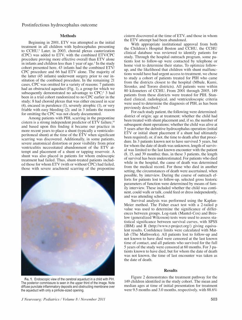
treatment in all children with hydrocephalus presenting to CCHU.5 Later, in 2003, choroid plexus cauterization (CPC) was added to ETV, with the combined ETV/CPC procedure proving more effective overall than ETV alone in infants and children less than 1 year of age.3 In the study cohort presented here, 43 infants had the combined ETV/CPC procedure and 66 had ETV alone. The majority of the latter (45 infants) underwent surgery prior to our in-stitution of the combined procedure. In the remaining 21 cases, CPC was omitted for a variety of reasons: 7 patients had an obstructed aqueduct (Fig. 1), a group for which we subsequently demonstrated no advantage to CPC;3 3 had been in a trial cohort randomized to no CPC earlier in the study; 8 had choroid plexus that was either encased in scar (4), encased in purulence (1), severely atrophic (1), or very friable with easy bleeding (2); and in 3 infants the reason for omitting the CPC was not clearly documented.
Among patients with PIH, scarring in the prepontine cistern is a strong independent predictor of ETV failure,10 and based upon this finding it became our practice in more recent years to place a shunt (typically a ventriculo-peritoneal shunt) at the time of the ETV when significant scarring was discovered. Additionally, in some patients, severe anatomical distortion or poor visibility from prior ventriculitis necessitated abandonment of the ETV at-tempt and placement of a shunt or tapping reservoir. A shunt was also placed in patients for whom endoscopic treatment had failed. Thus, shunt-treated patients includ-ed those for whom ETV (with or without CPC) had failed, those with severe arachnoid scarring of the prepontine
cistern discovered at the time of ETV, and those in whom the ETV attempt had been abandoned.
With appropriate institutional approval from both the Children’s Hospital Boston and CCHU, the CCHU clinical database was reviewed to identify patients for study. Through the hospital outreach program, some pa-tients lost to follow-up were contacted by telephone or home visit to determine their status. To optimize follow-up, and the likelihood that children with shunt malfunc-tions would have had urgent access to treatment, we chose to study a cohort of patients treated for PIH who came from the districts closest to the hospital (Mbale, Kumi, Sironko, and Tororo districts). All patients were within 80 kilometers of CCHU. From 2001 through 2005, 149 patients from these districts were treated for PIH. Stan-dard clinical, radiological, and ventriculoscopic criteria were used to determine the diagnosis of PIH, as has been previously described.5
For each study patient, the following were determined: district of origin; age at treatment; whether the child had been treated with shunt placement and, if so, the number of subsequent shunt operations; whether the child was alive at 5 years after the definitive hydrocephalus operation (initial ETV or initial shunt placement if a shunt had ultimately been required), or, if not, the time to death after that proce-dure. In 3 patients known not to have survived 5 years, but for whom the date of death was unknown, length of surviv-al was limited to the last known encounter with the patient (8, 15, and 39 months); thus, in these 3 patients, the length of survival has been underestimated. For patients who died while in the hospital, the cause of death was determined from the medical record. For those who died in another setting, the circumstances of death were ascertained, when possible, by interview. During the course of outreach ef-forts for patients lost to follow-up, selected gross historic parameters of function were determined by means of fam-ily interview. These included whether the child was conti-nent, could walk or talk, could feed or dress independently, and was attending school.
Survival analysis was performed using the Kaplan-Meier method. The Fisher exact test with a 2-tailed p value was used to determine the significance of differ-ences between groups. Log-rank (Mantel-Cox) and Bres-low (generalized Wilcoxon) tests were used to assess sta-tistical significance between survival curves, with SPSS (IBM) and R (http://www.r-project.org/) giving equiva-lent results. Confidence limits were calculated with Mat-lab (The Mathworks). All patients lost to follow-up and not known to have died were censored at the last known time of contact, and all patients who survived for the full 5 years of the study were censored at 60 months. For 3 pa-tients known to have died, but for whom the date of death was not known, the time of last encounter was taken as the date of death.
ResultsFigure 2 demonstrates the treatment pathway for the
149 children identified in the study cohort. The mean and median ages at time of initial presentation for treatment were 9.5 months and 3.0 months, respectively, with 88.6%
Fig. 1. Endoscopic view of the cerebral aqueduct in a child with PIH. The posterior commissure is seen in the upper third of the image. Note diffuse punctate inflammatory deposits and obstructing membrane over the aqueduct with only a pinhole-sized opening.

B. C. Warf et al.
504 J Neurosurg: Pediatrics / Volume 8 / November 2011
(132) being 1 year old or younger at initial treatment. At the time of the study, 84 (56.4%) children remained shunt-independent following ETV with or without CPC (includ-ing the 3 patients who underwent simultaneous shunt placement noted below), and a shunt had been placed in 65 (43.6%). Of the total of 68 shunts placed, 65 were ven-triculoperitoneal, 2 were subdural–peritoneal, and 1 was a ventriculosagittal sinus shunt. The operative mortality (death from any cause within 30 days of the procedure) was 1.2% (1 death in 81 procedures) for ETV and 4.4% (3 in 68) for shunt placement, which does not represent a significant difference (p = 0.3). Twenty-eight patients (18.8%) were lost to follow up before 5 years at a mean of 20.2 months and a median of 20 months. The outcome for 121 (81.2%) patients was known for at least 50 months.
In 37 (54.4%) of 68 shunt-treated patients, a shunt was placed because the ETV attempt had been abandoned. This included 6 of 9 patients in whom the ETV attempt had been initially abandoned for reservoir placement due to poor visibility resulting from murky CSF. In 28 (41.2%) the shunt was placed after failure of the endoscopic pro-cedure. In another 3 patients a VP shunt was placed at the time of successful ETV completion because of scarring noted in the prepontine cistern, based upon our finding of increased risk for ETV failure when cisternal scarring is encountered.10 These 3 patients were successfully treated with no additional operations at 25.5, 48, and 54 months of follow-up, respectively. Twelve of 40 patients with failed ETVs were successfully treated by repeat ventriculoscopy for reopening of the ETV. Three patients in whom ETV was initially abandoned for reservoir placement were able to have an ETV completed at a subsequent operation after the CSF had cleared. One of these had required no further treatment at 66 months follow-up, and the other 2 died at 2 and 4.3 months. Thus, of all 149 infants presenting for treatment of PIH, 84 (56.4%) were treated successfully without shunting and 65 (43.6%) required a shunt. Of 109 patients with a completed ETV (with or without CPC), 69 (63.3%) required no further surgery (Fig. 2).
The 5-year survival rate was 70.5% for the entire cohort with a mean survival of 46.2 months (95% CI 42.6–49.9 months). The 5-year survival rate in the non–shunt-treated group was 72.8%, with mean survival of 47.4 months (95% CI 42.7–52.2 months). For the shunt-treated patients, 5-year survival was 67.6% with a mean survival of 44.8 months (95% CI 39.2–50.4 months) (Fig. 3). There was no signifi-cant difference in survival between the groups as assessed by log rank (p = 0.43) and Breslow (generalized Wilcoxon p = 0.46) methods. Twenty-one (48.8%) of 43 known deaths occurred in the 1st year after the initial ETV or shunt place-ment. The causes of death, as ascertained from the medi-cal records or family interviews, are presented in Tables 1 and 2. Among patients with shunts, 5 (45%) of 11 known deaths in the 1st year were related to a shunt complication, while 1 death after that time was suspicious for a shunt malfunction. Among patients without a shunt, 2 deaths in the 1st year were secondary to ventriculitis (one of which was identified as a gram-negative infection). These deaths occurred at 1.2 months after the initial ETV and 1.0 month after a repeat ETV.
Shunt-treated patients had an average of 1.06 revi-sion operations per patient (median 0). At least 1 shunt revision was performed in 25 (37%) of the shunt-treated patients, and 14 (21%) required 2–10 revisions during the follow up period (Table 3). Excluding those lost to follow-up, the average for 5-year survivors was 0.97 revisions per patient, and for nonsurvivors the average was 1.61. There was no statistically significant difference (p = 0.4) between the number needing at least 1 revision among survivors (11 of 32) and that among nonsurvivors (11 of 23). Flexible ventriculoscopy was invaluable in many cas-es for the fenestration of intraventricular septations that had caused multiloculated hydrocephalus.
Table 4 presents a gross assessment of overall func-tional outcome in 42 children between the ages of 5 and 11 years (mean age 7.3 years, median 7.2 years) in whom the information could be obtained. Although most were continent of urine, slightly less than 2 of 3 could walk,
Fig. 2. Flowchart demonstrating the treatment path for 149 patients in the study cohort.

J Neurosurg: Pediatrics / Volume 8 / November 2011
Postinfectious hydrocephalus outcome
505
talk, or feed and dress themselves independently. Only 38% of those for whom the information was available at-tended school. There was a statistically significant differ-ence in functional outcomes between shunt-treated and non–shunt-treated patients. The majority of those without shunts could walk, talk, and feed and dress themselves; whereas, the majority of those with shunts could not.
Discussion
Postinfectious Hydrocephalus
In sub-Saharan Africa, probably between 80,000 and 375,000 infants develop hydrocephalus each year, and, the most conservative estimates suggest a potential eco-nomic burden to the region, in US dollars, ranging from $1 billion (using a human capital approach) to as much as
$56 billion (using the value of a statistical life approach) per year.6 In 2005, we provided the first report of neonatal infection as the major cause of infant hydrocephalus in an African country.5 In addition to the hydrocephalus (PIH), which can be treated, some of these children have dev-astating brain injuries from the initial ventriculitis (Fig. 4). Thus, the treated population includes survivors with varying degrees of neurocognitive and motor disabilities.
Although the major pathogens for infections that lead to neonatal ventriculitis in Uganda are not known, we recently reported that, of the 94% of PIH patients who had bacterial DNA recovered from their CSF at the time of surgery for hydrocephalus treatment, the most com-monly detected DNA was that of gram-negative bacteria in the phylum Proteobacteria.1 Furthermore, within this phylum, Gammaproteobacteria predominated in patients who had developed their initial illness during the rainy season, with Acinetobacter species identified in the ma-jority. By contrast, Betaproteobacteria predominated in
Fig. 3. Kaplan-Meier survival curves. Left: Graph showing survival curves for ETV with or without CPC and shunt treat-ment. The time points at which patients were lost to follow-up (without known subsequent death) are shown as censored (short vertical marks). For those patients known to have died but for whom the date of death was not known, the time of last encounter was considered the length of survival. Patients who reached 60 months’ survival and who might have subsequently died were also censored for statistical calculations (but not marked). Note that there is a trend toward early and late shunt failures, but global statistical comparisons of the curves show that there is no overall statistical difference between the survival groups (p > 0.4 for both log-rank and Breslow generalized Wilcoxon tests). Right: Graph showing 95% CIs (broken lines, blue for ETV and green for shunt treatment) for the cumulative survival functions using Greenwood’s formula (Mathworks implementation), demonstrating that the survival curves at all survival times remain within each others’ CIs with the solitary exception of the shunt curve at 12 months.
TABLE 1: Causes of death in 23 patients with shunts
Death w/in 1 yr Death After 1 yr
shunt infection “hydrocephalus” (?shunt malfunction)shunt infection malariashunt infection malariashunt infection/malfunction measlesexposed shunt/aspiration vomitingmalaria, anemia vomiting & diarrheamalaria, anemia respiratory infectionfebrile illness, cardiac arrest malnutritioncardiac arrest, unknown illness neglect, “rejected by clan”unknown unknownunknown unknown
unknown
TABLE 2: Causes of death in 20 patients without shunts
Death w/in 1 yr Death After 1 yr
ventriculitis malariaventriculitis malaria, anemia, malnutrition (marasmus)malaria, diarrhea severe malnutritionmalaria, anemia severe malnutritionmalaria, pneumonia diarrhea, vomitingmalaria, cardiac arrest sudden death in sleepgastroenteritis, hematuria unknowntachycardia, cardiac arrest unknownunknown unknownunknown unknown

B. C. Warf et al.
506 J Neurosurg: Pediatrics / Volume 8 / November 2011
the CSF of children who had become ill during the dry season. Work is in progress to better characterize the sea-sonal patterns, modes, and specific pathogens of these in-fections to develop public health strategies for effective prevention and treatment.
Treatment Paradigm and SuccessGiven the lack of urgent access to treatment for shunt
failure in rural Uganda, we have emphasized the benefit of avoiding shunt-dependence whenever possible. Treatment with ETV/CPC has proven very effective for congenital infant hydrocephalus of several etiologies including that associated with myelomeningocele3,7 encephalocele,12 the Dandy-Walker complex,9 and congenital aqueductal ste-nosis.3 In the case of PIH, the outcome is very dependent upon whether there is obstruction from subarachnoid scar within the prepontine cistern. During the time of treat-ment for the patient cohort presented here, most infants with cisternal scarring were given an opportunity for shunt independence. Subsequently, we demonstrated that cisternal scar was an independent risk factor that more than doubled the likelihood of ETV failure.10 Therefore, at CCHU, most patients with PIH and significant cisternal scar now receive shunts at the time of the ETV. Unfortu-nately MR imaging with fast imaging employing steady-state acquisition (FIESTA), which can demonstrate cis-ternal scarring prior to surgery, is not available in Uganda and most other developing countries.8
In the present study, 56.4% of the children had re-mained shunt-independent over the course of follow-up (including the 3 who underwent simultaneous ETV and shunt placement because of cisternal scarring). Some in-
fants presenting with PIH were simply unable to undergo ETV because of severe anatomical distortion from epen-dymal and parenchymal destruction and intraventricular scarring, and a shunt was placed. In this study cohort, 54.5% of shunt placements were performed for that rea-son. Among 109 patients with a completed ETV with or without CPC, 69 had no additional operations, including 55 with good outcomes, 3 with good outcomes who had simultaneous shunt placement when a scarred prepontine cistern was encountered, 1 who died, and 10 who were lost to follow-up. Ninety-five of the 109 patients with a completed ETV were neither lost to follow-up nor treated with shunt placement at the time of the initial ETV or ETV/CPC procedure; in 55 (57.9%) of these 95, the proce-dure was successful and no further surgery was required. There was no significant difference in success between those with ETV alone (32 [59.3%] of 54) and those with ETV/CPC (23 [56.1%] of 41, p = 0.835). This corresponds to the lack of significant difference in outcome between ETV and ETV/CPC we reported previously for a larger cohort of infants with PIH.3 Of the 40 patients in whom endoscopic treatment failed, 12 were treated successfully by repeat ETV, and 28 required shunt placement (Fig. 2).
The shunt-treated group had an average of 1.06 revi-sion operations per patient. At least 1 shunt revision was performed in 37% of these patients, and 21% required 2–10 revisions during the follow-up period (Table 3). This does not include those patients, known and unknown, who died of a shunt malfunction or infection without the benefit of operative treatment. Excluding those lost to follow-up, 5-year survivors had an average of 0.97 shunt reoperations per patient, whereas the average for nonsur-vivors was 1.61. However, we found no statistically sig-nificant difference between survivors and nonsurvivors in the proportion undergoing at least 1 shunt revision (p = 0.4). In this study group the operative mortalities (4.4% for shunt placement and 1.2% for ETV) were very similar to those previously reported from CCHU.2,3,5
SurvivalThe 5-year survival rate was 67.6% for shunt-treated
patients and 72.8% for those without a shunt; however, this difference was not statistically significant. For com-parison, the 5-year survival rate of Ugandan infants in the general population is estimated at 84%.13 As with the functional outcomes discussed below, a trend toward better survival in the non–shunt-treated group might be
TABLE 3: Shunt revisions in 68 shunt-treated patients
No. of Shunt Revisions No. of Patients (%)
0 43 (63)1 11 (16)2 4 (5.9)3 4 (5.9)5 1 (1.5)6 2 (2.9)7 2 (2.9)
10 1 (1.5)
TABLE 4: Functional outcomes in 42 patients 5–11 years of age with and without a shunt*
No Shunt ShuntMeasure No Yes No Yes p Value Total “Yes”
continent? 3 25 (89.3) 1 13 (92.9) 1.000 38 (90.5)walks? 6 19 (76.0) 8 5 (38.5) 0.035 24 (63.2)talks? 6 21 (77.8) 9 6 (40.0) 0.021 27 (64.3)feeds self? 5 22 (92.6) 10 5 (33.3) 0.003 27 (64.3)dresses self? 8 19 (70.4) 10 5 (33.3) 0.027 24 (57.1)in school? 13 9 (40.9) 9 3 (25.0) 0.465 13 (38.2)
* Mean age 7.3 yrs, median 7.2 yrs. Values represent numbers of patients (%) unless otherwise indicated.

J Neurosurg: Pediatrics / Volume 8 / November 2011
Postinfectious hydrocephalus outcome
507
expected due to treatment selection bias, given that shunt-treated patients most likely had more severe primary brain injuries and thus were more debilitated from the outset. Our work has previously suggested that in Uganda young children with disabilities may have higher mortal-ity from treatable conditions, such as infectious diseases and malnutrition, than their unaffected peers by virtue of their disability,12,13 and that survival can be significantly enhanced by the “observer effect” of a community-based program that entails home visits.4,13 The 70.5% overall 5-year survival for PIH is similar to that for infants from the same region of Uganda treated for myelomeningocele (63%)13 and encephalocele (61%).12 This study did not demonstrate a significant difference in survival between shunt-treated and non–shunt-treated patients. We had similarly demonstrated no significant difference in 5-year survival among infants with myelomeningocele treated at CCHU in regard to whether or how hydrocephalus had been treated.13
These findings suggest that sufficient equipoise ex-ists to evaluate the clinical outcomes and assess the rela-tive costs and benefits of primary CSF shunting for PIH compared with the primary endoscopic approach in a prospective randomized trial. It is worth noting, however, that nearly half of all known deaths occurred within the 1st year, and that in shunt-treated patients nearly half of the deaths were related to a shunt complication (mostly infection). Also, the patients in this study were, by design, relatively close to the treating facility, with better access to treatment of shunt complications than the majority of children treated at CCHU. Furthermore, in a recent as-sessment of hydrocephalus treatment in Uganda, we con-cluded that avoiding shunt dependence decreased the to-tal cost of care over time.6
Functional OutcomeThere was a statistically significant difference in
functional outcome between the shunt-treated and non–shunt-treated patients who were surveyed (Table 4). This is not surprising, given the fact that shunt-treated patients
would have had the most severe ventricular and cister-nal scar and anatomical deformity, which would likely be consistent with a more severe primary brain insult. Thus, this most likely reflects a treatment selection bias, and does not suggest that patients who undergo shunt place-ment have a worse functional outcome by virtue of the fact that they are treated by means of CSF shunting. In a previous study we did not find an overall statistically significant difference in early neurocognitive outcome measures between infants with myelomeningocele whose hydrocephalus had been treated by ETV/CPC and those treated by VP shunt placement.11 Infants with PIH who were able to be treated endoscopically were likely, on the whole, to have had a less severe inflammatory brain in-sult. In this group, the majority were able to walk, talk, and feed and dress themselves, in contrast to the majority of those with shunts, who could not. The number attend-ing school could be confounded by economic, social, and environmental factors, and there was no difference be-tween groups in that regard.
It is notable here that of patients successfully treated for PIH and surviving more than 5 years, more than 1 in 3 appear to be severely permanently disabled from the initial infection as determined by their inability to inde-pendently perform basic activities of daily living. The only way to avoid this is by prevention, or at least timely effective treatment, of neonatal sepsis in the region. This can only be accomplished after the pathogens and routes of infection have been identified. We emphasize that in-fants with PIH represent a subset of children affected by neonatal sepsis, many of whom did not survive to develop hydrocephalus.
ConclusionsNeonatal infection is the most common cause of hy-
drocephalus in Ugandan infants. This study provides the first assessment of an aggressive program of PIH treat-ment with an emphasis on preventing shunt dependence, which was ultimately avoided in more than half of the
Fig. 4. Noncontrast axial CT scan of the brain obtained in a 5-month-old girl with a history of fever and seizures at 1 month of age and rapid development of macrocephaly. Note the severe, diffuse parenchymal destruction. A tapping reservoir was placed to externally drain CSF for 2 weeks until it was sufficiently clear. Ventriculoscopy revealed severe, diffuse ependymal scar, yellow intraventricular inflammatory deposits, and an occluded aqueduct. Endoscopic third ventriculostomy revealed arachnoid scarring within the prepontine cistern. Treatment with ETV/CPC failed and a VP shunt was placed 1 month later.

B. C. Warf et al.
508 J Neurosurg: Pediatrics / Volume 8 / November 2011
patients. Around one third of treated infants died within 5 years, and at least one-third of the survivors were severely disabled. There was no significant difference in survival between those with and those without shunts. Although functional outcomes appeared worse for shunt-dependent children, this most likely reflected treatment selection bias because those with the worst sequelae of ventriculi-tis were more likely to have undergone shunt placement. Nonetheless, half of the deaths occurred within 1 year of the original ETV or shunt placement, and shunt com-plication was the single most common cause of death for shunt-treated patients during that period. Our results suggest that a randomized prospective trial to study sur-vival, functional outcomes, and cost-benefit analysis for primary ETV compared with primary shunt placement in African infants with PIH would be useful. These findings also lend urgency to the call for public health measures that will lead to prevention and effective early treatment of the infections leading to PIH, which is a significant and under-recognized global health problem.
Disclosure
The authors report no conflict of interest concerning the mate-rials or methods used in this study or the findings specified in this paper.
Author contributions to the study and manuscript preparation include the following. Conception and design: Warf. Acquisition of data: Warf, Dagi, Kaaya. Analysis and interpretation of data: Warf, Dagi, Schiff. Drafting the article: Warf. Critically revising the article: all authors. Reviewed submitted version of manuscript: all authors. Approved the final version of the manuscript on behalf of all authors: Warf. Statistical analysis: Schiff. Administrative/technical/material support: Warf, Dagi. Study supervision: Warf.
References
1. Li L, Padhi A, Ranjeva SL, Donaldson SC, Warf BC, Mugamba J, et al: Association of bacteria with hydrocephalus in Ugandan infants. Clinical article. J Neurosurg Pediatr 7:73–87, 2011
2. Warf BC: Comparison of 1-year outcomes for the Chaabra and Codman-Hakim Micro Precision shunt systems in Uganda: a prospective study in 195 children. J Neurosurg 102 (4 Sup-pl):358–362, 2005
3. Warf BC: Comparison of endoscopic third ventriculostomy alone and combined with choroid plexus cauterization in in-fants younger than 1 year of age: a prospective study in 550 African children. J Neurosurg 103 (6 Suppl):475–481, 2005
4. Warf BC: Hydrocephalus associated with neural tube defects: characteristics, management, and outcome in sub-Saharan Africa. Childs Nerv Syst (in press)
5. Warf BC: Hydrocephalus in Uganda: predominance of infec-tious origin and primary management with endoscopic third ventriculostomy. J Neurosurg 102 (1 Suppl):1–15, 2005
6. Warf BC, Alkire BC, Bhai S, Hughes C, Schiff S, Vincent JR, et al: Costs and benefits of neurosurgical intervention for infant hydrocephalus in sub-Saharan Africa. Clinical article. J Neurosurg Pediatr 8:509–521, 2011
7. Warf BC, Campbell JW: Combined endoscopic third ventric-ulostomy and choroid plexus cauterization as primary treat-ment of hydrocephalus for infants with myelomeningocele: long-term results of a prospective intent-to-treat study in 115 East African infants. Clinical article. J Neurosurg Pediatr 2:310–316, 2008
8. Warf BC, Campbell JW, Riddle E: Initial experience with com-bined endoscopic third ventriculostomy and choroid plexus cauterization for post-hemorrhagic hydrocephalus of prema-turity: the importance of prepontine cistern status and the predictive value of FIESTA MRI imaging. Childs Nerv Syst 27:1063–1071, 2011
9. Warf BC, Dewan M, Mugamba J: Management of Dandy-Walker complex–associated infant hydrocephalus by combined endoscopic third ventriculostomy and choroid plexus cauteriza-tion. Clinical article. J Neurosurg Pediatr 8:377–383, 2011
10. Warf BC, Kulkarni AV: Intraoperative assessment of cerebral aqueduct patency and cisternal scarring: impact on success of endoscopic third ventriculostomy in 403 African children. Clinical article. J Neurosurg Pediatr 5:204–209, 2010
11. Warf BC, Ondoma S, Kulkarni A, Donnelly R, Ampeire M, Akona J, et al: Neurocognitive outcome and ventricular volume in children with myelomeningocele treated for hydrocepha-lus in Uganda. Clinical article. J Neurosurg Pediatrics 4: 564–570, 2009
12. Warf BC, Stagno V, Mugamba J: Encephalocele in Uganda: ethnic distinctions in lesion location, endoscopic management of hydrocephalus, and survival in 110 consecutive children. Clinical article. J Neurosurg Pediatr 7:88–93, 2011
13. Warf BC, Wright EJ, Kulkarni AV: Factors affecting survival of infants with myelomeningocele in southeastern Uganda. Clinical article. J Neurosurg Pediatr 7:127–133, 2011
Manuscript submitted May 27, 2011.Accepted August 10, 2011.Address correspondence to: Benjamin C. Warf, M.D., Depart-
ment of Neurosurgery, Children’s Hospital Boston, 300 Longwood Avenue, Boston, Massachusetts 02115. email: [email protected].