arthrodesis techniques in horses - webnode
TRANSCRIPT

Vet Clin Equine 21 (2005) 691–711
Arthrodesis Techniques in Horses
Chad J. Zubrod, DVM, MSa,*,Robert K. Schneider, DVM, MSb
aOakridge Equine Hospital, 6675 East Waterloo Road, Edmond, OK 73034, USAbEquine Orthopedic Surgery, College of Veterinary Medicine, Washington State University,
Pullman, WA 99164, USA
Osteoarthritis is a common problem that occurs in all types of horsessecondary to joint injury, osteochondrosis, or infection or primarily fromthe wear and tear of repetitive use. It can cause progressive degenerationof the joint to a point where normal joint function is no longer possible.Affected horses develop chronic lameness that cannot be successfully treatedwith anti-inflammatory medications or surgical procedures aimed atrestoring joint function. When these treatments fail to return the animalto athletic performance or comfortable use of the limb, arthrodesis is a finaloption for some joints. In horses with osteoarthritis of low-motion joints,such as the proximal interphalangeal (PIP) joint or the distal tarsal joints,arthrodesis is performed with the goal of returning them to athleticperformance. In high-motion joints, arthrodesis is performed to improve thehorse’s use of the limb, with the goal of giving the horse long-term comfortrather than returning the horse to performance. Horses with fusion of high-motion joints have some degree of lameness (comfortable cripple) butsupport enough weight on the limb to avoid complications from overloadingthe contralateral limb.
Arthrodesis refers to the surgical fusion of a joint, resulting in bonyankylosis. Ankylosis of some joints can occur naturally in horses; however,in high-motion joints, bony bridging of the articular surface is rare. As jointdisease becomes advanced, articular cartilage degeneration results in bone-on-bone contact and severe lameness. Ankylosis removes the source of painin many cases; however, without surgical intervention, the process occursover a period of several years and may never result in complete bony unionand pain-free ambulation in some joints. There are currently accepted
* Corresponding author.
E-mail address: [email protected] (C.J. Zubrod).
0749-0739/05/$ - see front matter � 2005 Elsevier Inc. All rights reserved.
doi:10.1016/j.cveq.2005.07.004 vetequine.theclinics.com

692 ZUBROD & SCHNEIDER
methods for arthrodesis of several joints in the horse; however, not all jointsare amenable to arthrodesis. This article describes arthrodesis procedurescommonly used in horses.
Successful arthrodesis requires debridement of the articular cartilagethrough the calcified cartilage layer, exposing the subchondral bone;alignment of the joint into a weight-bearing position; and stabilization [1].The methods by which these steps are accomplished are variable, dependingon the individual joint. In some joints, fusion occurs after ablation of thearticular cartilage; however, in most joints, internal fixation is used tostabilize the joint.
Case selection is an important aspect of performing arthrodesis pro-cedures in horses. Some horses can live comfortable long lives with osteo-arthritis in one or several joints without requiring fusion. In low-motionjoints, horses with chronic lameness that is not responsive to manage-ment with anti-inflammatory medications are candidates for arthrodesis.In high-motion joints, the criteria become more complicated. The indicationfor arthrodesis must be based on the comfort level of the horse on thelimb with the affected joint. Based on our clinical experience, horses that aresupporting less than 50% of their normal weight-bearing force on the legat a walk are candidates for an arthrodesis procedure because they are atrisk for developing overload laminitis in the foot of the contralaterallimb. Foals with this degree of lameness can develop angular limb deformi-ties as the result of the abnormal forces created on the limb when the foalcenters its leg to support most of its weight on one limb.
Arthrodesis of low-motion joints
Proximal interphalangeal joint
Arthrodesis of the PIP joint is performed in horses with chronicosteoarthritis that is no longer responsive to anti-inflammatory medications,osteochondrosis, articular fractures, unstable joint injuries, or septicarthritis. Unlike arthrodesis of high-motion joints, arthrodesis of the PIPjoint is usually performed with the goal of returning the horse to athleticperformance. Careers requiring repetitive stopping and turning, such as thatoccurring in western event horses, can predispose horses to osteoarthritis ofthis joint. These same horses are exposed to acute overload forces whenperforming at high speeds that can cause fractures or destabilizing injury tothe supporting soft tissue structures of this joint. In addition to performancehorses, any horse that suffers trauma to the pastern region can have severedamage to this joint. The pastern is frequently injured when horses geta foot caught in a fence or under a panel. Also, any horse running in a fieldat high speed can injure the pastern joint.
The PIP joint has been arthrodesed using a wide variety of techniques[2–13]. The first procedure that was routinely used to arthrodese this joint

693ARTHRODESES TECHNIQUES
in horses was three parallel 4.5-mm cortical bone screws placed across thejoint in lag fashion [13]. This procedure was successful in a large number ofhorses; however, these horses were frequently maintained in a half-limb castfor 6 weeks because of postoperative discomfort. Despite the relativelyrecent publication of retrospective studies reporting the successful use oftwo cortical bone screws placed in lag fashion across the joint forarthrodesis of the PIP joint [6], most surgeons have moved towardtechniques that have increased the stability of the fixation used to stabilizethe joint. Stability was initially increased by using 5.5-mm cortical bonescrews and since has evolved to the combination of a dorsal plate inaddition to lag screws [2,14]. Greater stability has increased the horse’spostoperative comfort, minimized patient morbidity, and allowed the horseto be taken out of a cast at 2 weeks and released from the hospital,decreasing the expense of treatment. Application of a dorsal dynamiccompression plate (DCP) with three transarticular lag screws seems to bethe most stable technique currently used in cases of osteoarthritis andarticular fracture [2,14].
The patient is placed in lateral recumbency with the affected limbpositioned uppermost. The configuration of some second phalanx fracturesmay dictate positioning the horse with the affected leg down. The PIP jointis approached through an inverted ‘‘T’’-shaped incision centered on dorsalmidline, with the horizontal component created 1 cm proximal and parallelto the coronary band. The subcutaneous tissue is dissected free of theextensor tendon and remains with the skin flaps as they are reflected. Theextensor tendon and joint capsule are transected in an inverted ‘‘V’’ to openthe PIP joint. The base of the V-shaped incision is positioned at the levelof the joint to allow access to the collateral ligaments laterally and medially.The PIP joint is distracted using Kelly hemostatic forceps or an osteotome,and the collateral ligaments are transected. The joint is then luxateddorsally, the articular cartilage is removed, and the subchondral bone of theproximal and middle phalanges is foraged. The joint is reduced, and a four-hole narrow DCP is contoured to the dorsal surface of the first phalanx andthe proximal portion of the second phalanx. A 5.5-mm cortical bone screw isinserted through the distal hole of the plate into the second phalanx. Thisscrew should be positioned to allow room for the next screw proximal in theplate to be placed transarticularly into the proximal aspect of the secondphalanx. This 4.5-mm cortical bone screw is angled to cross the joint butalso angled to avoid the distal screw in the plate. The plate is attached to theproximal phalanx in load fashion, using a 4.5-mm cortical bone screw in oneof the two proximal holes. With the joint in anatomic alignment, 5.5-mmcortical bone screws are placed in lag fashion on the lateral and medial sidesof the plate. The screws should cross the joint palmar or plantar surface tothe center of the articular surface. Intraoperative radiographic control isnecessary to avoid penetrating the palmar or plantar cortex of the secondphalanx close to the navicular bone (Fig. 1). The proximal screw is placed in

694 ZUBROD & SCHNEIDER
the plate routinely, usually through a stab incision through the extensortendon.
The extensor tendon is reapposed with simple interrupted sutures usingmonofilament absorbable suture. The skin is closed routinely. The incision iscovered with a sterile nonadherent dressing, and a fiberglass half-limb cast isapplied. The cast is maintained for 2 weeks, when the sutures are removedand the horse is placed in a heavy support bandage. The support bandage ismaintained for an additional 2 weeks. The horse is stall rested for 90 days; atthat time, radiographs are taken to evaluate fusion of the joint. If there isbone bridging the joint, the horse may then be turned out in a smallpaddock (30 ft � 30 ft) for an additional 3 to 6 months. Generally, 6 to 12months is required after surgery before horses return to complete soundness.
Reported success rates of the procedure for horses with osteoarthritis ofthe PIP joint range from 50% to 85%; however, technical failures have beenassociated with chronic lameness in some horses [2,3,5,9,11]. Technicalfailures include placing the plate too far distally, resulting in inflammationand osteoarthritis of the distal interphalangeal (DIP) joint, and having thetransarticular lag screws exit the second phalanx too far distally, resultingin inflammation in the area of the navicular bone. Many horses havesuccessfully resumed athletic careers after fusion of the PIP joint. Thesuccess rate seems to be higher in horses that have had one rear pastern jointfused [2,6]. In our experience, the most common reason for horses todevelop lameness after successful pastern fusion in the forelimbs is thedevelopment of osteoarthritis in the DIP joint. Case selection is importantto avoid horses that may already have arthritis started in the DIP joint so asto improve the long-term prognosis for this procedure.
Fig. 1. Intraoperative fluoroscopic image demonstrating the positioning of lag screws medial
and lateral to the dorsal plate. The screws should not penetrate the palmar aspect of the second
phalanx near the navicular bone and supporting ligaments.

695ARTHRODESES TECHNIQUES
Open PIP joint injuries are fairly common in horses secondary to woundsthat occur in the pastern region. Horses frequently catch a foot undera panel or fence, suffering trauma and lacerations that can penetrate thejoint and injure the collateral ligaments. Because the joint is open, usinginternal fixation increases the risk of persistent infection occurring becauseof the addition of foreign material. If the joint can be placed in a weight-bearing position, the PIP joint can be fused. The joint can be opened andarticular cartilage debrided similar to the approach described previously, orsome of the articular cartilage can be removed by passing a 4.5-mm drill bitacross the pastern joint through two or three vertical dorsal arthrotomyincisions (Fig. 2). After cartilage removal, the joint is packed withautogenous cancellous bone graft and antibiotic-impregnated polymethyl-methacrylate (PMMA) implants and the limb is stabilized with a half-limbcast [15]. A transfixation pin cast may be used depending on the instabilityof the joint and the degree of discomfort of the horse. The horse is usuallymaintained in a cast for 10 to 12 weeks. Although these horses can havemore complications attributable to infection and decreased comfort on thelimb in the postoperative period, those horses that fuse can return tosoundness and their intended use.
Fig. 2. The articular cartilage of the PIP is debrided by passing a drill bit across the joint in
a dorsal-to-palmar or plantar direction, and the joint is packed with autogenous cancellous
bone graft and antibiotic-impregnated PMMA.

696 ZUBROD & SCHNEIDER
Arthrodesis of the distal tarsal joints
Osteoarthritis of the distal intertarsal (DIT) and tarsometatarsal (TMT)joints (bone spavin) is a common cause of lameness in equine athletes.Chronic repetitive compression, torsion, and shear strains frequently causeosteoarthritis; however, it can also occur secondary to osteochondrosis(juvenile spavin) or articular fracture. Distal tarsal osteoarthritis is commonin horses performing in events that increase torsional and shear forces on thedistal tarsal joints, such as those that run hard at a gallop, jump obstacles,or race at a trot or pace as well as in western performance horses used forreining, cutting, roping, and barrel racing. Osteoarthritis of the DIT andTMT joints can be managed successfully with anti-inflammatory medica-tions; however, the results of medical management can be disappointing,and lameness may persist in 25% to 50% of the cases [16].
In some horses, the cartilage degeneration associated with osteoarthritisprogresses to bony fusion of the DIT and TMT joints. These horsesfrequently become sound; however, degeneration only progresses to fusionof the distal tarsal joints in both hind limbs in a small number of horses.Therefore, a number of techniques to promote fusion of these joints havebeen developed. The diversity of techniques reflects the lack of a singlesuperior method of arthrodesis. Recently, successful outcomes have beenreported after fusion procedures using surgical drilling (SD), sodiummonoiodoacetate (MIA) injection, and laser surgery of the DIT and TMTjoints [17–24].
A recent retrospective study evaluating the use of SD to treatosteoarthritis of the DIT and TMT joints reported that 59% of the horsesreturned to their previous level of athletic performance without signs oflameness [19]. SD removes focal areas of articular cartilage and promotesosseous union between the bones [22]. The cunean tendon is identified bypalpation, and a 30-mm vertical skin incision is made over the distal twotarsal joints on the medial side of the tarsus distal to the cunean tendon. A0.9-mm � 25-mm (20-gauge, 1-inch) needle is then inserted into the DIT andTMT joints. The position of the needle within the joint is confirmed withfluoroscopy or intraoperative radiographs. A 3.2-mm drill bit is passed intoeach joint, and placement of the drill is again confirmed with fluoroscopy. A4.5-mm drill bit is then used to create three drill holes across each joint ina diverging pattern. Each tract is drilled to a depth between 2 and 3 cm; drillpositioning in the joint is confirmed with fluoroscopy. Subcutaneous tissueand skin are closed routinely. The distal tarsus is placed in a bandage toprotect the surgical incision.
Intra-articular injections of MIA, a chemical compound that causescartilage death and stimulates fusion, have also been used to fuse the DITand TMT joints [23]. Study results have shown that 40% to 90% of theanimals had successful outcomes after MIA injection and that 97% of thehorses had bone bridging the joints at 12 months [17,18,23,24]. Many horses

697ARTHRODESES TECHNIQUES
exhibit discomfort for 12 to 24 hours after MIA injection, however.Injections of MIA can be performed under general anesthesia or in thestanding sedated horse. A 0.7-mm � 25-mm (22-gauge, 1-inch) needle isinserted into the TMT joint proximal to the fourth metatarsal bone anddirected distally and medially at an angle of 45� [25]. A 0.7-mm � 25-mm(22-gauge, 1-inch) needle is inserted into the DIT joint at the junction of thefused first or second, third, and central tarsal bones on the medial side of thetarsus, distal to the cunean tendon [25]. Confirmation of needle placement ismade by retrieval of synovial fluid or via radiography. Each joint is injectedwith 100 mg MIA diluted in 0.9% sodium chloride (2 mL) that has beenaspirated through a 0.22-mm filter (Millex-GV; Millipore Corporation,Bedford, Massachusetts) to eliminate bacteria [17].
Laser-facilitated ankylosis has also been performed using a neodymium:yttrium aluminum garnet (Nd:YAG) or diode laser to destroy articularcartilage by superheating and vaporizing synovial fluid [20,25]. It has beenproposed that temperatures greater than 62�C result in chondrocyte deathas well as a collagen shift in the intertarsal ligaments and joint capsule[20,21,26]. A 1.1-mm � 40-mm (18-gauge, 1-inch) needle is placed on themedial and lateral sides of each DIT and TMT joint. Needle position isconfirmed with radiographs or fluoroscopy. The lateral needles serve asvents for plume evacuation during the application of laser energy to thejoints. A 5-mm skin incision is made over each joint in the location of theneedle on the medial side of the tarsus using a number 11 scalpel blade. A1.3-mm � 90-mm (16-gauge, 3-inch) stainless-steel needle is placed in theDIT and TMT joints through the skin incisions. A 600-mm contact laserfiber (600-mm Bare Fiber Assy, Flat Tip; BioLitec, East Longmeadow,Massachusetts) is inserted through the needle; the laser fiber and needle areadvanced across the joint as the laser (AccuVet 50D, 980-nm Diode SurgicalLaser; Lumenis, Santa Clara, California) is activated. Approximately 1200 Jof laser energy is applied to each joint. Needles are cooled by irrigation withchilled 0.9% sodium chloride during the procedure. Skin incisions are closedwith 2-0 polypropylene suture in a simple interrupted pattern. The distaltarsus is placed in a sterile adhesive bandage. Clinical articles on laser-facilitated ankylosis techniques have reported an increased percentage ofsound horses, minimal postoperative pain, and decreased convalescent timecompared with other methods of arthrodesis [20,21,26].
Assessment of these three arthrodesis techniques for distal tarsal jointarthrodesis have been evaluated by the authors by comparing the degree oflameness after treatment as well as by the amount of fusion of the distal twotarsal joints [27]. Twelve horses were evaluated and were split into twogroups. Group 1 (n ¼ 6) had laser surgery performed on the DIT and TMTjoints of one tarsus and MIA injection into the contralateral DIT and TMTjoints; these horses were evaluated for 6 months. Group 2 (n ¼ 6) had lasersurgery performed on the DIT and TMT joints of one tarsus and SD ofthe contralateral DIT and TMT joints; these horses were evaluated for 12
698 ZUBROD & SCHNEIDER
months. Postoperative comfort, lameness, radiographs, microradiographs,and histologic findings were compared between groups and between thejoints within groups.
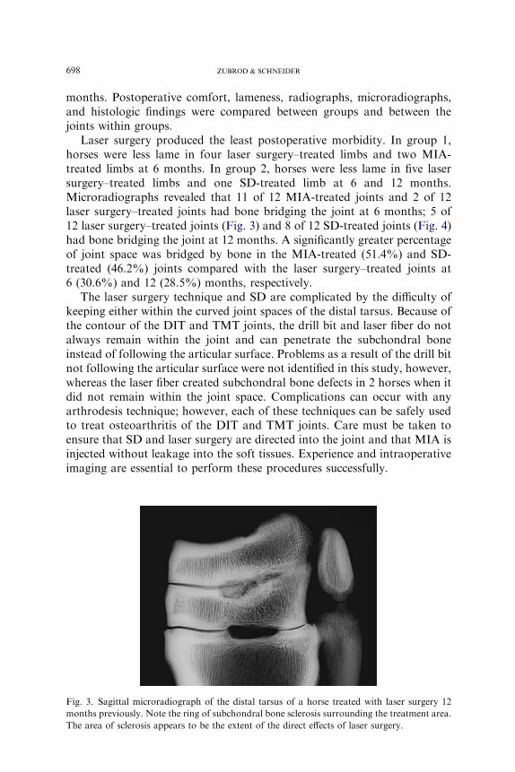
Laser surgery produced the least postoperative morbidity. In group 1,horses were less lame in four laser surgery–treated limbs and two MIA-treated limbs at 6 months. In group 2, horses were less lame in five lasersurgery–treated limbs and one SD-treated limb at 6 and 12 months.Microradiographs revealed that 11 of 12 MIA-treated joints and 2 of 12laser surgery–treated joints had bone bridging the joint at 6 months; 5 of12 laser surgery–treated joints (Fig. 3) and 8 of 12 SD-treated joints (Fig. 4)had bone bridging the joint at 12 months. A significantly greater percentageof joint space was bridged by bone in the MIA-treated (51.4%) and SD-treated (46.2%) joints compared with the laser surgery–treated joints at6 (30.6%) and 12 (28.5%) months, respectively.
The laser surgery technique and SD are complicated by the difficulty ofkeeping either within the curved joint spaces of the distal tarsus. Because ofthe contour of the DIT and TMT joints, the drill bit and laser fiber do notalways remain within the joint and can penetrate the subchondral boneinstead of following the articular surface. Problems as a result of the drill bitnot following the articular surface were not identified in this study, however,whereas the laser fiber created subchondral bone defects in 2 horses when itdid not remain within the joint space. Complications can occur with anyarthrodesis technique; however, each of these techniques can be safely usedto treat osteoarthritis of the DIT and TMT joints. Care must be taken toensure that SD and laser surgery are directed into the joint and that MIA isinjected without leakage into the soft tissues. Experience and intraoperativeimaging are essential to perform these procedures successfully.
Fig. 3. Sagittal microradiograph of the distal tarsus of a horse treated with laser surgery 12
months previously. Note the ring of subchondral bone sclerosis surrounding the treatment area.
The area of sclerosis appears to be the extent of the direct effects of laser surgery.

699ARTHRODESES TECHNIQUES
Because of the relatively focal area of severe cartilage and subchondralbone damage associated with laser surgery, it may be necessary to modifythe technique to promote more rapid and complete fusion. Performing lasersurgery at two or three locations within the joint would destroy morearticular cartilage, similar to that which occurs with surgical drilling. Thismay encourage more complete fusion, although still gaining the beneficialeffect that the laser energy seems to have on the innervation of the sub-chondral bone, synovium, and joint capsule.
This study demonstrates that laser surgery resulted in less fusion of theDIT and TMT joints compared with the other two techniques. MIA and SDproduced comparable amounts of bone bridging the joints when comparedat 6 and 12 months, respectively. Laser surgery resulted in bone bridging thejoint space in only 7 of 24 joints, and the smallest percentage of the jointspace was bridged by bone compared with the other two techniques. Despitethis observation, horses were more comfortable after surgery and mosthorses were less lame on the laser surgery–treated limb. This suggests thatlaser surgery may have a beneficial effect beyond fusion of the distal twotarsal joints.
Because of the difference in the postoperative comfort level between theSD- and laser surgery–treated joints in our study, it is difficult torecommend SD of the joint space. Therefore the authors currently usemodified laser treatment or MIA to fuse these two joints. In some horses,
Fig. 4. Sagittal microradiograph of the distal tarsus of a horse treated with SD 12 months
previously. There are focal areas of bone bridging within the drill tracts and bone lysis resulting
in enlargement of the drill tracts.

700 ZUBROD & SCHNEIDER
these two procedures are combined. In joints that are partially fused, a laseris passed through a drill hole across the joint in two or three locations; inmany horses, it is frequently the DIT joint that is partially fused. The TMTjoint is injected with MIA because it produces solid fusion of this joint withminimal risk of gaining access to the proximal intertarsal joint. Thisapproach requires evaluation and follow-up in a number of cases before itcan be recommended as the method of choice.
Arthrodesis of high-motion joints
Distal interphalangeal joint
Indications for arthrodesis of the DIP joint in horses are infrequent.Severe osteoarthritis, injury to the collateral ligaments that results in jointinstability, rupture of the deep digital flexor tendon, septic arthritis, orchronic articular fractures can all be a cause of chronic pain making affectedhorses candidates for arthrodesis of the DIP joint [28]. Successfularthrodesis of the DIP joint of the horse presents numerous challenges.Because this is a high-motion joint, rigid internal fixation or prolongedexternal coaptation is necessary to allow bone to bridge the joint. Thelocation of the DIP joint within the hoof capsule presents additionalchallenges for gaining access to the joint and for creating an asepticenvironment in which to place implants. Anatomically, there is a relativelysmall area of the distal phalanx that can hold implants. In addition, the hoofcapsule and laminae limit our ability to use DCPs to counteract the forcesacting on this joint.
The DIP joint is approached through two incisions [28,29]. The firstincision is made 1 cm proximal and parallel to the coronary band on thedorsal surface of the limb and is extended to the lateral and medial aspectsof the joint. The common or long digital extensor tendon is transected alongwith the joint capsule. The collateral ligaments are partially transected toallow enough space for curettes to be placed into the joint. The joint is thenpartially luxated, and the articular cartilage on the second and thirdphalanxes is debrided. It is not possible to remove all the articular cartilage;however, it is important to remove as much as possible. An alternativeapproach is to place an arthroscope in the dorsal and palmar or plantarjoint pouches and remove the articular cartilage with a motorized burr.
A second 8- to 10-cm longitudinal incision is made over the palmar orplantar surface of the pastern. The skin incision is made on the palmar orplantar surface midline of the limb from the proximal sesamoid bonesdistally to the heel bulbs. The incision is continued through the digitalannular ligaments and tendon sheath. The deep digital flexor tendon istransected to gain exposure to the palmar or plantar surface of the secondphalanx. The attachment of the straight distal sesamoidean ligament to thesecond phalanx is identified, and a vertical stab incision is created on

701ARTHRODESES TECHNIQUES
midline. The DIP joint is reduced and held in a weight-bearing position. A3.2-mm drill bit is used to drill through the second phalanx from the palmaror plantar surface proximal to the dorsal distal surface, bisecting thearticular surface from the dorsal to palmar or plantar surface. Positioning isconfirmed with fluoroscopy or radiographs. A 4.5-mm drill bit can be usedto make adjustments in the direction of the hole based on radiographs. Theglide hole in the second phalanx is then over-drilled with a 5.5-mm drill bit.A 4.0-mm drill bit is used to create the thread hole in the third phalanx. Adepth of 30 mm is sufficient in most horses and avoids penetrating thelaminae and hoof wall. A countersink is used to create a uniform surface forthe screw head to engage. The hole is measured and tapped in a routinemanner, and one 5.5-mm cortical bone screw is inserted. Screw placement isconfirmed with fluoroscopy or radiographs. Two additional screws areplaced in the same manner lateral and medial to the central screw. Once allthree screws are in position (Fig. 5), cancellous bone graft is harvested fromthe ipsilateral tuber coxae and placed into the dorsal and palmar or plantarsurfaces of the DIP joint [30]. Dorsally, the common digital extensor tendonand joint capsule are reapposed with absorbable suture material. Palmarly
Fig. 5. Postoperative radiograph of three 5.5-mm cortical bone screws placed in lag fashion
across the DIP joint. The screws do not penetrate the laminar surface of the third phalanx.

702 ZUBROD & SCHNEIDER
or plantarly, the deep digital flexor tendon is not reapposed. The tendonsheath is closed with absorbable suture in a simple interrupted pattern.Subcutaneous tissue and skin are closed routinely. The incisions are coveredwith a sterile nonadherent dressing, and a fiberglass cast is placed on thelimb from the carpus or tarsus distally. A transfixation pin cast can be usedto increase weight bearing on the affected limb when necessary. The cast ischanged, and sutures are removed at 2 weeks. The cast is changed every 4 to6 weeks depending on the horse’s comfort in the cast. Transfixation pin castsare usually maintained for 6 weeks, and the horse is then placed ina standard half-limb cast. Casts are normally maintained for approximately8 to 10 weeks. After removal of the cast, it is important to make appropriateshoeing adjustments to allow for comfort and use of the limb. Radiographsare taken at 90 days. If bone is present in the joint space, horses begin hand-walking. The horse’s use of the limb determines how soon the horse isturned out in a small paddock (30 ft � 30 ft).
Arthrodesis has also been performed using transarticular lag screws ina cruciate pattern with and without stainless-steel baskets [31]. Thesetechniques require a dorsal approach to the joint for debriding the articularcartilage; additionally, a flap in the dorsal hoof wall is necessary whenstainless-steel baskets are placed in the joint.
Arthrodesis of the DIP joint is accomplished without internal fixationwhen infection is present [32]. In these horses, the joint is approachedthrough a dorsal arthrotomy or with arthroscopy to debride the articularcartilage. The joint is packed with autogenous cancellous bone graft andantibiotic-impregnated PMMA implants, and the limb is placed in a half-limb transfixation pin cast [28]. The horse is usually maintained in atransfixation pin cast for 6 weeks and is then placed in a standard half-limbcast for another 6 weeks. The technique of using large cancellous bone graftsand external immobilization to fuse joints in the distal limb of the horse hasbeen reported [15].
Arthrodesis of the DIP joint is complicated by the anatomy and theconcentration of weight-bearing forces on the third phalanx in the mostdistal aspect of the limb. Internal fixation has been limited to lag screws,which do not always provide rigid immobilization. As a result, horses arenot always as comfortable after arthrodesis of the DIP joint as they are afterother fusion procedures. This increases the risk of laminitis in thecontralateral limb, especially when the horse has been treated for a longtime for chronic painful disease in the joint before performing the fusion.
Metacarpophalangeal and/or metatarsophalangeal joint
Arthrodesis of the metacarpophalangeal (MCP) and/or metatarsopha-langeal (MTP) joint is performed in horses with traumatic disruption of thesuspensory apparatus (biaxial sesamoid fractures, rupture of the suspensoryligament, or disruption of the distal sesamoidean ligaments) or chronic

703ARTHRODESES TECHNIQUES
osteoarthritis of the MCP and/or MTP joint, which has resulted in loss ofarticular cartilage and chronic pain because of exposure of the subchondralbone. In either case, the purpose of the procedure is to restore comfortableweight bearing on the limb for salvage of the animal rather than to returnthe horse to athletic performance.
Several techniques have been described for performing arthrodesis of thefetlock joint [33–42]. Although recent techniques offer some biomechanicaladvantages in vitro [41], the technique first described by Bramlage [33]continues to be the most routinely used, with some minor modifications.The skin and subcutaneous tissues are incised dorsolaterally over thecommon digital extensor tendon, extending from the proximal metacarpus/metatarsus to the PIP joint. The incision is continued through the commondigital extensor tendon, splitting it longitudinally, and through the dorsaljoint capsule of the MCP and/or MTP joint. A second incision is then madelaterally through the joint capsule and lateral collateral ligament of theMCP and/or MTP joint so that the joint can be luxated for debridement ofthe articular cartilage. Alternatively, a lateral condylar fracture can becreated with an oscillating bone saw (after predrilling holes for fracturerepair) to allow disarticulation of the joint; this approach is usually madethrough one dorsal skin incision. The articular cartilage of the first phalanx,the third metacarpal bone, and both sesamoid bones is removed witha curette or motorized burr. Osteostixis is performed on the subchondralbone plate of the third metacarpal bone and proximal phalanx. If there istraumatic disruption of the suspensory apparatus, a palmar or plantarfigure-of-eight tension band wire is placed at this time with the limb inpartial flexion to reconstruct palmar or plantar support for the jointbiomechanically. Placement of 1.5-mm wire is accomplished through3.2-mm holes drilled laterally to medially through the distal metacarpus/metatarsus and proximal phalanx; passing the wire in a figure-of-eightpattern is the most difficult part of the procedure. Use of an AO wire passercan greatly facilitate passage of the wire medially to laterally across thepalmar or plantar aspect of the joint.
The joint is then reduced and placed in 10� of dorsiflexion, and the dorsalsurface of the sagittal ridge of the third metacarpal bone is removed witha chisel to improve contact between the bone plate and the bone. A 14- to16-hole broad DCP is contoured to fit the dorsal surface. Alternatively,a fetlock arthrodesis plate has recently become available (LCP-ACP;Synthes, West Chester, Pennsylvania). If a condylar fracture was created, itis repaired with two 4.5-mm cortical bone screws at this time. The plateshould be placed so that screws in the distal four holes can be placed into thefirst phalanx. Minimal contouring of the plate is required. One screw is thenplaced through the plate and into the proximal phalanx in a routine manner.If the suspensory apparatus is intact, two 4.5-mm cortical bone screws areplaced in lag fashion through the third metacarpal bone into the proximalsesamoid bones while the proximal end of the plate is elevated

704 ZUBROD & SCHNEIDER
approximately 1.5 cm from the surface of the third metacarpal bone. Using4.5-mm screws increases the margin for error; if the positioning of the screwin the proximal sesamoid is not adequate, a 5.5-mm screw can be placed ina better position. Fixation of the proximal sesamoid bones allows the plateto be loaded against the distal sesamoidean ligaments, creating palmar orplantar support for the MCP and/or MTP joint. The plate is then loadedand attached to the third metacarpal bone with 5.5-mm cortical bonescrews. The 5.5-mm screws are recommended to increase the cyclic fatiguestrength of the bone-plate construct. Additional 5.5-mm transarticular lagscrews are then placed across the fetlock joint on each side of the plate(Fig. 6).
The incision is closed routinely in three layers: the common digitalextensor tendon, subcutaneous tissue, and skin. A fiberglass half-limb castis applied to the distal limb for anesthetic recovery. If there are nocomplications, the cast may be maintained for 2 weeks; at that time, it isremoved. The horse is routinely kept on broad-spectrum antimicrobials for3 days. If there is associated soft tissue damage or vascular compromise tothe skin, antibiotics may need to be continued until the soft tissues havehealed. The horse is confined to a stall for 90 days, and radiographs aretaken to evaluate bone bridging the joint. If bone is spanning the joint space,the horse may then be turned out into a small paddock for an additional90 days.
Approximately 65% of the horses treated with this technique return tocomfortable weight bearing [33,42]. Failures are typically related to laminitisof the contralateral limb or implant infection. Laminitis can be minimizedby performing MCP and/or MTP joint arthrodesis soon after traumaticdisruption of the suspensory apparatus or as soon as the animal beginsbearing less than 50% of the normal weight-bearing forces in cases ofchronic osteoarthritis. Implant infection greatly complicates this arthrodesisprocedure. It not only decreases the horse’s comfort on the operated limb,increasing the risk of contralateral laminitis, but greatly increases the costsof medical care and prolongs the horse’s stay in the hospital. Althougharthrodesis of the fetlock joint has become a more successful procedure, itstill has a higher infection rate than other orthopedic procedures performedin the horse. This is related to the minimal soft tissue coverage between theskin and the implants and the soft tissue damage that can occur in horseswith breakdown injuries. The management of complications like infectioncontinues to improve the success of this procedure.
Open joint injuries or horses with chronic infections of the MCP and/orMTP joint can also be successfully arthrodesed using the same conceptsalready described for the interphalangeal joints. The articular cartilage isdebrided through a lateral or medial arthrotomy incision that transects thecollateral ligament or through dorsal arthrotomy incisions. Enougharticular cartilage can be removed by passing a drill across the joint spaceto bring about fusion of the joint. The joint is packed with autogenous

705ARTHRODESES TECHNIQUES
cancellous bone and antibiotic-impregnated PMMA implants. The joint isstabilized by placing the distal limb in a transfixation pin cast with pinsplaced through the distal third of the metacarpus or metatarsus (Fig. 7).
Carpal arthrodesis
Partial or pancarpal arthrodesis is performed in horses with unstablelarge slab fractures of the carpal bones that result in carpal instability,multiple carpal bone fractures, or chronic severe osteoarthritis of the carpaljoints causing severe lameness at a walk. Partial or pancarpal arthrodesiscan be performed to restore comfortable weight bearing on the limb. Partial
Fig. 6. In cases of severe osteoarthritis of the MCP and/or MTP joint, arthrodesis can be
performed using a 14-hole plate. When the suspensory apparatus is intact, as demonstrated
here, a plantar tension band can be created by lag screw fixation of the proximal sesamoid
bones.

706 ZUBROD & SCHNEIDER
carpal arthrodesis is performed instead of pancarpal arthrodesis whenpossible, because maintaining partial range of motion in the carpus allowsincreased comfort and use of the limb. Partial carpal arthrodesis can beperformed to fuse the radiocarpal joint or to fuse the carpometacarpal andmiddle carpal joints. Arthrodesis of the carpus is a relatively uncommonprocedure, and there are few publications in the literature regarding thisprocedure [43–46].
One of the problems with carpal arthrodesis procedures is the difficulty inclosing the skin over the two plates that are routinely used for internalfixation. We have found it beneficial to place tension sutures in the skin overthe distal carpus and proximal one third of the metacarpus on the daybefore the surgical procedure. Presuturing the skin in this manner createsmore skin over the dorsal carpus as a result of chronic tension on the skinfor 24 hours before surgery. This relieves the tension on the skin incisionduring closure over the plates. The arthrodesis is typically performed withthe horse positioned in lateral recumbency, which is determined by thelocation of the fracture within the carpus; most frequently, the lateral side ofthe limb is positioned downward to allow access to the medial side of the leg,where comminuted fractures more typically occur. The carpus is approached
Fig. 7. Arthrodesis of the infected MCP and/or MTP joint can be performed by debridement of
the articular cartilage and packing the joint with large amounts of autogenous cancellous bone
graft and antibiotic-impregnated PMMA. A transfixation pin cast can be used to increase the
horse’s comfort on the limb.

707ARTHRODESES TECHNIQUES
through two vertical skin and subcutaneous incisions, one on either side ofthe extensor carpi radialis tendon. The incisions are extended through thejoint capsule over the affected joints. The articular cartilage is debridedusing a curette or motorized burr. One 4.5-mm broad DCP is contoured tofit the dorsolateral or dorsomedial aspect of the carpus, and a secondnarrow DCP plate is placed on the side opposite the broad DCP plate.Generally, six- or seven-hole plates are used (Fig. 8). Three screws in thethird metacarpal bone or the radius are usually all that is needed for stablefixation. Extending the plates into the diaphyseal region of either boneincreases the potential for stress concentration at the end of the plates. Themedial arthrotomy incision exposes the radiocarpal bone, whereas thelateral arthrotomy incision exposes the intermediate carpal bone. Plates arepositioned to gain maximum purchase in these two bones, preferably with5.5-mm screws. In horses with severely comminuted carpal bone fractures,bone dowels may be used to fill defects created by the severe comminution[46]. For carpal arthrodesis procedures, 5.5-mm screws should be used inevery hole unless marked angulation of the screw is necessary to avoida fracture line. Cancellous bone graft is harvested from the tuber coxae and
Fig. 8. A broad and narrow DCP in position to arthrodese the middle carpal and
carpometacarpal joints. Lag screws can also be used to reconstruct carpal bone fractures
before placement of the plates.
708 ZUBROD & SCHNEIDER
placed into the joints. The joint capsule is closed over the plates wheneverpossible. The subcutaneous tissue and skin are closed routinely.
The entire limb is then placed in a transfixation pin cast. Four or five0.125-inch (3.2-mm) pins are placed in a diverging pattern through stabincisions in the distal third of the radius. The pin cast increases the horse’spostoperative comfort and helps to protect the contralateral limb fromdeveloping supporting limb laminitis. The cast is typically changed at 2weeks to allow suture removal. The transfixation pin cast is maintained for6 weeks; at that time, the horse is changed to a sleeve cast for an additional4 to 6 weeks.
The horse remains confined to a stall until there is radiographic evidenceof bone bridging the joint. Once there is bone bridging the joint, the horsemay have exercise in a small paddock for an additional 90 days while thearthrodesis is maturing.
Complications are common with carpal arthrodesis in horses, includingsupporting limb laminitis and implant infection. Timing of the procedure isimportant, because the technique is more successful in horses that have notbeen non–weight bearing for a prolonged period because of osteoarthritisor attempted conservative management of comminuted carpal fractures.Approximately 67% of the horses survive and achieve comfortable weightbearing on the limb [46].
Scapulohumeral joint arthrodesis
Arthrodesis of the scapulohumeral joint has been performed in cases ofchronic severe osteoarthritis, subluxation, or articular fracture in miniatureor small horses to return them to nonpainful use of the limb [47–49].
A curvilinear incision is created through the skin and subcutaneous tissueon the craniolateral surface of the shoulder, extending from midscapula tomidhumerus. The incision is extended deep, caudal to the brachiocephalicusmuscle, and transects the omotransversarius muscle. A portion of the bicepstendon is removed from its origin on the supraglenoid tubercle toaccommodate the plate. Care must be taken to identify and avoid thesuprascapular nerve during dissection. The insertion of the supraspinatousmuscle is transected, and the muscle is retracted cranially away from thespine of the scapula. A portion of the origin of the deltoideus muscle on thehumerus may also need to be transected to accommodate the plate. The jointcapsule is then incised, and the articular cartilage of the humeral head andglenoid cavity is removed.
A 10 to 12-hole narrow DCP is contoured along the cranial scapularspine, over the supraglenoid tubercle, and along the humerus. Thescapulohumeral joint is positioned at an angle of approximately 120� forcontouring the plate. The plate is attached to the scapula and humerus using4.5-mm cortical bone screws. The screws into the scapula should be angledcaudomedially to obtain the greatest purchase along the spine of the

709ARTHRODESES TECHNIQUES
scapula. Two screws are placed in lag fashion across the joint through theplate to provide compression of the articular surface.
The biceps tendon and supraspinatus tendon are reapposed to theirorigin and insertion, respectively, using monofilament absorbable suture.Muscle layers are closed by apposing the fascia in a simple interruptedpattern of monofilament absorbable suture. The subcutaneous tissue andskin are closed routinely, and a stent is sutured over the incision. Horsesshould be assisted in recovery from general anesthesia to prevent unnec-essary stress on the fixation. The horse is confined to a stall until there isevidence of bone bridging the joint and is then turned out into a smallpaddock. The horse should not be turned out in a large pasture for at least6 months after the arthrodesis.
Horses that achieve stable bony fusion of the joint typically resumecomfortable weight bearing on the limb; however, complications during theankylosis process are not uncommon. Complications include implantinfection, implant failure, and scapular fracture. Laminitis is not a commoncomplication in this group of horses because of the small stature of thehorses that are candidates for scapulohumeral arthrodesis.
References
[1] Zamos DT, Honnas CM. Principles and applications of arthrodesis in horses. Compend
Contin Educ Pract Vet 1993;15(11):1533–41.
[2] Schaer R, Bramlage L, Embertson R, et al. Proximal interphalangeal arthrodesis in 22
horses. Equine Vet J 2001;33(4):360–5.
[3] DoranR,WhiteN,AllenD.Use of a bone plate for treatment ofmiddle phalangeal fractures
in horses: seven cases (1979–1984). J Am Vet Med Assoc 1987;191(5):575–8.
[4] Bukowiecki C, Bramlage L. Treatment of a comminuted middle phalangeal fracture in
a horse by use of a broad dynamic compression plate. J Am Vet Med Assoc 1989;194(12):
1731–4.
[5] Crabill M, Watkins J, Schneider R, et al. Double-plate fixation of comminuted fractures of
the second phalanx in horses: 10 cases (1985–1993). J Am Vet Med Assoc 1995;207(11):
1458–61.
[6] MacLellan K, Crawford W, MacDonald D. Proximal interphalangeal joint arthrodesis in
34 horses using two parallel 5.5-mm cortical bone screws. Vet Surg 2001;30(5):454–9.
[7] Watt B, Edwards R, Markel M, et al. Arthrodesis of the equine proximal interphalangeal
joint: a biomechanical comparison of two 7-hole 3.5-mm broad and two 5-hole 4.5-mm
narrow dynamic compression plates. Vet Surg 2002;31(1):85–93.
[8] Watt B, Edwards R, Markel M, et al. Arthrodesis of the equine proximal interphalangeal
joint: a biomechanical comparison of three 4.5-mmand two 5.5-mmcortical screws. Vet Surg
2001;30(3):287–94.
[9] Caron J, Fretz P, Bailey J, et al. Proximal interphalangeal arthrodesis in the horse
a retrospective study and a modified screw technique. Vet Surg 1990;19(3):196–202.
[10] Galuppo L, Stover S, Willits N. A biomechanical comparison of double-plate and Y-
plate fixation for comminuted equine second phalangeal fractures. Vet Surg 2000;29(2):
152–62.
[11] Martin G, McIlwraith C, Turner A, et al. Long-term results and complications of proximal
interphalangeal arthrodesis in horses. J Am Vet Med Assoc 1984;184(9):1136–40.

710 ZUBROD & SCHNEIDER
[12] Penraat J, Allen A, Fretz P, et al. An evaluation of chemical arthrodesis of the proximal
interphalangeal joint in the horse by using monoiodoacetate. Can J Vet Res 2000;64(4):
212–21.
[13] Schneider J, Carnine B, Guffy M. Arthrodesis of the proximal interphalangeal joint in
the horse: a surgical treatment for high ringbone. J Am Vet Med Assoc 1978;173(10):
1364–9.
[14] Watkins JP. Plate application for pastern arthrodesis in the management of degenerative
arthritis and simple fractures. In: AO/ASIF equine basic course. Columbus, OH: Ohio State
University; 2002.
[15] Bramlage L, Holcombe S, Embertson R. Surgical technique for massive cancellous bone
grafting for the treatment of end-stage infectious arthritis. In: Proceedings of the 38th
Annual Convention of the American Association of Equine Practitioners. Lexington (KY):
American Association of Equine Practitioners; 1992. p. 129–31.
[16] Baxter GM, Southwood LL, Dechant JE. Treatment of horses with distal tarsal
osteoarthritis. Compend Contin Educ Pract Vet 2003;25(2):148–56.
[17] Schneider RK. Chemical arthrodesis of the distal tarsus. In: Proceedings of the Eighth
Annual American College of Veterinary Surgeons Symposium. Rockville (MD): American
College of Veterinary Surgeons; 1998. p. 117–8.
[18] Bohanon TC. Chemical fusion of the distal tarsal joints with sodium monoiodoacetate in
horses clinically affected with osteoarthritis. In: Proceedings of the 41st Annual Meeting of
the American Association of Equine Practitioners. Lexington (KY): American Association
of Equine Practitioners; 1995. p. 148–9.
[19] Dechant JE, Baxter GM, Southwood LL, et al. Use of a three-drill-tract technique for
arthrodesis of the distal tarsal joints in horses with distal tarsal osteoarthritis: 54 cases (1990–
1999). J Am Vet Med Assoc 2003;223(12):1800–5.
[20] Hague BA,GuccioneA. Clinical impressions of a new technique utilizing aNd:YAG laser to
arthrodese the distal tarsal joints. In: Proceedings of the Ninth Annual Meeting of the
American College of Veterinary Surgeons. Rockville (MD): American College of Veterinary
Surgeons; 2000. p. 35.
[21] Hague BA, Guccione A. Laser-facilitated arthrodesis of the distal tarsal joints. Clin Tech
Equine Pract 2002;1(1):32–5.
[22] McIlwraith CW, Robertson JT. Arthrodesis of the distal tarsal joints. In: McIlwraith CW,
Robertson JT, editors. Equine surgery: advanced techniques. Philadelphia: Lea & Febiger;
1998. p. 193–7.
[23] Bohanon TC, Schneider RK, Weisbrode SE. Fusion of the distal intertarsal and tar-
sometatarsal joints in the horse using intra-articular sodiummonoiodoacetate. Equine Vet J
1991;23(4):289–95.
[24] Dowling BA, Dart AJ, Matthews SM. Chemical arthrodesis of the distal tarsal joints using
sodium monoiodoacetate in 104 horses. Aust Vet J 2004;82(1):38–41.
[25] Kraus-Hansen AE, Jann HW, Derr DV, et al. Arthrographic analysis of communication
between the tarsometatarsal and distal intertarsal joints of the horse. Vet Surg 1992;21(2):
139–44.
[26] GuccioneA,Hague BA. Clinical impressions of a new technique utilizing aNd:YAG laser to
arthrodese the distal tarsal joints. In: Proceedings of the 28th Annual Meeting of the
Veterinary Orthopedic Research Society. Okemos (MI): Veterinary Orthopedic Society;
2001. p. 33.
[27] Zubrod CJ, Schneider RK, Hague BA, et al. Comparison of three methods for arthrodesis
of the distal intertarsal and tarsometatarsal joints in horses. Vet Surg 2004;34, in press.
[28] Schneider R. Arthrodesis of the distal interphalangeal joint of the horse. In: Proceedings
of the 14th Annual American College of Veterinary Surgeons Symposium. Rockville (MD):
American College of Veterinary Surgeons; 2004. p. 82–5.
[29] Schneider R, Bramlage L, Hardy J. Arthrodesis of the distal interphalangeal joint in two
horses using three parallel 5.5-mm cortical screws. Vet Surg 1993;22(2):122–8.

711ARTHRODESES TECHNIQUES
[30] Markel MD. Bone grafts and bone substitutes. In: Nixon AJ, editor. Equine fracture repair.
1st edition. Philadelphia: WB Saunders; 1996. p. 87–93.
[31] Honnas C, Vacek J, Schumacher J. Arthrodesis of the distal interphalangeal joint in a horse
using stainless steel baskets and transarticular 4.5-mm cortical screws. Vet Comp Orthop
Traumatol 1995;8(1):46–51.
[32] Honnas C, Schumacher J, Kuesis B. Ankylosis of the distal interphalangeal joint in a horse
after septic arthritis and septic navicular bursitis. J Am Vet Med Assoc 1992;200(7):964–8.
[33] Bramlage LR. An initial report on a surgical technique for arthrodesis of the meta-
carpophalangeal joint in the horse. In: Proceedings of the 27th Annual Convention of the
American Association of Equine Practitioners. Lexington (KY): American Association of
Equine Practitioners; 1981. p. 257–61.
[34] Bramlage LR. Fetlock arthrodesis. In: Nixon AJ, editor. Equine fracture repair. 1st edition.
Philadelphia: WB Saunders; 1996. p. 172–8.
[35] Auer JA.Arthrodesis techniques. In:Auer JA, Stick JA, editors. Equine surgery. 2nd edition.
Philadelphia: WB Saunders; 1999. p. 696–704.
[36] Auer JA. Application of the dynamic condylar screw (DCS) and dynamic hip screw (DHS)
implant systems in the horse. Vet Comp Orthop Traumatol 1988;1(1):18–25.
[37] Crawley GR, Grant BD, White KK, et al. A modified Cloward’s technique for arthrodesis
of the normal metacarpophalangeal joint in the horse. Vet Surg 1988;17(3):117–27.
[38] Richardson DW, Nunamaker DM, Sigafoos RD. Use of an external skeletal fixation device
and bone graft for arthrodesis of the metacarpophalangeal joint in horses. J Am Vet Med
Assoc 1987;191(3):316–21.
[39] Whitehair KJ, Adams SB, Toombs JP, et al. Arthrodesis for congenital flexural deformity of
the metacarpophalangeal and metatarsophalangeal joints. Vet Surg 1992;21(3):228–33.
[40] Bowman KF, Leitch M, Nunamaker DM, et al. Complications during treatment of
traumatic disruption of the suspensory apparatus in Thoroughbred horses. J Am Vet Med
Assoc 1984;184(6):706–15.
[41] Sod GA, Martin GS. An in vitro biomechanical comparison of a prototype intramedullary
pin-plate with a dynamic compression plate for equine metacarpophalangeal arthrodesis.
Vet Surg 2004;33(1):83–91.
[42] McIlwraithCW,Robertson JT.Arthrodesis of the fetlock joint. In:McIlwraith andTurner’s
equine surgery advanced techniques. 2nd edition. Baltimore: Williams & Wilkins; 1998.
p. 198–204.
[43] Bertone AL, Schneiter HL, Turner AS, et al. Pancarpal arthrodesis for treatment of carpal
collapse in the adult horse, a report of two cases. Vet Surg 1989;18(5):353–9.
[44] Barr AR, Hillyer MH, Richardson JD. Partial carpal arthrodesis for multiple carpal
fractures and subluxation in a pony. Equine Vet Educ 1994;6(5):255–8.
[45] Auer JA, Taylor JR, Watkins JP, et al. Partial carpal arthrodesis in the horse. Vet Comp
Orthop Traumatol 1990;3(1):51–60.
[46] Lewis RD. Carpal arthrodesis: technique and prognosis. In: Proceedings of the 14th Annual
American College of Veterinary Surgeons Symposium. Rockville (MD): American College
of Veterinary Surgeons; 2004. p. 94–7.
[47] Semevolos SA, Watkins JP, Auer JA. Scapulohumeral arthrodesis in miniature horses. Vet
Surg 2003;32(5):416–20.
[48] Arighi M, Miller CR, Pennock PW. Arthrodesis of the scapulohumeral joint in a miniature
horse. J Am Vet Med Assoc 1987;191(6):713–4.
[49] MacDonald DG, Bailey JV, Fowler JD. Arthrodesis of the scapulohumeral joint in a horse.
Can Vet J 1995;36(5):312–5.