aptamer-based technologies in foodborne pathogen detection
TRANSCRIPT
fmicb-07-01426 September 8, 2016 Time: 17:38 # 1
REVIEWpublished: 12 September 2016
doi: 10.3389/fmicb.2016.01426
Edited by:Andrea Gomez-Zavaglia,
Center for Researchand Development in Food
Cryotechnology – The NationalScientific and Technical Research
Council, Argentina
Reviewed by:Kiiyukia Matthews Ciira,
Mount Kenya University, KenyaLearn-Han Lee,
Monash University Malaysia Campus,Malaysia
*Correspondence:Feng Xue
[email protected] Chen
Specialty section:This article was submitted to
Food Microbiology,a section of the journal
Frontiers in Microbiology
Received: 08 July 2016Accepted: 29 August 2016
Published: 12 September 2016
Citation:Teng J, Yuan F, Ye Y, Zheng L,Yao L, Xue F, Chen W and Li B
(2016) Aptamer-Based Technologiesin Foodborne Pathogen Detection.
Front. Microbiol. 7:1426.doi: 10.3389/fmicb.2016.01426
Aptamer-Based Technologies inFoodborne Pathogen DetectionJun Teng1, Fang Yuan2, Yingwang Ye1, Lei Zheng1, Li Yao1, Feng Xue2*, Wei Chen1* andBaoguang Li3
1 College of Food Science and Engineering, Hefei University of Technology, Hefei, China, 2 Animal Quarantine Laboratory,Jiangsu Entry-Exit Inspection and Quarantine Bureau, Nanjing, China, 3 Center for Food Safety and Applied Nutrition,U.S. Food and Drug Administration, Laurel, MD, USA
Aptamers are single stranded DNA or RNA ligands, which can be selected by amethod called systematic evolution of ligands by exponential enrichment (SELEX); andthey can specifically recognize and bind to their targets. These unique characteristicsof aptamers offer great potentials in applications such as pathogen detection andbiomolecular screening. Pathogen detection is the critical means in detecting andidentifying the problems related to public health and food safety; and only the rapid,sensitive and efficient detection technologies can enable the users to make theaccurate assessments on the risks of infections (humans and animals) or contaminations(foods and other commodities) caused by various pathogens. This article reviews thedevelopment in the field of the aptamer-based approaches for pathogen detection,including whole-cell SELEX and Genomic SELEX. Nowadays, a variety of aptamer-based biosensors have been developed for pathogen detection. Thus, in this review, wealso cover the development in aptamer-based biosensors including optical biosensorsfor multiple pathogen detection by multiple-labeling or label-free models such asfluorescence detection and surface plasmon resonance, electrochemical biosensorsand lateral chromatography test strips, and their applications in pathogen detection andbiomolecular screening. While notable progress has been made in the field in the lastdecade, challenges or drawbacks in their applications such as pathogen detection andbiomolecular screening remain to be overcome.
Keywords: aptamers, SELEX, ligands, aptamer-based biosensors, bacterial pathogen detection, dissociationconstants, biomolecular screening, high affinity
INTRODUCTION
Bacteria are microorganisms that are a few micrometers in length and morphologically describedas rod, sphere or spiral. They can sense and respond to temperature and pH changes, nutritionalstarvation or new food sources, toxins, stresses, and quorum sensing signals (Salis et al.,2009). Pathogens are harmful species that cause infections and contagious diseases that resultin many serious complications. Common bacterial pathogens and their complications includeEscherichia coli and Salmonella (food poisoning),Helicobacter pylori (gastritis and ulcers),Neisseriagonorrhoeae (sexually transmitted disease), N. meningitides (meningitis), Staphylococcus aureus(boils, cellulitis, abscesses, wound infections, toxic shock syndromes, pneumonia, and foodpoisoning), and Streptococcus spp. (pneumonia, meningitis, ear infections, and pharyngitis).Worldwide, infectious diseases account for nearly 40% of the estimated total 50 million deathsannually (Ivnitski et al., 1999).
Frontiers in Microbiology | www.frontiersin.org 1 September 2016 | Volume 7 | Article 1426

fmicb-07-01426 September 8, 2016 Time: 17:38 # 2
Teng et al. Aptamer Technologies in Pathogen Detection
Detection and identification of microbial pathogens arecrucial for public health and food safety (Law et al., 2015).Areas where detection of microbial pathogens is critical includeclinical diagnosis, water and environmental analysis, food safetyand biodefense. Currently, microbial culture-based tests andmolecular assays (immunological or nucleic acid technologies)are among the most commonly used methodologies in detectionand identification of microbial pathogens (Torres-Chavolla andAlocilja, 2009).
Aptamers are single stranded DNA or RNA ligands thatcan be selected for different targets starting from a hugelibrary of molecules containing randomly created sequences(Tombelli et al., 2005); and these specifically selected nucleicacid sequences can bind to a wide range of non-nucleicacid targets with high affinity and specificity (Jayasena, 1999).Aptamers usually vary in length from 25 to 90 bases, andtheir typical structural motifs can be classified into stems (Tokand Cho, 2000), internal loops, purine-rich bulges, hairpinstructures, hairpins, pseudoknots (Tuerk et al., 1992), kissingcomplexes (Boiziau et al., 1999), or G-quadruplex structures(Bock et al., 1992). The unique characteristics of aptamers suchas their highly specific binding affinity to non-nucleic acidtargets offer great potentials in the development of fast andefficient point-of-care assays for pathogen detection (Jayasena,1999).
The selection process of aptamers is called systematicevolution of ligands by exponential enrichment (SELEX),which was developed by two independent groups in 1990(Ellington and Szostak, 1990; Tuerk and Gold, 1990). Suchwork laid out the foundation for later developments ofaptamers and aptamer-based technologies. Since then, SELEXhas become a vital tool in selection of aptamers, transformingthe great potential of aptamers and their related technologiesin pathogen detection and biomolecular screening to areality.
SELECTION OF APTAMERS AGAINSTBACTERIAL PATHOGENS
Conventional SELEXAptamers is evolved via an iterative process of SELEX (Hamulaet al., 2006). The methodology consists screening large randomoligonucleotide libraries through iterative cycles of in vitroselection and enzymatic amplification (Ellington and Szostak,1990; Tuerk and Gold, 1990). Briefly, the selection consistsof numerous cycles, and each cycle includes three steps: (i)an in vitro synthesized DNA or RNA library is incubatedwith the target; (ii) the target-bound and unbound nucleic-acid sequences are separated and the sequences that are notbound to the target are removed; and (iii) the target-boundsequences are used as the template for the subsequent PCRamplification. The selected sequences are used as the inputsin the next round of selection; and such selection cyclewill continue until the desired sequence purity is achieved.In general, a random oligonucleotide library contains 40–100 single-stranded nucleotide sequences with a randomized
stretch of nucleotide in the center and fixed sequences oneach end. As many as 20 rounds of selection are carriedout until a pool of aptamer sequences with high targetaffinity is obtained. These aptamers can then be cloned andsequenced (Hamula et al., 2006). After SELEX technologywas established, a variety of aptamer-based methodologieshave been developed for pathogen detection and biomolecularscreening.
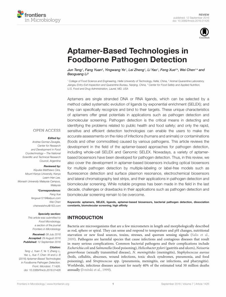
Most of the aptamers selected against pathogenic bacteriahave been evolved using the conventional SELEX proceduresas demonstrated in Figure 1 (Zhang et al., 2015). Zhanget al. (2015) described the selection of DNA aptamers targetedE. coli. Two high-affinity aptamers to E. coli was obtainedwith totally eight rounds of SELEX selections. Furthermore,these conventional SELEX procedures have been used in thedetection of various pathogens such as Vibrio parahaemolyticus(Hamula et al., 2011), Salmonella typhimurium (Duan et al.,2012), Listeria monocytogenes (Duan et al., 2013). Theseaptamers to a single bacterial species can be created within10 rounds of selection, while those to multiple bacterialspecies also can be achieved within 20 rounds of selectionas shown by the aptamer selection scheme in Figure 2(Liu G.Q. et al., 2014). As a result, Liu G.Q. et al. (2014)achieved selection of aptamers against various M-types ofS. pyogenes by using these SELEX procedures rather than theconventional aptamer selection procedures, which use purifiedmolecules of monoclonal cells as targets. It turned out thatthe aptamers selected through these procedures demonstratedhigh affinity and specificity to the targets (Liu G.Q. et al.,2014).
FIGURE 1 | Schematic showing the aptamer selection against livebacterial cells using whole-cell SELEX (Zhang et al., 2015).
Frontiers in Microbiology | www.frontiersin.org 2 September 2016 | Volume 7 | Article 1426
fmicb-07-01426 September 8, 2016 Time: 17:38 # 3
Teng et al. Aptamer Technologies in Pathogen Detection
FIGURE 2 | Schematic of bacterial cell SELEX against a mixture of the 10 most prevalent GAS M-types in Canada (Liu G.Q. et al., 2014).
OTHER TYPES OF SELEXS
Whole-Cell SELEXIn addition to the conventional SELEX method, several novelSELEX methods have been developed. For example, a series ofstudies aimed to shorten the selection rounds in the aptamermanufacturing process. As a result, various methodologies withsingle round aptamer selection procedure have been developed,e.g., the ASExp (Aptamer Selection Express) method, which usesthe magnetic mechanism for separation with microbeads (Fanet al., 2008).
A method called artificially expanded genetic informationsystems-SELEX (AEGIS-SELEX) was introduced in 2014 (Sefahet al., 2014). As the name suggests, this method uses artificiallyexpanded genetic information systems for aptamer selection.An AEGIS-SELEX is started with a GACTZP DNA library,consisting of randomized sequences, primer sites and twomodified nucleotides (ZP); and then a standard protocolfor whole-cell SELEX is followed for the selection cycle.After the 12th selection round, the aptamers are sequencedand the aptamers’ affinity is evaluated. As a result, theAEGIS-SELEX method empowered the system with higherbinding variations. The sequential aptamers can reach thenanomolar range and are expected to achieve higher sequencediversities nearer to that displayed by proteins (Sefah et al.,2014).
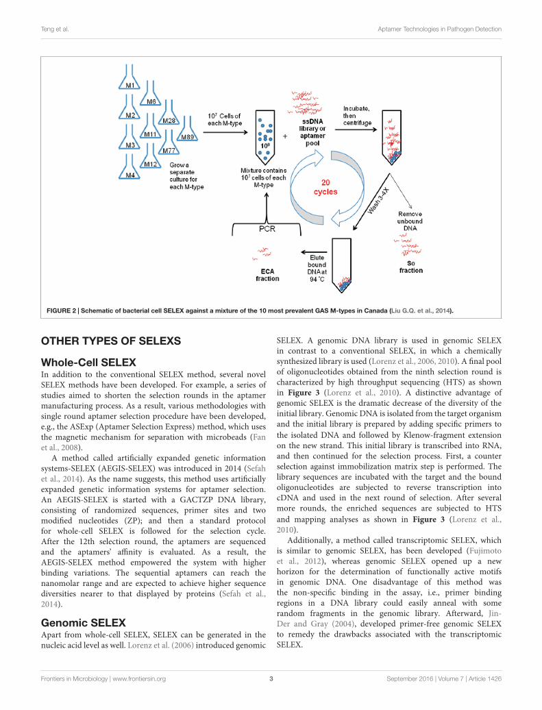
Genomic SELEXApart from whole-cell SELEX, SELEX can be generated in thenucleic acid level as well. Lorenz et al. (2006) introduced genomic
SELEX. A genomic DNA library is used in genomic SELEXin contrast to a conventional SELEX, in which a chemicallysynthesized library is used (Lorenz et al., 2006, 2010). A final poolof oligonucleotides obtained from the ninth selection round ischaracterized by high throughput sequencing (HTS) as shownin Figure 3 (Lorenz et al., 2010). A distinctive advantage ofgenomic SELEX is the dramatic decrease of the diversity of theinitial library. Genomic DNA is isolated from the target organismand the initial library is prepared by adding specific primers tothe isolated DNA and followed by Klenow-fragment extensionon the new strand. This initial library is transcribed into RNA,and then continued for the selection process. First, a counterselection against immobilization matrix step is performed. Thelibrary sequences are incubated with the target and the boundoligonucleotides are subjected to reverse transcription intocDNA and used in the next round of selection. After severalmore rounds, the enriched sequences are subjected to HTSand mapping analyses as shown in Figure 3 (Lorenz et al.,2010).
Additionally, a method called transcriptomic SELEX, whichis similar to genomic SELEX, has been developed (Fujimotoet al., 2012), whereas genomic SELEX opened up a newhorizon for the determination of functionally active motifsin genomic DNA. One disadvantage of this method wasthe non-specific binding in the assay, i.e., primer bindingregions in a DNA library could easily anneal with somerandom fragments in the genomic library. Afterward, Jin-Der and Gray (2004), developed primer-free genomic SELEXto remedy the drawbacks associated with the transcriptomicSELEX.
Frontiers in Microbiology | www.frontiersin.org 3 September 2016 | Volume 7 | Article 1426
fmicb-07-01426 September 8, 2016 Time: 17:38 # 4
Teng et al. Aptamer Technologies in Pathogen Detection
FIGURE 3 | Illustration of genomic SELEX (Lorenz et al., 2010). Genomic DNA is isolated from the target organism.
APPLICATIONS IN PATHOGENDETECTION AND BIOMOLECULARSCREENING
Currently, aptamer-based detection methods can be used inpublic health and food safety are limited. A primary reasonfor that might be the complexity of the methods since thesemethods involve a variety of techniques in the sample preparationand detection processes such as sample’s extraction, purification,enrichment, and separation (Pitcher and Fry, 2000; Stevens andJaykus, 2004).
Pathogen detection is important for public health and foodsafety. Three areas of application account for over two thirdsof all research in the field of pathogen detection (Lazcka et al.,
2007), including the food industry (Patel, 2002; Leonard et al.,2003), water and environment quality control (Emde et al., 1992;Theron et al., 2000), and clinical diagnosis (Atlas, 1999). Theremaining efforts go into fundamental studies (Herpers et al.,2003; Gao et al., 2004), method performance studies (Dominguezet al., 1997; Taylor et al., 2005), and development of new appliedmethods (Ko and Grant, 2003; Yoon et al., 2003).
Aptamers and antibodies are commonly used reagents invarious detection assays; the affinity of aptamers to their targetsis comparable to, or even higher than most of the monoclonalantibodies to their targets; typical dissociation constants ofaptamer-target complexes are found to be in the picomolar tolow micromolar ranges (Hamula et al., 2006). Therefore, nucleic-acid aptamers demonstrate numerous advantages as recognition
Frontiers in Microbiology | www.frontiersin.org 4 September 2016 | Volume 7 | Article 1426
fmicb-07-01426 September 8, 2016 Time: 17:38 # 5
Teng et al. Aptamer Technologies in Pathogen Detection
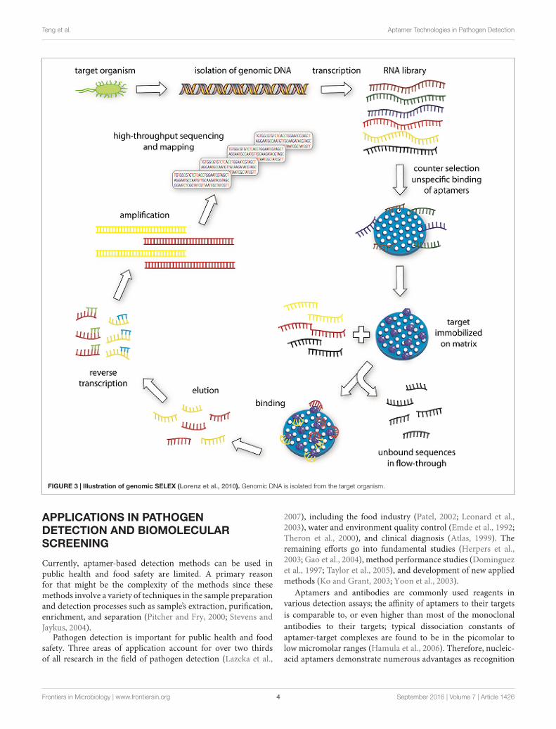
FIGURE 4 | Schematics of the present microbe detection system. (A) Preparation of aptamer conjugated fluorescence nanoparticles (A-FNPs). (B) Detectionof A-FNP-bound E. coli by the microchannel and optical particle counter.
elements in biosensing over the traditional antibodies. Moreover,aptamers are small in size, chemically stable and cost effective.More importantly, aptamers provide remarkable flexibility andconvenience in engineering their structures, which have ledto the development of novel biosensors that exhibited highsensitivity and specificity (Song et al., 2008). In addition to theseabove-mentioned advantages, aptamers offer some distinctivecharacteristics as a biological reagent, i.e., once selected, itcan be synthesized with high reproducibility and purity fromcommercial sources. Furthermore, in contrast to protein-basedantibodies or enzymes, aptamers (made of DNA) usually arechemically stable and often undergo significant conformationalchanges upon target binding (Song et al., 2008; Binnin et al.,2011). Nucleic acid aptamers are widely used in the field ofbiosensors. Therefore, numerous aptamer-based biosensors aredeveloped to detect bacterial pathogens. Hence, in this review,we also summarize the most commonly used aptamer-basedbiosensors and bioassay methods for detection of bacterialpathogens.
OPTICAL BIOSENSORS
Optical biosensors are probably the most popular in bioanalysis,due to their selectivity and sensitivity. Optical biosensors havebeen developed for rapid detection of contaminants (Willardsonet al., 1998; Tschmelak et al., 2004), toxins, drugs (Bae et al., 2004),and bacterial pathogens (Baeumner et al., 2003).
LABEL-FREE DETECTION OF BACTERIA
Several techniques have been described that allow direct, label-free monitoring of cells at solid-liquid interfaces (Ebato et al.,
1994; Morgan et al., 1996; Piehler et al., 1996; Medina et al.,1997; Ghindilis et al., 1998; Fratamico et al., 1998). Thesetechniques are based on direct measurement of a physicalphenomenon occurring during the biochemical reactions ona transducer surface (Ivnitski et al., 1999); changes in pH,oxygen consumption, potential difference, current, resistance, ionconcentrations, and optical properties can be used as measures ofsignal parameters in certain detection systems.
FLUORESCENCE DETECTION
Fluorescence occurs when a valence electron is excited fromits ground state to an excited singlet state. The excitation isproduced by the absorption of light of sufficient energy. Whenthe electron returns to its original ground state it emits aphoton at lower energy (Chung et al., 2014). Nowadays, thefluorescent material is also broadly used (Vigneshvar et al.,2015). Bacteria can be detected by using fluorescent beads asshown in Figure 4 (Chung et al., 2014). Fratamico et al. (1998)developed a real-time, continuous, and non-destructive single celldetection method that uses target specific aptamer-conjugatedfluorescent nanoparticles (A-FNPs) and an optofluidic particle-sensor platform to detect bacterial pathogens.
SURFACE PLASMON RESONANCEBASED DETECTIONS
Surface plasmon resonance (SPR) biosensors (Cooper, 2003)measure changes in refractive index caused by structuralalterations in the vicinity of a thin film metal surface. Given itshigh sensitivity and fingerprinting capability, surface-enhanced
Frontiers in Microbiology | www.frontiersin.org 5 September 2016 | Volume 7 | Article 1426

fmicb-07-01426 September 8, 2016 Time: 17:38 # 6
Teng et al. Aptamer Technologies in Pathogen Detection
FIGURE 5 | Flowchart of S. aureus detection using SERS (Wang et al., 2015). (A) Synthesis of monodispersed silver-coated magnetic nanoparticles and theirconjugation with aptamer 1. (B) Synthesis of core-shell plasmonic nanoparticles (AuNR-DTNB@Ag-DTNB) and their conjugation with aptamer 2. (C) Schematicillustration of the operating principle for S. aureus detection.
Raman scattering (SERS) has been applied in various fields (Nieand Emory, 1999; Cao et al., 2002; Li et al., 2010). The detectionand identification of pathogenic microorganisms by SERS haverecently drowned attention because of the potential applicationof this technology in single-cell detection (Chang et al., 2013).The flow chart in Figure 5 demonstrates the process of S. aureusdetection by using SERS (Wang et al., 2015). A plan f or theconjunction of aptamers to Ag-MNPs is shown in Figure 5A; anovel SERS tag (DioPNPs) was designed as shown in Figure 5B;and the operating principle of the SERS biosensor for bacterialdetection that is based on aptamer recognition is shown inFigure 5C.
ELECTROCHEMICAL BIOSENSORS
Electrochemical sensors have several advantages over optical-based systems in that they can operate in turbid media, offercomparable instrumental sensitivity, and are more amenable tominiaturization. Modern electroanalytical techniques can reachan extremely low limit of detection (up to 10−9 M), whichcan be achieved by using small volumes (1–20 mL) of samples(Mahmoud et al., 2012). A typical electrochemical method, whichis illustrated in Figure 6 (Jenkins et al., 1988), provides a brandnew avenue for the aptamer-based viability detection of variousmicroorganisms, particularly viable but non-cultural (VBNC)
Frontiers in Microbiology | www.frontiersin.org 6 September 2016 | Volume 7 | Article 1426

fmicb-07-01426 September 8, 2016 Time: 17:38 # 7
Teng et al. Aptamer Technologies in Pathogen Detection
FIGURE 6 | Schematic diagram of the aptamer-mediated electrochemical detection of live Salmonella Typhimurium bacteria (Jenkins et al., 1988).
bacteria, using a rapid, economic, and label-free electrochemicalplatform as Jenkins et al. (1988) reported.
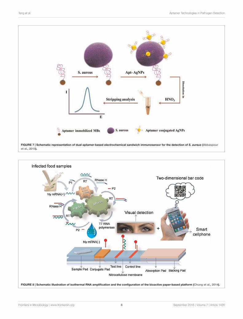
Recently, a newly developed aptamer-based biosensorsystem has been developed to detect pathogen (Abbaspouret al., 2015). This system uses a sensitive and highly selectivedual-aptamer-based sandwich immunosensor in conjunctionwith electrochemical means for the detection of S. aureusas shown by Figure 7 (Abbaspour et al., 2015). As a result,excellent discriminatory power of the biosensor was achievedby utilizing the two specific aptamer sequences against thetarget bacteria and the magnetic beads to capture S. aureusin a liquid phase. The electrochemical detection methoddemonstrated a few advantages in term of simplicity, turn-around time, low cost, and limit of detection compared tothe conventional detection methods. The superior sensitivityof this method provides the possibility to use aptamersto detect extremely low number (in single digital) ofpathogenic bacteria in foods, which is hardly achievable bythe conventional methods. Also, researchers have tried touse biotinylated single-stranded (ss) DNA aptamers withL. monocytogenes binding specificity to capture the bacteria,and subsequently detected the organism in qPCR assay.Biotinylated ssDNA aptamers are promising ligands that canbe used for concentrating foodborne pathogens prior to usingthe conventional molecular approaches for detection (Suh andJaykus, 2013).
LATERAL CHROMATOGRAPHY TESTSTRIPS
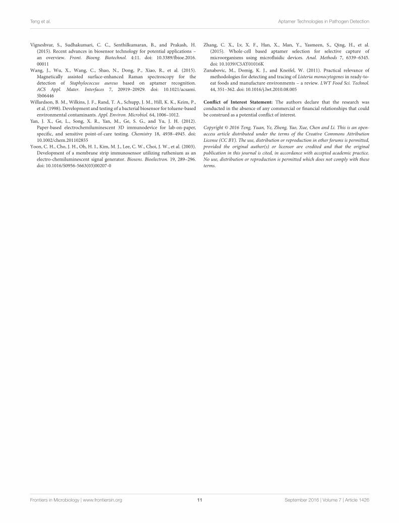
Lateral chromatography test strips, whose mechanism isillustrated in Figure 8, are also widely used. For example, LiuH.X. et al. (2014) demonstrated a simple and sensitive methodfor visual detection of viable pathogenic bacteria based on anisothermal RNA amplification reaction-based bioactive paper-based platform by using a two-dimensional barcode as thereceiving/transmitting media for rapid detection.
Numerous assays based on the specific binding of anantibody to an antigen, such as enzyme-linked immunosorbentassay (ELISA) (Zunabovic et al., 2011; Hsu et al., 2014)and immunochromatographic lateral flow test strips (Geet al., 2012; Nash et al., 2012; Yan et al., 2012; Cho andIrudayaraj, 2013), have been developed. However, rapidimmune tests, which are widely used in low resource settings,are not suitable for fast foodborne pathogen detection dueto their low sensitivity. Finally, a low-cost platform wasconstructed for viable pathogen detection with the nakedeyes (Liu H.X. et al., 2014). In that system, specificallyamplified products were applied to a paper-based platformto perform sandwich hybridization and followed by avisual exam. This method is suitable for point-of-careapplications to detect foodborne pathogens (Liu H.X. et al.,2014).
Frontiers in Microbiology | www.frontiersin.org 7 September 2016 | Volume 7 | Article 1426
fmicb-07-01426 September 8, 2016 Time: 17:38 # 8
Teng et al. Aptamer Technologies in Pathogen Detection
FIGURE 7 | Schematic representation of dual-aptamer-based electrochemical sandwich immunosensor for the detection of S. aureus (Abbaspouret al., 2015).
FIGURE 8 | Schematic illustration of isothermal RNA amplification and the configuration of the bioactive paper-based platform (Chung et al., 2014).
Frontiers in Microbiology | www.frontiersin.org 8 September 2016 | Volume 7 | Article 1426

fmicb-07-01426 September 8, 2016 Time: 17:38 # 9
Teng et al. Aptamer Technologies in Pathogen Detection
CONCLUSIONS AND PERSPECTIVES
In this review, we summarize the most commonly used SELEXmethods in selection of aptamers against bacterial foodbornepathogens and the application of aptamer-based biosensorsin biomolecular screening. Although SELEX advanced ratherslowly initially, the selection of aptamers against pathogenicbacteria has been stably progressing in the last decade andnowadays, this technology has been evolved into a useful toolin pathogen detection and biomolecular screening. Initially,conventional steps and PCR were used in the SELEX proceduresin the early years and then, several novel approaches and newbiological materials were adapted in the SELEX procedures. Onthe other hand, targeting bacterial cells for detection purposeby SELEX also encounters some drawbacks, because bacteria’shighly variable and complex structures may influence theperformance of aptamers. Therefore, it is necessary to continueto develop simpler and more efficient SELEX methods (requiringfewer rounds in selection) to generate specific and/or universalaptamers against various bacterial pathogens.
Compared to traditional antibody generation process, SELEXcan efficiently generate specific nucleic acid probes againstvarious analytes in a relatively short period of time. Moreimportantly, the extraordinary properties of the selectedaptamers, such as easy scale-synthesis, easy modification andlong-term stability, make aptamers ideal alternatives to the
traditional antibodies. However, improvement in aptamerselection efficiency by SELEX is needed in the future work. Also,other platforms, including magnetic separation techniques, arrayor microfluidic chips, can be integrated with initial SELEX tofurther widen the applications of this promising technology. Forexample, at present, biosensor-based detection technologies canmerely meet the basic requirements for testing in the laboratoryand clinic. Obviously, it can be expected that simpler, faster, moreefficient, and more economic aptamer-based methods will bedeveloped for pathogen detection and biomolecular screening inthe future.
AUTHOR CONTRIBUTIONS
JT, FY, LZ, YY, LY, FX, and BL wrote the manuscript. WC and BLrevised the manuscript.
ACKNOWLEDGMENTS
This work is financially supported by the NSFC grant of21475030, 31301460, the S&T Research Project of AnhuiProvince 15czz03109, the National 10000 Talents-Youth Top-notch Talent Program, the National and Zhejiang Public BenefitResearch Project (201313010, 2014C32051).
REFERENCESAbbaspour, A., Norouz-Sarvestani, F., Noori, A., and Soltani, N. (2015).
Aptamer-conjugated silver nanoparticles for electrochemical dual-aptamer-based sandwich detection of Staphylococcus aureus. Biosens. Bioelectron. 68,149–155. doi: 10.1016/j.bios.2014.12.040
Atlas, R. M. (1999). Legionella: from environmental habitats to disease pathology,detection and control. Environ. Microbiol. 1, 283–293. doi: 10.1046/j.1462-2920.1999.00046.x
Bae, Y. M., Oh, B. K., Lee, W. W., and Choi, J. (2004). Detection of insulin-antibodybinding on a solid surface using imaging ellipsometry. Biosens. Bioelectron. 20,895–902. doi: 10.1016/j.bios.2004.03.032
Baeumner, A. J., Cohen, R. N., Miksic, V., and Min, J. (2003). RNA biosensor for therapid detection of viable Escherichia coli in drinking water. Biosens. Bioelectron.18, 405–413. doi: 10.1016/S0956-5663(02)00162-8
Binnin, J. M., Leung, D. W., and Amarasinghe, G. K. (2011). Aptamersin virology: recent advances and challenges. Front. Microbiol. 3:29. doi:10.3389/fmicb.2012.00029
Bock, L. C., Griffin, L. C., Latham, J. A., Vermaas, E. H., and Toole, J. J. (1992).Selection of single-stranded DNA molecules that bind and inhibit humanthrombin. Nature 355, 564–566. doi: 10.1038/355564a0
Boiziau, C., Dausse, E., Yurchenko, L., and Toulmé, J. J. (1999). DNAaptamers selected against the HIV-1 trans-activation-responsive RNA elementform RNA-DNA kissing complexes. J. Biol. Chem. 274, 12730–12737. doi:10.1074/jbc.274.18.12730
Cao, Y. W. C., Rongchao, J., and Mirkin, C. A. (2002). Nanoparticles withRaman spectroscopic fingerprints for DNA and RNA detection. Science 297,1536–1540. doi: 10.1126/science.297.5586.1536
Chang, Y. C., Yang, C. Y., Sun, R. L., Cheng, Y. F., Kao, W. C., and Yang,P. C. (2013). Rapid single cell detection of Staphylococcus aureus by aptamer-conjugated gold nanoparticles. Sci. Rep. 3:1863. doi: 10.1038/srep01863
Cho, I. H., and Irudayaraj, J. (2013). Lateral-flow enzyme immunoconcentrationfor rapid detection of Listeria monocytogenes. Anal. Bioanal. Chem. 405, 3313–3319. doi: 10.1007/s00216-013-6742-3
Chung, J. Y., Kang, J. S., Jurng, J. S., Jung, J. H., and Kim, B. C. (2014). Fast andcontinuous microorganism detection using aptamer-conjugated fluorescentnanoparticles on an optofluidicplatform. Biosens. Bioelectron. 67, 303–308. doi:10.1016/j.bios.2014.08.039
Cooper, M. A. (2003). Label-free screening of bio-molecular interactions. Anal.Bioanal. Chem. 377, 834–842. doi: 10.1007/s00216-003-2111-y
Dominguez, J. A., Matas, L., Manterola, J. M., Blavia, R., Sopena, N., Belda, F. J.,et al. (1997). Comparison of radioimmunoassay and enzyme immunoassaykits for detection of Legionella pneumophila serogroup 1 antigen in bothconcentrated and nonconcentrated urine samples. J. Clin. Microbiol. 35, 1627–1629.
Duan, N., Wu, S., Chen, X., Huang, Y., and Whang, Z. (2012). Selection andidentification of a DNA aptamer targeted to Vibrio parahaemolyticus. J. Agric.Food Chem. 60, 4034–4038. doi: 10.1021/jf300395z
Duan, N., Wu, S. J., Chen, X. J., Huang, Y. K., Xia, Y., Ma, X. Y.,et al. (2013). Selection and characterization of aptamers against SalmonellaTyphimurium using whole-bacterium systemic evolution of ligands byexponential enrichment (SELEX). J. Agric. Food Chem. 61, 3229–3234. doi:10.1021/jf400767d
Ebato, H., Gentry, C. A., Herron, J. N., Müller, W., Okahata, Y., Ringsdorf, H.,et al. (1994). Investigation of specific binding of antifluorescyl antibodyand Fab to fluorescein lipids in Langmuir-Blodgett deposited films usingquartz crystal microbalance methodology. Anal. Chem. 66, 1683–1689. doi:10.1021/ac00082a014
Ellington, A. D., and Szostak, J. W. (1990). In vitro selection of RNA molecules thatbind specific ligands. Nature 346, 818–822. doi: 10.1038/346818a0
Emde, K. M. E., Mao, H., and Finch, G. R. (1992). Detection and occurrence ofwaterborne bacterial and viral pathogens. Water Environ. Res. 64, 641–647.
Fan, M., Mcburnett, S. R., Andrews, C. J., Allman, A. M., Bruno, J. G., and Kiel,J. L. (2008). Aptamer selection express: a novel method for rapid single-stepselection and sensing of aptamers. J. Biomol. Tech. 19, 311–331.
Fratamico, P. M., Strobaugh, T. P., Medina, M. B., and Gehring, A. G. (1998).Detection of Escherichia coli O157:H7 using a surface plasmon resonancebiosensor. Biotechnol Tech. 12, 571–576. doi: 10.1023/A:1008872002336
Frontiers in Microbiology | www.frontiersin.org 9 September 2016 | Volume 7 | Article 1426

fmicb-07-01426 September 8, 2016 Time: 17:38 # 10
Teng et al. Aptamer Technologies in Pathogen Detection
Fujimoto, Y., Nakamura, Y., and Ohuchi, S. (2012). HEXIM1-binding elementson mRNAs identified through transcriptomic SELEX and computationalscreening. Biochimie 94, 1900–1909. doi: 10.1016/j.biochi.2012.05.003
Gao, L. Y., Guo, S., Mclaughlin, B., Morisaki, H., Engel, J. N., and Brown, E. J.(2004). A mycobacterial virulence gene cluster extending RD1 is requiredfor cytolysis, bacterial spreading and ESAT-6 secretion. Mol. Microbiol. 53,1677–1693. doi: 10.1111/j.1365-2958.2004.04261.x
Ge, L., Yan, J. X., Song, X. R., Yan, M., Ge, S. G., and Yu, J. H. (2012).Three-dimensional paper-based electrochemiluminescence immunodevicefor multiplexed measurement of biomarkers and point-of-care testing.Biomaterials 33, 1024–1031. doi: 10.1016/j.biomaterials.2011.10.065
Ghindilis, A. L., Atanasov, P., Wilkins, M., and Wilkins, E. (1998). Immunosensors:electrochemical sensing and other engineering approaches. Biosens. Bioelectron.13, 113–131. doi: 10.1016/S0956-5663(97)00031-6
Hamula, C. L., Chris, L. X., and Xing-Fang, L. (2011). DNA Aptamers bindingto multiple prevalent M-types of Streptococcus pyogenes. Anal. Chem. 83,3640–3647. doi: 10.1021/ac200575e
Hamula, C. L., Guthrie, J. W., Zhang, H., Li, X. F., and Le, X. C. (2006). Selectionand analytical applications of aptamers. TrAC Trends Analyt. Chem. 25, 681–691. doi: 10.1016/j.trac.2006.05.007
Herpers, B. L., Jongh, B. M. D., van der Zwaluw, K., and Hannen, E. J. V.(2003). Real-time PCR assay targets the 23S-5S spacer for direct detection anddifferentiation of Legionella spp. and Legionella pneumophila. J. Clin. Microbiol.41, 4815–4816. doi: 10.1128/JCM.41.10.4815-4816.2003
Hsu, M. Y., Yang, C. Y., Hsu, W. H., Lin, K. H., Wang, C. Y., Shen, Y. C.,et al. (2014). Monitoring the VEGF level in aqueous humor of patients withophthalmologically relevant diseases via ultrahigh sensitive paper-based ELISA.Biomaterials 35, 3729–3735. doi: 10.1016/j.biomaterials.2014.01.030
Ivnitski, D., Atanasov, P. E., and Abdel-Hamid, I. (1999). Biosensors for detectionof pathogenic bacteria. Biosens. Bioelectron. 14, 599–624. doi: 10.1016/S0956-5663(99)00039-1
Jayasena, S. D. (1999). Aptamers: an emerging class of molecules that rivalantibodies in diagnostics. Clin. Chem. 45, 1628–1650.
Jenkins, S. H., Heineman, W. R., and Halsall, H. B. (1988). Extending the detectionlimit of solid-phase electrochemical enzyme immunoassay to the attomole level.Anal. Biochem. 168, 292–299. doi: 10.1016/0003-2697(88)90321-1
Jin-Der, W., and Gray, D. M. (2004). Selection of genomic sequences that bindtightly to Ff gene 5 protein: primer-free genomic SELEX. Nucleic Acids Res.32:e182. doi: 10.1093/nar/gnh179
Ko, S., and Grant, S. A. (2003). Development of a novel FRET method fordetection of Listeria or Salmonella. Sens. Actuators B Chem. 96, 372–378. doi:10.1016/S0925-4005(03)00572-0
Law, J. W.-F., Mutalib, N.-S. A., Chan, K.-G., and Lee, L.-H. (2015). Rapidmethods for the detection of foodborne bacterial pathogens: principles,applications, advantages and limitations. A review. Front. Microbiol. 5:770. doi:10.3389/fmicb.2014.00770
Lazcka, O., Campo, F. J. D., and Muñoz, F. X. (2007). Pathogen detection: aperspective of traditional methods and biosensors. Biosens. Bioelectron. 22,1205–1217. doi: 10.1016/j.bios.2006.06.036
Leonard, P., Hearty, S., Brennan, J., Dunne, L., Quinn, J., Chakraborty, T., et al.(2003). Advances in biosensors for detection of pathogens in food and water.Enzyme Microb. Technol. 32, 3–13. doi: 10.1016/S0141-0229(02)00232-6
Li, J. F., Huang, Y. F., Ding, Y., Yang, Z. L., Li, S. B., Zhou, X. S., et al. (2010). Shell-isolated nanoparticle-enhanced Raman spectroscopy. Nature 464, 392–395. doi:10.1038/nature08907
Liu, G. Q., Lian, Y. Q., Gao, C., Yu, X. F., Zhu, M., Zong, K., et al. (2014).In vitro Selection of DNA aptamers and fluorescence-based recognition forrapid detection Listeria monocytogenes. J. Integr. Agric. 13, 1121–1129. doi:10.1016/S2095-3119(14)60766-8
Liu, H. X., Zhan, F. F., Liu, F., Zhu, M. J., Zhou, X. M., and Xing, D. (2014). Visualand sensitive detection of viable pathogenic bacteria by sensing of RNA markersin gold nanoparticles based paper platform. Biosens. Bioelectron. 62, 38–46. doi:10.1016/j.bios.2014.06.020
Lorenz, C., Gesell, T., Zimmermann, B., Schoeberl, U., Bilusic, I., Rajkowitsch, L.,et al. (2010). Genomic SELEX for Hfq-binding RNAs identifies genomicaptamers predominantly in antisense transcripts. Nucleic Acids Res. 38, 3794–3808. doi: 10.1093/nar/gkq032
Lorenz, C., Pelchrzim, F. V., and Schroeder, R. (2006). Genomic systematicevolution of ligands by exponential enrichment (Genomic SELEX) for theidentification of protein-binding RNAs independent of their expression levels.Nat. Protoc. 1, 2204–2212. doi: 10.1038/nprot.2006.372
Mahmoud, L., Zamay, A. S., Kolovskaya, O. S., Reshetneva, I. T., Zamay, G. S.,Kibbee, R. J., et al. (2012). Aptamer-based viability impedimetric sensor forbacteria. Anal. Chem. 84, 8966–8969. doi: 10.1021/ac302902s
Medina, M. B., Houten, L. V., Cooke, P. H., and Tu, S. (1997). Real-time analysis ofantibody binding interactions with immobilized E. coli O157:H7 cells using theBIAcore. Biotechnol. Tech. 11, 173–176. doi: 10.1023/A:1018453530459
Morgan, C. L., Newman, D. J., and Price, C. P. (1996). Immunosensors: technologyand opportunities in laboratory medicine. Clin. Chem. 42, 193–209.
Nash, M. A., Waitumbi, J. N., Hoffman, A. S., Paul, Y., and Stayton, P. S. (2012).Multiplexed enrichment and detection of malarial biomarkers using a stimuli-responsive iron oxide and gold nanoparticle reagent system. ACS Nano 6,6776–6785. doi: 10.1021/nn3015008
Nie, S., and Emory, S. R. (1999). Probing single molecules and singlenanoparticles by surface-enhanced Raman scattering. Science 275, 1102–1106.doi: 10.1126/science.275.5303.1102
Patel, P. D. (2002). (Bio) sensors for measurement of analytes implicated in foodsafety: a review. TrAC Trends Analyt. Chem. 21, 96–115. doi: 10.1016/S0165-9936(01)00136-4
Piehler, J., Brecht, A., Geckeler, K. E., and Gauglitz, G. (1996). Surface modificationfor direct immunoprobes. Biosens. Bioelectron. 11, 579–590. doi: 10.1016/0956-5663(96)83293-3
Pitcher, D. G., and Fry, N. K. (2000). Molecular techniques for the detectionand identification of new bacterial pathogens. J. Infect. 40, 116–120. doi:10.1053/jinf.2000.0635
Salis, H., Tamsir, A., and Voigt, C. (2009). Engineering bacterial signals and sensors.Contrib. Microbiol. 16, 1–32.
Sefah, K., Yang, Z. Y., Bradley, K. M., Hoshika, S., Jiméneza, E., Zhang, L. Q., et al.(2014). In vitro selection with artificial expanded genetic information systems.Proc. Natl. Acad. Sci. U.S.A. 111, 1449–1454. doi: 10.1073/pnas.1311778111
Song, S., Wang, L., Li, J., Fan, C., and Zhao, J. (2008). Aptamer-based biosensors.Trends Analyt. Chem. 27, 108–117. doi: 10.1016/j.trac.2007.12.004
Stevens, K. A., and Jaykus, L. A. (2004). Bacterial separation and concentrationfrom complex sample matrices: a review. Crit. Rev. Microbiol. 30, 7–24. doi:10.1080/10408410490266410
Suh, S. H., and Jaykus, L. A. (2013). Nucleic acid aptamers for capture and detectionof Listeria spp. J. Biotechnol. 167, 454–461. doi: 10.1016/j.jbiotec.2013.07.027
Taylor, A. D., Yu, Q., Chen, S., Homola, J., and Jiang, S. (2005). Comparisonof E. coli O157: H7 preparation methods used for detection with surfaceplasmon resonance sensor. Sens. Actuators B Chem. 107, 202–208. doi:10.1016/j.snb.2004.11.097
Theron, J., Cilliers, J., Du, P. M., Brözel, V. S., and Venter, S. N. (2000). Detection oftoxigenic Vibrio cholerae from environmental water samples by an enrichmentbroth cultivation-pit-stop semi-nested PCR procedure. J. Appl. Microbiol. 89,539–546. doi: 10.1046/j.1365-2672.2000.01140.x
Tok, J., and Cho, J. R. (2000). RNA aptamers that specifically bind to a 16Sribosomal RNA decoding region construct. Nucleic Acids Res. 28, 2902–2910.doi: 10.1093/nar/28.15.2902
Tombelli, S., Minunni, M., and Mascini, M. (2005). Piezoelectric biosensors:strategies for coupling nucleic acids to piezoelectric devices. Methods 37, 48–56.doi: 10.1016/j.ymeth.2005.05.005
Torres-Chavolla, E., and Alocilja, E. C. (2009). Aptasensors for detection ofmicrobial and viral pathogens. Biosens. Bioelectron. 24, 3175–3182. doi:10.1016/j.bios.2008.11.010
Tschmelak, J., Proll, G., and Gauglitz, G. (2004). Sub-nanogram per litredetection of the emerging contaminant progesterone with a fully automatedimmunosensor based on evanescent field techniques. Anal. Chim. Acta 519,143–146. doi: 10.1016/j.aca.2004.06.031
Tuerk, C., and Gold, L. (1990). Systematic evolution of ligands by exponentialenrichment: RNA ligands to bacteriophage T4 DNA polymerase. Science 249,505–510. doi: 10.1126/science.2200121
Tuerk, C., Macdougal, S., and Gold, L. (1992). RNA pseudoknots that inhibithuman immunodeficiency virus type 1 reverse transcriptase. Proc. Natl. Acad.Sci. U.S.A. 89, 6988–6992. doi: 10.1073/pnas.89.15.6988
Frontiers in Microbiology | www.frontiersin.org 10 September 2016 | Volume 7 | Article 1426
fmicb-07-01426 September 8, 2016 Time: 17:38 # 11
Teng et al. Aptamer Technologies in Pathogen Detection
Vigneshvar, S., Sudhakumari, C. C., Senthilkumaran, B., and Prakash, H.(2015). Recent advances in biosensor technology for potential applications –an overview. Front. Bioeng. Biotechnol. 4:11. doi: 10.3389/fbioe.2016.00011
Wang, J., Wu, X., Wang, C., Shao, N., Dong, P., Xiao, R., et al. (2015).Magnetically assisted surface-enhanced Raman spectroscopy for thedetection of Staphylococcus aureus based on aptamer recognition.ACS Appl. Mater. Interfaces 7, 20919–20929. doi: 10.1021/acsami.5b06446
Willardson, B. M., Wilkins, J. F., Rand, T. A., Schupp, J. M., Hill, K. K., Keim, P.,et al. (1998). Development and testing of a bacterial biosensor for toluene-basedenvironmental contaminants. Appl. Environ. Microbiol. 64, 1006–1012.
Yan, J. X., Ge, L., Song, X. R., Yan, M., Ge, S. G., and Yu, J. H. (2012).Paper-based electrochemiluminescent 3D immunodevice for lab-on-paper,specific, and sensitive point-of-care testing. Chemistry 18, 4938–4945. doi:10.1002/chem.201102855
Yoon, C. H., Cho, J. H., Oh, H. I., Kim, M. J., Lee, C. W., Choi, J. W., et al. (2003).Development of a membrane strip immunosensor utilizing ruthenium as anelectro-chemiluminescent signal generator. Biosens. Bioelectron. 19, 289–296.doi: 10.1016/S0956-5663(03)00207-0
Zhang, C. X., Lv, X. F., Han, X., Man, Y., Yasmeen, S., Qing, H., et al.(2015). Whole-cell based aptamer selection for selective capture ofmicroorganisms using microfluidic devices. Anal. Methods 7, 6339–6345.doi: 10.1039/C5AY01016K
Zunabovic, M., Domig, K. J., and Kneifel, W. (2011). Practical relevance ofmethodologies for detecting and tracing of Listeria monocytogenes in ready-to-eat foods and manufacture environments – a review. LWT Food Sci. Technol.44, 351–362. doi: 10.1016/j.lwt.2010.08.005
Conflict of Interest Statement: The authors declare that the research wasconducted in the absence of any commercial or financial relationships that couldbe construed as a potential conflict of interest.
Copyright © 2016 Teng, Yuan, Ye, Zheng, Yao, Xue, Chen and Li. This is an open-access article distributed under the terms of the Creative Commons AttributionLicense (CC BY). The use, distribution or reproduction in other forums is permitted,provided the original author(s) or licensor are credited and that the originalpublication in this journal is cited, in accordance with accepted academic practice.No use, distribution or reproduction is permitted which does not comply with theseterms.
Frontiers in Microbiology | www.frontiersin.org 11 September 2016 | Volume 7 | Article 1426