approach to hematuria - handout.ppt to hematuria - 2.pdf4 detection of hematuria-the dip stick •...
TRANSCRIPT

1
Isabelle Ayoub, MDAssistant Professor-Clinical
Department of Internal MedicineDivision of Nephrology
The Ohio State University Wexner Medical Center
Evaluation of Hematuria
DISCLOSURE STATEMENTS
Nothing to disclose
2
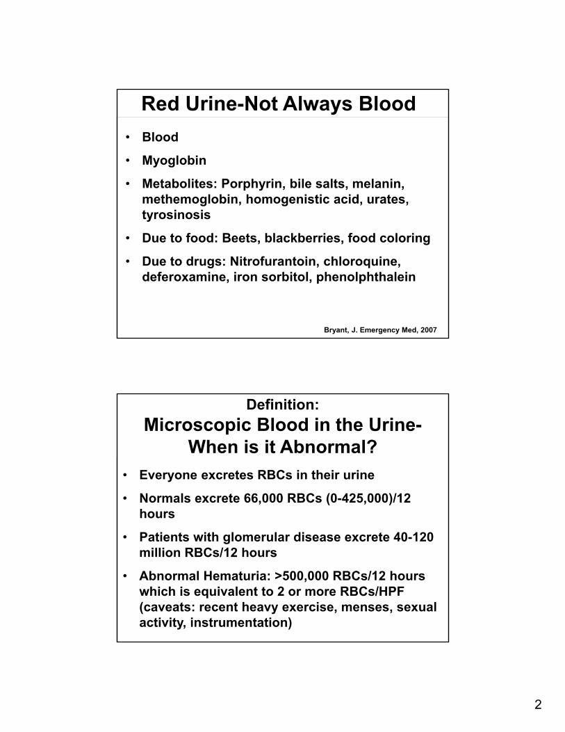
• Blood
• Myoglobin
• Metabolites: Porphyrin, bile salts, melanin, methemoglobin, homogenistic acid, urates, tyrosinosis
• Due to food: Beets, blackberries, food coloring
• Due to drugs: Nitrofurantoin, chloroquine, deferoxamine, iron sorbitol, phenolphthalein
Bryant, J. Emergency Med, 2007
Red Urine-Not Always Blood
• Everyone excretes RBCs in their urine
• Normals excrete 66,000 RBCs (0-425,000)/12 hours
• Patients with glomerular disease excrete 40-120 million RBCs/12 hours
• Abnormal Hematuria: >500,000 RBCs/12 hours which is equivalent to 2 or more RBCs/HPF (caveats: recent heavy exercise, menses, sexual activity, instrumentation)
Definition:
Microscopic Blood in the Urine-When is it Abnormal?

3
DefinitionDefinition• Macroscopic• Microscopic• Asymptomatic: Not associated with pain
(dysuria, loin pain, renal colic), renal dysfunction, hypertension, proteinuria, or macroscopic hematuria.
• Asymptomatic Microscopic Hematuria (AMH) is common and presents the most significant diagnostic and therapeutic challenges
Micro-Hematuria: Scope of the Problem
Using >3 RBC/hpf on 3 occasions over 2-3 weeks:
• Prevalence
Children: 2-6%
Adults: 4%
Men: 2-5%
Women 5-11%
39% may have single episode
Potential kidney donors: 12%
4
Detection of Hematuria-The Dip Stick• Dipstick relies on oxidation of an organic peroxide on the
test strip by the peroxidase-like activity of hemoglobin
• False Positives: Myoglobinuria, hemoglobinuria, povidone-iodine, H2O2, bacterial peroxidases, semen, Ph>9
• False Negatives: Presence of ascorbic acid (supplements), formaldehyde (preservative), low pH
• Test Performance
Sensitivity-93-100%
Specificity-60-80%
Negative predictive value ~98%
Schroder, BMJ, 1994; Huussen J, Neth J Med, 2004
Use Urine Microscopy to Confirm RBCsGlomerular Hematuria
Capillary
Note: Alkaline urine dissolves casts!
Acanthocytes:
• 98% specific, 52% sensitive if >5% of RBCs in a urine sample; sensitivity >80% if found in 3 consecutive urine samples
• Not inducible by changes in pH, osmolality
• Urine RBCs can be dysmorphic but not indicate glomerular bleeding, such as these (B) commonly found crenated RBCs, caused by osmotic shifts in RBC water
Acanthocytes
Dysmorphic RBC

5
Diagnosis Microscopic Macroscopic(n>2000) (n>1200)
Cancer 0.5-5% 23%
Nephrolithiasis 5% 5-11%
Infection 1.7-4% 33%
BPH 3-13% 13%
Intrinsic Renal 2-11%
No Diagnosis 43-57% 8-21%
Sutton, JAMA, 263:2475, 1990; Boman, Scand J Urol Neph, 2001; Murakami, J Urol, 144:49, 1990, Sultana, Br J Urol, 78:691,1999
Hematuria in Adults
Approach to HematuriaIdentify Origin of the Blood
Approach to HematuriaIdentify Origin of the Blood
Glomerular Hematuria
Micro- or Macroscopic
Abnormal Morphology Proteinuria, active sediment
May be familial
- Check first degree relatives
- Look for hearing loss
Non-Glomerular Hematuria
Micro- or Macroscopic
Normal morphology
Isolated finding
6
To biopsy….or not to biopsy?
What nephrologists think about during the evaluation of microscopic
hematuria
Case #1-Isolated Microscopic Hematuria
A 22 year old Asian male was found to have hematuriaduring a routine school evaluation. The patient wasotherwise healthy, had no complaints, no significant PMHand physical exam was unremarkable including a normalblood pressure on no medications. No FH of kidneydisease. SCr was 0.8 mg/dl. Urinalysis showed no proteinbut did show acanthocytes. 24-hour urine contained 115mg protein. Would you do a kidney biopsy?
a. No, because the patient does not have abnormalproteinuria and kidney function is normal
b. Yes, because the UA indicates glomerular bleeding
c. Yes, because the patient appears to have a systemicprocess and the kidney may be involved

7
Case #2-Isolated Microscopic Hematuria
A 69 year old white female developed muscle aches oneyear ago, was diagnosed with polymyalgia rheumatica. Shewas treated with prednisone, felt better, but upon tapersymptoms became much worse. She then developed leftfoot drop. A tentative diagnosis of mononeuritis multiplexwas made. SCr was 0.7 mg/dl. Urinalysis showed no proteinbut did show acanthocytes. 24-hour urine contained 178 mgprotein. Would you do a kidney biopsy?
a. No, because the patient does not have proteinuria andkidney function is normal
b. Yes, because the UA indicates glomerular bleeding
c. Yes, because the patient appears to have a systemicprocess and the kidney may be involved
• No biopsy was done. Patient was followed and after several years hematuria resolved, kidney function remained normal. Presumptive diagnosis of IgAN
Case #2 - Answer
• ANCA was 1:80
• Sural nerve biopsy was non-diagnostic
• A kidney biopsy was performed and showed pauci-immune crescentic GN
Case #1 - Answer

8
Renal Biopsy for Hematuria?Renal Biopsy for Hematuria?• Yes
Proteinuria Present (≥ 500 mg/day)
Abnormal Renal Function (Cr≥1.3)
Possible Systemic Process
Potential Kidney Donor
• No
No Proteinuria
Normal (stable) Renal Function
No Systemic Process
RATIONALE FOR NOT DOING A BIOPSY: The glomerular diseases that are most likely to cause isolated hematuria have no proven treatments, and in the absence of proteinuria
carry an excellent renal prognosis
Pathologic Diagnosis of Hematuria
Pathologic Diagnosis of Hematuria
Pathologic Diagnosis None
IgAN
Thin GBM
Mesangial Proliferation
FSGS
HTN, Membranous, Int Nephritis
Topham et al, Q.J. Med., 7:329:1994
% of Patients
53 (but 13%-no EM)
30
4
7
3
3
Microscopic Hematuria in 165 patientswith no other renal or systemic findings:

9
Pathology of Common Causes of Isolated Microscopic Glomerular Hematuria
Average age of onset 20-30, male preponderance, prevalence higher in Asia than US, UK, Canada, rare in people of African descent. Most common form of GN in Japan, China, Singapore, Taiwan.
Thin GBM
<250 nm
IgAN Alport’s
GBM thick and thin, basket weave appearance in thick areas
IgA in mesangium
TBM/Familial benign hematuria is due to an autosomal dominant defect in the alpha 3 or 4 chains of collagen type IV, with heterozygous expression.
Isolated thin GBM disease may develop proteinuria and renal insufficiency
ALPORT’S: X-linked is most common. Defect in the alpha 5 chain of type IV collagen. Affected males, female carriers. Many males will develop proteinuria and renal insufficiency within first 2 decades
Natural History of Isolated Hematuria
Biopsy IgAN Thin GBM Normal*# of patients 12 13 20
Mean Age 30 35 30
Macroscopic Hematuria 6 1 1
Cr Clearance 109 115 113
11 Year Follow-upHematuria 10 13 7**
Cr Clearance 100 110 113
*Mean Duration Hematuria 4 years; **5 of the 7 patients developed stones over the 11 year follow-up, suggesting they may have had crystaluria to start.
Niewuhof et al., KI, 49:222, 1996

10
Natural History of Isolated HematuriaNatural History of Isolated Hematuria
Kim et al, KJIM, 2009
100 patients with AMH followed for an average of 32 months
107,192 Japanese were screened with a single urine dipstick:
18-29 >80
Men 0.9% 8.5%
Women 7.3% 15.3
10 years later the odds ratio for developing ESRD
was calculated:
Men vs. Women 1.4
Hematuria vs. no hematuria 2.3
Proteinuria vs. no proteinuria 15
Iseki et al, Kidney Int, 1996
Isolated Hematuria and ESRD-Adults

11
Effect of Proteinuria on theDifferential Diagnosis of Hematuria
Microscopic Hematuria in 135 patients:
Effect of Proteinuria on theDifferential Diagnosis of Hematuria
Microscopic Hematuria in 135 patients:
Proteinuria <0.3 g/d
Thin GBM 43%
IgAN 20%
Normal 37%
Proteinuria up to 2.4 g/d
IgAN 46%
FSGS 13%
Membranous, MPGN, AIN
Acute prolif, Alport’s
In IgAN:
Proteinuria (g/d) ESRD over 7-10yrs
0.3-0.99 10%
1-1.99 25-35%
2-2.99 40%
>3 60%
Hall et al, Clin Nephrol 2004
Proteinuria Changes Everything

12
Iseki et al, Kidney Int, 1996
Natural History of Hematuria with Proteinuria
YES NO
Proteinuria Present (≥ 500 mg/day)
Abnormal Renal Function
Possible Systemic Process
Potential Kidney Donor
No Proteinuria
Normal (stable) Renal function
No Systemic Process
Kidney Biopsy For Microscopic Hematuria

13
Case #3-Systemic Disease and Macroscopic Hematuria
A patient with a past history of SLE (no nephritis) and clotting due to anti-phospholipid syndrome was taken off AZA 3 months ago. She called to say she saw blood in her urine. SCr was 0.7mg/dl, P/C ratio was 0.9, and INR was 3.5 on her usual dose of warfarin. What is the next step?
a. Stop the anticoagulation because she is bleeding due to a high INR
b. Restart immunosuppression with AZA, and add high dose prednisone 1mg/kg/d
c. Do a kidney biopsy
d. Perform a urinalysis
e. Do cystoscopy
WARFARIN RELATED NEPHROPATHY
AKI appears shortly after INR acutely increases to >3.0.• WRN is common: Seen in 33% (CKD) and 16% (no-
CKD) of warfarin-treated patients whose INR acutely rises to >3.0
• Patients with WRN have increased mortality (one-year mortality rate 31.0% versus 18.9% in no-WRN patients).
• WRN accelerates the progression of CKD• WRN should be suspected on biopsy of patients on
warfarin if the RBC casts are disproportionate to the degree of underlying glomerular injury
• WRN may be part of a broader Anticoagulant-Related Nephropathy-ARN, so switching anticoagulants is questionable

14
• 103 CKD patients on warfarin therapy with serial measures of INR and serum creatinine
• Of these, 49 patients experienced at least one INR>3.0 and had Scr measured before and after the INR.3.0
• 18 of these patients (37%) had an unexplained increase in Scr>0.3 mg/dl associated with INR>3.0
Brodsky; Nephrology Clinical practice 2010
WRN is Common
• RBC in Bowman’s space • Glomeruli normal in appearance• Dense RBC casts causing tubular obstruction • RBC casts do not contain Tamm-Horsfall protein
Biopsy Findings in WRN

15
Kidney Biopsy Showed IgAN
No Histologic Evidence of LN or WRN
Case #3-Answer
Case #4-Macroscopic Non-Glomerular Hematuria
A 58 year old African American male complained of redurine and was found to have new hematuria. He had a SCrof 1.5 mg/dL and about a 500 mg/d urine protein excretion.These levels have been stable for years, and were attributedto long-standing, poorly-controlled hypertension. Bloodpressure was now controlled. He was a former smoker, quit5 years ago. He developed a DVT 2 months ago, and was onwarfarin with an INR of 2.5. Urinalysis showed no bacteria,WBC, or casts, but he did have many eumorphic RBCs thatwere of uniform size. Renal ultrasound showed echogenic,9cm kidneys. What next?a. Perform a kidney biopsy for suspected GNb. Strain urine for kidney stonesc. Send urine cytologyd. Do cystoscopy

16
• GU Cancer• Nephrolithiasis (also hypercalcuria,
hyperuricosuria)• BPH• Cysts• Infection• Anatomic Lesions (a-v fistula/malformation;
hemangioma; angiomyolipoma; renal varicies)• Hematologic (coagulopathy; platelet dysfunction;
hemoglobinopathy)• Ischemia/infarct; emboli; exercise; malignant HTN
Differential Diagnosis of Non-Glomerular Hematuria
Approach to Patients with Asymptomatic Non-Glomerular Hematuria
17
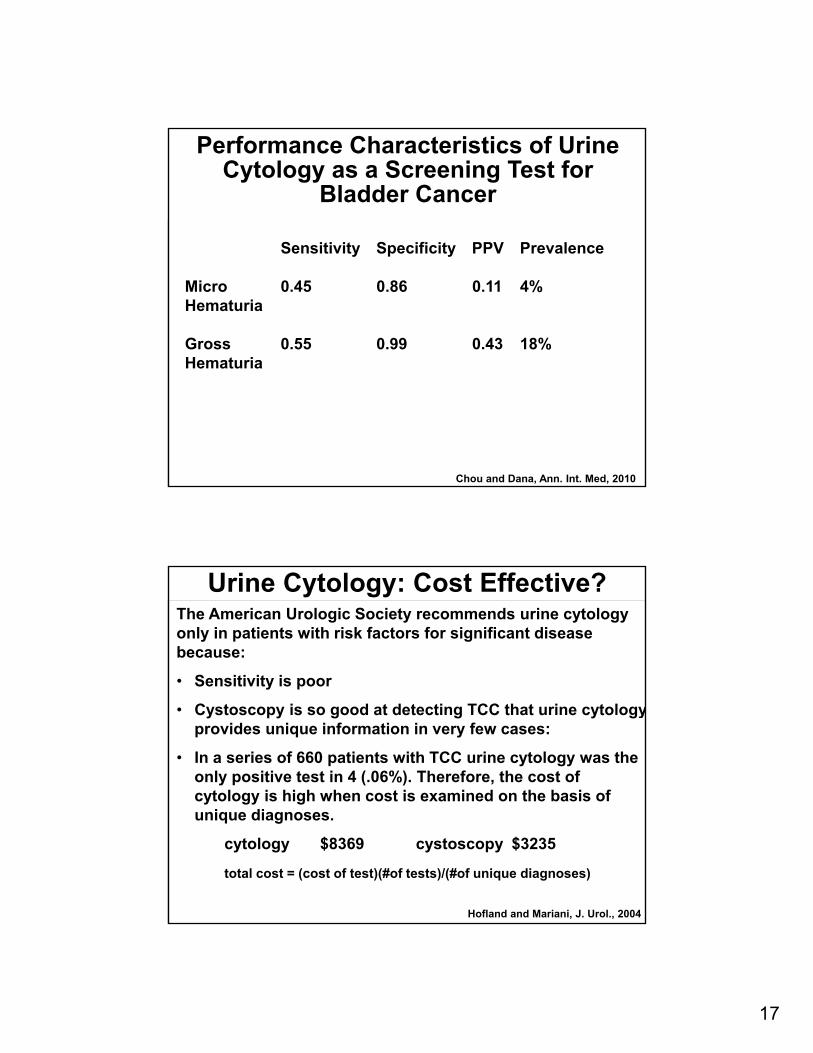
Performance Characteristics of Urine Cytology as a Screening Test for
Bladder Cancer
Sensitivity Specificity PPV Prevalence
Micro 0.45 0.86 0.11 4%Hematuria
Gross 0.55 0.99 0.43 18%Hematuria
Chou and Dana, Ann. Int. Med, 2010
Urine Cytology: Cost Effective?The American Urologic Society recommends urine cytology only in patients with risk factors for significant disease because:
• Sensitivity is poor
• Cystoscopy is so good at detecting TCC that urine cytologyprovides unique information in very few cases:
• In a series of 660 patients with TCC urine cytology was the only positive test in 4 (.06%). Therefore, the cost of cytology is high when cost is examined on the basis of unique diagnoses.
cytology $8369 cystoscopy $3235
total cost = (cost of test)(#of tests)/(#of unique diagnoses)
Hofland and Mariani, J. Urol., 2004

18
• Age (>50)
• Male sex
• Smoking
• Episodes of macroscopic hematuria
• Analgesic abuse (Phenacetin)
• Irritative voiding symptoms, previous GU history
• Exposure to aromatic amines/benzenes
• Exposure to cyclophosphamide
• Pelvic irradiation
• Exposure to aristolochic acid (herbal weight-loss)
• Parasitic infection (Schistosoma haematobium)
Risk Factors for Urothelial Cancers
Hematuria in Adults-Cancer as a Function of Age, Symptoms, and Type of Hematuria
Sultana et al, Br. J. Urol., 78:691, 1996
Age 50+ and gross hematuria are worrisome combination

19
• This patient had cystoscopy that revealed a transitional cell carcinoma of the bladder
• It was felt that anticoagulation unmasked the cancer
• The proteinuria and elevated SCr along with smaller, echogenic kidneys on US were felt to be consistent with hypertensive nephrosclerosis
• The TCC was successfully removed
Case #4-Answer
Bonus CaseA 40 year old white woman presented with flank pain and red urine. She had similar episodes twice before. She did not recall if she had other symptoms with these, specifically colds or other acute illnesses, but this time she had a sore throat that began about 3 days ago. She has not seen a physician regularly. Someone on her father’s side of the family required dialysis. Her father died of a stroke at age 45. Blood pressure was 145/95. Exam showed a red throat, clear lungs, unremarkable heart, and obesity, with a tender left flank. Urine dipstick showed large blood, 1+ protein, and no leukocytes. Urine sediment had too many RBCs to count, and they appeared to be eumorphic. SCr was 1.3 mg/dL. Which is correct?
a. You should quantify proteinuria and set up a kidney biopsy to rule out GN
b. You should send urine studies for calcium, oxalate, citrate, and sodium
c. You should get a detailed neurologic historyd. You should check complement component C3 and C4 levelse. You should hospitalize, push fluids, and give narcotics for pain
control

20
• The glomerular diseases most commonly associated with upper respiratory tract infections are IgAN and post-strep GN.
• IgAN occurs during the infection, usually soon after it is apparent
• Post-sterp occurs after the infection is resolved; the lag is usually several days to a couple of weeks
• There is not much proteinuria and this amount could be accounted for by the hematuria
• The RBCs do not appear to be dysmorphic
• Loin Pain Hematuria Syndrome is a diagnosis of exclusion
• Stones are possible, and a flat plate could be helpful, unless the stones were radio-lucent; also would not expect an increased SCr with stones under most circumstances
• But this patient appears to have either CKD or AKI, and a relevant family history ANSWER: C→ PCKD with cyst rupture
A Word About Loin-Pain Hematuria Syndrome• Unexplained, disabling chronic flank pain and hematuria:• Syndrome characterized by flank pain and micro- or
macroscopic hematuria, often in Caucasian (93%) females (70%). No clear urologic etiology, although 50% have a history of nephrolithiasis, and most have abnormal urine risk factors for stones.
• Renal biopsy of these patients shows hemorrhage into multiple tubules. Glomeruli are normal on light and immunofluorescence, but EM often shows thin (51%) or thick (20%) GBMs.
Presumed Mechanism: Glomerular hematuria causes tubular obstruction, back-leak of glomerular filtrate, renal parenchymal swelling, with stretching of the renal capsule causing pain, plus an abnormal pain response. Correlation with stones remains obscure.
α3 chain of collagen IV α5 chain of collagen IVBlood-filled tubules Thin GBMs

21
When No Diagnosis is MadeWhen No Diagnosis is Made
• If no diagnosis is made after initial evaluation,
patients should be followed every six months
• It is not clear how often to repeat urologic studies.
• In one large study of 225 patients (Murakami et al,
1990) 91% of the serious (eg cancer, stones)
lesions were found at the initial visit.
• An additional 9% (22 cases, 4 malignancies) were
discovered over the next 1.5 years with extensive
urologic testing every 6 months.