appendix to sheehan w,thurber s. review of two...
TRANSCRIPT
Appendix to Sheehan W, Thurber S. Review of Two Years of Experiences with SPECT AmongPsychiatric Patients in a Rural Hospital Setting. J Psychiatr Pract 2008;14:318–23
1
APPENDIX to Sheehan W, Thurber S. Review of Two Years of Experiences with SPECTAmong Psychiatric Patients in a Rural Hospital Setting. J Psychiatr Pract 2008;14:318–23
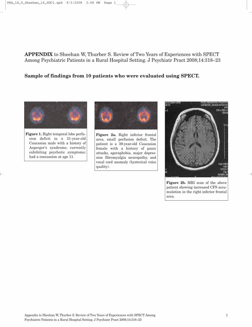
Sample of findings from 10 patients who were evaluated using SPECT.
Figure 1. Right temporal lobe perfu-sion deficit in a 31-year-oldCaucasian male with a history ofAsperger’s syndrome; currentlyexhibiting psychotic symptoms;had a concussion at age 11.
Figure 2a. Right inferior frontalarea, small perfusion deficit. Thepatient is a 39-year-old Caucasianfemale with a history of panicattacks, agoraphobia, major depres-sion fibromyalgia neuropathy, andvocal cord anomaly (hysterical voicequality).
Figure 2b. MRI scan of the abovepatient showing increased CFS accu-mulation in the right inferior frontalarea.
PRA_14_5_Sheehan_16_SDC1.qxd 9/3/2008 3:08 PM Page 1

Appendix to Sheehan W, Thurber S. Review of Two Years of Experiences with SPECT AmongPsychiatric Patients in a Rural Hospital Setting. J Psychiatr Pract 2008;14:318–23
2
Figure 3. Patchy perfusion deficits in the right frontal-parietal area. The patient is a 56-year-old Caucasian male with ahistory of unspecified behavioral problems and urea cycle disorder.
PRA_14_5_Sheehan_16_SDC1.qxd 9/3/2008 3:09 PM Page 2

Appendix to Sheehan W, Thurber S. Review of Two Years of Experiences with SPECT AmongPsychiatric Patients in a Rural Hospital Setting. J Psychiatr Pract 2008;14:318–23
3
Figure 4. Large contiguous perfusion deficits in the right frontal-parietal area and a large defect in the area of the pituitary fossa.The patient is a 40-year-old Caucasian male with a history of abasilar skull fracture and an extended period of being comatose.He also had hypopituitarism secondary to the head injury. He washospitalized for paranoid thinking and outbursts of anger.
Figure 5a. (Axials) Grossly asymmetricactivity between the right and left cere-bral hemispheres, including severelydiminished activity of the right hemi-sphere (occipital, temporal, parietal andfrontal areas). The patient is a 72-year-old Caucasian male who experienced aright hemisphere subdural hematoma ina motor vehicle accident 50 years earlier.He was referred for medical manage-ment of irritable, argumentative behav-iors.
PRA_14_5_Sheehan_16_SDC1.qxd 9/3/2008 3:09 PM Page 3

Appendix to Sheehan W, Thurber S. Review of Two Years of Experiences with SPECT AmongPsychiatric Patients in a Rural Hospital Setting. J Psychiatr Pract 2008;14:318–23
4
Figure 5b. Coronals of the above.
PRA_14_5_Sheehan_16_SDC1.qxd 9/3/2008 3:09 PM Page 4

Appendix to Sheehan W, Thurber S. Review of Two Years of Experiences with SPECT AmongPsychiatric Patients in a Rural Hospital Setting. J Psychiatr Pract 2008;14:318–23
5
Figure 6. Large areas of diminished orabsent activity in the left cerebral hemi-sphere (e.g., complete absence of activityin the temporal lobe). The patient is a 44-year-old Caucasian female living in agroup home because of low cognitivefunctioning following an attack of herpesencephalitis. Has a history of partialcomplex seizures. Referred because ofirritability and obsessive-compulsiveand purging behaviors.
Figure 7. Sagittals show patchy perfusion deficits in the leftfrontal-parietal area near the vertex. The patient is a 44-year-oldCaucasian male with a history of bipolar disorder, disorientation,suicidality, and nocturnal seizures. He had experienced severalclosed head injuries in boxing and snowmobile accidents.
PRA_14_5_Sheehan_16_SDC1.qxd 9/3/2008 3:10 PM Page 5
Appendix to Sheehan W, Thurber S. Review of Two Years of Experiences with SPECT AmongPsychiatric Patients in a Rural Hospital Setting. J Psychiatr Pract 2008;14:318–23
6
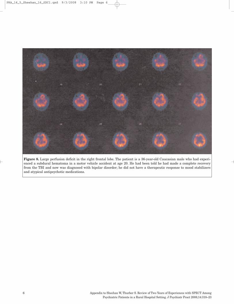
Figure 8. Large perfusion deficit in the right frontal lobe. The patient is a 36-year-old Caucasian male who had experi-enced a subdural hematoma in a motor vehicle accident at age 20. He had been told he had made a complete recoveryfrom the TBI and now was diagnosed with bipolar disorder; he did not have a therapeutic response to mood stabilizersand atypical antipsychotic medications.
PRA_14_5_Sheehan_16_SDC1.qxd 9/3/2008 3:10 PM Page 6

Appendix to Sheehan W, Thurber S. Review of Two Years of Experiences with SPECT AmongPsychiatric Patients in a Rural Hospital Setting. J Psychiatr Pract 2008;14:318–23
7
Figure 9. Large perfusion deficit sugges-tive of a functional disconnection of theleft temporal lobe from the rest of thecerebrum. The patient is a 34-year-oldCaucasian male who sustained a headinjury in an automobile accident thatalso resulted in a broken back and para-plegia. He developed an iatrogenic addic-tion to prescription medications for painand a paranoid delusional disorder thatwas unresponsive to medication.
Figure 10. Large perfusion defect in the left medial orbito-frontalcortex. The patient is a 56-year-old Caucasian male with posttrau-matic stress disorder related to experiences in Vietnam and a headinjury from a motor vehicle accident in the early 1970s; history ofmoodiness, anger, and work-related accidents that rendered himunemployable.
PRA_14_5_Sheehan_16_SDC1.qxd 9/3/2008 3:10 PM Page 7